* To whom all correspondence should be addressed. Mob.: +91-9545559861;
E-mail:[email protected]
A Quick and Efficient Method to Quantify
Baculo Virus by Quantitative Real Time PCR
Mahammad Azhar1*, R. Somashekhar2 and P.P.B.S. Kumar3
1Department of Biotechnology, Jawaharlal Nehru Technological University,
Hyderabad - 500 072, India
2Department of Biotechnology, Azyme Biosciences, Bangalore - 560 069, India. 3Department of Biotechnology, Acharya Nagarjuna University, Guntur - 522 510, India.
DOI: http://dx.doi.org/10.13005/bbra/1178
(Received: 25 September 2013; accepted: 22 November 2013)
Recombinant protein expression using the Baculovirus Expression System is widely used for the production of therapeutics, diagnostics, and reagents. The current “gold standard” for Baculovirus titration, the plaque assay, is labor intensive and time consuming. The real-time PCR assay has the advantage of speed and accuracy. We determined Baculovirus tire by a novel quantitative PCR (qPCR) method, using Baculovirus gene gp64 (Coat protein) specific primers and a recombinant gene-specific primers. Virus specific amplification and gene specific amplification were compared, and both values were found to be very close, suggesting that most of the virus were expressing our gene of interest. This method is useful to determine the percentage of non-recombinant virus particles in virus solution. Further, we compared real-time PCR results with plaque assay, and found that the values were much similar. This method has the advantage of time saving as it completes in 2-3 hours, and not only quantifies the virus, but also quantify the virus, containing the gene of interest.
Key words: QPCR; Baculovirus, AcMNPV, MOI, gp64.
The Baculovirus expression vector system (BEVS), is widely used for the over expression of recombinant protein in insect cells, providing protein post-translational modifications, similar to those obtained when using higher eukaryotic cells. Furthermore, insect cells can be grown to a very high density in serum-free medium as a suspension culture, enabling the production of large quantities of recombinant protein1.
To maximize efficiency of recombinant protein production, determining the best multiplicity
of infection (MOI) is very crucial. Conventionally, Baculoviruses have been titrated by plaque assay2-4 end-point dilution5, and antibody-based assays6,7. But, all of these methods are labor intensive, time consuming, and need a degree of expertise in virus handling and cell culture, sometimes producing results that can be hard to interpret8. Therefore, a quick, easy, and accurate quantification of Baculoviruses is required for the establishment of multiplicities of infection, which is critical for establishing a productive infection cycle.
Real Time quantitative PCR has been used widely for the quantification of DNA9 and RNA10. It has been used for the quantification of several viruses11 and has been very well demonstrated to be a sensitive and accurate method, for virus detection and quantification. Its main advantages are its rapidity and the accuracy of quantification12.
using two different Primers set for quantification. The quantification of accuracy obtained using QPCR, was verified by plaque assay.
Material and Methods
reagents
CFX96 real time PCR machine, qPCR 96 well white plates from Bio-Rad. QiAMP DNA and blood mini kit from Qiagen MESA GREEN qPCR Master Mix Plus for SYBR Assay No ROX from Eurogentec.Cellfectine,Grace insect media, FBS, 4%Agarose, 2xGrace insect media from Invitrogen (Gibco), Cell culture flask and plates from Corning. Primers were synthesized at Eurofins genomics, India.
Cell Culture and recombinant Baculovirus Production
Sf9 cells (1×10⁶) were removed from an exponentially growing culture, and seeded in 6well cell culture plate. Enterokinase gene containing recombinant bacmids (Viral DNA) previously generated in our lab, sequence and insert presence, was confirmed by sequencing. Using Cellfectine (Invitrogen), Cells were transfected with recombinant bacmid DNA (1µg), and incubated at 27oC. Five days post-transfection, the culture medium containing recombinant virus was harvested, and 0.5 mL (P1 Virus) was used to inoculate T-25 flasks containing 2.5x10⁶ Sf9 cells, and were incubated at 27oC in non-humidified incubator. Three days post-infection, the culture medium containing recombinant virus was harvested, and 0.5 mL (P2 Virus) was used to inoculate T-75 flasks containing 8x10⁶ Sf9 cells, and were incubated at 27oC in non-humidified incubator. Recombinant virus stocks were harvested three days post-infection (P3 Virus), clarified by centrifugation at low speed, and titrated by plaque assay (in duplicates) in Sf9Cells. Plaque assay
Plaque assays were carried out as described by King and Possee [3]. Briefly, six well cell culture plates were seeded with Sf9 cells at a density of 0.8x10⁶ cells/well, in a final volume of 2 mLmedia and allowed to settle for 30 min. P3 Viral stocks was serially log diluted (1:10) to 10-8 in Grace insect media supplemented with 10% fetal bovine serum (FBS). Media was removed from six well plates and diluted viral stock added in wells,
starting from 10-4 to10-8. In control well only media was added and incubated at room temperature. After 1 h, the virus inoculum was removed and 2 mL low gelling temperature agarose (4%) was overlaid onto the cells. After the agarose overlay had solidified, cells were incubated at 27 oC for 8 days. The cells were then stained with Neutral Red (Sigma-Aldrich) to visualize plaques.
The following calculation yielded the virus titer in plaque-forming units (pfu) per milliliter:
Pfu/mL= average plaque count x dilution factor x 10
Viral dna extraction
Under sterile conditions, 200 µL virus solutions was removed from the viral stock to be titrated (P3 Virus stock) and used for DNA extraction. Viral DNA was extracted and purified using a Genomic DNA isolation Kit (Qiagen-India) as described by the manufacturer’s Protocol. Briefly, 200 µL of virus was lysed by addition of 20µL Proteinase K and 200 lysis buffer, immediately mixed by pulse vortexing for 15 sec, and incubated at 56°C for 10 min. 200 µL ethanol (96-100%) was then added to the sample. The sample was then applied on the QiAamp Mini spin column and centrifuged at 8000 rpm for 1 min, followed by two washes with wash buffers and centrifugation (8,000 rpm for 1 min). A final spin at 13,000 rpm was carried out to remove residual buffers, and the DNA was then eluted from the column in a final volume of 200 µL elution buffer.
Primers
Primers (Table.1) for amplification of a 72-bp region of the Autographa californica nucleopolyhedrovirus (AcNPV) gp64, and 78-bp region of the Bovine Enterokinase gene, were designed by using Oligonucleotide Properties Calculator online software.
Quantitative PCr Conditions
data analysis
After qPCR run was finished, Data analysis was done with the CFX software manager version 2.0 installed on the system computer. Unknown samples titer was calculated by extrapolating unknown samples values with standard curve.
results and disCussion
Baculovirus Autographa californica multicapsid nucleopolyhedrovirus (AcMNPV) envelope glycoprotein gp64 gene was selected as the target for amplification because it is specific to AcMNPV, and is responsible for coding a viral envelope fusion protein that is necessary for receptor binding and membrane fusion, in the event of viral infection. Along with gp64 gene,
we choose one more target, Enterokinase gene, for amplification, which gene was incorporated in bacmids. Then we compared both results and correlation between both gene amplification. Comparing both results of gp64 and EK gene amplification, we found that the results were very close - qPCR determined Virus titer with EK was 0.94 ×10⁸ pfu/ml, and for GP64 was 1.1×10⁹. In another experiment, EK titer was 0.994 × 10⁸ and gp64 was 1.22 × 10⁹ these results indicate that there is no or very less non recombinant virus (i.e. virus without our gene of interest), and results were consistent from one assay to another. Further PCR results were analyzed, and we found results were matching with plaque assay (Table 2). So, this is a novel and useful method to determine functional viral titer in sample.
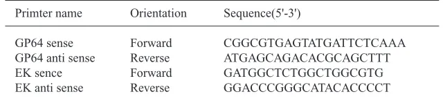
table 1. Sequence of the primers
Primter name Orientation Sequence(5'-3')
GP64 sense Forward CGGCGTGAGTATGATTCTCAAA GP64 anti sense Reverse ATGAGCAGACACGCAGCTTT EK sence Forward GATGGCTCTGGCTGGCGTG EK anti sense Reverse GGACCCGGGCATACACCCCT
table 2. QPCR and Plaque assay titer comparison
Target Cq mean QPCR titer Plaque SQ mean (pfu/mL) titer (pfu/mL)
Enterokinase 16.53 9.44E+0.8 1.3×10⁹
Gp64 16.25 1.14E+0.9
Note: Cq-Quatification cycle; SQ-Starting quantity;Pfu-Plaque forming unit
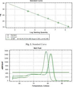
Standard curve was generated by using Baculovirus sample, which virus titer was previously determined by Plaque assay, and gp64 gene was the target for amplification in standards. Standards amplification duplicates were very close (Fig. 1) and standard curve, all dilutions were correlating with amplification, its regression square was 0.99 (Fig. 2). This emphasizes that, much care needs to be taken while setting up experiments in q PCR, to get reliable results.
In this experiment, we used SYBR green chemistry for detection of amplified product and Melt curve generated two separate peaks, each specific to gp64 and EK (Fig. 3). Looking at this data, we found that results were much reliable and cost-effective, compared to probe-based assay, to determine viral titer.
Fig. 2. Standard Curve
Fig. 3. Melt curve peaks of gp64 and EK
ConClusion
In this study, it was established that quantitative PCR can be applied for precise and quick quantification of recombinant Baculovirus. It is apparent especially when this method is employed for quantification of huge number of recombinant Baculovirus samples.
reFerenCes
1. Kost TA, Condreay JP, Jarvis DL. Baculovirus as versatile vectors for protein expression in insect and mammalian cells. Nature Biotechnology,
2005; 23: 567–575.
2. hink WF, Vail PV. A plaque assay for titration of alfalfa looper nuclear polyhedrosis virus in a cabbage looper (TN-368) cell line. J Invertebr Pathol , 1973; 22: 168-174.
3. King LA, Possee RD. The Baculovirus Expression System: A Laboratory Guide. London: Chapman and hall, 1992.
4. O’Reilly DR, Miller LK, Luckow VA.. Baculovirus Expression Vectors: A Laboratory Manual. New York: Oxford University Press. 1994.
5. Lynn DE. Improved efficiency in determining the titer of the Autographa californica baculovirus nonoccluded virus. Bio Techniques, 1992; 13: 282–285.
6. Kitts PA, Green G. An immunological assay for determination of baculovirus titers in 48 hours.
Anal Biochem, 1999; 268: 173–178.
7. Kwon MS, Dojima T, Toriyama M, Park EYDevelopment of an antibody-based assay for determination of baculovirus titer in 10 h. Biotechnology Prog, 2002; 18: 647-651. 8. hunt IFrom gene to protein: A review of new and
enabling technologies for multi-parallel protein expression. Protein Expression & Purification, 2005; 40: 1–22. Review.
9. Kaltenboeck, B., & Wang, C. M. Advances in Real Time PCR: Application to clinical laboratory diagnostics. Elsevier Academic Press INC. 2005; pp. 219-259
10. Bustin, S. A. Absolute quantification of mRNA using real-time reverse transcription polymerase chain reaction assays. Journal of Molecular Endocrinology, 2000; 25(2): 169-193.
11. Mackay, I. M., Arden, K. E., & Nitsche, A. Real-time PCR in virology. Nucleic Acids Research, 2002; 30(6): 1292-1305.