Original Research Article
Effect of primary percutaneous coronary intervention on ventricular
repolarization through evaluation of QT dispersion in patient with
acute myocardial infarction
Behzad Babapour
1, Bita Shahbazzadegan
2*, Bahareh Khademi
3INTRODUCTION
Cardiovascular disorders are the most common cause of death worldwide. AMI, induced by complete or incomplete blockage of arteries supplying blood to the myocardium (coronary artery disease), is the most common cause of these disorders. About one million people in the United States of America faced to myocardial infarction and more than one million patients
admitted to coronary care unit (CCU), suspicion to acute myocardial infarction.1 Patients with acute myocardial infarction with elevated ST-segment candidate tore-perfusion of blood flow in the arteries supplying blood to the heart. This re-perfusion performs with fibrinolytic agents such as streptokinase or by means of PCI.2
Myocardial ischemia alters the electrical and mechanical activity of the myocardium. In the heart's electrical
ABSTRACT
Background: Cardiovascular disease is the most common cause of death around the world. QT dispersion is one of the parameters that used for evaluation of ventricular arrhythmia. Primary PCI increases probability of coronary artery and reperfusion of the ventricular arrhythmia. The aim of this study was to determine effect of primary percutaneous coronary intervention (PCI) on ventricular repolarization through evaluation of QT dispersion in patient with acute myocardial infarction.
Methods: In this pre-post test study, 77 patients with acute ST with elevated myocardial infarction under primary PCI were investigated. The ECG and ST dispersion before PCI and 24 hours after PCI were determined and then the amount of QTd was calculated. The repeated measurement ANOVA was used to compare QTd of pre- PCI treatment and QTd in 24 hours after PCI. Data analysis was performed using statistical software SPSS ver.17.
Results: From 77 participants, 60 were male and 17 were female. 43 (55.8%) had a MI position in ANT, PRE, and EXT, 33 (42.9%) had in the INF, and only one person (1.3%) had a MI position in LAT. The results showed that mean QT dispersion in ECG, 24h after primary PCI, for most of measured variables was deceased compare to before primary PCI, but the difference was not significant.
Conclusions: The amount of QTd 24 hours after PCI decreased but its decline was not significant. With regards to lack of convenience data, more researches are recommended in this field.
Keywords: Acute coronary syndrome, Percutaneous coronary intervention, Cardiac care unit
1
Department of Internal Medicine, 2Social Determinants of Health (SDH) Research Center, School of Medicine, Ardabil University of Medical Sciences, Ardabil, Iran
3General Practitioner, School of Medicine, Islamic Azad University, Ardabil, Iran
Received: 02 December 2017
Revised: 02 January 2018
Accepted: 03 January 2018
*Correspondence:
Dr. Bita Shahbazzadegan,
E-mail: [email protected]
Copyright: © the author(s), publisher and licensee Medip Academy. This is an open-access article distributed under the terms of the Creative Commons Attribution Non-Commercial License, which permits unrestricted non-commercial use, distribution, and reproduction in any medium, provided the original work is properly cited.
activity disorder that induces ventricular repolarization abnormalities causing the heart arrhythmias.3 Reperfusion to the heart after ischemia may change the heart performance, profoundly. QT dispersion (QTd) in ECG is an important parameter for evaluation of ventricular repolarization heterogeneity.4 QTd means the difference between maximum and minimum QT interval derived from the 12-lead ECG.5 Is higher and more prolonged QTd related to higher ventricular repolarization
heterogeneity and associated to arrhythmias
susceptibility.2 QT interval on electrocardiograms (ECG) reflects the ventricular repolarization. QTd reflects homogeneity of ventricular repolarization and important evidences exist that indicates increased QTd associates with some heart disease, especially malignant arrhythmias.6,7
Roukema showed a direct correlation between QTd and
myocardial ischemia. Ischemia increases the
repolarization time in myocardial infarction patients and thus prolongs QT in ECG.8 Previous studies have shown that QTd obtained immediately after exercise testing was significantly higher in patients with coronary artery stenosis than patients without stenosis (˃50%).6 Primary
PCI is one of the important treatments, PCI carried out with or without stent of angioplasty. If PCI used as a primary method of reperfusion therapy without of fibrinolytic prescription and before it is called primary PCI. When the Primary PCI is performed by a person with experience in a specialist center, is beneficial over fibrinolysis.1 Chander compared the effect of PCI on QTd and fibrinolytic in 45 acute coronary syndrome patients in 2005. The QTd in patients undergoing PCI were decreased, tremendously (75±21 ms to 38±20 ms, p<0.0001). Its values in patients who were treated with fibrinolytic, also declined (78±19 ms to 67±22ms, p<0.05), but in lower amounts compare to PCI.9 Alasti investigated the effect of PCI on the QT dispersion on 96 patients in 2010. Effect of complete reopen of coronary arteries in patients with stable angina and uni-arterial disease were studied. ECG before PCI and 24 hours after PCI were assessed and the parameters of duration of QRS, QT interval, QT, modified QT, JT and modified JT was calculated. There were a distinct difference between the duration of the QRS, modified QTd, and modified JT dispersion ECG before and after PCI.10
A significant number of patients suddenly die after myocardial infarction (MI) and it showed that the measurement of QTd (an important factor for cardiovascular mortality) is a very simple and non-invasive way to predict the risk of sudden death in these patients. Regarding to the importance of ventricular repolarization activities including QT in creation of dangerous arrhythmias after myocardial infarction, factors that affects ventricular repolarization changes are important. Reperfusion with PCI is one of these factors. Therefore, in this study, patients with acute myocardial infarction with elevated ST-segment treated with PCI
PCI and the size of QT in the various leads was calculated. The interval between the minimum and maximum QT (QTd) was measured before and after PCI. This parameter was used to investigate the effect of PCI on ventricular repolarization activity through evaluation of QTd.
METHODS
In a pre-posttest study, all patients admitted in heart center of Urmia’s Seyedoshohada Hospital from date 2015/3/21 to 2016/3/20 with acute myocardial infarction who were treated with PCI were investigated (77 patients). Patients' medical records in hospital record center were collected. Demographic characteristics (age, sex, etc.) and ECG before and 24 hours after PCI were extracted from the documents.
Inclusion criteria were acute myocardial infarction patients with elevated ST-segment by clinical symptoms, laboratory findings (increased cardiac troponin1) and proven ECG changes that treated with PCI.
Exclusion criteria were electrolyte abnormalities, absence of sinus rhythm, heart conduction block, at least in 8-lead ECG inability to review and use of medications that affects the QTD such as digitalis, depressants, anti-psychotic, respectively. ECG photographs of patients were assessed and their QT on millisecond (msec) was calculated manually.
To calculate the QT, the interval between Q wave or beginning of the QRS complex to the end of the T wave was calculated. When a U wave existed, the lowest point of curve between T and U waves was considered as the QT interval. QTc was calculated with the formula Bazzet. The longest and shortest QTc interval in ECGT12 Leady was calculated and their difference was considered as QTd.
Descriptive characteristics of patients were investigated using descriptive statistics (mean and standard deviation) and presented in frequency tables and charts. The repeated measurement ANOVA was used to compare QTd of pre- PCI treatment and QTd in 24 hours after PCI. Data analysis was performed using statistical software SPSS ver.17. The study proposal was approved by the Research Board and Ethics Committee of Islamic Azad University, Ardabil Branch.
RESULTS
From 77 participants, 60 were male and 17 were female. 39 cases had ≤55 years old and 38 patients were older than 55 years. The average age of the patients was 56.13±11.26 and their age range was 26-86 years (Table 1).
person (1.3%) had a MI position in LAT. In 29 of 77 patients (37.7%) the RCA artery was involved, 38
(49.4%) had involvement in LAD artery, and in 10 (13%) person the arteries of LCX and OM were involved.
Table1: Frequency of demographic information of participant.
Frequency Gender Age
High blood pressure
High blood
pressure Diabetic Smoking
Female Man 55≥ ˃55 Yes No Yes No Yes No
Number 60 17 39 38 29 48 12 65 45 32
percent 77.9 22.1 50.6 49.4 37.7 62.3 15.6 84.4 58.4 41.6
Distribution of EF in patients is shown in Table 2. The highest and lowest prevalence of EF were 30%-45% and 45% or more, respectively.
Table 2: Frequency of EF of patients participated in the study.
EF Frequency Frequency
Non reported 15 19.5
50%≥ 5 6.5
45%-50% 5 6.5
30%-45% 31 40.3
˃30% 21 27.3
Total 77 100
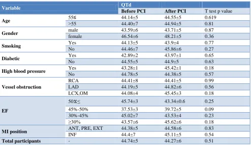
Mean QTd before PCI (QTd1) for all of participants was 0.44±0.06 and the corresponding values for QTd after PCI (QTd2) was 0.44±0.05. The difference between these values was not significant (Table 3). The results of paired
T-test showed that the differences QTd1 and QTd2 for within groups for variables of age, gender, smoking status, diabetic, and high blood pressure was not statistically significant (5% level). In addition, the difference of QTd1 and QTd2 in vessel obstruction groups, EF values, and MI position was also not significant (Table 3).
According to Table 4 and the results of the independent T-test except for variable of gender after PCI, mean QTd between the groups for variables of gender before the PCI, and for other variable (diabetes, hypertension and smoking) before and after PCI were not statistically significant. The QTd value for males was statistically higher than females (0.48 versus 0.44).
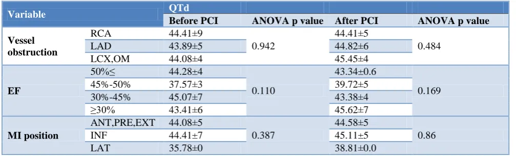
The results of analysis of variance (ANOVA) also showed that the differences between the groups for the variables of Vessel obstruction, EF, and MI position were not significant at 5% level (Table 5).
Table 3: Results of T test for mean QTd before and after PCI within groups of studied variables.
QTd Variable
T test p value
After PCI Before PCI
0.619 44.55±5
44.14±5 55≥
Age
0.81 44.94±5
44.40±7 ˃55
0.87 43.71±5
43.59±6 male
Gender
0.36 48.21±5
46.54±6 female
0.77 43.9±4
44.13±5 Yes
Smoking
0.27 45.86±6
44.46±7 No
0.65 43.97±1
42.89±2 Yes
Diabetic
0.63 44.9±5
44.55±5 No
0.18 45.42±1
43.28±1 Yes
High blood pressure
0.57 44.38±5
44.78±5 No
0.99 44.41±5
44.41±8 RCA
Vessel obstruction LAD 44.19±5 44.82±6 0.56
0.18 45.45±3
44.08±4 LCX,OM
0.25 43.34±0.6
45.74±3 50 ≥%
EF 45%-50% 37.53±3 39.72±5 0.09
0.23 43.53±4
45.02±7 30%-45%
0.18 45.62±6
43.57±6 ≥30%
0.83 44.58±6
44.38±5 ANT, PRE, EXT
MI position
0.54 45.11±5
44.4±7 INF
0.51 44.27±6
44.74±5 -
Table 4: Result of T test for QTd for between groups for different variables before and after PCI.
Variable QTd
Before PCI T test p value After PCI T test p value
Gender Female 43.45±6 0.077 43.71±5 0.002
Man 46.43±6 48.21±5
Diabetic Yes 42.89±9 0.455 43.97±5 0.581
No 44.34±5 44.90±5
High blood pressure
Yes 43.03±7
0.233 45.42±5 0.437
No 44.76±6 44.38±5
Smoking Yes 44.04±5 0.865 43.90±4 0.122
No 44.25±7 45.86±6
Table 5: Result of T test for QTd for between groups for different variables before and after PCI.
Variable QTd
Before PCI ANOVA p value After PCI ANOVA p value
Vessel obstruction
RCA 44.41±9
0.942
44.41±5
0.484
LAD 43.89±5 44.82±6
LCX,OM 44.08±4 45.45±4
EF
50%≥ 44.28±4
0.110
43.34±0.6
0.169
45%-50% 37.57±3 39.72±5
30%-45% 45.07±7 43.38±4
≥30% 43.41±6 45.62±7
MI position
ANT,PRE,EXT 44.08±5
0.387
44.58±5
0.86
INF 44.41±7 45.11±5
LAT 35.78±0 38.81±0.0
DISCUSSION
The mean QTd values for before and after PCI were 44.74±5 and 44.27±6, respectively. These values were in normal range. Malik and Batchvarov stated that the range of QT in normal subjects is 10-71 msec.11
In our study the differences between QTd before and after PCI within the group for all study variables were not significant. This result is in opposition with the results of studies of Chander, Cavusoglu and Nikiforos that reported PCI and fibrinolytic, both, successfully reduced the QTd via reperfusion of blood, but PCI was over superior to the fibrinolytic.9,12,13 In the study of Marasia and Alasti the effect of PCI on QTd was significant, but in our study the difference of QTd before and 24 hours after PCI was not statistically significant.10,14 Giedrimiene and Hamze reported that QT dispersion in patients, who were treated with PCI, out of artery type and the location of MI, was recovered.15,16 In our study changes in QTd after PCI in all three groups of the artery clogging (LAD, RCA, LCX, OM) increased, insignificantly
In the study of Nikoforos the effectiveness of fibrinolytic and PCI in reducing of the QTd were investigated.13 They reported QTd was decreased and concluded that PCI was
results. They also reported that changes in QTd in patients with or without hypertension were not significant. This result is in agreement with our study results. Eslami also confirmed the effectiveness of PCI in reduction of QTd.17
Differences QTd before and after PCI for variables of age, gender, smoking status, diabetic, and high blood pressure was not statistically significant. Saveliova reported that differences for variables of age and sex) was insignificant.18,19
In the study of Chander the difference of QTd among the genders after reperfusion, was not significant (9). This result is in the opposition with our study that the QTd before PCI was slightly higher in the women compared to men, but 24 hours after PCI men had higher QTd than that of women (p value, 0.002). Aydinlar reported that there is a dearth of information on the influence of elective PCI on ECG parameters, especially QT.20 Saveliova reported that there are a little or no sex differences in QTd among normal subjects.18
CONCLUSION
convenience data, more researches are recommended in this field.
ACKNOWLEDGEMENTS
Thus, the authors are thanked of all staff and patients in Urmia hospitals that cooperated in this study.
Funding: No funding sources Conflict of interest: None declared
Ethical approval: The study was approved by the Institutional Ethics Committee
REFERENCES
1. Ridker MP, Genest J, Lippy P. Risk factors for atherosclerotic heart disease. In: Braunwald E, Zipes DP, Libby P, editors. Heart Disease: A Textbook of Cardiovascular Medicine. 6th ed. Philadelphia: W.B: Saunders Company; 2001: 1010–1039. 2. Szyma s i wia t ows i ezler J, Budaj A.
The relationship between diastolic function of the left ventricle and QT dispersion in patients with
myocardial infarction. Int J Cardiol.
1999;69(3):245–9.
3. Day CP, McComb JM, Campbell RWF. QT
dispersion: An indication of arrhythmia risk in patients with long QT intervals. Br Heart J. 1990;63(6):342-4.
4. Yosefian S, Farshidi H, Sobahni M, Rahimi S. QT-Dispersion as a potential marker in prognosis of acute myocardial infarction. HMJ. 2009;12(4):223-30.
5. Giedrimiene D, Giri S, Giedrimas A, Kiernan F, Kluger J. Effects of ischemia on repolarization in patients with single and multivessel coronary
disease. Pacing Clin Electrophysiol.
2003;26(1p2):390–3.
6. Jensen BT, Abildstrom SZ, Larroude CE, Agner E, Torp-Pedersen C, Nyvad O, et al. QT dynamics in risk stratification after myocardial infarction. Heart Rhythm. 2005;2(4):357–64.
7. Segerson NM, Litwin SE, Daccarett M, Wall TS, Hamdan MH, Lux RL. Scatter in repolarization timing predicts clinical events in post-myocardial infarction patients. Heart Rhythm. 2008;5(2):208-14.
8. Bonow RO, Mann DL, Zipes DP, Libby P.
Braunwald’s Heart Disease: A Textboo of Cardiovascular Medicine, 2-Volume Set. Elsevier Health Sciences. 2011.
9. Chander S, Kumar R, Jorapur V, Desai N, Rao M, Yeragani VK. Effect of mechanical coronary reperfusion on QT dispersion in acute coronary syndrome. Indian Heart J. 2004;57(3):233–6. 10. Alasti M, Adel MH, Torfi E, Noorizadeh M,
Bahadoram S, Moghaddam MA, et al. QT
Dispersion: Does It Change after Percutaneous Coronary Intervention? J Tehran Heart Center. 2011;6(1):19-23.
11. Malik M, Batchvarov VN. Measurement,
interpretation and clinical potential of QT dispersion. J Am Coll Cardiol. 2000;36(6):1749-66. 12. Cavusoglu Y, Gorenek B, Timuralp B, Unalir A,
Ata N, Melek M. Comparison of QT dispersion between primary coronary angioplasty and thrombolytic therapy for acute myocardial infarction. Isr Med Assoc J. 2001;3(5):333-7. 13. Nikiforos S, Hatzisavvas J, Pavlides G, Voudris V,
Vassilikos VP, Manginas A, et al. QT-interval dispersion in acute myocardial infarction is only shortened by thrombolysis in myocardial infarction grade 2/3 reperfusion. Clinical Cardiol. 2003;26(6):291-5.
14. Marazia S, Zimarino M, Torge G, Pasquale M, Caputo M, Floris F, et al. QT dispersion and myonecrosis after stent percutaneous coronary intervention. Ital Heart J Suppl. 2004;5(11):861-7. 15. Giedrimiene D, Giri S, White CM, Giedrimas E,
Kluger J. The immediate and short-term effect of successful percutaneous coronary intervention on repolarization in acute myocardial infarction patients. Ann Noninvasive Electrocardiol. 2002;7(4):357-62.
16. Hamza O, Mouffok M, Bouzid A, Merad-Boudia Kh. CRT-108 Evaluation of Corrected QT and QT Dispersion Changes in Acute ST-Elevation Myocardial Infarction after Primary Percutaneous Coronary Intervention. J Am Coll Cardiol Intv. 2014;7(2):9.
17. Eslami V, Safi M, Taherkhani M, Adibi A, Movahed MR. Evaluation of QT, QT dispersion, and T-wave peak to end time changes after primary percutaneous coronary intervention in patients presenting with acute ST-elevation myocardial infarction. J Invasive Cardiol. 2013;25(5):232-4. 18. Savelieva I, Camm AJ, Malik M. Gender-specific
differences on QT dispersion measured in 1100 healthy subjects (abstr). Pacing Clin Electrophysiol. 1999;22:885.
19. Savelieva I, Camm AJ, Malik M. Do we need age-adjustment of QT dispersion? Observations from 1096 normal subjects (abstr). Heart. 1999;81:47. 20. Aydinlar A, Senturk T, Ozdemir B, Kaderli AA,
Aydin O. Effect of percutaneous transluminal coronary angioplasty on QT dispersion and heart rate variability parameters. Cardiovasc J Afr. 2009;24(4):240–4.