*Corresponding author: saida@uthm.edu.my 2018 UTHM Publisher. All right reserved.
e-ISSN: 2600-7924/penerbit.uthm.edu.my/ojs/index.php/jst
Effect of Calcination Temperature on (N, Fe) Doped TiO2 Nanoparticles
Siti Aida Ibrahim
1*, Abdul Hadi Zainal Alam
2, Rosniza Hussin
1, Zakiah Kamdi
2, Mohamed
Nasrul Mohamed Hatta
2,
Ainun Rahmahwati Ainuddin
2and Muhamad Zaini Yunos
21* Faculty of Engineering Technology, Universiti Tun Hussein Onn Malaysia, Hab Pendidikan Tinggi Pagoh,
KM 1, Jalan Panchor, 84600 Panchor, Johor Darul Takzim, Malaysia
2Faculty of Mechanical & Manufacturing Engineering, Universiti Tun Hussein Onn Malaysia, Parit Raja, 86400 Batu Pahat, Johor Darul Takzim, Malaysia.
Received 30 September 2017; accepted 7 March 2018; available online 1 August 2018
1. Introduction
Environmental problem such as water pollution is one of the major issues today due to a substantial population growth which generates more waste products through industries, transportation and households. These pollutants are dangerous to our health as it is soluble in water and easily consume by human beings and animals. An extensive research work by various researchers have been made in order to improve the water quality including by employing trees and microbial as bio-indicator for heavy metal, filtration and advanced oxidation processes (AOPs) as alternative methods [1,2]. However, the existing technology that has been used to overcome this problem is unable to remove these pollutants completely. A majority of non-degradable organic pollutants in the water are not treatable due to their high chemical stability and/or low biodegradability. Some method is very expensive, high maintenance and applicable only in selective environment or condition. Recent studies have established that the developments of new catalytic and
photocatalytic processes provide great help to address this problem [3,4].
TiO2 is one of the most promising
photocatalysts that can be employed for the purification of water and air [5,6]. It is also
proven that TiO2 can be employed in various
environmental applications due to its superior photoreactivity, long term stability, inert and low cost [3,4]. The photocatalytic activity of
TiO2 depends on several factors such as
crystallinity, surface area, impurities and density of surface hydroxyl groups [5,7]. In
general, TiO2 required the presence of light to
decompose organic pollutant. Due to its large
band gap energy (3.0 - 3.2 eV), TiO2 is only
active under UV light irradiation, which limits
the light utilization rate of TiO2 to only 5% of
solar energy. Considerable efforts have been
made by many researchers to tune TiO2 with
metals and/or non-metal elements that may alter the wavelength absorption of the photocatalyst. Surface modification such as metal ion (i.e. Ag, Fe and Zr) and non-metal doping (i.e. C, F and N), or by coupling with other narrow band gap semiconductors have been adopted to enhance the performance of
TiO2 under visible light irradiation [6,8-10].
Abstract: TiO2 is one of the most promising photocatalysts that is widely used for environmental clean-up due to its ability to degrade organic pollutants in air or water in the presence of UV light irradiation. In this study, the sol-gel method followed by calcination process was employed to synthesize N, Fe doped TiO2 nanoparticles. The effect of calcination temperature on the structural, morphology and optical properties of the as-prepared samples was analyzed. Titanium tetra isopropoxide (TTIP) was used as Ti precursor and urea and ferric nitrate nonahydrate were the employed precursors to obtain N and Fe, respectively. X-ray diffraction pattern displayed a transformation of anatase structure to biphasic of anatase and rutile structure as the calcination temperature was increased from 300 to 700 °C. FESEM images indicated an agglomeration of particles with the grain size was estimated at 50-170 nm. UV-Vis analysis revealed that the increment of calcination temperature induced a red shift in the absorption spectra from 485 nm to 664 nm. Hence, the results indicate that N, Fe doped TiO2 is a highly potential visible-driven photocatalyst to degrade pollutants under the presence of visible light irradiation
Keyword: TiO2; Sol-gel; Photocatalytic Activity; Optical Property.
45
More recently, co-doping TiO2 with two kinds
of element has been proposed to promote the photocatalytic performance further benefiting from a synergistic effect of both dopants
[11,12]. Study by Ashkarran et al. revealed
that TiO2 co-doped with Ag and N improved
the photocatalytic efficiency under visible light irradiation due to the generation of two different electronic states acting as electron traps [13].
In this work, nanocrytalline N, Fe doped
TiO2 was synthesised by using sol-gel method
and subsequently followed by a calcination process. Titanium (iv) isopropoxide was used as Ti precursor while iron (iii) nitrate and urea act as the sources of Fe and N dopant, respectively. The calcination temperatures were varied from 300 to 700 °C. The characteristics of the as-prepared samples were investigated using XRD, FESEM and UV-Vis. The effects of calcination temperature towards
N, Fe doped TiO2 characteristic were
investigated and discussed.
2. Experimental Method
All chemicals used in this study were analytical reagent without further purification. Distilled water was used in all experiments.
The preparation of N, Fe doped TiO2 was a
modification method from Teck and Ibrahim (2016) [14]. In a typical synthesis, 20 ml titanium isopropoxide (TTIP) and 80 ml isopropanol were added dropwise into a hydrolysis medium which contains 8 ml of acetic acid, 6 ml of distilled water iron (III) nitrate (0.15 wt%) and urea (0.3 wt%). The resulting mixture was stirred slowly for 3 hours at room temperature and centrifuged at 3000 rpm for 20 minutes in order to obtain the white precipitate. The white precipitate was dried at 80 °C for 12 hours in a drying oven. The precipitate was granulated by using agate mortar to get fine granules. Finally, the sample was calcined at 300, 500 and to 700 °C for 2 h, respectively.
The as-prepared samples were
characterised by x-ray diffraction (XRD), field
emission scanning electron microscope
(FESEM) and ultraviolet-visible
spectrophotometer (UV-VIS). The XRD was performed on a D8 Advanced Bruker System with Cu kα radiation as the x-ray source. Morphologies of the samples were observed by using a high-resolution field emission
environmental scanning electron microscope (FESEM, JSM-7600F). Optical properties of samples were measured on a Shimadzu
UV-VIS spectrometer (UV-1800) in the
wavelength of 300-800 nm.
3. Results and Discussion
X-ray powder diffraction (XRD) testing is used to determine the structure and crystallite
size of N, Fe-TiO2 samples. Fig. 1 shown XRD
patterns for N-Fe-TiO2 samples at different
calcination temperatures of 300 °C, 500 °C and 700 °C. The anatase and rutile phase used were similar to the Joint Committee on Powder Diffraction Standard (JCPDS) file no. 08-2082 and file no. 03-6142, respectively.
The results shown that N, Fe doped TiO2
samples were anatase at temperature 300 °C and 500 °C while rutile phase occurred at 700 °C. It was observed that with increasing temperature from 300 to 500 °C, the peak intensities of anatase and rutile increased
gradually, implying improvement of
crystallization and growth of crystallites [15,16]. Generally, the transformation of anatase to rutile occurs when the temperature was increased from 400 to 800 °C [16]. This can be attributed to the fact that when sufficient thermal treatment is provided, the phase transformation of meta-stable anatase to
a more stable rutile phase in the TiO2
specimen can be achieved [14]. It is noted that all samples did not show the presence of iron or nitrogen compound. This may be due to two reasons, one is the limitation of XRD to detect low concentration foreign atoms in the composition and the other is the similarity of ionic radii of Ti4+ (0.68 Å) and Fe3+ (0.64 Å)
[17].
Fig. 1 XRD patterns of N, Fe-TiO2 at (a) 300 °C, (b) 500 °C and (c) 700 °C.
(A: Anatase, R: Rutile)
2 Theta
20 30 40 50 60 70 80
Inten
si
ty
(a.
u)
A R
A A
A
A A
R R
46
The average crystallite size was calculated according to Scherrer’s equation as shown in Eq (1). Table 1 shown the summary table of XRD analysis for all samples. Based
on the result, the crystallite size of N, Fe-TiO2
was in the range of 14 to 46 nm. The
crystallite size of N, Fe doped TiO2 at 700 °C
which consist of biphasic phase of anatase and rutile is larger than the single phase samples. The trend shows that the crystallite size was increased with the increment of calcination
temperature. Li et al. [18] reported similar
behaviour as the increment calcination temperature accelerated the crystallite growth due to high-phase transformation heat.
𝐷 = 0.9𝜆
𝛽 cos 𝜃 (1)
where D denotes the average crystallite size (nm); K is the Scherrer constant, somewhat arbitrary value that falls in the range 0.8–1.0 (it has been assumed to be 0.9 in present work); λ is wavelength of X-ray radiation (0.154 nm); θ is the diffraction angle and β is full width at half maximum (FWHM).
Table 1 Summary table of XRD analysis
Samples Phase Crystallite
size (nm) N, Fe-TiO2 (300 °C) Anatase 14 N, Fe-TiO2 (500 °C) Anatase 20 N, Fe-TiO2 (700 °C) Anatase &
Rutile
46
Fig. 2 shows the morphology of N,
Fe-TiO2 calcined at temperatures of 300 °C, 500
°C, and 700 °C. It is found that all samples have rough surface and agglomerated. The grain size of all samples were estimated in the range of 50 to 170 nm. According to Brinker and Scherer, small particles incline to agglomerate due to high surface energy and draws other particles to colesce together to
form bigger particle size [19].
Fig. 2 FESEM image of TiO2 powders: a) N-Fe-TiO2 (300 °C), b) N-Fe-TiO2 (500 °C), c) N-Fe-TiO2 (700 °C)
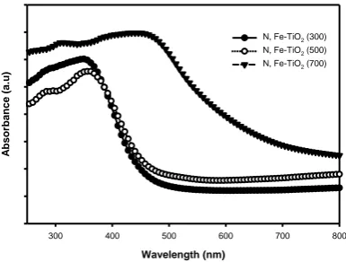
Fig. 3 shows the optical properties of N,
Fe doped TiO2 using UV-Vis
Spectrophotometer. The values of the light absorption were obtained by extrapolating the steepest slope of the UV-Vis spectra. The
intersection point between X-axis and
extrapolation line shows the excitation wavelength of the as-prepared samples. Based on the figure, an intense absorption of all samples found at wavelengths from 485 nm to 664 nm which can be assigned to the band gap
absorption of N, Fe-TiO2 due to the electronic
transition from the valence band to the conduction band. The extension of absorption wavelength in visible light region may attribute to the increment of calcination temperature from 300 to 700 °C, resulting from crystal growth and crystallinity of
N,Fe-TiO2 . Thus, the synthesis parameter such as
the calcination temperature is important for optical characteristics as these aspects increase
the ability of TiO2 to work under broader
spectrum of light.
Fig. 3 UV-Vis Spectral of N, Fe-TiO2 calcined at varies temperature of 300 to 700 °C.
4. Conclusion
In conclusion, N, Fe-TiO2 were successfully
synthesised by using sol-gel method. The XRD pattern shown that calcination
a
100 nm
100,000 x b
100 nm
100,000 x
Wavelength (nm)
300 400 500 600 700 800
A
bsorbance
(a.
u)
N, Fe-TiO2 (300)
N, Fe-TiO2 (500)
47
temperatures plays a centre role for phase transformation from anatase to rutile as the temperature increased from 300 to 700 °C. The grain size is also affected when the calcination temperature was increased. Based on UV-Vis
result, the increment of calcination
temperature broadened the range of response of visible light. Therefore, it is found that the temperature plays an important role towards
TiO2 characteristic.
Acknowledgements
The authors would like to express their utmost gratitude and acknowledgment to Universiti Tun Hussein Onn for providing funding support.
References
[1] Hedberg, Y., Herting, G., & Wallinder, I.O. (2011). “Risks of using Membrane Filtration for Trace Metal Analysis and Assessing the Dissolved Metal Fraction of Aqueous Media – A Study on Zinc,
Copper and Nickel” in Environmental
Pollution, Vol 159. No 5. pp. 1144-1150. [2] Arslan-Alaton, I., (2007). “Degradation of
a Commercial Textile Biocide with
Advanced Oxidation Processes and
Ozone” in Journal of Environmental
Management, Vol 82. No 2. pp. 145-154. [3] Dahl, M., Liu, Y., & Yin, Y. (2014)
“Composite Titanium Dioxide
Nanomaterials” in Chemical Reviews, Vol
114. No 19. pp. 9853-9889.
[4] Wang, Y., He, Y., Lai, Q., & Fan, M. (2014) “Review of the Progress in
Preparing Nano Tio2: an Important
Environmental Engineering Material” in
Journal of Environmental Sciences, Vol 26. No 11. pp. 2139-2177.
[5] Di Paola, A., Bellardita, M., Palmisano, L., Barbieriková, Z., & Brezová, V. (2014) “Influence of Crystallinity and OH Surface Density on the Photocatalytic
Activity of Tio2 Powders” in Journal of
Photochemistry and Photobiology A: Chemistry, Vol 273. pp. 59-67.
[6] Ibrahim, S.A., & Sreekantan, S. (2014) “
Fe-TiO2 Nanoparticles by Hydrothermal
Treatment with Photocatalytic Activity
Enhancement” in Advanced Materials
Research, Trans Tech Publ, Vol 1024. pp. 39-43.
[7] Suttiponparnit, K., Jiang, J., Sahu, M., Suvachittanont, S., Charinpanitkul, T., & Biswas P. (2011) “Role of Surface Area, Primary Particle Size, and Crystal Phase
on Titanium Dioxide Nanoparticle
Dispersion Properties” in Nanoscale Res
Lett, Vol 6. No 1. pp. 27.
[8] Harikishore, M., Sandhyarani, M.,
Venkateswarlu, K., Nellaippan, T.A., & Rameshbabu N. (2014) “Effect of Ag
Doping on Antibacterial and
Photocatalytic Activity of Nanocrystalline
TiO2” in Procedia Materials Science, Vol
6. pp. 557-566.
[9] Zaleska, A. (2008) “Doped-TiO2: A
Review” in Recent Patents on
Engineering, Vol 2. No 3. pp. 157-164. [10] Hussin, R., Choy, K.L., & Hou, X.H.
(2014) “Fabrication of multilayer
ZnO/TiO2/ZnO Thin Films with
Enhancement of Optical Properties by Atomic Layer Deposition (ALD)” in
Applied Mechanics and Materials, Vol 465. No 2. pp. 916-921.
[11] Fu, C., Gong, Y., Wu, Y., Liu, J., Zhang, Z., Li, C., & Niu, L. (2016)
“Photocatalytic Enhancement of Tio2 by
B and Zr Co-Doping and Modulation of
Microstructure” in Applied Surface
Science, Vol 379. pp. 83-90.
[12] Dolat, D., Mozia, S., Ohtani, B., & Morawski, A.W. (2013) “Nitrogen,
Iron-Single Modified (N-Tio2, Fe-Tio2) and
Co-modified (Fe, N-Tio2) Rutile Titanium
Dioxide as Visible-Light Active
Photocatalysts” in Chemical Engineering
Journal, 225 pp. 358-364.
[13] Ashkarran, A.A., Hamidinezhad,
Haddadi, H., & Mahmoudi, M.
(2014)“Double-doped TiO2 Nanoparticles
as an Efficient Visible-Light-Active Photocatalyst and Antibacterial Agent
under Solar Simulated Light” in Applied
Surface Science, Vol 301. pp. 338-345. [14] Teck, K.M., & Ibrahim, S.A. (2016)
“Effect of Fe Addition towards TiO2
Formation for Photocatalytic Activity” in
ARPN Journal of Engineering and Applied Sciences, Vol 11. No 14. pp. 8704 - 8709.
[15] Chen, Q., Liu, H., Xin, Y., & Cheng, X.
(2013) “TiO2 Nanobelts – Effect of
48
Properties” in Electrochimica Acta, Vol
111. pp. 284-291.
[16] Atitar, F.M., Ismail, A.A., Al-Sayari, S.A., Bahnemann, D., Afanasev, D., &
Emeline, A.V. (2015) “Mesoporous TiO2
Nanocrystals as Efficient Photocatalysts: Impact of Calcination Temperature and Phase Transformation on Photocatalytic
Performance” in, Chemical Engineering
Journal, Vol 264. pp. 417-424.
[17] Safari, M., Nikazar, M., & Dadvar, M. (2013) “Photocatalytic Degradation of Methyl Tert-Butyl Ether (MTBE) by
Fe-TiO2 Nanoparticles” in Journal of
Industrial and Engineering Chemistry, Vol 19. No 5. pp. 1697-1702.
[18] Li, D., Cheng, X., Yu, X., & Xing, Z. (2015) “Preparation and Characterization
of TiO2-Based Nanosheets for
Photocatalytic Degradation of
Acetylsalicylic Acid: Influence of
Calcination Temperature” in Chemical
Engineering Journal, Vol 279. pp. 994-1003.
[19] Brinker, C.J., & Scherer, G.W (1990)
Sol-gel Science: The Physics and
Chemistry of Sol-gel Processing,