*Corresponding Author: Payman Hejazi (Ph.D), Department of Medical Physics, Semnan University of Medical Sciences, Semnan, Iran E-mail: hejazip@semums.ac.ir, Tel: 09126474163
Objective: Using Megavoltage photons generated by medical linear accelerator is a
common modality for the treatment of malignant. The crucial problem for using photon beams >8MV is the photoneutron yields that increase the risk of secondary cancer that treated with high-energy photon beams. The contaminated neutrons produced in different components of the accelerator head and rely on many parameters. The purpose of this study was to determine the effect of field size on the neutron dose equivalent in center and far from it at the Elekta Sl 75/25 18 MV linear accelerator. Methods: Neutron dosimetry was carried out with CR-39 films with using of chemical etching technique. The measurement was done at isocenter, 25 cm and 50 cm far from it at 100 cm SSD for squared field with 5 up to 30 cm side. Results: The results revealed that the neutron dose equivalent increased with increasing field sizes especially for 5*5 cm2 field size. It was decreased with increasing distance from the isocenter. Conclusion: The effect of field size on neutron contamination depend on amount of field aperture where in small field size 5×5 cm2 less variation need for significant change but for larger field size 10×10 cm2 this variation must be larger. The contaminated neutron outside photon field is independent of field size.
A R T I C L E I N F O A B S T R A C T
Article history:
Received: 15 March, 2015 Revised: 10 April, 2015 Accepted: 19 April, 2015 Published: 30 April, 2015
Key words:
Medical linear accelerator Photon field size
Neutron contamination CR-39 dosimeter
INTRODUCTION
Recently, the use of high-energy photon beams generated by a medical linear accelerator has become the common method for treating deep body tumours and cancers in patients (Rudd et al., 2007). Compared to the low-energy accelerators, high-energy accelerators advantages including less skin dose, more depth dose, and less scattered radiation dose outside of the field (Hashemi et al., 2007; Mesbahi et al., 2010a). Despite of these advantages, the interaction of high-energy photons with energies higher than 7 - 8 MV with high atomic number materials in the head of accelerator such as target,
flattening filter, the collimation system, as well as walls and other parts inside of treatment room produce unwanted neutrons (Zabihzadeh et al., 2009; Facure et al., 2005). So the linear accelerator with high photon energy can produce undesirable neutrons directly at both head of accelerator and patient body and that is why during a course of routine treatment, neutrons are not negligible and significant contribution to the total dose and they can be a danger to adjacent normal tissues, and they participate in the destruction and secondary harmful malignancies (Zanini et al., 2004; Ongaro et al., 2001). According to previous study we can say that the head of linear accelerator has the largest share in the IJABBR- 2014- eISSN: 2322-4827
International Journal of Advanced Biological and Biomedical Research
Journal homepage: www.ijabbr.com
Original Article
The Effect of Field Size and Distance From the Field Center on Neutron Contamination in
Medical Linear Accelerator
Mahdi Jahangiri1, Payman Hejazi1*, Seyed Mehdi Hashemi2, Abbass Haghparast3, Bardia Hajizadeh4
1Department of Medical Physics, Semnan University of Medical Sciences, Semnan, Iran
2Second Standard Dosimetry Laboratory, Karaj Institute of Nuclear Science and Technology, Karaj, Iran
3Department of Medical Physics, Kermanshah University of Medical Sciences, Kermanshah, Iran
production of neutrons (Mesbahi et al., 2010b; Vega-Carrillo et al., 2011). The majority of the neutron contamination was produced by photonuclear interactions with head components of the medical linear accelerator with high atomic number when the incident photon energy exceeds above the threshold energy of interaction (Zanini et al., 2004; Sohrabi et al., 1999). Many studies have been performed on photoneutron production at variety of medical linear accelerators and effects of various parameters such as field size on the neutron contamination in isocenter (center of treatment field on patient bed) and far from it. By means of Monte Carlo code MCNPX a 18 MV photon beam Elekta SL 75/25 accelerator for open fields is simulated and shown that the neutron dose equivalent decreases with increasing of fieldsize, especially for field size larger than 20×20 cm2. This study also shown that the neutron dose equivalent for open field decreases with increasing distance from the central axis (Mesbahi et al., 2010b). Ghiasi and Mesbahi, with simulation of the Varian 18 MV accelerator taking into the account the effect of flattening filter and the secondary collimator jaws. Their study were shown that the neutron flux decreases with increasing field size (Ghiasi et al., 2010). Hashemi and et al. were measured neutron dose with using the polycarbonate film on Elekta SL 75/25 18 MV photon field and they were shown that neutron dose equivalent increases with increasing field size. Also they were indicated that with increasing distance from the beam center neutron dose equivalent decreases for open fields (Hashemi et al., 2008). As well as, Al-Ghamdi and et al. were studied variations of photoneutron intensity with field size by means of CR-39 film dosimeters and similar results were obtained (Al-ghamdi et al., 2008). In a simulation study using the Monte Carlo code by Mao and et al. on the field size of a few energies and accelerators were performed, it was shown that neutron dose increases by reducing the field size (Mao et al., 1997). Kim and et al. performed a simulation on a Varian accelerator energies of 10 MeV and 15 MeV in the various fields of 0×0 to 40×40 cm2 was showed that the maximum dose of neutrons is in the field size of 20×20 cm2 and this value decreases with increasing field size (Kim et al., 2007). However, with this interpretation it seems difficult and obscure to conclude that the effect of field size on neutron dose is additive or deductive. Therefore, to investigate further in this study investigated to establish the effect of field size on neutron contamination in open photon beams.
MATERIALS AND METHODS
In this study, the neutron contamination of photon fields was investigated at Elekta SL 75/25 18 MV medical linear accelerator, situated at Imam Reza Hospital in Kermanshah. Considered Field sizes were 5×5, 10×10, 20×20 and 30×30 cm2. In all exposures target to surface patient bed distance (SSD) were considered 100 cm, and the Monitor unit (MU) was set equal to 200 units. Changing of the field size causes the change in the photon
dose received by the patient. So to calculate the neutron dose equivalent, also it considered phantom collimator scatter factor. Using the linear accelerator, exposures were performed to film dosimeters (figure 1), as the neutron dose measurements were performed on the bed and for three distance of 0, 25 and 50 cm from central axis of field.
The CR-39 films in dimensions of 2.5×2.5 cm2 and thickness of 1.5 mm was used as fast neutron dosimeter, because these dosimeters are very sensitive to fast neutrons and insensitive to high-energy X-ray photons. These film dosimeters were calibrated by neutron fission of Cf-252 source in Atomic Energy Organization of Iran. Used doses for the calibration of the dosimeters were 0.5, 1, 2, 4 and 5 mSv.
To determine the neutron dose equivalent, traces of neutrons should to appear or develop on the CR-39 dosimeters after exposure and the chemical etching. The chemical etching technique was used for this purpose. Films set in an alkaline solution with a concentration of 6.25 M NaOH for 3 hours in the 85°C water bath in this technique. Then the films were analysed and interpreted by using an optical microscope with a magnification of 275X and Dino Capture 2.0 software. Figure 2 shows an appeared traces of neutron by means of this reading system.
In this study data analysis was used one-way Anova with tukey tests for comparisions in the SPSW Statistics 18 program.
RESULTS
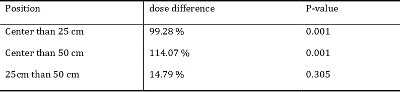
showed the neutron dose equivalent difference were significant for distance of center with 25 cm and center with 50 cm. But this difference was not significant for distance of 25 cm with 50 cm. The results are presented in tables of 3 to 6.
DISCUSSION AND CONCLUSIONS
Many studies were done on the effect of field size on the neutron contamination. Some of them were shown additive trend (Hashemi et al., 2008), and the others were agreed with decreasing trend (Ghavami et al., 2010). Linac kind, beam definer dosimeter and the simulation methods are parameters could influence on the result. But some studies were done on the same linac, set up and modifier but different trend. Our result was indicated the increasing trend in total. With changing field size from 5×5 cm2 to 10×10 cm2 (a four fold increasing in area) we saw a meaningful change in the neutron dose equivalent but the same increment in area from 10×10 cm2 to 20×20 cm2 had a meaningless change in the neutron dose equivalent. On the other hand, after 10×10 cm2 field size, the effect of field size on neutron contamination is significant that the variation to be large enough (from 10×10 cm2 to 30×30 cm2). According to these result field sizes had no absolute additive or deductive effect on field size but this effect depend on the amount of the collimator opening. In small field, the effect of field size on contaminated neutron is significant. It is may be because of neutron flux that produced from linac head components before secondary jaws especially flattening and differentiated filters. These results are partially agree with Kim (Kim et al., 2007). Mesbahi demonstrated that a free flattening filter linac with the same set up and collimator aperture has less neutron fluence than a linac with flatteing filter. This status is more pronounce for small field size (Mesbahi, 2009). If the neutron flux produced in the head component was uniform through the field we witness a perfect increasing trend in neutron contamination with increasing field size. It is may be because of neutron flux that produced from linac head components before secondary jaws especially flattening and differentiated filters. Because of the shape of these filters (cone shape) the amount of mass in the way of the photons increase slowly with increasing field size and as a result we were onlooker of the rapid production of contaminated neutron with size. After 10×10 cm2 field size, it needs more increasing to show significant variation. The production of the neutron contamination outside the field is isotropic and filed size has no effect on it that our results approve it. With increasing field size the edge of field became nearer, from 22.5 cm to 10 cm, to first the position of detectors (25 cm) but has no effect on neutron flux outside the field. Also, because the neutron dose decreases with increasing distance from the canter, this can be realized that most of the produced fast neutrons in the head of accelerator are forward, and their risks decreases in far distances; Because of successive scattering with bed, patient, room
walls and other objects in the room, their energies lost and convert to thermal neutrons.
The unwanted neutron mainly produce in linac head in forward and isotropic way. The forward neutron chiefly yield in photon field and rely on the flattening and differentiated filter material and shape. Because of cone shape of them the variation of contaminated neutron flux with field size is not linear and uniform. But the approving needs measurement with small increments in field size and Monte Carlo simulation.
ACKNOWLEDGEMENTS
This paper outcome a part from M.Sc project carried out at Semnan University Medical Sciences. We would like to express our special thanks to Semnan University Medical Sciences and the Radiotherapy Department of Kermanshah Imam Reza Hospital and Karaj Institute of Nuclear Science and Technology, as well as Atomic Energy Organization of Iran for providing financial support and clinical and technical assistance required for performing this research.
REFERENCES
Al-ghamdi, H., Fazal ur, R., Al-Jarallah, M. I., Maalej, N. (2008). Photoneutron Intensity Variation with Field Size Around Radiotherapy Linear.
Facure, A., Falcao, R. C., Silva, A. X., Crispim, V. R., Vitorelli, J. C. (2005). A study of neutron spectra from medical linear accelerators. Appl Radiat Isot, 62(1), 69-72.
Ghavami, S.M., Mesbahi, A., Mohammadi, E. (2010). The impact of automatic wedge filter on photoneutron and photon spectra of an 18-MV photon beam. Radiation Protection Dosimetry, 138(2), 123-128.
Ghiasi, H., Mesbahi, A. (2010). Monte Carlo characterization of photoneutrons in the radiation therapy with high energy photons: a Comparison between simplified and full Monte Carlo models. Iran. J. Radiat. Res., 8 (3), 187-193.
Hashemi, S. M., Hashemi-Malayeri, B., Raisali, G., Shokrani, P., Sharafi, A. (2007). A study of the photoneutron dose equivalent resulting from a Saturne 20 medical linac using Monte Carlo method. NUKLEONIKA, 52(1), 39−43.
Kim, H. S., Park, Y. H., Koo, B. C., Kwon, J. W., Lee, J. S., Choi, H. S. (2007). Evaluation of the photoneutron field produced in a medical linear accelerator. Radiat Prot Dosimetry, 123(3), 323-328.
Mao, X. S., Kase, K. R., Liu, J. C., Nelson, W. R., Kleck, J. H., Johnsen, S. (1997). Neutron sources in the Varian Clinac 2100C/2300C medical accelerator calculated by the EGS4 code.. Health Phys, 72(4), 524-529.
Mesbahi, A. (2009). A Monte Carlo study on neutron and electron contamination of an unflattened 18-MV photon beam. Applied Radiation and Isotopes, 67(1), 55-60.
Mesbahi, A., Ghiasi, H., Mahdavi, S. R. (2010a). Photoneutron and capture gamma dose equivalent for different room and maze layouts in radiation therapy. Radiat Prot Dosimetry, 140(3), 242-249.
Mesbahi, A., Keshtkar, A., Mohammadi, E., Mohammadzadeh, M. (2010b). Effect of wedge filter and field size on photoneutron dose equivalent for an 18MV photon beam of a medical linear accelerator. Applied Radiation and Isotopes, 68(1), 84-89.
Ongaro, C., Zanini, A., Nastasi, U., Rodenas, J., Ottaviano, G., Manfredotti, C. (2001). Analysis of photoneutron spectra produced in medical accelerators (vol 45, pg L55, 2000). Physics in Medicine and Biology, 46(3), 897-897.
Rudd, P. J., Prior, D., Austin-Smith, S. (2007). Neutron contamination of 10 MV X-rays: its relevance to treatment room door and maze design. Br J Radiol, 80(954), 469-475.
Sohrabi, M., & Mostofizadeh, A. (1999). Measurement of photoneutron doses in and out of high-energy X-ray beam of a SATURNE-20 medical linear accelerator by ECE polycarbonate detectors. Radiat Meas, 31(1), 479-482.
Vega-Carrillo, H., & Baltazar-Raigosa, A. (2011). Photoneutron spectra around an 18 MV LINAC. Journal of Radioanalytical and Nuclear Chemistry, 287(1), 323-327.
Zabihzadeh, M., Ay, M. R., Allahverdi, M., Mesbahi, A., Mahdavi, S. R., Shahriari, M. (2009). Monte Carlo estimation of photoneutrons contamination from high-energy X-ray medical accelerators in treatment room and maze: a simplified model. Radiat Prot Dosimetry, 135(1), 21-32.
Fig. 1. View of the examined Elekta linac
Fig. 3. Comparison of neutron dose equivalent for different field sizes
Table 1.
The neutron dose equivalent (mSv/Gy) in the center, 25cm and 50cm far from center of open photon field.
distance (cm)
Field Size
5×5 10×10 20×20 30×30
0 1.33 ± 0.04 1.70 ± 0.07 1.92 ± 0.06 2.13 ± 0.07
25 0.87 ± 0.06 0.98 ± 0.06 1.05 ± 0.06 1.13 ± 0.07
50 0.82 ± 0.04 0.85 ± 0.06 0.96 ± 0.05 0.99 ± 0.07
* Means ± SE
Table 2.
The percentage of neutron dose equivalent difference between the field sizes in center of field
First field (cm2) Second field (cm2)
Percentage change in dose
(first – second)
P-value
5×5 10×10 -39.59% 0.001
5×5 20×20 -57.65% 0.001
5×5 30×30 -76.50% 0.001
10×10 20×20 -18.07% 0.156
20×20 30×30 -18.85% 0.091
30×30 10×10 36.91% 0.001
Table 3.
The percentage of difference between measuremented neutron dose equivalent in different positions in the field of 5 ×5 cm2
Position dose difference P-value
Center than 25 cm 47.13% 0.001
Center than 50 cm 52.35% 0.001
Table 4.
The percentage of difference between measuremented neutron dose equivalent in different positions in the field of 10 ×10 cm2
Position dose difference P-value
Center than 25 cm 74.55 % 0.001
Center than 50 cm 87.57 % 0.001
25cm than 50 cm 13.02 % 0.262
Table 5.
The percentage of difference between measuremented neutron dose equivalent in different positions in the field of 20 ×20 cm2
Position dose difference P-value
Center than 25 cm 84.04 % 0.001
Center than 50 cm 100.46 % 0.001
25cm than 50 cm 16.42 % 0.116
Table 6.
The percentage of difference between measuremented neutron dose equivalent in different positions in the field of 30 ×30 cm2
Position dose difference P-value
Center than 25 cm 99.28 % 0.001
Center than 50 cm 114.07 % 0.001