Analysis of Normal and Malignant Human B Cell
Growth Using Gene Transfer
Michael Joachim Buschle
Being a thesis submitted for the degree of DOCTOR OF PHILOSOPHY
to the Faculty of Medicine University of London
October 1992
Royal Free Hospital School of Medicine
m e d i c a l l i b r a r y, ROYÆ! ' k-cE HOSPI TA L
All rights reserved
INFO RM A TIO N TO ALL U SER S
The quality of this reproduction is dependent upon the quality of the copy submitted.
In the unlikely event that the author did not send a complete manuscript
and there are missing pages, these will be noted. Also, if material had to be removed, a note will indicate the deletion.
uest.
ProQuest U540555
Published by ProQuest LLC(2016). Copyright of the Dissertation is held by the Author.
All rights reserved.
This work is protected against unauthorized copying under Title 17, United States Code. Microform Edition © ProQuest LLC.
ProQuest LLC
789 East Eisenhower Parkway P.O. Box 1346
The aim of this thesis was to investigate the effects of individual oncogenes on human B lymphocytes. To permit these studies, a gene transfer and expression technique for normal B lymphocytes and cells from patients with B cell chronic lymphocytic leukaemia (B-CLL) was developed. O f the numerous techniques tested, only electroporation proved to have reasonable efficiency and in due course, a powerful promoter/enhancer element containing cytomegalovirus and human T cell leukaemia virus I sequences was identified. Several eukaryotic expression vectors based on this promoter/enhancer element were constructed: a cassette vector was designed and used to subclone the oncogenes c-myc, v-Ha-ras, v-fos, v-raf and v-mos. As the mahgnant B lymphocytes B-CLL constitute a homogeneous, monoclonal B cell population with large number of cells available for experiments, gene transfer studies were initiated with material derived from patients with this disease. Transfection of B-CLL samples with the constructs alone or in combination did not result in cell transformation, however, prolonged cell survival was consistently observed in c-myc transfected samples. Subsequent analysis revealed that interferon-y was secreted at high levels following c-myc gene transfer.
Addition of recombinant IFN-y to cultured B-CLL cells demonstrated that this
cytokine promotes B-CLL cell survival by preventing programmed cell death, or apoptosis, and may therefore be responsible for the slow and progressive accumulation of the Go arrested malignant B lymphocytes observed in vivo. Furthermore, IFN-y was shown to be produced by B-CLL cells in an autocrine marmer by analysis of IFN-y transcripts and double colour flow cytometry. Synthesis of IFN-y by B-CLL cells may also explain why
high levels of this cytokine were found in serum samples from patients with this disease. In conclusion, IFN-y may be an important growth factor contributing to the development of this common form of leukaemia. Inhibition of IFN-y mediated effects may lead to novel
Table o f Contents...3
List of Figures... 7
Abbreviations...9
Acknowledgements... 11
C hapter 1: General introduction._____________________________________ The Immune System... 13
Human B Lymphocytes...16
Surface molecules of B lymphocytes 19 CD5^ B lymphocytes may represent a separate B cell lineage 21 Immunoglobulin gene rearrangement and expression 22 Immunoglobulin isotype switching 26 Cytokines involved in human B cell growth and differentiation 29 B ceU activation at the molecular level 33 B Cell Malignancies... 35
Chromosomal abnormalities and genes involved in B cell malignancies 36 B Cell Chronic Lymphocytic Leukaemia (B-CLL)... 39
Clinical features 39 Cellular charactaistics of B-CLL 40 B-CLL cells and their relation to CD5^ B lymphocytes 40 Chromosomal abnormalities and oncogenes in B-CLL 41 B-CLL response to recombinant cytokines 42 Proto-oncogene expression in B-CLL cells following mitogenic activation44 Aims of TTiis Project...46
Chapter 2: Gene Transfer Into Human B Cell Progenitors, Normal B ____________Lymphocytes and B-CLL Cells.____________________________ 4 7 Introduction...48
Materials and Methods... 50
B-CLL patients 50
Evaluation of purity 50
In vitro stimulation experiments 51
Nucleotide incorporation assay 53
Electroporation 53
CAT assays 54
Double colour analysis 55
Simultaneous detection of 6-galactosidase activity and cell surface
antigens 55
Double colour immunofluorescence staining of pCMV-LTR-BZLF1
transfected cells 56
Expression vectors 57
Results...58
Techniques of transfection 58
Characterization of transfected tonsillar B cells 59
Gene transfer into bone marrow B cell progenitors 59
Transfection of B-CLL cells 61
Discussion... 79
Chapter 3: Construction of Eukaryotic Expression Vectors for Gene Transfer
____________into Primary B Cells.______________________________________8 3
Introduction... 84
Selection of genes for transfection of primary B lymphocytes 84
Gene expression systems 90
Materials and Methods... 95
DNA sources 95
Large scale preparation of plasmids 96
Digestion of DNA with restriction enzymes 96
Dephosphorylation of 5' termini 96
Creation of blunt ends 97
Phenol/chloroform extraction and ethanol precipitation of DNA 97
Isolation of DNA from agarose gels 98
Ligation of DNA fragments 98
Preparation of competent cells and transformation of R coll with plasmids99
Detection of recombinant plasmids 99
Evaluation of recombinant DNA 100
Construction of the CM V-LTR cassette vector pCMV-LTR-poly 104
Design of the reporter gene containing plasmid pCMV-LTR-BZLF1 105
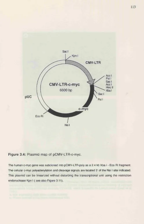
Construction of pCMV-LTR-myc 106
Construction of pCMV-LTR-Ha-ras 106
Design of pCMV-LTR-fos 107
Construction of pCMV-LTR-raf 108
Design of pCMV-LTR-mos 109
Conclusions... 122
Chapter 4: Effects of Oncogene Transfection on the Monoclonal B
____________Lymphocytes of B-CLL__________________________________ 1 2 3
Introduction... 124 Materials and Methods... 127
B-CLL patients 127
Electroporation 127
Expression vectors 128
Detamination of transfection efficiency 129
Feeder layer cultures for transformation experiments 129
Viability counts after c-myc transfection 131
Cell proliferation analysis 131
Analysis of phenotypic changes after c-myc transfection 132
RNA extraction from transfected cells 133
Slot blot analysis of RNA 135
Radiolabeling of probes by nick-translation 136
DNA probes 137
Measurement of soluble cytokines by ELISA 137
Results... 138
Transfection of B-CLL cells with oncogene constructs 138
Detailed analysis of the effects of transfected c-myc 140
Transfected c-myc prolongs survival of cultured B-CLL cells 140
C-myc transfection induces phenotypic changes in B-CLL cells 140
C-myc gene transfer induces EFN-y synthesis 141
C-myc transfection results in down-regulation of IL -10 message 142
Introduction...155 Materials and Methods... 157
Samples 157
Cell culture 158
Measurement of soluble 158
Analysis of apoptosis 159
Determination of ceU numbers 160
Ki-67 staining 161
Northern blot analysis 161
Radiolabeüng of DNA inserts by random priming 164
DNA probes 165
Detection of intracellular IFN-y 165
Results... 167
IFN-y inhibits apoptosis of cultured B-CLL cells 167
IFN-y is present in B-CLL serum samples 167
B-CLL cells may synthesize IFN-y 168
Discussion... 176
C hapter 6: Conclusions. 1 7 9
References quoted ... 184
Publications arising from this thesis and related work...207
Figure 1.2
Figure 1.3
Figure 1.4
Figure 2.1:
Figure 2.2:
Figure 2.3:
Figure 2.4:
Figure 2.5 a:
Figure 2.5 b:
Figure 2.6:
Figure 2.7 a:
Figure 2.7 b:
Figure 2.7 c:
Figure 2.8:
Figure 2.9:
Figure 2.10:
Figure 2.11:
Figure 2.12:
Figure 3.1:
Figure 3.2:
Figure 3.3:
Figure 3.4:
Figure 3.5:
Figure 3.6:
Diagram of B cell development...18
Schematic diagram of Ig heavy chain rearrangement... 25
Immunoglobulin isotype switching... 28
Eukaryotic expression vectors used for transfection studies in chapter 2 ...64
Voltage dependence of transfection efficiency... 65
Photomicrographs of Percoll separated tonsil B lymphocytes... 66
CAT expression of transfected Percoll separated B lymphocytes ..67
Bone marrow mononuclear cells transfected with pCM V-P... 68
Double colour immunofluorescence staining of TdT positive bone marrow B cell progenitors after BZLFl transfection... 69
CAT expression of transfected B-CLL cells... 70
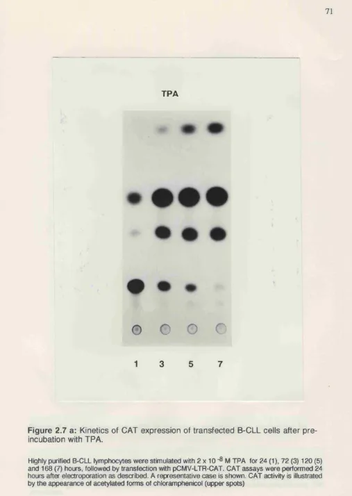
Kinetics of CAT expression of transfected B-CLL cells after pre incubation with TPA... 71
Delayed induction of maximum reporter gene expression by TNF.72 CAT expression of uninduced B-CLL cells after transfection... 73
Activation of B-CLL cells after transfection does not allow reporter gene expression...74
CAT expression and nucleotide incorporation of TPA activated B-CLL cells... 75
Influence of DNA concentration and cuvette volume on CAT expression...76
Double colour immunofluorescence staining of B-CLL cells transfected with pCMV-LTR-BZLFl... 77
Promoter usage of B-CLL cells... 78
Construction of pCM V-LTR-poly... 110
Restriction maps and gel-electrophoretic analysis of plasmids pCMV-LTR-poly andpCMV-LTRpoly A ... I l l Map of plasmid pCM V-LTR-BZLFl... 112
Plasmid map pCMV-LTR-c-myc...113
Myc expression of transfected N1H3T3 fibroblasts... 114
Figure 3.9: Plasmid map of pC M V-LTR -raf...118 Figure 3.10: Plasmid map of pCMV-LTR-mos...119
Figure 3.11: Restriction analysis of plasmids c-myc,
pCMV-LTR-Ha-ras and pC M V-LTR-BZLF 1... 120
Figure 3.12: Restriction analysis of plasmids pCMV-LTR-fos, pCMV-LTR-raf
and pCMV-LTR-mos...121
Figure 4.1: p-galactosidase expression of B-CLL cells 24 hours after gene transfer... 143
Figure 4.2: B-CLL cells cultured on bone marrow stroma for 4 weeks after gene
transfer... 144 Figure 4.3: Transfected c-myc promotes survival of B-CLL cells...145 Figure 4.4: Histograms of c-myc and control transfected cells stained with
CD19, CD5 and ICAM-1 antibodies...146 Figure 4.5: Histograms of B-CLL cells transfected with c-myc and a control
plasmid stained with LFA-1 antibodies...147
Figure 4.6: C-myc transfection of B-CLL cells induces IFN-y synthesis 148
Figure 4.7: C-myc transfected B-CLL cells down-regulate IL-10 message... 149
Figure 5.1 : Apoptotic cell death of B-CLL cells is prevented by IFN -y 169
Figure 5J2: IFN-y inhibits DNA degradation in cultured B-CLL cells 170
Figure 5.3: Analysis of apoptosis by flow cytometry... 171 Figure 5.4: Survival of B-CLL cells after 5 days in culture in the presence or
absence of 100 U/ml IFN -y... 172 Figure 5.5: Sera from B-CLL patients contain IFN-y...173
Figure 5.6: Activated B-CLL cells synthesize high levels of IFN-y m RNA.. 174
APAAP: alkaline phosphatase anti-alkaline phosphatase
A P I : activator protein 1
ATP: adenosine 5'-triphosphate
B -C LL: B-cell chronic lymphocytic leukaemia
bcr: breakpoint cluster region
P-gal: beta-galactosidase
bp: base pairs
BSA: bovine serum albumin
B Z L F l: Bam H I Z leftward reading frame 1
cAMP: cyclic adenosine monophosphate
CAT: chloramphenicol acetyltransferase
CD: cluster of differentiation
C M L: chronic myeloid leukaemia
C M V: cytomegalovirus
cDNA: complementary DNA
c-onc: cellular (proto)-oncogene
CSF: colony stimulating factor
CTP: cytidine 5-triphosphate
D N A : deoxyribonucleic acid
DNase: deoxyribonuclease
D TT: dithiothreitol
EBV: Epstein-Barr virus
£ . cob: Escherichia cob
E LIS A : enzyme linked immunosorbent assay
Ep: immunoglobulin heavy chain enhancer
EDTA: ethylene diamine tetraacetate
FCS: foetal calf serum
F IT C : fluorescein isothiocyanate
GAP: GTPase activating protein
GDP: guanosine 5'-diphosphate
GTP: guanosine 5'-triphosphate
Ha-ras: Harvey-ras
H T L V I: human T ceU leukaemia virus I
IFN:
Ig:IL:
kb:
Ki-ras:
LFA:
LTR:
MHC:
mRNA:
MSV:
NK cells:
NZB mice:
PBS:
P h i:
RB:
RITC:
RNA:
RNase:
RSS:
SDS:
SV40:
TdT:
TNF;
TPA:
TTP:
U:
UTP:
v-onc:
interferon immunoglobulin interleukin kilo bases Kirsten-rasleucocyte fimction associated antigen long terminal repeat
major histocompatibility complex messenger RNA
mouse sarcoma virus natural killer cells
New Zealand Black mice phosphate buffered saline Philadelphia chromosome retinoblastoma gene rhodamine isothiocyanate ribonucleic acid
libonuclease
recombinase signal sequence sodium dodecyl sulfate simian virus 40
terminal deoxynucleotidyl transferase tumour necrosis factor
12-Otetradecanoylphorbol 13-acetate thymidine 5'-triphosphate
units
Acknowledgements:
I am profoundly grateful to Professor Malcolm Brenner for giving me the opportunity to initiate the work on this thesis, for constant support, and most of all, for his encouragement and advice throughout its course.
I am most grateful to Professor A. Victor Hoffbrand for supervising me during the second half of this thesis. Without his support and advice I would not have been able to carry out the studies presented here.
I would like to thank Dr. Dario Campana for sharing with me his wide knowledge of cellular immunology and the biology of human leukaemias. I am indebted to Dr. Cliona Rooney for her support and many helpful discussions. I am grateful to Dr. Paul Farrell for providing me with laboratory space and for valuable advice. I am further grateful to Dr. Hans Drexler for many helpful discussion and for his constant support.
I would also like to thank Dr. Concha Bello-Fernandez for moral support and tolerating me in times of despair, Elaine Coustan-Smith, Atsushi Manabe, Donna Rill, Dr. Shulamit Katzav and Dr. John Cleveland for technical advice and for their pleasant company. Thanks to Drs. Mark Larché, Peter Dias and Sally Sarawar for most valuable proof-reading and for company during "mental relaxation" after work.
The Immune System:
Although mature peripheral blood cells are extremely specialized with no
morphological resemblance between different cell types, all cells, including the constituents
of the immune system, are derived from a common set of haemopoietic stem cells. These
pluripotent stem cells are the initial cells in a cascade of sequential differentiation and
proliferation events giving rise to committed progenitor cells, which in turn proliferate and
differentiate, eventually generating mature cells (Dexter and Spooncer 1987).
Haemopoiesis including B lymphopoiesis in adults takes place in the bone marrow. The
majority of T lymphocytes mature in the microenvironment of the thymus (Ezine et al.
1984, Ikuta et al. 1992, Lepault and Weissman 1981) (Figure 1.1).
pluripotent stem cell
bone marrow
com m ited progenitor ceils thymus
□
□
□
□
c□
□
Mega karyocyteF ig u re 1.1: S ch e m e of haem opoiesis.
( ^ ) T lymphocyte
( ^ ) B lymphocyte Monocyte Erythrocytes Platelets Neutrophil Eosinophil Basophil
secreting antigen specific immunoglobulin (Ig), but can also act as antigen presenting cells (Chestnut and Grey 1981, Chestnut and Grey 1986, Lanzavecchia 1985).
Human B Lymphocytes:
B lymphocytes synthesize immunoglobulins which bind to microorganisms, toxins, proteins, carbohydrates and other potentially harmful antigens. In addition, B cells are able to present antigen to T helper cells and interact with other cell types by cell-cell contact or through cytokines. In peripheral blood, B lymphocytes represent 5% - 15% of the circulating lymphoid cells, but in peripheral lymphoid organs approximately 50% of the cells are B lymphocytes.
Antibodies are Y-shaped glycoproteins consisting of 2 identical heavy and light Ig chains held together by disulphide bonds (Burton 1987). Ig chains are subdivided into constant and variable regions. Heavy chain genes contain information for variable regions of the Ig molecule binding antigen (including the complementarity determining regions), and several constant region domains which are responsible for effector functions such as transmembrane signalling, complement activation or binding of Fc receptors (Burton
1987). light chain genes code for only one variable and one constant region. Both, light and heavy chain variable regions contribute to antigen binding. Two Ig light chain classes (k and X) have been described, and 9 different heavy chain constant regions are known. The constant region of the 9 heavy chain molecules (p, Ô, Y1» Y2. Y3, Y4, « i, «2, e) determines the isotype of antibodies, independent of the type of light chain associated. Each antibody isotype has different biological properties (Jeske and Capra 1984): IgM antibodies, which are the first immunoglobulins synthesized, activate complement most efficiently. IgG molecules (IgGi-IgG4) bind to receptors of phagocytic cells, facilitating phagocytic elimination of pathogens. IgA binds to antigen along mucous membranes. The resulting antigen-IgA complexes are removed by selective transport mechanisms across the mucous membranes. Binding of IgE molecules to the surface receptor of eosinophils and basophils triggers the release of histamines and lytic enzymes to destroy parasitic invaders.
of differentiation are initiated by multiple cell division, but the proliferative activity of the cells decreases towards the end of each phase (Figure 1.2). The first phase starts with the differentiation of B cell progenitor cells in the bone marrow until they reach the stage of the mature, resting B lymphocyte. This process is antigen independent, generating a large number of B cells which are diverse with regard to their antigen receptors. Although it is likely that pre-B cells possess their own differentiation program, there is evidence that surrounding bone marrow stromal cells influence B cell maturation (Kincade et al. 1989). During these processes, the cells first rearrange their immunoglobulin heavy chain genes followed by light chain genes (see below). Successfully rearranged Ig genes are translated into protein and can first be detected in the cytoplasm. Finally, the B cells express surface IgM and IgD molecules becoming mature, resting B lymphocytes. Surface lgM+ and lgD+ positive cells leave the bone marrow and migrate to the spleen or other secondary lymphoid organs, but may also return to the bone marrow (Brenner et al. 1981).
72 37 dim
34
20 dim CjLi
proliferation
IgM
IgM
peripheral blood
bone marrow
II peripheral | | IIlymphoid | | organs |
I
antigent
proliferation
antigen-independent antigen-dependent
F ig u re 1.2: D iagram of B cell developm ent.
Surface molecules o f B lymphocytes:
Differentiating B cells sequentially express a number of specific surface antigens depending on the stage of maturation (Figure 1.2). Monoclonal antibodies recognizing such proteins are available and have been used to study B cell development (Campana et al. 1985, Zola 1987). In addition, comparison of antigens expressed on B leukaemic cells, which are arrested at specific stages of differentiation, with their normal counterparts helped to accumulate information about the development of the B cell compartment (Greaves 1986). Depending on the antigen bound, monoclonal antibodies to leukocyte antigens have been grouped into 'clusters of differentiation' (CD) in the course of the four International Workshops on Leukocyte Differentiation Antigens held so far (Knapp et al. 1989). The function of B cell antigens has been reviewed extensively (Clark and Lane 1991, Zola 1987). In the following, only a selection of clustered antigens will be described.
CD19, as the most broadly expressed surface marker for B cells, is commonly used as a pan-B cell marker. This antigen is exclusively found on the surface of B lymphocytes and is expressed by virtually all B cell types except plasma cells. The gene for the CD 19 antigen has been cloned and sequenced (Stamenkovic and Seed 1988, Tedder and Isaacs 1989). CD 19 is a glycosylated integral transmembrane protein and belongs to the immunoglobulin superfamily. Monoclonal antibodies to CD 19 inhibit anti-lg mediated activation as well as DNA synthesis of B lymphocytes (Barrett et al. 1990, Pezzutto et al.
1987).
The CD20 cluster of antibodies defines a membrane embedded phosphoprotein.
CD20 expression is restricted to cells of the B lineage, but the antigen appears at later stages of B cell differentiation than CD 19 (Figure 1.2). CD20 tends to be lost when B cells are activated. Antibodies to CD20 appear to pre-activate B lymphocytes (Clark and Ledbetter 1986a).
lida and Nadler 1983) and it is a ligand for CD23 (Aubry et al. 1992). CD21 expression is confined to B lymphocytes, but is found more mature B cells than CD 19 and CD20 (Figure 1.2). This antigen may be involved in T cell mediated B lymphocyte activation, since CD21 antibodies induce proliferation in the presence of T cells (Clark and Ledbetter 1986b).
CD22 antibodies react with a pair of highly glycosylated proteins. Sequence analysis of the CD22 gene revealed that it most likely codes for a cellular adhesion molecule (Stamenkovic and Seed 1990). The antigen is found on the surface of most (75%) peripheral blood B lymphocytes. Functional studies demonstrated that CD22 antibodies enhance anti-Ig induced DNA synthesis (Pezzutto et al. 1988).
CD23, the low-affinity IgE receptor, is expressed on a variety of cells including B cells, some T cells and monocytes (Delespesse et al. 1991). The extracellular part of this transmembrane glycoprotein can be cleaved into different soluble forms which may act as an autocrine B cell growth factor (Gordon et al. 1989) or may prevent cell death of germinal centre B lymphocytes (Liu et al. 1991). (2D23 expression is restricted to mature B cells and is not found on bone marrow B lymphocytes. Cytokines influence CD23 expression: while IL-4 displays a stimulatory effect, interferon-y (IFN-y) down-regulates CD23 in normal B cells (Delespesse et al. 1991).
CD5^ B lymphocytes may represent a separate B cell lineage:
Antibodies in the CD5 cluster of differentiation recognize a 67 kD glycoprotein, which is present on the surface of all T cells. CD5 is also expressed by the malignant B lymphocytes of B cell chronic lymphocytic leukaemia (B-CLL) and a subset of normal B cells (Gadol and Ault 1986). Recently, a B cell antigen, CD72, has been identified as the ligand of CD5 (Van de Velde et al. 1991). It has been proposed that CD5/CD72 interaction may be important in communication between antigen presenting B lymphocytes and T helper cells (DeFranco 1991).
Small numbers of normal CD5Vsurface IgM+ B lymphocytes were first identified in cells derived from lymph nodes (Caligaris-Cappio et al. 1982). In adults CD5+ B lymphocytes represent approximately 10% of the total peripheral blood B cell population (Freedman et al. 1987, Hardy et al. 1987), while a much larger proportion of CD5+ B lymphocytes is found in foetal tissue (Antin et al. 1986, Freedman et al. 1987) and following bone marrow transplantation (Ault et al. 1985). Increased numbers of CD5^ B cells are also found in patients with autoimmune disease (Hardy et al. 1987, Taniguchi et al. 1987).
cells. Possible functions of CD5^ B cells will be discussed in the context of B-CLL (see below).
Immunoglobulin gene rearrangement and expression.
The most important molecules expressed on the surface of B lymphocytes are Igs, which could be regarded as antigen receptors. The work of Tonegawa led to the discovery that B lymphocytes rearrange separate Ig gene segments and thereby enable B cells to synthesize antibodies against virtually every natural, and even artificial antigens (Tonegawa 1983).
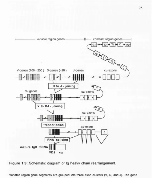
The segments coding for Ig heavy and the two light chains are located on three different chromosomes: heavy chain genes map to chromosome 14, K-light chain genes to chromosome 2 and X-light chain genes to chromosome 22. Like all eukaryotic genes, sequences coding for Igs are organized in coding exons separated by intervening, non coding introns. Ig exons for heavy and light chain genes are clustered in related groups spread along the chromosomes (Figure 1.3). Segments coding for Ig variable regions are located 5' of constant region exons. The individual gene segments coding for Ig variable regions are contained in three exon clusters in the case of the heavy chain locus, and in two clusters on the light chain locus.
pairs (bp). The length of the spacer is recognized by the recombinase: only RSS with 12 and 23 bp spacer can be joined (Tonegawa 1983). The reaction is initiated by joining of a
Dh segment with a Jh exon on both chromosomes (Alt et al. 1986). D y segments are flanked by two RSS containing 12 bp spacers, but V y and Jy elements have a single RSS with 23 bp spacer at their 3' and 5' end, respectively. Thus, assembly of a potentially functional V y D y J y heavy chain gene must be mediated by joining of the Jy and V y
elements to D y segments. V y to D y rearrangements have not been observed (Yancopoulos and Alt 1986). At present, the factors responsible for Ig gene assembly are not known, but recently, two "recombinase activating genes" (RAG 1 and RAG 2) which display recombinase activity have been cloned and partially characterized (Schatz et al. 1992). Joining of individual segments is not precise. Only one of three rearrangements will be in frame and bases are sometimes deleted, or nucleotides added by the enzyme terminal deoxynucleotidyl transferase (TdT). This so called N-region addition further increases diversity (Tonegawa 1983).
The translocation of segments changes the physical relation of two gene regulatory sequences. A promoter area located 5' of every V y segment comes in closer proximity to a enhancer segment which is contained in the intron between Jy and constant region exons (Figure 1.3). Transcription factors bind to the gene regulatory regions and initiate synthesis of RNA (Calame 1985). Provided that V y D y and Jy exons have been assembled correctly and in frame and do not contain premature stop codons, the nuclear RNA is spliced and translated into protein. Ig heavy chains then accumulate in the endoplasmic reticulum (Figure 1.2).
Productive V y - D y J y joining on one chromosome also triggers a feedback mechanism to prevent rearrangement of heavy chain variable sequences on the other allele. This control mechanism, termed allelic exclusion, ensures that an individual B cell will synthesize only one antigen specific Ig molecule (Alt et al. 1986).
that Ig light chain variable regions do not contain D segments. Therefore, only V l and J l elements are recombined. In accordance with the 12/23 rule, V l and J l elements are flanked by 12 bp or 23 bp spacers (Tonegawa 1983). Initially, V l and J l k light chain
genes on chromosome 2 are joined and after productive rearrangement transcribed and translated. Failure to achieve a productive V Jk rearrangement on one or the other
chromosome results in the assembly of X genes on chromosome 22. As in the case of
heavy chain gene rearrangement, a feedback mechanism prevents assembly of more than one light chain variable region gene (Alt et al. 1986). Successful rearrangement results in the production of light chains in the endoplasmic reticulum. Formation of disulphide bonds between the two Ig chains leads to the dislocation of an endoplasmic reticulum retention protein (Hendershot et al. 1987). Completed Ig molecules are transported to the cell
membrane, resulting in aphenotypically identifiable B cell (Figure 1.2).
variable region genes constant region genes(X2
-V-genes (100 - 200 ) D-genes («20 ) J-genes
D to J - joining
V- genes cu-exons
V to DJ - joining
cu-exons
tra n s c rip tio n
cu-exons
RNA splicing
cjLi-exons
DDOO
mature IgH mRNA
VDJ CM
F ig u re 1.3: S chem atic diagram of Ig heavy chain rearrangem ent.
Although gene rearrangements create an extremely diverse B cell pool, synthesizing antibodies to as many as 10^ or more different antigens at a time, this process also leads to a substantial loss of pre-B cells during differentiation. As mentioned above, assembly of Ig variable region gene segments is not precise. In fact, statistical analysis lead to the conclusion that the majority of cells (>90%) w ill fail to rearrange their Ig genes successfully (Cohen and Duke 1992). The destiny of these non-productive B cells is not known at present. Recently, it has been suggested, that these cells activate a suicidal mechanism (apoptosis) (Cohen and Duke 1992) and are quickly removed from the bone marrow by scavenger cells such as macrophages. In addition, B lymphocytes circulating in the secondary lymphoid organs have also been shown to die by apoptosis unless rescued by stimulation with antigen (Liu et al. 1989). Failure to activate apoptosis following unsuccessful gene rearrangement could result in the abnormal accumulation of B lymphocytes (see below).
Immunoglobulin isotype switching:
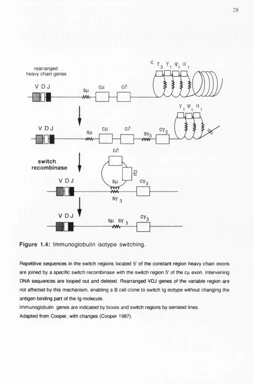
During development, B cells first express IgM on their surface followed by co expression of IgD (Figure 1.2). At later stages of B cell differentiation a clone can switch Ig synthesis from IgM to different Ig heavy chain classes (isotypes) without changing the antigen binding part of the molecule (Figure 1.4). Two molecular mechanisms contribute to the switching of Ig isotypes (Shimitzu and Honjo 1984): IgM and IgD expressing B cells generate long nuclear VDJ-Cp-Cd transcripts. The RNA is spliced into VDJ-Cp or VDJ-Cd by alternative splicing mechanisms and translated into protein.
s w itch re c o m b in a s e
V u , ^ a
V D J
i
caV D J
MM
V D J
1 A A A
1 r > n ^
I
SM sy 3SY 3
—vw<---F ig u re 1.4: Im m unoglobulin isotype switching.
Repetitive sequences In the switch regions located 5' of the constant region heavy chain exons are joined by a specific switch recombinase with the switch region 5' of the c^ exon. Intervening DNA sequences are looped out and deleted. Rearranged VDJ genes of the variable region are not affected by this mechanism, enabling a B cell clone to switch Ig isotype without changing the antigen binding part of the Ig molecule.
Cytokines involved in human B cell growth and differentiation:
Much information about the role of individual cytokines on B cells has been obtained by in vitro studies using highly purified B lymphocytes. However, the in vitro situation is much less complex compared to the environment encountered by B cells in vivo, where B cells are regulated by a network type interaction of different cell types. Cell cell contact and soluble factors synthesized by a large variety of cells mediate B lymphocyte differentiation and the immune response (O'Garra et al. 1988).
Molecular analysis revealed, that cytokines in general are glycosylated proteins of low molecular weight. Binding of cytokines to their respective receptors expressed on the surface of cells triggers intracellular mechanisms which in turn lead to activation of genes and finally result in altered cell function. Growth factors can activate cells through autocrine (the target cells themselves synthesize the stimulating cytokine), paracrine (surrounding cells produce the growth factor) or endocrine (the cytokine is produced by a distant organ) stimulation.
Cloning of cytokine genes and production of recombinant growth factors has helped to define B cell growth factors and their function in B lymphocytes. Initially, growth factor activity was believed to be cell lineage restricted. More recent studies, however, demonstrated that cytokines have multiple effects on cells of different lineages. Furthermore, it was found that any one cytokine may have different effects, depending on the stage of differentiation or what other signals the cell has received (Gordon and Guy 1987, O'Garra et al. 1988). In some instances, in vitro studies have resulted in conflicting reports about the role of certain cytokines in B cells, which may be due to different assay systems applied, or the methods used to purity B cells (Gordon and Guy 1987, O'Garra et al. 1988). There follows a brief description of the function of the most relevant cytokines for human B cells. The activity of recombinant molecules is given.
Two distinct molecular forms of IL-1 (IL -la and IL -lp ) are known. Both
responses and is synthesized by a variety of cells including T cells, B cells, macrophages and fibroblasts (Dinarello 1991). IL -la , together with recombinant CD23, inhibits programmed cell death of germinal centere B lymphocytes and EBV transformed B cells may use IL-1 as an autocrine growth factor (Gordon and Cairns 1990, Scala et al. 1984).
The T lymphocyte derived T cell growth factor IL-2 also appears to be an important regulator of B lymphocyte growth and function (O'Garra et al. 1988, Smith 1988). Despite initial controversy, it is now accepted that lL-2 is involved in the regulation of human B lymphocyte proliferation (Nakagawa et al. 1986, O'Garra et al. 1988). lL-2 may also induce differentiation of B cells into antibody secreting cells and this effect is enhanced by IFN-y (Nakagawa et al. 1985, O'Garra et al. 1988).
IL-4 , a cytokine synthesized by activated T cells, has multiple effects on B lymphocytes (Yokota et al. 1988). Addition of IL-4 to cultured B lymphocytes induces proliferation of B cells costimulated with submitogenic doses of anti-lgM antibodies (Defrance et al. 1987b) and increases expression of CD23 (Defrance et al. 1987a) as well as CD40 molecules (Clark and Lane 1991). Moreover, lL-4 is an important cytokine for Ig isotype regulation: murine B cell cultures costimulated with mitogens and IL-4 synthesize IgG i as well as high levels of IgE (Snapper et al. 1988). Except for lL-4 induced proliferation, IFN-y antagonizes the effects of IL-4 on B lymphocytes (Defrance et al. 1987a). IL-4 in turn has negative effects on lL-2 dependent proliferation and differentiation of B cells (Defrance et al. 1988).
IL-7 was isolated and its cDNA cloned from an SV40 transformed bone marrow stroma cell line (Goodwin et al. 1989, Namen et al. 1988). Although characterization of the function of this growth factor is not completed, investigations using recombinant IL-7 demonstrated, that IL-7 supports growth of pre-B and pre-T cells (Kincade et al. 1989). In contrast to B cells, IL-7 activity is not restricted to immature cells of the T lineage (Henney
1989)
Cytokine synthesis inhibitory factor (CSIF), or /L-iO , was recently discovered by its ability to suppress IFN-y production of a subset of mouse T helper cells. Interestingly, an EBV encoded protein termed BCRFl and human IL-10 share high homology at the nucleic acid and the amino acid level (Vieira et al. 1991). Both human and mouse IL-10 are able to suppress IFN-y synthesis of activated peripheral blood mononuclear cells, implying that IL-10 is an important modulator of immune responses in mouse and man (Vieira et al. 1991). Only a limited number of investigations have been conducted yet to determine the function of IL-10 in human B lymphocytes, but a recent report suggests that IL-10 may be a novel growth and differentiation factor for activated human B lymphocytes (Rousset et al.
1992).
Tumour necrosis factor a (TNF-a) causes the necrosis of certain tumours in animals and is cytotoxic for a number of cell lines in vitro (Beutler and Cerami 1989). TN F-a is synthesized by a variety of cells including macrophages as well as T and B
lymphocytes (Sung et al. 1988a, Sung et al. 1988b). TNF has been shown to prolong proliferation of anti-lgM and mitogen stimulated B lymphocytes (Kehrl et al. 1987b).
TNF-p (lymTNF-photoxin) has similar activities as TN F-a (Paul and Ruddle 1988), including
augmentation of mitogen activated B cell proliferation (Kehrl et al. 1987a). Lymphotoxin is a T cell derived cytokine.
Interferon-a (IFN-a) is a family of homologous molecules with at least 15 known functional proteins, while only one form of IFN-P has been identified yet. Both molecules
(Balkwill 1989). IFN -a and -6 have been reported to enhance proliferation of anti-lgM
stimulated human B lymphocytes (Morikawa et al. 1987).
In contrast to the primarily antiviral effects of IFN -a and IFN-P, release of IFN-y
by activated T lymphocytes and NK cells has pronounced consequences on the regulation of immune responses (Balkwill 1989). It differs structurally and functionally from the two other IFNs and binds to a separate receptor (Balkwill 1989). IFN-y is a potent activator of macrophages as well as cytotoxic T cells and increases MHC class II expression on antigen presenting cells (Trichineri and Perussia 1985). IFN-y has multiple effects on B cells:
antibody production of IL-2 stimulated B lymphocytes is enhanced (Nakagawa et al. 1985, Trichineri and Perussia 1985) and proliferation of IL-4 stimulated B lymphocytes is augmented. Human B lymphocytes also proliferate when costimulated with anti-lgM antibodies and IFN-y (Morikawa et al. 1987, O'Garra et al. 1988). Although this molecule can synergize with IL-4 to induce proliferation, it counteracts IL-4 effects on CD23 expression and suppresses IL-4 mediated augmentation of IgG l and IgE expression (Defrance et al. 1988, Snapper et al. 1988), suggesting a reciprocal regulation of effects mediated by these two cytokines.
B cell activation at the molecular level:
Binding of growth factors to their cognate receptors results in transient activation of receptor molecules. This in turn induces intracellular proteins involved in the growth stimulatory pathway and the synthesis of a number of so called second messenger molecules. Ultimately, these signals are transmitted to the nucleus where transcription of specific genes is initiated, resulting in cell division or differentiation.
Most information about signal transduction in haemopoietic cells has been obtained using cell types other than B cells, including T lymphocytes (Linch et al. 1987). Certain receptors, including surface Igs, are coupled to membrane bound proteins with high affinity to guanine nucleotides (G proteins) (Cambier and Ransom 1987). Ligand binding activates the intracellular G proteins which, depending on the type of receptor, induce the enzymes adenylate cyclase, resulting in higher intracellular levels of cyclic (c)AMP, or phospholipase C (Berridge and Irvine 1984). Phospholipase C in turn generates two more second messengers: inositol triphosphate and diacylglycerol. Inositol triphosphate induces the release of calcium from intracellular stores whereas diacylglycerol stimulates protein kinase C, a key enzyme for cellular activation. Hydrolisis of phophoinositides and activation of protein kinase C appears to be particularly important for B cell activation (Cambier and Ransom 1987). Other receptors, including many growth factor receptors, induce protein tyrosine phosphorylation upon ligand binding (Ullrich and Schlesinger 1990). The activated receptors transmit signals directly by phosphorylation of tyrosine residues of specific substrates or by interacting with intracellular tyrosine kinases associated with the receptor molecule, such as the members of the src family. In B cells, the IL-4 receptor has tyrosine kinase activity (Cambier and Ransom 1987) and protein tyrosine phosphorylation may be an alternative mode of transmembrane signalling by membrane Ig (Gold et al. 1990).
B Cell Malignancies:
arrest to allow repair mechanisms to operate. By contrast, mutated p53 forms found in tumour cells are no longer functional. As a result, malignant ceUs harbouring mutations and chromosomal rearrangements accumulate.
Chromosomal abnormalities and genes involved in B cell malignancies:
B cells give rise to a number of distinct leukaemias and lymphomas, which appear to be arrested at a specific stage of differentiation and may represent a transitory cell type normally found in small numbers (Greaves 1986). Such malignancies include for example pie B acute lymphoblastic leukaemia (B-ALL), mature B cell stage Burkitt's lymphoma and B cell chronic lymphocytic leukaemia, and plasma cell stage multiple myeloma. These properties, combined with the fact that chromosomal translocations involving immunoglobulin loci as well as various proto-oncogenes are common features of many B cell malignancies, has made them an attractive model for studying the molecular genetics of cancer and has also helped to identify novel proto-oncogenes (Korsmeyer 1992b).
Pre B cell ALL is the most common form of childhood leukaemia. The chromosomal translocation t(l;19), found in approximately 30% of the cases is associated with a poor prognosis of the patients (Carroll et al. 1984). The E2A gene codes for a ubiquitously expressed transcription factor and is located on chromosome 19. The PBX gene mapping to chromosome 1 belongs to a novel family of homeobox genes. In the course of the translocation process, a fusion gene between E2A and PBX is created, resulting in transcription and translation of a chimeric E2A/PBX protein coding for a potential transcription factor with the ability to transform cells (Kamps et al. 1990, Nourse et al. 1990).
chromosome 22, termed the breakpoint cluster region (her). The her region is part of the her gene which occupies approximately 90 kb on chromosome 22. The t(9;22) results in a fusion protein encoded by 5' bcr sequences and 3' abl sequences on the Ph^ chromosome (Kurzrock et al. 1988). In contrast to CML, the bcr gene can be rearranged in two areas in ALL. The original breakpoint region, where CM L rearrange, is located within bcr sequences (exons 10-14). The ALL specific breakpoint is located in intron 1 creating a much shorter transcript and fusion protein (Hermans et al. 1987). Both fusion protein forms, the p210 found in CM L and ALL, and the short pl90 ALL type protein transform cells because of augmented tyrosine kinase activity of the fusion protein compared to wild-type c-abl (Lugo et al. 1990). As suggested by the human disease, the molecular forms of the bcr-abl fusion protein may affect the type of malignancy generated. Animal experiments confirmed the oncogenic potential of bcr-abl fusion constructs, but demonstrated that other, secondary events are also likely to influence the outcome of this malignancy (Elefanty et al.
1990).
Interchromosomal rearrangements involving the uninterrupted coding sequence of the c-myc gene on chromosome 8 and various Ig heavy and light chain loci are commonly found in Burkitt's lymphoma and mouse plasmacytomas (Cole 1986). Both, overexpression due to introduction of the cp intron enhancer and differential promoter usage may contribute to c-myc activation in the different forms of Burkitt's lymphoma (Kelly and Siebenlist 1986). The function of the c-myc gene and its possible role in B cell malignancies is discussed further in chapter 2.
shown to be involved in cell cycle regulation (Motokura et al. 1991). Reexamination of B cell tumours bearing the t(l 1 ;14) translocation using PRAI>1 probes revealed that PRAD-1 message was abundant in cases with bcl-1 rearrangements (Rosenberg et al. 1991). Therefore, PRAD-1 or a gene located in the direct vicinity may be the candidate oncogene located on the breakpoint of chromosome 11.
B Cell Chronic Lymphocytic Leukaemia:
Clinical features:
B-CLL accounts for approximately 30% of all adult leukaemias in Europe and the United States, but is much less frequent in Asia (Dighiero et al. 1991, Gale and Foon 1987). The incidence of B-CLL increases with age: 90% of patients are older than 50 years. Males are 1.5 times more affected by the malignancy than females and multiple families with an increased occurence of this type of leukaemia have been reported (Gunz 1977). In the majority of cases (95%), CLL develops from the expansion of a single malignant B cell clone, but on rare occasions (<5%) can also originate from T lymphocytes. Clonality has been demonstrated using a variety of approaches, but was shown perhaps most convincingly by analysis of Ig gene rearrangements (Foroni et al. 1987).
Hypogammaglobulinaemia, anaemia, thrombopaenia and immune neutropaenia are frequently associated with B-CLL (Dighiero et al. 1991). Autoimmune diseases, in particular haemolytic anaemia and thrombocytopaenia, evolve in about 10 - 25% of patients (Dighiero et al. 1991). Many patients die because of age related complications or infections due to impaired immune response, but expansion of the tumour cell mass is the most frequent cause of death. A small proportion of cases undergo transformation to more aggressive forms: approximately 3 - 5 % of B-CLL patients develop Richters syndrome, a lymphoproliferative disease with extremely poor prognosis (Dighiero et al. 1991). Interestingly, a recent study suggested that activation of the p53 gene, a so called tumour suppressor gene, may be involved in Richter's transformation of B-CLL (Gaidano et al. 1991). Accumulation of prolymphocytes is observed in about 10% of B-CLL, again accompanied by poor response to chemotherapy and limited survival.
comprise high-risk patients, who have a median survival of less than 2 years after diagnosis. Rai et al. reported an overall median survival of 71 months. Alternative staging systems have been proposed (e.g. (Binet et al. 1981).
The variable course of the disease and the high median age of B-CLL patients have hampered the development of a general treatment schedule for B-CLL. Usually, patients at early stage of the diseaæ remain untreated, unless the disease progresses. By contrast, high risk patients receive various forms of chemotherapy (Dighiero et al. 1991).
Cellular characteristics o f B-CLL:
Cell cycle analysis by flow cytometry of a large number of peripheral blood cells from B-CLL patients confirmed previous studies reporting that the neoplastic cells are non dividing, Go arrested lymphocytes (Orfao et al. 1992). Morphologically, the malignant cells resemble mature peripheral blood B lymphocytes. Phenotypic analysis with monoclonal antibodies, however, revealed a number of differences between B-CLL cells and most normal B lymphocytes (Caligaris-Cappio and Janossy 1985, Freedman and Nadler 1990). B-CLL cells express surface IgM often together with IgD, but the intensity is weaker than on normal B lymphocytes, whereas intracellular Ig levels are increased. Antibodies to pan B cell antigens (such as CD 19, CD20, CD 21, and CD37) normally react with B-CLL cells, but antigens found on immature B lymphocytes, including CD 10, are not present. Although B-CLL cells resemble resting B lymphocytes, B cell activation markers such as CD23 and CD25 (the low affinity receptor for IL-2) are frequently detected. In contrast to normal peripheral blood B lymphocytes, B-CLL cells express receptors to mouse red blood cells (Caligaris-Cappio and Janossy 1985). Furthermore, B-CLL cells express the CD5 antigen, a membrane protein initially described as a pan T cell marker (Caligaris-Cappio and Janossy 1985, Freedman and Nadler 1990).
B-CLL cells and their relation to CDS'*' B lymphocytes:
autoreactive antibodies (Hayakawa and Hardy 1988). Interestingly, New Zealand Black Mice (NZB), an inbred mouse strain with distinctively higher levels of Ly-1+ B cells compared to normal strains, develop autoimmune diseases at high frequency (Hayakawa and Hardy 1988). This observation could not only be of importance for the pathology of human autoimmune diseases with increased numbers of CD5^ B lymphocytes, but also for B-CLL: CD5^ B-CLL cells synthesize autoreactive IgM antibodies after stimulation (Sthoeger et al. 1989) and autoimmunity against blood cell components occurs with relatively high frequency (10% - 25%) during the course of the disease (Dighiero et al.
1991).
Although experimental evidence suggests the existence of a unique Ly-1+/CD5+ B cell lineage, this subject needs further study. It remains uncertain, whether B-CLL originates from normal CD5 B lymphocytes which have acquired the ability to express this antigen as part of the neoplastic process or whether they derive directly from cells of the hypothetical CD5+ B lineage.
Chromosomal abnormalities and oncogenes in B-CLL:
abnormalities contribute to the pathogenesis of B-CLL, or if they constitute secondary effects.
Activated proto-oncogenes have not been found yet in a substantial proportion of B-CLL cases. Two studies reported rearrangement of the bcl-2 gene in 3 of 32 and 3 of 44 cases, respectively (Adachi et al. 1990, Raghoebier et al. 1991). In contrast to follicular lymphoma, where trarislocations of the bcl-2 gene occur in narrowly defined major and minor breakpoint cluster regions (see above), B-CLL rearrange bcl-2 in the 5' area of the gene. Whether bcl-2 rearrangements define a more aggressive B-CLL subtype or has other consequences is uncertain at present. Other, extremely rare, oncogene rearrangements have also been observed, including the retinoblastoma locus, bcl-1 and bcl-3 genes (Dighiero et al. 1991).
B-CLL response to recombinant cytokines:
In general, the response of B-CLL cells to recombinant cytokines is more refractory than normal B cells. Reports about the role of individual growth factors are often controversial, which may reflect the heterogeneity of the clinical material studied. In vitro studies using recombinant growth factors may ultimately lead to a better understanding of the pathology of the disease and may also be useful for the development of novel strategies for the treatment of B-CLL.
Although little information is available about the function of IL-1 in B-CLL, this cytokine may be of importance: IL-1 is synthesized by B-CLL clones (Morabito et al. 1987). However, a role of IL-1 as an autocrine growth factor in B-CLL is unlikely because it does not induce DNA synthesis even when the cells are coactivated with various stimuli.
DNA synthesis (Karray et al. 1987), and IL-4 suppresses the response of B-CLL cells to IL-2 (Karray et al. 1988a).
Unlike normal B lymphocytes, B-CLL cells do not proliferate in response to lL-4
(Karray et al. 1988a) and IL-4 inhibits IL-2 induced DNA synthesis (Karray et al. 1988a). IL-4 is a potent activator of CD23 expression in normal B lymphocytes and B-CLL cells. This effect is enhanced by the addition of IFN-y, TN F-a and IL-2 to B-CLL cells
stimulated with IL-4 (Fournier et al. 1992), but not in normal B lymphocytes.
IL-6 may be involved in the regulation and development of B-CLL cells , since a high proportion of B-CLL clones are able to synthesize this growth factor (Biondi et al. 1989). However, experiments using unpurified tisssue culture supernatant containing IL-6 activity did not have any effects (Drexler et al. 1988) and the function of this cytokine has to be reexamined using recombinant material.
Although interferon-a and -y are not able to stimulate proliferation of B-CLL cells on their own, they enhance the effects of IL-2 induced DNA synthesis (Karray et al. 1988b, Karray et al. 1987). It has also been proposed that interferons induce differentiation of B-CLL cells (Totterman et al. 1988, Totterman et al. 1987).
Proto-oncogene expression in B-CLL cells following mitogenic activation:
Studies of gene activation in B-CLL cells following mitogenic induction are difficult, because of the poor response of the malignant lymphocytes to many B cell growth factors. The growth and differentiation arrest of B-CLL cells can be overcome with a variety of agents, but phorbol esters, such as TPA (12-0-tetradecanoylphorbol 13-acetate) are the most potent stimuli (Totterman et al. 1980). TPA stimulation results in morphological changes coupled with the induction of Ig secretion and slightly increased DNA synthesis (Totterman et al. 1980). At the molecular level, transient expression of the proto-oncogenes c-myc, c-jun and c-fos becomes apparent after TPA stimulation (Larsson et al. 1987, Murphy et al. 1990). TPA directly activates protein kinase C, a key enzyme of intracellular activation (Berridge and Irvine 1984), but calcium levels are not increased. This can be achieved by the use of calcium ionophores such as A23187. TPA and calcium ionophore A23187 act synergistically in the activation of B-CLL cells inducing morphological changes, DNA synthesis and differentiation more efficiently than TPA alone (Drexler et al. 1988, Drexler et al. 1987). Again, maturation and cell cycle progression are characterized by a rapid increase of mRNA levels for the nuclear proto-oncogenes c-myc, c-jun and c-fos (Drexler et al. 1989, Gignac et al. 1990b). Treatment of B-CLL cells in vitro with TNF also induces transcription of these proto-oncogenes. However, TNF induced increase of mRNA levels is markedly delayed compared to TPA/A23187 stimulated cells: for example maximum c-myc RNA levels are observed within 4 hours following TPA/A23187 treatment. In contrast, TNF induced c-myc is detectable after 72-120 hours (Gignac et al. 1990a). These results indicate, that the effects of TNF may be mediated by other cytokines or suggest an auto-induction of TNF itself (Cordingley et al. 1988).
Aims of this project:
Our knowledge of genes regulating growth and differentiation in normal and malignant B lymphocytes is limited in part due to the lack of suitable gene transfer and expression systems allowing manipulation of the genetic material and expression of inserted genes in B cells. For this reason the effects of proto-oncogenes including the c-myc gene in normal and the mahgnant B lymphocytes of B-CLL are unclear, even though c-myc is rapidly activated following mitogenic stimulation in these cells. Similarly, we do not know whether the c-myc and other proto-oncogenes, which influence B cell growth and development in transgenic mice or EBV transformed B cell lines, are indeed important for these processes in primary B lymphocytes. The aims of this thesis were to begin to address these questions by
( 1 ) developing a method for transfection of genes into the mahgnant B cells of patients with B-CLL and into normal B lymphocytes.
(2) transfecting the c-myc gene into B-CLL cells following the estabhshment of electroporation as a method with reasonable efficiency, in order to study its effects on cell growth, survival, cytokine production and phenotypic changes.
(3) attempting to obtain permanently growing B-CLL derived cell lines by introducing the oncogenes Ha-ras, fos, raf, jun and mos alone or in combination with c-myc into B-CLL lymphocytes.
Gene Transfer into Human B Cell
Progenitors, Normal B
Introduction:
Gene transfer and expression in primary cells and cell lines has increased our understanding of gene function and is a particularly important tool in analyzing the mechanisms leading to malignancy. DNA transfection studies have allowed the detection of oncogenes derived from primary tumours and the determination of their role in the progression of carcinogenesis (Barbacid 1987, Bishop 1983, Bishop 1987, Land et al. 1983, Varmus 1984, Weinberg 1989). Transfection has also been used to identify the transforming sequences of carcinogenic viruses (Graham and Van der Eb 1973, H ill and Hillowa 1972), while transfer of various growth factor genes permits autonomous growth of factor dependent cell lines (Lang et al. 1985). Finally, efficient gene transfer techniques, allowing introduction of individual genes into suitable host cells, will be increasingly useful for the production of therapeutic agents and are one of the requirements for future gene therapy.
However, the range of cell types in which gene transfer and expression can be successfully achieved is restricted, imposing limitations on the potential power of gene transfer experiments. For example, although methods have been published for the transfection of primary T cells (Cann et al. 1988) and a number of different techniques have been applied to lymphoid cell lines, so far there have been no reports of successful transfection of purified B cell progenitors, mature cells of the B lineage or primary B leukaemia cells.
cells, but also in bone marrow B cell progenitors and the malignant, CD5^ lymphocytes of patients with B cell chronic lymphocytic leukaemia.
Materials and Methods:
B-CLL patients:
B-CLL samples were obtained from 12 patients. Diagnosis of B-CLL was based on morphological, clinical and immunological criteria and was unequivocal. None of the patients was treated for at least 3 months prior to investigation.
Twenty ml peripheral blood were obtained by venipuncture and prevented from clotting by the addition of 10 U/ml of preservative free Heparin. Blood was diluted 1:1 with R P M I1640 (Whittaker Bioproducts, Walkersville, MD, USA) and mononuclear cells separated from red blood cells as well as granulocytes by Ficoll-Hypaque (Lymphoprep, Nycomed, Birmingham, U.K.) density gradient centrifugation at 450 x g for 30 minutes. The mononuclear cell-containing interface was further purified by depleting monocytes by adherence to plastic (Nunc tissue culture plates, Roskilde, Denmark) for 90 minutes at 37®C at a cell concentration of 4 x W cells/ml RPMI supplemented with 10% foetal calf serum (PCS; Flow Laboratories, Herts, U. K.; Whittaker Bioproducts, Lot # 9M 057). Non-adherent cells were harvested by gentle pipetting. T lymphocytes were removed from the non-adherent cell fraction by rosetting with sheep red blood cells. One x 10? mononuclear cells were incubated with a 1 % suspension of washed sheep red blood cells (Tissue Culture Services, Buckingham, U.K.) at 4^C for 45 minutes followed by a second Ficoll-Hypaque centrifugation (Falkow et al. 1982). The interface, containing highly enriched B lymphocytes was harvested, resuspended in RPMI supplemented with 10% PCS, 100 I.U . penicillin/ml, 100 pg/ml streptomycin, 2 mM L-glutamine (Whittaker Bioproducts) and the cell number determined by counting trypan blue excluding cells in a haemocytometer. Cells were cultured at a final concentration of 2 x 10^/ml at 37^C, 5% CO2 in a humidified atmosphere. Identical conditions were used for the culture of tonsillar B lymphocytes and bone marrow mononuclear cells.
antigen CD 19 (Sera Labs, Crawley Down, Sussex, U.K.). For detection of monocytes a mouse monoclonal antibody recognizing CD14 (JMC-17, Dr. H. Zola, Adelaide, Australia) was used. A microtiter plate method (Campana and Janossy 1986) was applied for indirect immunofluorescence staining. One x 10^ cells were transfered to a 96-well microtiter plate, pelleted by acceleration to 1000 x g in a centrifuge and the supernatant discarded. Cells were washed three times with PBS A (phosphate buffered saline (PBS), 0.2% sodium azide, 0.2% bovine serum albumin (BSA); Sigma, St. Louis, MO, USA) applying the same procedure. Carefully titrated antibodies were added to the cell pellet, resuspended gently in a total volume of 50 pi and incubated for 10 minutes at room temperature. Cells were washed three more times with PBS A followed by incubation with a 1:100 dilution in PBSA of fluorescein isothiocyanate (FITC) or tetramethyl rhodamine (RITC) conjugated goat anti-mouse isotype specific antisera (Southern Biotechnology, Birmingham, AL, USA) for another 10 minutes at room temperature. After three washes in PBSA, cells were transfered to glass slides, fixed by exposure to formaldehyde vapour for 10 minutes and subsequently air dried. Cells were viewed under a Zeiss axiophot photomicroscope (Carl Zeiss AG, Oberkochen, F.R.G.) equipped with filters suitable for FITC and RTTC.
This procedure was applied to all B-CLL and tonsil cell separation experiments. All preparations were >90% CD19 positive, containing less than 2% T lymphocytes and monocytes.
In vitro stimulation experiments: Purified B-CLL lymphocytes were stimulated
continuously with phorbol ester TPA (2 x M; Sigma) or tumour necrosis factor (TNF)