University of Pennsylvania
ScholarlyCommons
Publicly Accessible Penn Dissertations
Fall 12-22-2009
Differential Cellular Response to Linear and Strain
Stiffening Hydrogel Substrates
Jessamine P. Winer
University of Pennsylvania, [email protected]
Follow this and additional works at:http://repository.upenn.edu/edissertations
Part of theBiomaterials Commons, and theMolecular, Cellular, and Tissue Engineering Commons
This paper is posted at ScholarlyCommons.http://repository.upenn.edu/edissertations/50
For more information, please [email protected].
Recommended Citation
Winer, Jessamine P., "Differential Cellular Response to Linear and Strain Stiffening Hydrogel Substrates" (2009).Publicly Accessible Penn Dissertations. 50.
Differential Cellular Response to Linear and Strain Stiffening Hydrogel
Substrates
Abstract
The mechanical properties of the substrate upon which cells are cultured have been shown to influence a variety of cell properties including cell adhesion, spreading, protein expression and differentiation. The work presented here examines how the nonlinear mechanical properties of biopolymer gels affect the cellular responses to substrate stiffness. Cell spread area decreases with decreasing substrate stiffness when cells are cultured on linearly elastic polyacrylamide gels but display no spread area sensitivity when cultured on fibrin gels of various moduli. Fibrin gels, and other semiflexible biopolymer networks, exhibit strain stiffening, whereby the elastic modulus of the gel increases with increasing applied strain. Mechanosensitive cells and strain stiffening gels engage in a mechanical feedback loop with cells increasing their applied force and the gel modulus increasing as a result until the cells reach their maximum spread area. Cell applied forces locally induce anisotropy in an initially isotropic matrix providing a mechanism for cell/cell communication over a distance of ~5 cell lengths. This results in alignment of adjacent cells and formation of ring-like multicellular patterns. Finally, due in part to its mechanical properties, fibrin is an appealing scaffold for neural tissue repair. Initial animal studies confirm that salmon derived fibrin mitigates pain and inflammation after injury to the central nervous system.
Degree Type
Dissertation
Degree Name
Doctor of Philosophy (PhD)
Graduate Group
Bioengineering
First Advisor
Paul Janmey
Keywords
biophysics, nonlinear elasticity, fibrin, tissue mechanics
Subject Categories
Biomaterials | Molecular, Cellular, and Tissue Engineering
DIFFERENTIAL CELLULAR RESPONSE TO LINEAR AND STRAIN
STIFFENING HYDROGEL SUBSTRATES
Jessamine Winer
A DISSERTATION in
BIOENGINEERING
Presented to the Faculties of the University of Pennsylvania in partial fulfillment of the requirements for the degree of
Doctor of Philosophy 2009
_______________________________ _______________________________
Paul A. Janmey, PhD Susan Margulies, PhD
Professor of Physiology Professor of Bioengineering
Supervisor of Dissertation Graduate Group Chairperson
Dissertation Committee
Dennis Discher, PhD, Professor of Chemical and Biomolecular Engineering John Weisel, PhD, Professor of Cell and Developmental Biology
ii
ACKNOWLEDGEMENTS AND DEDICATION
I’d like to thank:
Ed and Lizzie for being there through thick and thin
My father for being the only family member to read all my articles
My mother for believing in me even though she thought I worked with slime mold My uncle Joe for getting me on this path
My family: nuclear, extended and acquired for never doubting my potential All the members of the Janmey Lab but in particular Jeff for making me laugh
and reminding me that experiments can work. Ilya for patiently answering my endless questions. Qi for being an awesome cubicle mate. Robert for
introducing me to buffalo grass. Kate for making me smile when things were dark. Shaina for counting dots and watching rearing rats.
My committee members for being sounding boards for my ideas and providing the resources which made this thesis possible.
My collaborators Anthony and Christine for their skills and time
Above all this dissertation would not have been possible without the guidance and support of my advisor Paul Janmey and his penchant for rescuing lost grad students.
iii ABSTRACT
DIFFERENTIAL CELLULAR RESPONSE TO LINEAR AND STRAIN
STIFFENING HYDROGEL SUBSTRATES
Jessamine P. Winer Supervisor: Paul A. Janmey
iv
TABLE OF CONTENTS
CHAPTER 1. INTRODUCTION 1
1.1 INTRODUCTION... 1
1.2 REFERENCES... 8
CHAPTER 1 – FIGURES... 14
CHAPTER 2. SUBSTRATE STIFFNESS REGULATES CELL CYCLE PROGRESSION IN HUMAN MESENCHYMAL STEM CELLS 15
2.1 ABSTRACT... 15
2.2 INTRODUCTION... 16
2.3 MATERIALS AND METHODS... 18
2.3.1CELL CULTURE... 18
2.3.2 1D POLYACRYLAMDIE GELS... 18
2.3.3 RHEOLOGY OF TISSUES AND GELS... 19
2.3.4 IMAGING AND IMAGE ANALYSIS... 20
2.3.5 QUASI 3D POLYACRYLAMIDE GELS... 21
2.4 RESULTS... 21
2.4.1 THE ELASTICITY OF BONE MARROW AND A DIFFERENTIATED TISSUE WITH COMPARABLE STIFFNESS... 21
2.4.2 SOFT GELS INDUCE A ROUND SHAPE AND CELL CYCLE ARREST IN HMSCS... 22
2.4.3 QUIESCENT CELLS ARE COMPETENT TO RESUME PROLIFERATION AND MAINTAIN MULTIPOTENCY... 23
2.5 DISCUSSION... 25
v
CHAPTER 2 – TABLES... 36
CHAPTER 2 – FIGURES... 37
CHAPTER 3. CONTRACTILE CELLS RESPOND TO THE HIGH STRAIN MODULUS OF STRAIN STIFFENING BIOPOLYMER GELS 41
3.1 ABSTRACT... 41
3.2 INTRODUCTION... 42
3.3 MATERIALS AND METHODS... 44
3.3.1CELL CULTURE... 44
3.3.2FIBRIN GEL PREPARATION... 44
3.3.3COLLAGEN GEL PREPARATION... 45
3.3.4POLYACRYLAMIDE GEL PREPARATION... 45
3.3.5DISPLACEMENT MICROSCOPY... 46
3.3.6RHEOLOGY... 47
3.3.7ATOMIC FORCE MICROSCOPY ... 48
3.3.8SCANNING ELECTRON MICROSCOPY... 48
3.3.9IMAGE ANALYSIS ... 48
3.3.10STATISTICS... 49
3.4 RESULTS... 49
3.4.1CELLS CULTURED ON SOFT FIBRIN SPREAD AS THOUGH ON A STIFF SUBSTRATE... 49
3.4.2A NETWORK OF FIBERS IS REQUIRED FOR CELLS TO SPREAD ON FIBRIN... 50
3.4.3CELL APPLIED DISPLACEMENTS ARE CELL TYPE, DISTANCE, GEL STIFFNESS AND INTEGRIN TYPE DEPENDENT... 51
3.4.4STRAINS APPLIED BY CELLS STIFFEN THE GEL GLOBALLY AND LOCALLY... 53
vi
3.5 DISCUSSION... 55
3.6 REFERENCES... 61
CHAPTER 3 – FIGURES... 67
CHAPTER 4. CELL APPLIED FORCES PROMOTE NONLINEAR GEL ASYMMETRY THAT IN TURN PROMOTES PATTERN FORMATION 74
4.1 ABSTRACT... 74
4.2 INTRODUCTION... 75
4.3 MATERIALS AND METHODS... 76
4.3.1CELL CULTURE... 76
4.3.2FIBRIN GEL PREPARATION... 76
4.3.3COLLAGEN GEL PREPARATION... 77
4.3.4POLYACRYLAMIDE GEL PREPARATION... 77
4.3.5.ATOMIC FORCE MICROSCOPY... 78
4.3.6IMAGE ANALYSIS ... 78
4.3.7STATISTICS... 79
4.4 RESULTS... 79
4.4.1CELL APPLIED FORCES INDUCE LOCAL ANISOTROPY IN FIBRIN GELLS... 79
4.4.2NONLIEARLY ELASTIC SUBSTRATES INDUCE ELONGATION ANG PATTERNING OF CELLS... 80
4.4.3PATTERN FORMATION OF CELLS SEEN ON FIBRIN BUT NOT POLYACRYLAMIDE GELS.... 81
4.5 DISCUSSION... 83
4.6 REFERENCES... 88
CHAPTER 4 – TABLES... 93
CHAPTER 4 – FIGURES... 95
vii
BETWEEN CONTRACTILE CELLS AND COLLAGEN BUT NOT FIBRIN
GELS 101
5.1 ABSTRACT... 101
5.2 INTRODUCTION... 102
5.3 MATERIALS AND METHODS... 104
5.3.1CELL CULTURE... 104
5.3.2FIBRIN GEL PREPARATION... 104
5.3.3COLLAGEN GEL PREPARATION... 105
5.3.4ATOMIC FORCE MICROSCOPY... 105
5.3.5DISPLACEMENT MICROSCOPY... 106
5.3.6IMAGE ANALYSIS ... 106
5.4 RESULTS... 107
5.4.1FILAMIN A IS REQUIRED FOR FORCE INDUCED ALIGNMENT OF COLLAGEN FIBERS... 107
5.4.2FILAMIN A IS REQUIRED FOR CELL INDUCED CONTRACTION OF COLLAGEN FIBERS... 107
5.4.3FILAMIN A IS NOT REQUIRED FOR CELL INDUCED CONTRACTION OF FIBRIN FIBERS... 108
5.5 DISCUSSION... 108
5.6 REFERENCES... 111
CHAPTER 5 – FIGURES... 115
CHAPTER 6. SALMON FIBRIN AS A TISSUE ENGINEERNIG CONSTRUCT FOR NERVOUS SYSTEM REPAIR 120
6.1 ABSTRACT... 120
6.2 INTRODUCTION... 121
6.3 MATERIALS AND METHODS... 123
viii
6.3.2CRANIAL ABLATION SURGERY... 124
6.3.3NERVE ROOT COMPRESSION INJURY... 124
6.3.4HISTOLOGY... 125
6.3.5BEHAVIOR TESTING ... 125
6.3.6MECHANICAL ALLODYNIA... 126
6.3.7STATISTICS... 127
6.4 RESULTS... 127
6.4.1SALMON FIBRIN IS A NEUTRAL SCAFFOLD FOR CNS REPAIR AFTER CORTICAL ABALTION ... 127
6.4.1SALMON FIBRIN MITIGATES PAIN AND INFLAMMATION AFTER NERVE ROOT COMPRESSION... 128
6.4.1SALMON THROMBIN IS SUFFICIENT TO MITIGATE PAIN AFTER NERVE ROOT COMPRESSION... 129
6.5 DISCUSSION... 129
6.6 REFERENCES... 134
CHAPTER 6 – FIGURES... 140
CHAPTER 7. CONCLUSIONS AND FUTURE DIRECTIONS 146
7.1 SUMMARY OF RESULTS... 146
7.2 FUTURE DIRECTIONS... 148
7.2.1CONTRIBUTION OF ENZYMATIC DEGRADATION OF FIBRIN TO CELL SPREADING... 148
7.2.2TIME LAPSE IMAGING OF FIBRIN DEFORMATION DURING CELL SPREADING AND MIGRATION.. 149
7.2.3DEVELOPMENT OF AN IN VITRO MODEL OF INITIAL LIVER FIBROSIS... 151
7.2.4SALMON FIBRIN AS A REGULATOR OF CYTOKINE PRODUCTION... 153
7.3 MATERIALS AND METHODS... 154
7.3.1CELL CULTURE... 154
7.3.2FIBRIN GEL PREPARATION... 154
ix
7.3.4COLLAGEN GEL PREPARATION... 155
7.3.5TIME LAPSE MICROSCOPY... 156
7.4 REFERENCES... 157
x
LIST OF TABLES
xi
FIGURES
1.1 Cartoon of a focal complex... 14 2.1 Substrate stiffness reglates hMSC shape, spread area and proliferation... 37 2.2 A soft substrate suppresses Bromodeoxyuridine uptake by hMSCs …… ... 38 2.3 Presentation of a hard substrate to cells on a soft gel stimulates spreading and cycle
re-entry …… ... 39 2.4 Cells retain differentiation potential ……... 40 3.1 Fibroblast spreading on fibrin is comparable to spreading on stiff polyacrylamide 67 3.2 Cell spreading on “soft” polyacrylamide requires a continuous network of fibrin.. 68 3.3 Cells actively contract fibrin gels over several cell lengths …… ... 69 3.4 Cellular restructuring of fibrin gels is cell type, distance and stiffness dependent .. 70 3.5 Integrin type mediates restructuring of fibrin gels …… ... 71 3.6 Contractile cells strain gels sufficiently to stiffen fibrin gels both in 2D and 3D … 72 3.7 Cell stiffness correlates with modulus of relaxed gel …… ... 73 4.1 Displacement fields are anisotropic and correlate with cell shape ... 95 4.2 Cellular contraction aligns adjacent filaments ……... 96 4.3 Fibrinogen concentration and distance to the nearest cell regulates cell shape but not spread area …… ... 97 4.4 Low density seeding of hMSCs on fibrin promotes formation of ring structures .. 98 4.5 Pattern formation is characterized by cell / cell orientation not alignment ……... 99 4.6 HMSCs exhibit high spread area, high axial ratio and strong orientation on collagen
xii
5.1 Cartoon of filamin A and its relavent binding domains … ... 115 5.2 Filamin A is critical for alignment of local collagen fibrils ……... 116 5.3 Cells lacking filamin A do not apply active forces to collagen but can on fibrin .. 117 5.4 Active contractility does not correlate with spread area or circularity …… ... 118 5.5 Melanoma cells spread better on fibrin gels than collagen gels …… ... 119 6.1 Cortical ablation technique ……... 140 6.2 Fibrin scaffold does not adversely affect behavior recovery after ablation of motor
cortex. ……... 141 6.3 Fibrin scaffold does not adversely affect gliosis or inflamation after ablation of
motor cortex.… ... 142 6.4 Salmon fibrin reduces pain and inflammation following a nerve root injury ……. 143 6.5 Salmon fibrin reduces inflammation following a nerve root injury ……... 144 6.6 Salmon thrombin is a critical component in reducing pain after nerve root injury145 7.1 Cellular contraction of fibrin gels does not require MMP activity ... 161 7.2 Time lapse imaging confirms coordinated cell movement on fibrin … ... 162 7.3 Collagen gel stiffness influences morphology of hepatic stellate cells … ... 163
1
CHAPTER 1
Introduction
1.1 Introduction
Until recently, with the exception of sensory nerves, the general consensus was that cell signaling was achieved solely through chemical messengers such as insulin and calcium. As cells were perceived to be insensitive to mechanical cues, the vast majority of cell culture was and is performed on glass coverslips or polystyrene dishes. While some cell types such as keratinocytes and fibroblasts flourish on these substrates, others such as endothelial cells adopt an aberrant phenotype or, like neurons, generally fail to thrive. Two main techniques are employed when cells can not be cultured on glass. In some cases cells are cultured on gels made from components of the extracellular matrix (ECM) (Schor et al. 1983) and in other cases they are grown on a feeder layer of supporting cells (Huettner and Baughman 1986). In keeping with the central dogma of the field, these methods were hypothesized to work by providing the necessary chemical environment such as ligands in the case of ECM gels and soluble factors in the case of cell feeder layers. Over the past decade it has been shown that in many cases a physiologic phenotype can be achieved simply by culturing cells on a more compliant matrix (Engler et al. 2004; Georges et al. 2006) suggesting that it is proper mechanical signals not just chemical signals that the alternative culture methods provide.
2
sensing the application of external force have been shown to be critical for many processes such as vasodilation (Naruse and Sokabe 1993) and bone remodeling (Misra and Samanta 1987). Yet cells are not just receivers of force, they can also actively apply force on their surroundings (Hellam and Podolsky 1969). This is true not just of muscle cells but for all motile cells because movement requires force. These traction forces have been quantified by measuring the ability of a cell to deform a substrate with known mechanical properties (Harris et al. 1980). To measure these traction forces new cell culture substrates had to be developed that were compliant enough to be deformed by cells (Pelham and Wang 1997). It was quickly recognized that cells behaved quite differently on these soft matrices than on traditional rigid substrates. They displayed altered motility, morphology and protein expression in response to matrix mechanical properties. While not all cell types exhibit substrate mechanosensing, most tissue forming cells including fibroblasts, mesenchymal stem cells, endothelial cells and epithelial cells exhibit increased spreading, increased traction forces and decreased motility on stiffer substrates (Dembo and Wang 1999; Engler et al. 2006; Pelham and Wang 1997; Pelham and Wang 1998; Yeung et al. 2005).
3
myosin II is the force generating component (Wakatsuki et al. 2003). Myosin utilizes the energy from ATP to exert force on filamentous actin, with a single myosin motor exerting 3-4 pN of force (Finer et al. 1994). The actin network serves as the scaffold upon which these forces are exerted and transmitted. A complex of proteins connects the actin filaments to clustered transmembrane integrin dimers which transiently bind to the extracellular matrix. This system works together to exert localized forces on the order of 1-10 nN/µm2 at distinct locations predominantly at the cells periphery. In one paper it was reported that cells adjusted the force they applied over two orders of magnitude as the stiffness of the substrate was varied from 1 - 100 kPa to maintain a constant applied strain of approximately 130 µm (Saez et al. 2005). This raises a critical question. Why do non muscle cells have the capacity to apply such significant forces?
4 Bard 1972).
It has been proposed that the purpose of the high forces that non muscle cells exert is to compact and align the fibers of the ECM, creating tissues with the necessary structural design to resist the forces nature applies (Harris et al. 1981). This dissertation begins the process of combining the existing knowledge of cell traction forces, cell substrate mechanosensing and the mechanics of biopolymer networks to construct a theory explaining the mechanical interactions between contractile cells and mechanosensitive gels.
To start, a new role for substrate mechanosensing in regulating cell cycle progression of human mesenchymal stem cells (hMSC) is described in Chapter 2. Culturing hMSCs on gels with a shear modulus below 200 Pa, the approximate modulus of bone marrow, leads to almost complete cessation of proliferation without compromising the ability of these cells to subsequently re-enter the cell cycle and/or terminally differentiate. This is consistent with a recent report of decreased proliferation of dermal fibroblasts in less dense, more compliant 3D collagen matrices compared to denser, stiffer gels (Hadjipanayi et al. 2008). Work subsequent to that published in this dissertation has shown that FAK dependent activation of Rac and induction of cyclin D1 is suppressed on soft matrices indicating a stiffness dependent G1 cell cycle checkpoint (Klein et al. 2009).
5
adhesive so the researcher can select and control the ligand type and density (Reinhart-King et al. 2003). Additionally, for studies of traction forces, gels made from these materials are linearly elastic making it mathematically tractable to determine the amount of force required to produce the observed strain. The problem, as Chapter 3 reveals, is that the substrate mechanosensing behavior observed on synthetic gels can not always be reproduced on biopolymer gels such as fibrin.
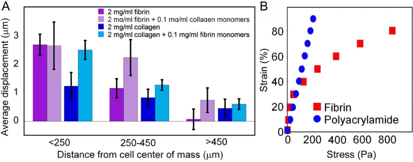
The strength and elasticity of fibrin gels allow them to resist external forces without tearing. These properties come from the monomer, fiber and bulk structure. At the monomer level it has been shown that each coiled coil domain can reversibly unfold, stretching 23 nm, when a force of approximately 94 pN (Brown et al. 2007) or 20 myosin powerstrokes (Ishijima et al. 1996) is applied. AFM data indicates that fibrin protofibrils are linearly elastic (Lim et al. 2008) with a modulus on the order of 1 MPa. As the fibrils laterally aggregate the structures increase in stiffness in the range of 10s of MPa (Collet et al. 2005) but the elastic modulus remains independent of strain. The low strain, less than 15%, elastic modulus of the gels formed by these fibers is 5 orders of magnitude lower than that of the individual fibers but the bulk modulus increases several orders of magnitude as the strain increases. This nonlinear elasticity occurs in gels made from many biopolymers and is thought to result from an open mesh network being formed by fibers of intermediate stiffness such that their persistence length is on the order of the distance between crosslinks (Storm et al. 2005).
6
fibroblasts and hMSCs apply to fibrin networks. Fibroblasts and hMSCs are shown to deform the fibrin matrix up to 5 cell lengths away from their periphery. This deformation not only leads to fiber alignment but also stiffening of the adjacent fibrin network. By applying sufficient force to engage the strain stiffening regime of the biopolymer networks, cells create a degree of mechanical anisotropy that cannot be obtained or explained by fiber orientation alone (Thomopoulos et al. 2007).
Chapter 4 investigates a role for this single cell induced local mechanical and structural anisotropy in driving multicellular pattern formation. Pattern formation has been observed in cells cultured in a variety of biopolymer networks including fibrin, collagen and matrigel (Nicosia and Ottinetti 1990; Vernon et al. 1992). Endothelial cells, which form non physiologic, cobblestone monolayers when cultured on tissue culture plastic, will form branching networks with interconnected lumen when cultured on or within collagen gels (Bach et al. 1998). Their ability to form these networks depends on the collagen concentration and the gel’s internal tension (Sieminski et al. 2004). The data in Chapter 4 correlates the deformability of the gel with the degree of pattern formation demonstrating how the ability of cells to sense the mechanical properties of their matrix and the nonlinear mechanical properties of biopolymer networks combine to facilitate formation of multicellular patterns.
7
substrates (Byfield et al. 2009). Thus it is filamin’s role in the collagen binding focal complex not its role as an actin crosslinker which is critical to substrate mechanosensing on collagen. The work done here suggests that this is also true for the ability of cells to compact their matrices since cells lacking filamin A retain the ability to deform fibrin but not collagen gels. Additionally, the data suggest that melanoma cells and HEK 293 cells apply smaller forces on collagen matrices than fibrin matrices.
8
1.2 References:
Adams RA, Bauer J, Flick MJ, Sikorski SL, Nuriel T, Lassmann H, Degen JL, Akassoglou K. 2007. The fibrin-derived gamma377-395 peptide inhibits microglia activation and suppresses relapsing paralysis in central nervous system autoimmune disease. J Exp Med 204(3):571-82.
Akassoglou K, Adams RA, Bauer J, Mercado P, Tseveleki V, Lassmann H, Probert L, Strickland S. 2004. Fibrin depletion decreases inflammation and delays the onset of demyelination in a tumor necrosis factor transgenic mouse model for multiple sclerosis. Proc Natl Acad Sci U S A 101(17):6698-703.
Bach TL, Barsigian C, Chalupowicz DG, Busler D, Yaen CH, Grant DS, Martinez J. 1998. VE-Cadherin mediates endothelial cell capillary tube formation in fibrin and collagen gels. Exp Cell Res 238(2):324-34.
Barbieri B, Balconi G, Dejana E, Donati MB. 1981. Evidence that vascular endothelial cells can induce the retraction of fibrin clots. Proc Soc Exp Biol Med 168(2):204-7.
Bell E, Ivarsson B, Merrill C. 1979. Production of a tissue-like structure by contraction of collagen lattices by human fibroblasts of different proliferative potential in vitro. Proc Natl Acad Sci U S A 76(3):1274-8.
Brown AEX, Litvinov RI, Discher DE, Weisel JW. 2007. Forced unfolding of the coiled-coils of fibrinogen by single molecule AFM. Biophysical Journal:524A-524A. Byfield FJ, Wen Q, Levental I, Nordstrom K, Arratia PE, Miller RT, Janmey PA. 2009.
9
coated with collagen but not fibronectin. Biophys J 96(12):5095-102.
Collet JP, Shuman H, Ledger RE, Lee S, Weisel JW. 2005. The elasticity of an individual fibrin fiber in a clot. Proc Natl Acad Sci U S A 102(26):9133-7.
Cunningham CC, Gorlin JB, Kwiatkowski DJ, Hartwig JH, Janmey PA, Byers HR, Stossel TP. 1992. Actin-binding protein requirement for cortical stability and efficient locomotion. Science 255(5042):325-7.
Dembo M, Wang YL. 1999. Stresses at the cell-to-substrate interface during locomotion of fibroblasts. Biophys J 76(4):2307-16.
Dubey N, Letourneau PC, Tranquillo RT. 2001. Neuronal contact guidance in magnetically aligned fibrin gels: effect of variation in gel mechano-structural properties. Biomaterials 22(10):1065-75.
Elsdale T, Bard J. 1972. Collagen substrata for studies on cell behavior. J Cell Biol 54(3):626-37.
Engler AJ, Griffin MA, Sen S, Bonnemann CG, Sweeney HL, Discher DE. 2004. Myotubes differentiate optimally on substrates with tissue-like stiffness: pathological implications for soft or stiff microenvironments. J Cell Biol 166(6):877-87.
Engler AJ, Sen S, Sweeney HL, Discher DE. 2006. Matrix elasticity directs stem cell lineage specification. Cell 126(4):677-89.
Finer JT, Simmons RM, Spudich JA. 1994. Single myosin molecule mechanics: piconewton forces and nanometre steps. Nature 368(6467):113-9.
10
complex development. J Cell Biol 159(4):695-705.
Georges PC, Miller WJ, Meaney DF, Sawyer ES, Janmey PA. 2006. Matrices with compliance comparable to that of brain tissue select neuronal over glial growth in mixed cortical cultures. Biophysical Journal 90(8):3012-3018.
Guidry C, Grinnell F. 1985. Studies on the mechanism of hydrated collagen gel reorganization by human skin fibroblasts. J Cell Sci 79:67-81.
Hadjipanayi E, Mudera V, Brown RA. 2008. Close dependence of fibroblast proliferation on collagen scaffold matrix stiffness. J Tissue Eng Regen Med.
Harris AK, Stopak D, Wild P. 1981. Fibroblast traction as a mechanism for collagen morphogenesis. Nature 290(5803):249-51.
Harris AK, Wild P, Stopak D. 1980. Silicone rubber substrata: a new wrinkle in the study of cell locomotion. Science 208(4440):177-9.
Hellam DC, Podolsky RJ. 1969. Force measurements in skinned muscle fibres. J Physiol 200(3):807-19.
Herbert CB, Nagaswami C, Bittner GD, Hubbell JA, Weisel JW. 1998. Effects of fibrin micromorphology on neurite growth from dorsal root ganglia cultured in three-dimensional fibrin gels. J Biomed Mater Res 40(4):551-9.
Huettner JE, Baughman RW. 1986. Primary culture of identified neurons from the visual cortex of postnatal rats. J Neurosci 6(10):3044-60.
11 70(1):383-400.
Ju YE, Janmey PA, McCormick ME, Sawyer ES, Flanagan LA. 2007. Enhanced neurite growth from mammalian neurons in three-dimensional salmon fibrin gels. Biomaterials 28(12):2097-108.
Klein EA, Yin L, Kothapalli D, Castagnino P, Byfield FJ, Xu T, Levental I, Hawthorne E, Janmey PA, Assoian RK. 2009. Cell-cycle control by physiological matrix elasticity and in vivo tissue stiffening. Curr Biol 19(18):1511-8.
Lim BBC, Lee EH, Sotomayor M, Schulten K. 2008. Molecular basis of fibrin clot elasticity. Structure 16(3):449-459.
Misra JC, Samanta S. 1987. Effect of material damping on bone remodelling. J Biomech 20(3):241-9.
Naruse K, Sokabe M. 1993. Involvement of stretch-activated ion channels in Ca2+ mobilization to mechanical stretch in endothelial cells. Am J Physiol 264(4 Pt 1):C1037-44.
Nicosia RF, Ottinetti A. 1990. Modulation of microvascular growth and morphogenesis by reconstituted basement membrane gel in three-dimensional cultures of rat aorta: a comparative study of angiogenesis in matrigel, collagen, fibrin, and plasma clot. In Vitro Cell Dev Biol 26(2):119-28.
Niewiarowski S, Goldstein S. 1976. Fibrin clot retraction by human skin fibroblasts: effects of ADP and thrombin. Proc Soc Exp Biol Med 151(2):253-6.
12
Pelham RJ, Jr., Wang YL. 1998. Cell locomotion and focal adhesions are regulated by the mechanical properties of the substrate. Biol Bull 194(3):348-9; discussion 349-50.
Quick AJ, Hussey CV. 1950. The mechanism of clot retraction. Science 112(2915):558-9.
Reinhart-King CA, Dembo M, Hammer DA. 2003. Endothelial Cell Traction Forces on RGD-Derivatized Polyacrylamide Substrata. Langmuir 19(5):1573-1579.
Saez A, Buguin A, Silberzan P, Ladoux B. 2005. Is the mechanical activity of epithelial cells controlled by deformations or forces? Biophys J 89(6):L52-4.
Schor AM, Schor SL, Allen TD. 1983. Effects of culture conditions on the proliferation, morphology and migration of bovine aortic endothelial cells. J Cell Sci 62:267-85.
Sieminski AL, Hebbel RP, Gooch KJ. 2004. The relative magnitudes of endothelial force generation and matrix stiffness modulate capillary morphogenesis in vitro. Exp Cell Res 297(2):574-84.
Storm C, Pastore JJ, MacKintosh FC, Lubensky TC, Janmey PA. 2005. Nonlinear elasticity in biological gels. Nature 435(7039):191-4.
Thomopoulos S, Fomovsky GM, Chandran PL, Holmes JW. 2007. Collagen fiber alignment does not explain mechanical anisotropy in fibroblast populated collagen gels. J Biomech Eng 129(5):642-50.
13
cellular networks in vitro. Lab Invest 66(5):536-47.
Wakatsuki T, Wysolmerski RB, Elson EL. 2003. Mechanics of cell spreading: role of myosin II. J Cell Sci 116(Pt 8):1617-25.
Weiss HJ. 1973. Bleeding disorders due to abnormal platelet function. Med Clin North Am 57(2):517-30.
14
Figure 1.1 Cartoon of a focal complex
15
CHAPTER 2
Substrate stiffness regulates cell cycle progression in
human mesenchymal stem cells
(Adapted from Winer JP, Janmey PA, McCormick ME, Funaki M. 2009 Tissue Engineering Part A15: 147-54.)
1.1 Abstract.
16
2.2 Introduction.
Many adult tissues contain a small population of stem cells that are capable of producing identical daughter cells and differentiating into multiple, although not all types of, cell lineages. These cells serve as reservoirs when tissues are turned over or are damaged by trauma, disease, aging, etc (Tarnowski and Sieron 2006). Mesenchymal tissues, such as bone marrow, adipose tissue and cartilage, have been shown to contain mesenchymal stem cells that can differentiate into not only cells in the tissue they originate from but also cells in other mesenchymal tissues (Pittenger et al. 1999). For instance, mesenchymal stem cells isolated from adult bone marrow can differentiate and regenerate fat, bone, cartilage, muscle, ligament, tendon and stroma (Beyer Nardi and da Silva Meirelles 2006; Friedenstein et al. 1970).
Due to their multipotency and capacity for self-renewal, adult somatic stem cells have received significant attention for use in clinical applications such as regenerative tissue engineering and gene therapy (Kassem 2006). Among adult somatic stem cells, bone marrow-derived human mesenchymal stem cells (hMSCs) are one of the most promising target populations, due to the relative ease with which they can be isolated from the bulk of the cells in bone marrow and their significant expansion capacity in vitro
17
bone marrow microenvironment such as the presence of soluble factors, cell-cell interactions and cell-matrix interactions help to regulate stem cell behavior (Minguell et al. 2001). For instance, extracellular matrix proteins have been shown to regulate differentiation of MSC into chondrocytes and osteoblasts (Bosnakovski et al. 2006; Kundu and Putnam 2006). Many of the precise features of the extracellular matrix required for regulating the pool of undifferentiated stem cells remain unclear.
Over the last 10 years, the importance of substrate stiffness as a mechanism for modulating cell shape and phenotype has become increasingly studied. Work on differentiated tissue-forming cell types suggests that each responds to a different and specific range of substrate stiffness and often exhibits the most in vivo like morphology when the gel stiffness matches their native tissue compliance (Georges and Janmey 2005). For instance neurons exhibit more neurite branching on gels mimicking the stiffness of brain tissue and myotubes exhibit striation only on gels whose compliance is comparable to muscle tissue (Engler et al. 2004; Flanagan et al. 2002). Fibroblasts adopt a round shape on a soft matrix, and stress fibers are apparent only when they are plated on matrices stiffer than 3600 Pa (Yeung et al. 2005). In multipotent hMSC matrix elasticity can drive morphological and protein expression changes toward neurogenic, myogenic or osteogenic profiles in a myosin II-dependent fashion (Engler et al. 2006). Most ex vivo studies on hMSCs have been carried out by seeding cells on plastic or glass surfaces, which are drastically stiffer than any non calcified adult tissue. Polyacrylamide offers a well characterized system for modulating substrate stiffness while maintaining
18
In this report we provide evidence that bone marrow-derived hMSCs become quiescent on soft polyacrylamide gels that mimic the elasticity of bone marrow. Quiescent hMSCs on soft gels remained competent to resume proliferation or initiate terminal differentiation when additional cues were delivered. Thus, the elasticity of bone marrow may contribute to the ability of the bone marrow microenvironment to maintain quiescent multipotent mesenchymal stem cells.
2.3 Materials and Methods.
2.3.1Cell Culture
Bone marrow-derived human mesenchymal stem cells were maintained in growth medium (GM), DMEM with 1 g/L D-glucose, 0.3 mg/ml L-glutamine and 100 mg/L sodium pyruvate and 10% heat inactivated fetal bovine serum on tissue culture plastic prior to seeding on gels. For adipocyte differentiation, cells were exposed to two chemical induction cycles consisting of 3 days in adipogenic induction media (GM, 1 µM dexamethasone, 200 µM indomethacin, 10 µg/ml insulin and 0.5 mM 3-Isobutyl-1-methylxanthine) and 1 day in adipogenic maintenance media (GM and 10 µg/ml insulin). Undifferentiated cells were kept in GM and re-fed every 3 days. For osteoblast differentiation cells were incubated with osteoblast induction media (GM, 50 µm ascorbic acid-2-phosphate, 10 mM β-glycerophosphate and 100 nM dexamethasone) for 24 days changing the media every 3-4 days. Cells were plated at a density of 5 x 10⁴ cells per 4.9 cm2 on gels, or 1 x 10⁵ cells per 4.9 cm2 (confluent) or 1 x 10⁴ cells per 4.9 cm2 (sparse) on glass.
19
Polyacrylamide gels for cell culture were prepared using a protocol derived from the seminal Pelham and Wang experiments(Wang et al. 2001; Wang and Pelham 1998). Solutions of 3.0% acrylamide and 0.2% bisacrylamide (for 250 Pa gel) or 7.5% acrylamide and 0.5% bisacrylamide (7500 Pa gel) were prepared in phosphate buffered saline (PBS), pH 7.4. Polymerization was initiated with N,N,N’,N’-tetramethylethylenediamine and ammonium persulfate. A droplet of 150 µl was deposited on a 25 mm glass coverslip previously modified with 3-aminopropyltrimethoxysilane and glutaraldehyde. 15 µl of 2% acrylic acid N-hydroxysuccinimide ester in toluene was applied to the droplet and a 25 mm chlorosilanized coverslip was placed on top of the droplet and removed after polymerization is completed. The gels were placed into an agarose coated six well polystyrene tissue culture plate and washed with 50 mM HEPES, pH 8.2. The N-succinimide crosslinker on the top of the gel was reacted for 1 hour with the ligand of interest diluted in HEPES buffer. Gels were washed and cells were seeded within 24 hours. Ligands used were either a mixture of 0.1 mg/ml rat tail collagen and 0.01 mg/ml human fibronectin.
2.3.3Rheology of Tissues and Gels
To measure the dynamic shear storage modulus (G') of polyacrylamide gels, 500
20
parameters were chosen to probe the elastic response of the tissues in the region where G’ is independent of strain. To measure the G’ of animal tissues, either 8 mm diameter, 2 mm thick samples (for rat tissues) or 25 mm diameter, 2 mm thick samples (for bovine tissues) were punched out. The shear moduli of the hydrated tissue samples were measured at 37°C using parallel plates with geometries matching the sample. Rat tissues were dissected from the animal, kept hydrated in PBS for no longer than 1 hour prior to the measurements. Bovine tissues were purchased from a commercial slaughterhouse, transported on ice, hydrated and warmed to 37°C before measurements were made. Three samples of each tissue were measured.
2.3.4 Imaging and Analysis
21 with DAPI. Only isolated cells were counted.
2.3.5 Quasi 3D polyacrylamide gels
Substrate sandwiches to mimic a 3D environment were composed as described by Beningo et al.(Beningo et al. 2004) with some minor modifications. Cells were seeded onto a 25 mm gel and allowed to adhere for 48 hours. Then a second coverslip, with or without a 250 Pa polyacrylamide gel on the bottom side, was placed on top of the seeded gel. To bring the cells into close proximity with a second coverslip, a sterilized 35 gram weight was placed on top of the sandwich for 30 seconds. The media was reintroduced after which the weight will be removed and the cells were left in the sandwiches for 2 days. The cells were imaged just before the top coverslip is applied and 2 days afterward.
2.4 Results.
2.4.1 The elasticity of bone marrow and a differentiated tissue with comparable stiffness.
22
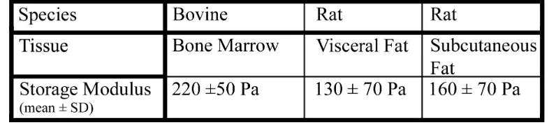
acrylamide and 0.1% bisacrylamide was adopted, which has a polymerized G’ of 250 ± 25 Pa, to mimic the elasticity of their natural environment. In some experiments a second substrate of 7.5% acrylamide and 0.5% bisacrylamide with a storage modulus of 7500 ± 250 Pa, which is in the range of muscle tissue, served as a control (Engler et al. 2004).
2.4.2 Soft gels induce a round shape and cell cycle arrest in hMSCs.
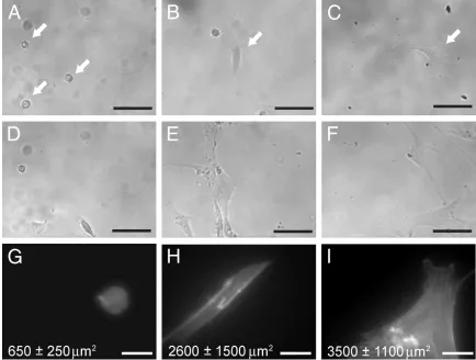
After two days on 250 Pa gels coated with a mixture of fibronectin and collagen I, hMSC had small, rounded morphologies with a disorganized F-actin cytoskeleton (Fig 2.1 A and G). They had an average projected cell area of only 650 ± 250 µm2 compared to a spread area of 3500 ± 1100 µm2 on glass coated with the same adhesion proteins. A similar phenotype has been observed in fibroblasts, endothelial cells and astrocytes on 250 Pa gels (Georges et al. 2006; Pelham and Wang 1997; Yeung et al. 2005). Extending the culture time to seven days resulted in some of the cells strengthening their adhesion to the compliant matrix and beginning to spread; however, there was no noticeable increase in cell number (Fig 2.1 D).
In comparison hMSC kept on 7500 Pa gels for two days were partially spread, with a projected cell area of 2600 ± 1500 µm2 and exhibited some actin organization but few stress fibers (Fig 2.1 B and E). After seven days they apparently increased their numbers and became increasingly spread upon cell-cell contact (Fig 2.1 H). On glass, hMSC exhibited a spread morphology, had abundant stress fibers and proliferated rapidly (Fig 2.1 C, F and I).
23
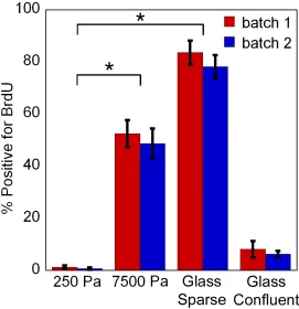
incorporation assay was performed on cells cultured on various substrates (Fig 2.2). After an overnight incubation with BrdU, virtually no hMSCs adhering to 250 Pa gels incorporated BrdU. Thus, despite the presence of serum, DNA replication, a step required for hMSC cell division, is suppressed. On 7500 Pa gels approximately half of the cells replicated their DNA in 24 hours and when sparsely seeded on glass over 80% of the cells exhibited nuclear BrdU staining. To check that the quiescent phenotype was not a donor specific phenomenon, the results were reproduced using a second batch of hMSCs. These results imply that substrate compliance can significantly affect progression of the cell cycle in hMSCs.
2.4.3 Quiescent cells competent to resume proliferation and maintain multipotency
24
cells from a second donor to ensure it was not a donor specific response. Therefore, hMSCs on 250 Pa gels are competent to resume proliferation, once they receive a mechanical signal from a stiffer substrate.
Terminal differentiation is achieved through a highly coordinated set of events that includes growth arrest and expression of cell lineage restricted phenotypes. Thus, the temporary growth arrest of hMSCs on 250 Pa gels motivated this investigation into whether these cells are competent to differentiate. Since substrate elasticity is reported to specify the lineage into which hMSC differentiate, adipocyte differentiation of hMSCs was tested on 250 Pa gels, which is an elasticity comparable to fat tissues (Engler et al. 2006). Morphological changes such as a round shape and disorganized F-actin observed in hMSCs on 250 Pa gels, have been associated with adipogenic differentiation (Koutnikova and Auwerx 2001; Spiegelman and Ginty 1983).
25
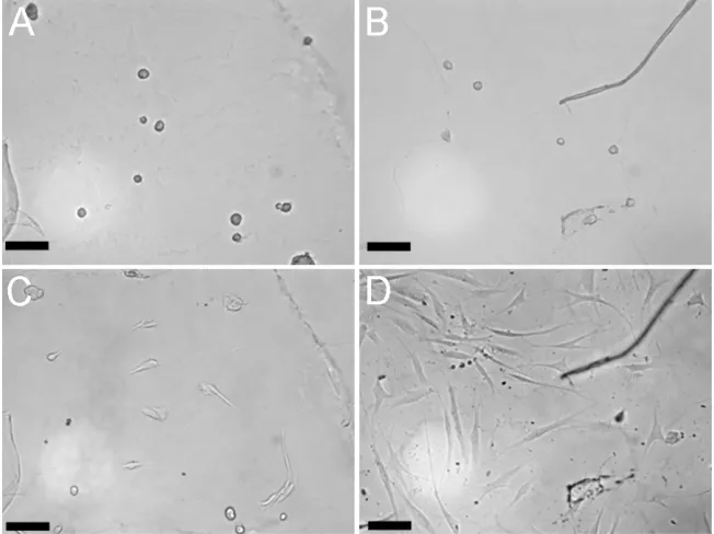
Efficient adipogenic differentiation of growth-arrested hMSCs on 250 Pa gels raises the possibility that these non-proliferative cells are differentiated into preadipocytes and committed to an adipogenic lineage, instead of staying quiescent as stem cells. To rule out this possibility, hMSCs were sparsely seeded on a soft gel for two days, then transferred to a glass coverslip using the quasi 3D method and finally cultured in osteoblast induction media for 24 days. As mentioned earlier cells on 250 Pa gels that came in contact with a stiff surface transferred to that substrate, resumed proliferation and took on the spindle phenotype typical of hMSCs on tissue culture plastic. After 24 days in osteoblast induction media calcium rich extracellular matrix was clearly visible in the culture dish and stained positive with Alizarin Red S (Fig 2.4 B) matrix mineralization is a late stage marker of osteoblast differentiation (Kundu and Putnam 2006). Without stimulation by osteoblast induction media the cells did not deposit calcified extracellular matrix (data not shown). These results demonstrate that hMSCs on 250 Pa gels maintain their multilineage potential, supporting the hypothesis that hMSCs seeded on soft gels stay quiescent as stem cells.
2.5 Discussion.
26
high number of cells accumulating lipid droplets, yet without chemical induction there was no lipid accumulation. Chemical induction was also required for osteoblast differentiation. This requirement for synchronized mechanical and chemical stimulation provides an explanation for how hMSCs can be compartmentalized into compliant tissues such as bone marrow and yet resist spontaneous differentiation.
In this study the polyacrylamide gel system developed by Pelham and Wang was employed to modify substrate elasticity without changing the chemical environment to which the cells are exposed (Pelham and Wang 1997). In addition to matrix elasticity, the choice of extracellular ligand can also strongly affect hMSC adhesion and differentiation (Salasznyk et al. 2004). Collagen type 1 is found in a variety of tissues including bone and adipose and is regularly used as a substrate for cell adhesion experiments; however, on the 250 Pa gel collagen alone was not sufficient to ensure adhesion of a majority of cells (unpublished observation). Fibronectin alone has been shown to inhibit adipogenesis by strengthening the actin cytoskeleton; however, a round shape, such as that induced by a compliant substrate, negates this inhibition (Spiegelman and Ginty 1983). A mixture of collagen type I and fibronectin (10:1) provided the best adhesion of cells to the 250 Pa gels without affecting differentiation potential. HMSCs have the capacity to remodel their microenvironment by altering the expression of matrix metalloproteases, which might help to promote efficient terminal differentiation after an initial strong adhesion is achieved (Neth et al. 2006; Urs et al. 2004).
27
with other proliferating cell types such as NIH 3T3 fibroblasts, bovine aortic endothelial cells and normal rat kidney epithelial cells which all continue to divide when cultured on gels soft enough to promote a round phenotype (Guo et al. 2006; Yeung et al. 2005). Thus stem cell quiescence on 250 Pa gels is not a general shape induced failure of cytokinesis but a specific sensitivity of hMSCs to substrate compliance.
When nonproliferating hMSCs were presented with a protein-coated glass substrate, the cells developed a spindle morphology and reentered the cell cycle. In addition to showing that the round cells on soft matrices were only quiescent and not senescent, this experiment also demonstrated that the presence of a stiff substrate overrides the physical cues from a compliant matrix. Thus, in agreement with experiments using NIH 3T3 fibroblasts on stiffness gradients, hMSCs select a stiffer substrate over a more compliant one (Lo et al. 2000). Further experiments will be required to show if this dominant signal from a stiff matrix true also occurs in a fully 3D and not just a quasi 3D environment.
28
still proliferating and had a 30% lower percent adipocyte differentiation after 1 week in differentiation media. A role for substrate stiffness in priming cells for adipogenesis has not previously been considered. The enhanced conversion of hMSCs on soft gels into adipocytes upon a chemical signal may help to explain why bone marrow becomes fatty with age (Gurevitch et al. 2007).
On the soft 250 Pa gels without any chemical induction, there was not a significant population of cells exhibiting a neuronal phenotype with neurite-like protrusions such as those described by Engler et al. on gels of similar compliances (Engler et al. 2006). On stiff 7500 Pa gels, cells did exhibit the elongated morphology described in that study (Engler et al. 2006). Although their conditions were quite similar to the ones used here, two key differences likely led to a round adipocyte-like shape over a neuronal phenotype. First, fibronectin, which made up 10% of the adhesion mixture used in this study, has been shown to decrease the differentiation of neural stem/precursor cells to neurons and those neurons that did commit on a fibronectin-coated substrate extended shorter neurites (Flanagan et al. 2006). Second, bone marrow donor variability might contribute to a predisposition toward adipocyte or neuronal differentiation as donor age and sex have been shown to effect osteoblast and myoblast differentiation capacity (Deasy et al. 2007; Siddappa et al. 2007). The results presented here indicate that soft gel induced quiescence is not strongly donor dependent because two independent batches produced similar BrdU incorporation profiles.
29
direct terminal stem cell differentiation. First, several tissues in the body have similar compliances; for example brain, fat and bone marrow tissues all have a storage modulus of approximately 200 Pa, yet all maintain unique populations of cells. Second, in vivo
30
2.6 References
Balaban NQ, Schwarz US, Riveline D, Goichberg P, Tzur G, Sabanay I, Mahalu D,
Safran S, Bershadsky A, Addadi L and others. 2001. Force and focal
adhesion assembly: a close relationship studied using elastic micropatterned
substrates. Nat Cell Biol 3(5):466-72.
Baxter MA, Wynn RF, Jowitt SN, Wraith JE, Fairbairn LJ, Bellantuono I. 2004.
Study of telomere length reveals rapid aging of human marrow stromal cells
following in vitro expansion. Stem Cells 22(5):675-82.
Beningo KA, Dembo M, Wang YL. 2004. Responses of fibroblasts to anchorage of
dorsal extracellular matrix receptors. Proc Natl Acad Sci U S A
101(52):18024-9.
Beyer Nardi N, da Silva Meirelles L. 2006. Mesenchymal stem cells: isolation, in
vitro expansion and characterization. Handb Exp Pharmacol(174):249-82.
Bonab MM, Alimoghaddam K, Talebian F, Ghaffari SH, Ghavamzadeh A, Nikbin
B. 2006. Aging of mesenchymal stem cell in vitro. BMC Cell Biol 7:14.
Bosnakovski D, Mizuno M, Kim G, Takagi S, Okumura M, Fujinaga T. 2006.
Chondrogenic differentiation of bovine bone marrow mesenchymal stem cells
(MSCs) in different hydrogels: influence of collagen type II extracellular
matrix on MSC chondrogenesis. Biotechnol Bioeng 93(6):1152-63.
Chawla A, Schwarz EJ, Dimaculangan DD, Lazar MA. 1994. Peroxisome
proliferator-activated receptor (PPAR) gamma: adipose-predominant
31
135(2):798-800.
Deasy BM, Lu A, Tebbets JC, Feduska JM, Schugar RC, Pollett JB, Sun B, Urish
KL, Gharaibeh BM, Cao B and others. 2007. A role for cell sex in stem
cell-mediated skeletal muscle regeneration: female cells have higher muscle
regeneration efficiency. J Cell Biol 177(1):73-86.
El-Jack AK, Hamm JK, Pilch PF, Farmer SR. 1999. Reconstitution of
insulin-sensitive glucose transport in fibroblasts requires expression of both
PPARgamma and C/EBPalpha. J Biol Chem 274(12):7946-51.
Engler AJ, Griffin MA, Sen S, Bonnemann CG, Sweeney HL, Discher DE. 2004.
Myotubes differentiate optimally on substrates with tissue-like stiffness:
pathological implications for soft or stiff microenvironments. J Cell Biol
166(6):877-87.
Engler AJ, Sen S, Sweeney HL, Discher DE. 2006. Matrix elasticity directs stem cell
lineage specification. Cell 126(4):677-89.
Flanagan LA, Ju YE, Marg B, Osterfield M, Janmey PA. 2002. Neurite branching
on deformable substrates. Neuroreport 13(18):2411-5.
Flanagan LA, Rebaza LM, Derzic S, Schwartz PH, Monuki ES. 2006. Regulation of
human neural precursor cells by laminin and integrins. J Neurosci Res
83(5):845-56.
Freytag SO, Paielli DL, Gilbert JD. 1994. Ectopic expression of the
CCAAT/enhancer-binding protein alpha promotes the adipogenic program
32
Friedenstein AJ, Chailakhjan RK, Lalykina KS. 1970. The development of
fibroblast colonies in monolayer cultures of guinea-pig bone marrow and
spleen cells. Cell Tissue Kinet 3(4):393-403.
Funaki M, Randhawa P, Janmey PA. 2004. Separation of insulin signaling into
distinct GLUT4 translocation and activation steps. Mol Cell Biol
24(17):7567-77.
Georges PC, Janmey PA. 2005. Cell type-specific response to growth on soft
materials. J Appl Physiol 98(4):1547-53.
Georges PC, Miller WJ, Meaney DF, Sawyer ES, Janmey PA. 2006. Matrices with
compliance comparable to that of brain tissue select neuronal over glial
growth in mixed cortical cultures. Biophysical Journal 90(8):3012-3018.
Guo WH, Frey MT, Burnham NA, Wang YL. 2006. Substrate rigidity regulates the
formation and maintenance of tissues. Biophys J 90(6):2213-20.
Gurevitch O, Slavin S, Feldman AG. 2007. Conversion of red bone marrow into
yellow - Cause and mechanisms. Med Hypotheses.
Kassem M. 2006. Stem cells: potential therapy for age-related diseases. Ann N Y
Acad Sci 1067:436-42.
Koutnikova H, Auwerx J. 2001. Regulation of adipocyte differentiation. Ann Med
33(8):556-61.
Kundu AK, Putnam AJ. 2006. Vitronectin and collagen I differentially regulate
osteogenesis in mesenchymal stem cells. Biochem Biophys Res Commun
33
Liu L, DiGirolamo CM, Navarro PA, Blasco MA, Keefe DL. 2004. Telomerase
deficiency impairs differentiation of mesenchymal stem cells. Exp Cell Res
294(1):1-8.
Lo CM, Wang HB, Dembo M, Wang YL. 2000. Cell movement is guided by the
rigidity of the substrate. Biophys J 79(1):144-52.
McBeath R, Pirone DM, Nelson CM, Bhadriraju K, Chen CS. 2004. Cell shape,
cytoskeletal tension, and RhoA regulate stem cell lineage commitment. Dev
Cell 6(4):483-95.
Minguell JJ, Erices A, Conget P. 2001. Mesenchymal stem cells. Exp Biol Med
(Maywood) 226(6):507-20.
Neth P, Ciccarella M, Egea V, Hoelters J, Jochum M, Ries C. 2006. Wnt signaling
regulates the invasion capacity of human mesenchymal stem cells. Stem Cells
24(8):1892-903.
Pelham RJ, Jr., Wang Y. 1997. Cell locomotion and focal adhesions are regulated by
substrate flexibility. Proc Natl Acad Sci U S A 94(25):13661-5.
Pittenger MF, Mackay AM, Beck SC, Jaiswal RK, Douglas R, Mosca JD, Moorman
MA, Simonetti DW, Craig S, Marshak DR. 1999. Multilineage potential of
adult human mesenchymal stem cells. Science 284(5411):143-7.
Reiser J, Zhang XY, Hemenway CS, Mondal D, Pradhan L, La Russa VF. 2005.
Potential of mesenchymal stem cells in gene therapy approaches for inherited
and acquired diseases. Expert Opin Biol Ther 5(12):1571-84.
34
to Vitronectin and Collagen I Promotes Osteogenic Differentiation of Human
Mesenchymal Stem Cells. J Biomed Biotechnol 2004(1):24-34.
Shao D, Lazar MA. 1997. Peroxisome proliferator activated receptor gamma,
CCAAT/enhancer-binding protein alpha, and cell cycle status regulate the
commitment to adipocyte differentiation. J Biol Chem 272(34):21473-8.
Siddappa R, Licht R, van Blitterswijk C, de Boer J. 2007. Donor variation and loss
of multipotency during in vitro expansion of human mesenchymal stem cells
for bone tissue engineering. J Orthop Res.
Spiegelman BM, Ginty CA. 1983. Fibronectin modulation of cell shape and
lipogenic gene expression in 3T3-adipocytes. Cell 35(3 Pt 2):657-66.
Tarnowski M, Sieron AL. 2006. Adult stem cells and their ability to differentiate.
Med Sci Monit 12(8):RA154-63.
Tontonoz P, Hu E, Spiegelman BM. 1994. Stimulation of adipogenesis in fibroblasts
by PPAR gamma 2, a lipid-activated transcription factor. Cell 79(7):1147-56.
Urs S, Smith C, Campbell B, Saxton AM, Taylor J, Zhang B, Snoddy J, Jones Voy
B, Moustaid-Moussa N. 2004. Gene expression profiling in human
preadipocytes and adipocytes by microarray analysis. J Nutr 134(4):762-70.
Wang N, Naruse K, Stamenovic D, Fredberg JJ, Mijailovich SM, Tolic-Norrelykke
IM, Polte T, Mannix R, Ingber DE. 2001. Mechanical behavior in living cells
consistent with the tensegrity model. Proc Natl Acad Sci U S A
98(14):7765-70.
35
substrate for mechanical studies of cultured cells. Methods Enzymol
298:489-96.
Yeung T, Georges PC, Flanagan LA, Marg B, Ortiz M, Funaki M, Zahir N, Ming
W, Weaver V, Janmey PA. 2005. Effects of substrate stiffness on cell
morphology, cytoskeletal structure, and adhesion. Cell Motil Cytoskeleton
36
Table 2.1 Rheological properties of bone marrow and fat tissue
Animal tissues were freshly harvested and their shear storage modulus was measured on a strain controlled rheometer. At least three samples of each tissue type were measured
37
Figure 2.1 Substrate stiffness regulates hMSC shape, spread area and proliferation
A-C: cells were plated on collagen type I and fibronectin-coated substrates and imaged in bright field after 24 hours of incubation in growth media (GM). Arrows indicate location of cells. D-F: Images at the same locations after 7 days of incubation in GM. Cells were kept at 37°C during imaging. Representative images from 2 independent experiments are shown. Scale bar = 100 µm. G-I: F-actin in hMSC was labeled with FITC conjugated phalloidin two days after seeding. Also reported is the mean projected area ± standard
38
Figure 2.2 A soft substrate suppresses Bromodeoxyuridine uptake by hMSCs
Cells were plated on substrates for 48 hours before incubating the cells in media containing BrdU overnight. Cells were kept in serum-containing media for the entire period, except for the Glass Confluent, which was serum starved for 24 hours before the BrdU was added in serum free media. Cells were stained with anti-BrdU and the nuclei were counterstained with DAPI. At least 40 cells were counted on each substrate and
39
Figure 2.3 Presentation of a hard substrate to cells on a soft gel stimulates
spreading and cycle re-entry
Cells were seeded onto 250 Pa gels and imaged after two days in serum containing media (A,B). Then a top substrate of either another 250 Pa gel (C) or a glass coverslip (D) was
used to sandwich the cells in a quasi 3D environment. Two days later the cells were imaged again at the same locations. Results were repeated in an independent experiment.
40
Figure 2.4 Cells retain differentiation potential
41
CHAPTER 3
Contractile cells respond to the high strain modulus of
strain stiffening biopolymer gels
(Adapted from Winer JP, Oake S, Janmey PA.2009. PLoS One4: e6382)
3.1 Abstract
42
3.2 Introduction
Over the last decade it has been demonstrated that a variety of tissue-forming cells can both sense the stiffness of their substrate and apply a controlled force onto that substrate. Not all cell types respond to stiffness changes in the same way, but many including endothelial cells (Yeung et al. 2005), fibroblasts (Pelham and Wang 1998), mammary epithelial cells (Paszek et al. 2005) astrocytes (Georges et al. 2006), macrophages (Fereol et al. 2006) and mesenchymal stem cells (Engler et al. 2006), exhibit increased spreading and adhesion on stiffer substrates compared to softer ones. Numerous proteins and protein complexes required for responding to substrate stiffness, such as the actomyosin network, small GTPases, protein phosphatases, and integrin adhesion sites have been identified, but the mechanism by which forces govern the interactions among these proteins is not yet defined.
43
displacement fields. Other synthetic systems with linear elasticity include silicone films (Harris et al. 1980) and flexible PDMS micropillars (Tan et al. 2003).
Studies done on synthetic gels have been used to understand how cells respond to the mechanical properties of the tissue microenvironment; however, extracellular matrix proteins such as collagen type I and fibrin display nonlinear mechanical properties such as strain stiffening (Storm et al. 2005) and negative normal stress (Janmey et al. 2007). In these materials the elastic modulus of the gel increases orders of magnitude as the applied strain increases such that the resistance that a cell feels would be a strong function of the strain that it applies. Many cell types also modulate the force they apply according to the stiffness of the gel, applying smaller forces when cultured on softer gels (Pelham and Wang 1999). Using responses to linearly elastic materials to predict a cell's behavior on nonlinear gels is further complicated since it is not known what property the cell’s mechanosensor is measuring. For example, whether cells attempt to exert a constant deformation and monitor the required stress, or whether they exert a constant stress and respond to the degree of strain remains an open question (Saez et al. 2005).
44
to the material's high strain modulus. On fibrin with a low strain modulus of 100 Pa the cells spread as if on a much stiffer gel using actomyosin contraction to strain the fibrin gel, locally increase its modulus, and achieve optimal spreading through a force-limited mechanism.
3.3 Materials and Methods
3.3.1Cell Culture
Bone marrow-derived human mesenchymal stem cells (Cambrex) were maintained in DMEM (GIBCO) with 1 g/L D-glucose, 0.3 mg/ml L-glutamine and 100 mg/L sodium pyruvate, 100 units/ml penicillin, 100 µg/ml streptomycin and 10% heat inactivated fetal bovine serum (GIBCO) on tissue culture plastic prior to seeding on gels. NIH 3T3 fibroblast’s (ATCC) culture media was identical except it contained 4.5 g/L D-glucose and 10% calf serum (GIBCO) instead of fetal bovine serum. All cells were maintained at 37ºC and 5% CO2. Unless otherwise specified all reagents are analytical
grade and purchased from Sigma.
3.3.2 Fibrin Gel Preparation
Lyophilized salmon fibrinogen (Wang et al. 2000) and thrombin (Michaud et al. 2002) were provided by Sea Run Holdings. Fibrin gels were prepared by diluting the stock solution of fibrinogen with T7 buffer (50 mM Tris, 150 mM NaCl at a pH of 7.4) to make a working solution of desired concentration. Polymerization was initiated in a 24 well tissue culture plate by adding 5 µl of thrombin (activity = 100 NIH units/ml) to 250
45
the rheometer and found to be approximately 1 mm at the thinnest point. In some cases 0.15 mM glycine-proline-arginine-proline (GPRP) was added to the working solution or the already polymerized gel was incubated in a 0.01 mg/ml solution of type 1 rat tail collagen for four hours at 4°C
3.3.3 Collagen Gel Preparation
Acid solubilized Type I rat tail collagen was purchased from BDBioscience. Collagen gels were prepared by adding a 1:10 dilution of 10x PBS to the stock solution of collagen and then bringing the volume to the desired concentration with 1x PBS. Gelation was initiated by adding NaOH until the solution reached pH 7.5. 250 µl of the working solution was added to each well of a 24 well tissue culture plate. The plate was then placed at 37˚C to accelerate gelation. After 1 hr, 1ml PBS was added to each of the wells. In some case the already polymerized gel was incubated with a 0.01 mg/ml fibrinogen solution for 1 hour at 4˚C, washed with PBS and then incubated with 1 U/ml of salmon thrombin for 10 min at room temperature.
3.3.4 Polyacrylamide Gel Preparation
46
U/ml thrombin for 30 minutes. For the two network coated substrates, the polyacrylamide was first ligated to fibrinogen monomers which act as nucleation sites and then coated with either 0.1 mg/ml fibrinogen and 0.5 U/ml thrombin for 30 minutes or with 0.5 mg/ml fibrinogen and 0.5 U/ml thrombin for 30 minutes. The networks were 5-10 fibers high and less than 2 microns thick. The thin fibers were produced by preparing a solution of 0.1 mg/ml fibrinogen and 0.1 U/ml thrombin 1 hour in advance and then allowing it to react with the gel for 1.5 hours. The thick fibers were prepared by allowing a solution of 0.5 mg/ml fibrinogen to react with 0.2 U/ml thrombin for 2 minutes and then adding excess of the thrombin inhibitor p-nitrophenyl-p’-guanidinobenzoate. This solution of fibers was then reacted with the surface of the polyacrylamide for 1.5 hours.
3.3.5 Displacement Microscopy
47
DMSO, 5 µM blebbistatin, 20 µM Cytochalasin D or 10 µM Nocodazole) was added. After 30 minutes the beads were imaged again. Image J (NIH) and Adobe Photoshop were used to generate a map of bead displacements for each pair of images. Due to the low magnification needed to capture the scale of the gel deformation the pixel resolution resulted in a error of ± 0.25 µm. To minimize the effect of cells other than the one of interest, only 50-100 cells were seeded per well of the 24 well dish. In addition the imaged cells were selected because they were at least 1 mm from the nearest cell.
3.3.6 Rheology
48
estimated elastic modulus were measured at each point.
3.3.7 Atomic Force Microscopy
Force indentation curves of fibrin gels were performed as previously described (Solon et al. 2007) on a Veeco Bioscope I using a silicon nitride probe with a cantilever spring constant of 0.01 N/m and a 5 µm polystyrene particle attached. Relative stiffnesses of different points on the gel are estimated by fitting the first 500 nm of the indentation curves to the Hertz model (Hertz 1882).
3.3.8 Image Analysis Scanning Electron Microscopy
For scanning electron microscopy samples were fixed with 2% glutaraldehyde in sodium cacodylate buffer, dehydrated with ethanol, critical point dried and coated with gold/palladium as described previously(Langer et al. 1988). The images were taken on a Philips XL20 scanning electron microscope (Philips Electron Optics, Eindhoven, The Netherlands) in the electron microscopy facility at the Department of Cell Biology University of Pennsylvania, PA, USA.
3.3.9 Image Analysis
49
determined using Image J’s analyze particles command.
3.3.10 Statistics
All statistics was preformed using Kaleidagraph software and unless otherwise mentioned significance was determined using a one way ANOVA and then applying a Tukey HSD post test with an α threshold of 0.05
3.4 Results
3.4.1 Cells cultured on soft fibrin spread as though on a stiff substrate
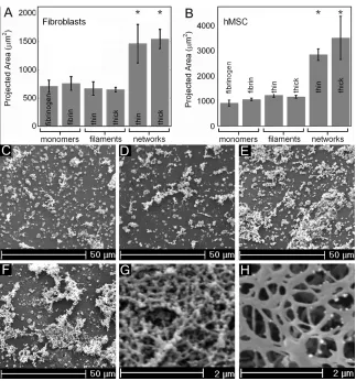
Consistent with previous reports (Yeung et al. 2005) fibroblasts are round on fibrinogen-coated polyacrylamide gels with a shear modulus of 100 Pa and become increasingly spread as the gel stiffness increases until they reach a spread area of approximately 2200 µm2 on 16 kPa polyacrylamide gels (Fig. 3.1 A). In contrast, on 1 mm thick fibrin gels of 1, 2, 4 and 8 mg/ml, with low strain shear moduli of 30, 60, 140 and 350 Pa respectively, the fibroblasts spread to areas statistically similar to each other and to the average spread area on the stiffest fibrinogen-coated polyacrylamide gel. Similar results were obtained using human mesenchymal stem cells (hMSCs) on the two kinds of substrates. Cells on fibrin displayed actin stress fibers, which are noticeably absent in cells cultured on soft polyacrylamide (Fig. 3.1 B-D). Cells on fibrin also had large focal adhesions consistent with a contractile phenotype (Fig. 3.1 E).
50
Therefore, if application of a small strain to the substrate is sufficient for the cells to respond to substrate mechanics, then 2 mg/ml fibrin and 70 Pa polyacrylamide gels will appear equally soft, but if the cells apply strains large enough to enter the strain-stiffening regime of fibrin elasticity, then they will respond to fibrin as a stiffer matrix than 70 Pa polyacrylamide. The latter hypothesis is consistent with the data, for figure 3.1 E shows that the modulus of 2 mg/ml fibrin at 80% strain is 3.7 kPa and the average spread area of cells on these gels is ~1900 µm2; this is comparable to cells on a 3.2 kPa fibrinogen-coated polyacrylamide gel which have an average spread area of ~1800 µm2.
3.4.2 A network of fibers is required for cells to spread on fibrin