A Comparative Study
of the Structures of Electron-Transfer Proteins
Using NMR
A thesis
submitted for the degree of Doctor of Philosophy;
in partial fulfilment of the requirements
of the University of London
Alfred Daniel Jesse Lee
July 1990
Department of Chemistry
University College London
Sir Christopher Ingold Laboratories
Gordon Street, London
All rights reserved INFORMATION TO ALL USERS
The qu ality of this repro d u ctio n is d e p e n d e n t upon the q u ality of the copy subm itted.
In the unlikely e v e n t that the a u th o r did not send a c o m p le te m anuscript and there are missing pages, these will be note d . Also, if m aterial had to be rem oved,
a n o te will in d ica te the deletion.
uest
ProQuest 10797784
Published by ProQuest LLC(2018). C op yrig ht of the Dissertation is held by the Author.
All rights reserved.
This work is protected against unauthorized copying under Title 17, United States C o d e M icroform Edition © ProQuest LLC.
ProQuest LLC.
789 East Eisenhower Parkway P.O. Box 1346
ABSTRACT
This thesis is a study of the structure of horse-heart cytochrome-c, and the relationship of its structure to function, using primarily nmr1 spectroscopy. It is demonstrated that nmr spectroscopy is a useful tool for the study of proteins in solution, and an assignment procedure for nmr spectra of proteins is described. By the use of this procedure, the nmr spectrum of native ferricytochrome-c is assigned. The assignment of the nmr spectrum of tfa- modified2 cytochrome-c is given, and compared to that of the native protein.
Using the assignments prepared for native ferri- and ferrocytochrome- c, and for the modified protein in both oxidation states, an attempt is made to describe and explain the structure and dynamics of cytochrome-c. Comparisons are made of NH/ND exchange rates, the aromatic ring flip of Tyr97, Gd3* ion binding sites and denaturation temperatures between the native and tfa-modified cytochrome-cs. The effect of amino acid substitutions between horse and tuna cytochrome-cs upon chemical shift of NH and CaH protons is also studied.
The results from these experiments provide a basis for the discussion of the relative importance of electrostatic interactions in determining the structure and dynamics of cytochrome-c. It is concluded that the structure of cytochrome-c is extremely rigid, and that its structure is determined more by close range forces than long-range electrostatic forces. It is believed that the techniques described in this thesis will be of general applicability to the study of electron-transfer proteins.
1 NMR is nuclear magnetic resonance.
ACKNOWLEDGEMENTS
I would like to express my gratitude to Dr Williams, for his patience and perseverance with me over the last four years, and to the British people for funding my tenure in London. It is to Julie that I owe much: for keeping me on an even keel, and for loving me despite my writing up on our honeymoon!
I thank UCL Chemistry Department for providing the equipment and laboratory space for this research, and the Chemical and Physical Society for providing the scientific-minded community in which to socialise.
It is the fate of those who toil at the lower employments of life ... to be exposed to censure,
without hope of praise; to be disgraced by miscarriage or punished for neglect ... . Among these unhappy mortals is the writer of PhD.s ... .
Every other author may aspire to praise; he (the PhD. writer) can only hope to escape reproach.
CONTENTS
Page
C hapter 1. Introduction 14
C hapter 2. Electrostatics 34
C hapter 3. JH nmr Assignments 45
native horse heart ferricytochrome-c
C hapter 4. *H nmr Assignments 79
modified cytochrome-cs
C hapter 5. Thermodynamics 122
modified cytochrome-cs
Gd3f Ion Binding 124
Denaturation and Partial Unfolding 137
C hapter 6. Dynamics 144
modified cytochrome-cs
Aromatic Ring Flip 146
NH/ND Exchange Rates 162
C hapter 7. Structural comparisons 173
between cytochrome-cs
C hapter 8. Conclusions 185
Bibliography. 191
Figure 1.1 1.2 1.3 1.4 1.5 1.6 1.7 1.8 3.1 3.2 3.3 3.4 3.5 3.6 3.7 3.8
INDEX TO FIGURES
Title Page
Pseudomonas c ^ . 20
Tuna c. 21
Paracoccus cS50. 22
Cytochrome-c. The a-helices are
represented by barrels, 310bends by blocks. 24 Side-chain exposure in cytochrome-c. 25 The haem and ligands of cytochrome-c. 26 Haem-packing amino acid side-chains. 27 Reaction scheme of electron transfer. 32 Cytochrome-c. The barrels represent
a-helices. 47
Data collection using the Hoult Parameter. 55 Before and After
Application of weighting functions to the fid. 57 Tl-cosy spectrum of horse ferricytochrome-c
(pH6.0, 40°C, 7mM, 99.8%D20 ). 59 ‘H-Recsy spectrum of horse ferricytochrome-c
(pH6.0, 40°C, 7mM, 90%H2O). 61
‘H-Noesy spectrum of horse ferricytochrome-c (pH6.0, 40°C, 7mM, 90%H2O)
showing C-terminal helix traced out. 63 Tl-Noesy spectrum of horse ferricytochrome-c (pH6.0, 40°C, 7mM, 90%H2O)
showing C/N terminal helix crossing point 64 ‘H-Noesy spectrum of horse ferricytochrome-c (pH6.0, 40°C, 7mM, 90%H2O)
Figure 3.9 3.10 3.11 3.12 3.13 3.14 4.1 4.2 4.3 4.4 4.5
Title Page
xH-cosy spectrum of horse ferricytochrome-c
(pH6.0, 40°C, 7mM, 99.8%D20 ). 68 ‘H-Recsy spectrum of horse ferricytochrome-c
(pH6.0, 40°C, 7mM, 90%H2O). 69
‘H-Noesy spectrum of horse ferricytochrome-c
(pH6.0, 40°C, 7mM, 90%H2O). 70
‘H-Noesy spectrum of horse feiricytochrome-c (pH6.0, 40°C, 7mM, 90%H2O)
showing N-terminal helix traced out. 71 Tl-Noesy spectrum of horse ferricytochrome-c (pH6.0, 40°C, 7mM, 90%H2O)
showing C-terminal helix. 76
Tl-Noesy spectrum of horse ferrocytochrome-c (pH7.8, 40°C, 8mM, 90%H2O)
showing C-terminal helix. 77
!H-Cosy spectrum of AocB and T6y region in tfa-ferrocytochrome-c
(pH7.8, 6mM, 99.8%D20 , 40°C). 84 Tf-Noesy spectrum of AaB and TBy region
in native ferrocytochrome-c
(pH7.8, 8mM, 90%H2O, 40°C). 85
Tl-Noesy spectrum showing the NH/CaH region of tfa-modified ferrocytochrome-c
(pH7.8, 6mM, 40°C). 86
Ti-Noesy spectrum showing the NH/CaH region of native horse heart ferrocytochrome-c
(pH7.8, 6mM, 40°C). 87
19F spectrum of tfa-ferricytochrome-c
Figure 4.6 4.7 4.8 4.9 4.10 4.11 4.12 4.13 4.14 4.15 4.16 4.17 4.18 5.1 Title
19F NMR Spectrum of tfa-ferrocytochrome-c (pH7.8, 40°C, 6mM).
Non phase sensitive xH-Cosy spectrum of tfa-ferricytochrome-c
(pH7.12, 40°C, 7.1mM).
Phase sensitive ^ -C o sy spectrum of tfa-ferricytochrome-c
(pH6.8, 8mM, 99.8%D20 , 40°C).
19F NMR Spectrum of tfa-ferricytochrome-c (pH6.3, 40°C, 7mM).
Wide Sweep Width NMR Spectrum of tfa-ferricytochrome-c (40°C, 7mM, pH6.3). 19F NMR Spectrum of a redox mixture of cytochrome-c (40°, 7mM, pH6.3).
UV-Vis spectra of native and tfa-modified ferricytochrome-cs.
Resonance Raman spectrum of tfa-modified ferricytochrome-c.
Resonance Raman spectrum of native ferricytochrome-c.
Met80-Haem band in native and modified cytochrome-cs.
!H NMR Spectrum of native ferrocytochrome-c (40°C, pH7.8, 7mM).
NMR Spectrum of tfa-ferrocytochrome-c (40°C, pH7.8, 7mM).
!H-Noesy spectrum of native ferricytochrome-c at pH 10.5.
Tfa-ferrocytochrome-c
(40°C, pH7, 6mM, 0% Gd3*).
Figure Title Page
5.2 Tfa-ferrocytochrome-c
(40°C, pH7, 6mM, 6% Gd3"). 127
5.3 Native ferrocytochrome-c
(40°C, pH7, 6mM, 0% Gd3*). 128
5.4 Native ferrocytochrome-c
(40°C, pH7, 6mM, 6% Gd3*). 129
5.5 Native ferrocytochrome-c
(21°C, pH7, 6mM, 12% Gd3"). 130 5.6 19F nmr spectmm of tfa-ferrocytochrome-c
at 60, 65, and 70°C. 139
5.7 19F nmr spectmm of tfa-ferricytochrome-c
at 40, 45, and 50°C. 138
6.1 Graph of IJ IB vs 1/t for
native ferrocytochrome-c. 153
6.2 Graph of IJ IB vs 1/t for
tfa-modified ferrocytochrome-c. 154
6.3 Graph of Ink vs 1/T for
native ferrocytochrome-c. 155
6.4 Graph of Ink vs 1/T for
tfa-modified ferrocytochrome-c. 156
6.5 Stacked Plot of ID spectra from
tfa-ferrocytochrome-c at 21-55°C. 157 6.6 Native ferrocytochrome-c after 10 hours
NH/ND Exchange experiment. 165
6.7 Native ferrocytochrome-c after 20 hours
NH/ND Exchange experiment. 166
6.8 Tfa-ferrocytochrome-c after
1, 6, 12, 18, 56, and 720 minutes in the
Figure
7.1
7.2 7.3
Title Page
The differences in chemical shift of equivalent protons in horse and tuna cytochrome-cs
Table 2.1 3.1 4.1 4.2 4.3 4.4 5.1 5.2 6.1 6.2 6.3 6.4
INDEX TO TABLES
Title Page
Non-helical hydrogen bonds in
tuna ferrocytochrome-c. 37
[H nmr Assignments for horse
feiricytochrome-c (8.6mM, pH6.0, 40°C). 73 Legend to spectra in figures 4.3 and 4.4
listing residues according to the number
given by the cross peak. 88
*H nmr Assignments for native and modified horse ferrocytochrome-c at various pHs
(40°C, 6mM). 89
Assignments of some tfa-ferricytochrome-c
*H resonances (pH7.8, 6mM, 99.8%DzO, 40°C). 94 Horse ferricytochrome-c (7mM, 40°C)
pH dependence of *H nmr assignments. 115 Gd3* Binding Sites in
tfa-modified ferrocytochrome-c. 131
Ratios of denaturation temperatures for native and tfa-modified
ferri- and ferrocytochrome-cs. T*(A)/T*(B) 140 Enthalpy, entropy, ring flip rate, and T t
from Tyr97 ring flip experiment in
native and modified cytochrome-cs. 149 Native ferrocytochrome-c.
Intensities of peaks from difference spectra. 150 Tfa-modified ferrocytochrome-c.
Intensities of peaks in the difference spectra. 151 Native ferrocytochrome-c.
Values of T, 1/T, Ia/Ib, k, and Ink.
Footnotes, rather than endnotes, have been used throughout this thesis to assist the reader and researcher in easy referencing. A complete bibliography of sources used is provided at the end of the thesis.
AMINO ACID ABBREVIATIONS USED
A Ala alanine
C Cys cysteine
D Asp aspartic acid E Glu glutamic acid F Phe phenylalanine
G Gly glycine
H His histidine
I lie isoleucine
K Lys lysine
L Leu leucine
M Met methionine
N Asn asparagine
P Pro proline
Q Gin glutamine
R Arg arginine
S Ser serine
T Thr threonine
V Val valine
w
Tip tryptophanINTRODUCTION
Proteins are composed of amino acids linked together by peptide bonds to form a polypeptide chain. The sequence of amino acids in this chain is known as the primary structure. This chain is often wrapped into further, locally-ordered, three-dimensional, structural units - such as a-helices or B- pleated sheets - which are termed secondary structure. The overall three- dimensional structure of the protein, resulting from the folding together of these groups from lower structural levels into an indivisible unit, is termed the tertiary structure of the protein. Thus the tertiary structure includes a description not only of local structural units (the secondary structure) but also, in so far as is possible, of the spatial relationships of these structural units and all other residues.
Proteins fulfil a variety of functions. These include providing structure, as does keratin in hair and nails; being catalysts, such as enzymes, or serving as chemical messengers, as do the hormones. Whilst much is known about the properties of small peptides in solution, it is not possible from this knowledge to account for the reactivity of proteins. For example the pK. of certain labile protons or the rotation rate of an amino acid side chain, measured in a small peptide, does not necessarily bear any resemblance to those values measured from a similar amino acid that is buried in a protein3. It is the overall structure of the protein, rather than the amino acid sequence, that has a determining influence on the function. The multiplicity of roles performed by proteins results from the combining of only twenty amino acids into a variety of different sequences, so that it is perhaps not surprising that the physical and chemical properties of those amino acids are modified. Furthermore protein structures are cooperative,4 and therefore it is not a simple matter to describe the relevance of any particular amino acid to the function of the whole protein.
When metal ions are included in a protein, their properties can be
3 Acc. Chem. Res., 1989 22 268-275, Charles L. Perrin.
modified and controlled by the protein, and the reverse is also true. Through the evolution of proteins, and metal ions in proteins, the properties of these biologically useful molecules have been sculptured, often at the expense of introducing strain in certain areas of the protein or metal ion/protein association. It is often this structural strain that is important for the successful functioning of the biological molecule.
This thesis is concerned with the metal-centred, electron-transfer protein cytochrome-c. It is hoped that the facts uncovered will be of general use in understanding other proteins. An electron-transfer protein is a protein with a redox centre capable of transferring electrons to a substrate, which may be another redox protein or a small molecule, such as: superoxide dismutase and plastocyanin (copper centred), cytochrome-c (iron centred), and nitrogenase (molybdenum centred). Metal redox centres can support odd electron species, and can therefore be used in one-electron transfer steps. There is no requirement to transfer a proton at the same time. Non-metal centres, such as flavins, quinones, or 0 2, usually transfer two electrons and use protons to compensate the charge. These non-metal centres are often associated with the generation of a proton gradient in the respiratory chain5.
Although many things are known about what proteins do in the biological environment and about their structure, little is still known about the "why" in the structure/function relationship. It is only possible to take the natural situation and postulate why it exists. For example the redox potentials of cytochromes are high compared to those of other metal-ion centred complexes, and cytochromes have no charged ligating groups at the haem, and also wrap the whole haem in a hydrophobic environment. It is necessary for cytochrome-
c to transfer electrons at the terminal end of the electron transfer chain, in which electrons are moved from NADH(-320mV) to oxygen(+820mV). Because of the high redox potential of cytochrome-c oxidase, which is the final member of the electron transport chain, it is necessary that the penultimate member of
5 Faraday Discuss. Chem. Soc., 1982 74 311-329; G R Moore, Z-X Haung,
the chain, cytochrome-c, has a high redox potential as well6. Could a biological chain be devised around a system of lower redox potentials? Indeed this is the case in the Desulfovibro bacteria, which have an anaerobic respiration pathway using sulphate as the oxidant and yielding H2S as the waste product. The midpoint redox potential of sulphate respiration is -250mV7. The Green Sulphur bacteria Chlorobiaceae use H2S in anaerobic photosynthesis8. These examples show that in the case of cytochrome-c, the need to transfer an electron to cytochrome oxidase, and thence to oxygen, results in the demand for a high redox potential at cytochrome-c.
However not all physical properties of the cytochromes are so easily rationalised. Another example of a varying physical attribute of cytochrome- cs is the N-terminal helix, which is of varying length in cytochromes from different sources9. Perhaps these small variations are important in fulfilling different roles in the different animals, or perhaps the variations are redundant bits from evolutionarily historic needs. It is known that all the plant cytochrome-cs have an extension of eight amino acids at the N-terminal end of the polypeptide chain10. These extra amino acids might not be important for the functioning of cytochrome-c, but might have a role in the transfer of protein across the inner-mitochondrial membrane, during the building stage of cytochrome-c before the haem is added. We need first to know more about the independent aspects of function and structure, before we can start hypothesising on the possible interactions of the two.
One approach to this problem involves determining as precisely as
6 Alexander Tzagoloff, Mitochondria, (1982, Plenum, New York)
1 R E Dickerson and R Timkovich in "The Enzymes", vol 11 (P Boyer, ed) 3rd ed, p402, (Academic Press, New York, 1975).
8 Scientific American, 1980 242 136-153, R E Dickerson. The Enzymes, pp 526-534.
9 The Enzymes, p 537.
possible the structure of the haem protein under as many different conditions as possible, and for all stable intermediates along the reaction pathway. However it is not sufficient to look at structure alone. The proteins have a kinetic role in the physiological environment. Kinetic control of the protein function is determined by the amino acid sequence of the polypeptide. Therefore a study of time-dependent aspects of the cytochrome structure is also necessary.
The role of cytochrome-c is to transfer electrons within the electron transport chain that is part of the oxidative phosphorylation pathway. The conversion of energy from reduced organic molecules to ATP has been proposed as a five step pathway. The five step procedure suggested by Mitchell11 and Williams12 involves: the transfer of electrons, the flow of electrons to 0 2, transfer of protons separately, the generation of a proton gradient across the ATP synthesis site, and the conversion of the electrochemical gradient into chemical energy by driving the synthesis of ATP.
The design of cytochrome-c must fulfil a number of conflicting criteria in order that it can be effective in its action in the physiological environment. Firstly the redox potential must be high (26Q±20mV). This will involve the location of the haem in a deep non-polar cleft in the polypeptide fold and linked to neutral axial ligands. Secondly the rate of oxidation and reduction must be sufficiently fast to support a turnover rate (0 2 —> H20 ) of approximately 500s'1 at cytochrome-flflj. Since the haem must be largely protected from the solvent, this will require the orientation of the cytochrome-c molecule in its electron- transfer complexes so as to have the most accessible area of the haem presented to its oxidant or reductant. This restriction may also require that any conformational changes in the protein on oxidation or reduction be minimised. Finally the cytochrome-c molecule must react with a limited number of potential oxidants and reductants within the mitochondrion, and must prevent access to the haem of potential ligands such as H20 , Cl', or 0 2. It might be assumed that
11 Nature (London), 1961 191 144; P Mitchell.
flexibility would be required in order that molecular recognition with redox partners could be facilitated, and binding to those partners achieved. It would appear that some balance is occurring between needs for rigidity, to enhance the electron transfer rate, and flexibility, to help binding to redox partners.
Two topics of significance in the study of the cytochrome-c molecule and its biological function are the control of the redox potential, and the electron-transfer rate. By understanding structure and dynamics of the molecule, it should be possible to cast some light on these important aspects. Because proteins are cooperative molecules, properties of biological interest can be understood only in terms of the whole structure13.
An understanding of cytochrome-c design and function requires knowledge of the energies and geometries of the dynamic states available to the structure. Structural changes occurring on the time-scale of seconds, such as protein unfolding or ligand exchange, can not be important to the activation process which must occur at a rate of at least 500s'1. At the other extreme, motions with time constants of the order of lO ^s1, such as rotations of methyl groups, are unlikely to be coupled to the vibronic activation of the protein matrix. So it is necessary to study only vibrations, librations, ring flips, etc. with activation energies <20kT.
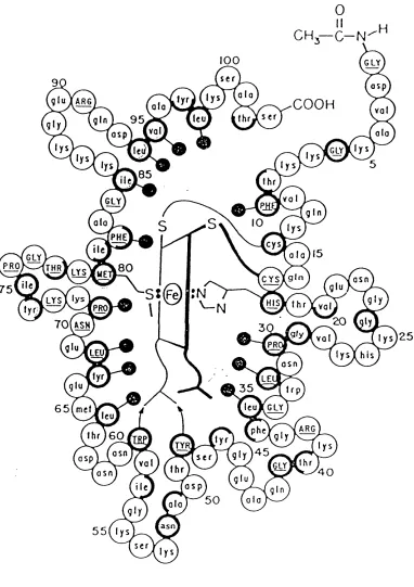
Cytochromes taken from different species show a high degree of structural homology. As can be seen in the figures 1.1, 1.2, and 1.314 the burying of the haem and the propionic side chains is achieved in cytochromes with as many as one hundred and thirty amino acids (Paracoccus c550), through one hundred and three amino acids (Tuna c), to as few as eighty amino acids CPseudomonas c55l). In cytochrome-c the polypeptide chain is organized into five a-helices that contain =45% of the amino acids. There is no B-strand
13 Comments on Inorg Chem, 1985 4_ 55-98; G Williams, G R Moore, R J P Williams.
67|
48
40
20 29
33 56
105
29
112
119 78
40
84 24
131
48 130
126
structure but there are six, type-II 310 bends, most of which are located at the base and right-hand side of the structure, according to the orientation of figure 1.4. The amino acids in the cytochrome-c15 can be classified into three groups as is demonstrated in figure 1.5: solvent exposed (normal circles), partly solvent exposed (half-bold circles), and buried (bold circles). The polypeptide chain is wrapped around the haem in such a way as to only leave =4% of the haem exposed, through an area representing only =0.06% of the total protein surface16. The haem is bound to the protein through four covalent bonds to the amino acids: Cysl4, Cysl7, Hisl8, and Met80, as seen in figure 1.617. The haem and polypeptide are further held in relation to one another by an extensive array of non-covalent interactions, as is demonstrated in the figure 1.718. Besides the structural homology there is also a high degree of sequence homology among cytochrome-cs. That is to say that cytochrome-cs from different species often have the same amino acids in the same positions (or equivalent positions in the case of those cytochromes with a greater or lesser number of amino acids in total), or have at least a substituted amino acid of the same kind; for example hydrophil ic or hydrophobic side chain.
The study of proteins should follow the pathway: sequence —> structure —> dynamics —> function. Identification of those regions that have unusual dynamic properties may yield information on the activity of the protein. The structure of cytochrome-c has been investigated by a variety of techniques, which have revealed much information upon which the experiments and conclusions of this thesis are based19.
15 R E Dickerson and R Timkovich, in the Enzymes, vol 11 (P Boyer, ed) 3rd ed (Academic Press, New York, 1975), pp 397-547.
16 Nature, 1978 275 73-74; E Stellwagen.
17 Scientific American, 1980 242 136-153, R E Dickerson.
18 The Porphyrins, vol 7, p 268, R Timkovich. (D Dolphin, ed., Academic Press, New York, 1979).
19 R Lemberg and J Barret, Cytochromes (Academic Press, New York,
1973)
88
104
[60
21 28,
3 3
75
50
43
Figure 1.4
The a-helices are represented bv barrels, 3inbends by blocks.
0
100 GLY
s er
asp a la
C 0 0 H
ala val
leu 9 5
val ala
asp
l y s 8 5
I hr
vo I
GLY
g I n ala
PHE
,c*s,
a I a ) l 5 GLY
PRO
^ r s V ^ S p
asn 7 5
gl y PRO
20
7 0 ( ASH
PRi
LEU his
as n
3 5 t r p leuJfGLY 6 5 ( m e t
leu
ARG I h r ) 6 0
TYI
>•“% 5(
glU ) y
( a l a h
asn s e r
asp
val
Ihr 4 0
as n
ile a s p g I n
5 0 ala
asn
5 5 ( 1 ys ser
f
Lys
P h
Ltu
M«1
.80.
T h r
T h r
T y r
,48,
A»n
.52,
Horse cy toc hr om e £
There is general agreement between the different techniques that the structures deduced from X-ray diffraction studies20 are largely conserved in solution21. The haem propionate groups are buried, which prevents them from ionising as the pH is changed in the physiological ranges, and thus limiting pH- dependent redox properties22. There may of course still be pH-dependent structural changes (see chapter 4). A major redox-dependent structural change occurs at the back of the protein around Ile57 (from X-ray23, nmr24 and chemical modification work25). Nmr studies also indicate that there are changes in the region around PhelO and Tyr97M (both haem packing residues).
Pseudocontact shifts have been calculated using the low-spin, iron-III ion as a rhombic nmr shift probe27. It is then possible to express the observed shift as a sum of two terms, describing the dipolar (through space) and Fermi- contact (through bond) interactions of the nucleus and the unpaired electron28. The contact term is scalar and rapidly attenuated by intervening bonds, and therefore only has an observable effect on the protons of the haem group and its ligands. It has been estimated that only approximately 0.2% of the unpaired
20 J. Mol. Biol., 1981 153 79-115; T Takano and R E Dickerson.
21 Eur J Biochem, 1980 103 533-541; G R Moore, R J P Williams.
22 Biochim. Biophys. Acta, 1980 590 261; G R Moore, G W Pettigrew, R C Pitt, R J P Williams.
23 T Takano and R E Dickerson in Electron Transport and Oxygen
Utilisation, Elsevier, 1982, Chien Ho, ed.
24 J. Inorg. Biochem., 1980 12 1-15, G R Moore, R J P Williams, J C W Chien, L C Dickson.
25 See chapter four.
26 Faraday Discuss Chem Soc, 1982 74_ 311-329: G R Moore, Z-X Huang,
C G S Eley, H A Barker, G Williams, M N Robinson, R J P Williams.
27 J. Mol. Biol., 1985 183 447-460, G Williams, N J Clayden, G R Moore, R J P Williams.
28 K Wiithrich, Nmr in Biological Research: Peptides and Proteins
electron density is delocalised into the orbitals of the ligands29. Thus for all those protons, other than haem and haem ligand protons, the delocalisation of the electron does not affect the treatment of the unpaired electron as a single point dipole at the centre of the iron atom.
Thermal B-factors and molecular-dynamics calculations30 have been used to assessthe dynamics of the structure, as have the motions of aromatic side- chains about their CB-CY bonds31. Another probe of protein mobility is the NH/ND exchange rate. Ulmer and Kagi32 found that the NH exchange rates of the ferri protein are faster than for the ferro protein, and thus there are more dynamic states available to the ferri protein. Trp59 NH is particularly resistant to exchange. The most mobile protons are those at the surface of the protein, but there are differences even between the mobility of these, indicating that factors other than the degree of exposure are important in determining the dynamic properties of cytochrome-c.
For reactions of cytochrome-c with its physiological redox partners, the rate of electron transfer within the active complex is in the range 10M 0V 1, and we can therefore neglect slow processes (rate <102s'1) such as protein unfolding, ligand exchange, and rotations of many aliphatic and aromatic side-chains within the protein. Rates occurring in the range 1012-1014 s'1 are in the range of those studied by X-ray crystallography33 (B-factors) and arise from small oscillations
29 Biochim Biophys Acta, 1973 322 38-44; W Horrocks, W De, Jr, and S Greenberg.
30 Nature (London), 1980 286 304: S H Northrup, M R Pear, J A
McCammon, M Karplus.
J Mol Biol, 1981 153 1087; S H Northrup, M R Pear, J D Morgan, J A McCammon, M Karplus.
31 FEBS Letts., 1976 70 96; I D Campbell, C M Dobson, G R Moore, S J Perkins, R J P Williams.
32 Biochemistry, 1968, 7 2710; D D Ulmer, J H R Kagi.
33 T Takano and R E Dickerson, in "Electron Transport and Oxygen
Utilisation" (C Ho, ed) (Elsevier, Amsterdam, 1982),pp 17-26.
of atoms or small groups of atoms about potential energy minima. It has been suggested that Tyr74 and Phe82 are involved in the activation/relaxation of the reaction centre. This is because Tyr74 is in a redox-state, conformationally- sensitive position, while Phe82 lines the left side of the haem crevice34.
The rates of conformational changes may be studied using nmr. In cytochrome-c the large chemical shift differences between pairs of methyl groups from the same leucine or valine residue have confirmed that most of the interior of cytochrome-c has a lack of mobility34. The interior might be considered as a crystalline solid rather than the conventional "oil-drop" model of protein interiors. In these regions asymmetric side-chains flip at rates less than 1 0 V .34
In the Marcus Hush model of electron transfer,35 a five step mechanism is envisaged. First the reactants diffuse together. This is followed by some structural reorganisation of bond lengths and angles of the complex and the surrounding solvent, in order that the electron transfer step may occur isoenergetically. Subsequent relaxation yields the equilibrium configuration of the product complex, and is followed by diffusion apart to give the separated products. The motion of the electron must satisfy the Frank-Condon principle that electronic and nuclear motions are not coupled. The mechanism proposed for the transfer of electrons in electron-transfer proteins has elements in common with both transition-state theory and the description of transitions in electronic spectra. The reactive complex {AB}' (figure 1.8) in which the electron is transferred has the property that its energy is independent of the position of the electron. Thus no energy is absorbed or emitted during the electron transfer step.
The same reaction scheme may be applied to the electron-transfer reactions of proteins such as cytochrome-c. In such a system a wide range of
34 Comments on Inorganic Chemistry, 1985 4 55-98; G Williams, G R
Moore, R J P Williams.
35 Ann. Rev. Phys. Chem., 1964 75 155-196; R A Marcus. Inorg Chem, 1975 14 213-216; R A Marcus, N Sutin.
vibrational and torsional modes are available, through which the energy of the electron at the acceptor and donor sites may be controlled. It is necessary to establish to what extent the polypeptide serves to prevent structural changes in the haem environment on changing between the oxidised and reduced states, as in the Marcus-Hush model, or whether it is more important that the polypeptide protect the haem from potential ligands other than those supplied by the polypeptide itself. The Marcus Hush model36 makes the assumption that changes affecting the energy of the electron at the haem in the reactive complex are not dependent on the precise nature of the redox partner37. It has been demonstrated in chapter four of this thesis that this is a justified assumption because the structure of tfa-modified cytochrome-c is not different from the native protein. In studies comparing different cytochromes38, attention has been drawn to the fact that certain residues, the structure, and the redox potential of cytochrome- cs are highly conserved. In chapter seven of this thesis the effect of amino acid sequence on the structure and dynamics of cytochrome-c will be investigated, and in chapters four to six an attempt will be made to establish the relevance of electrostatic versus hydrophobic effects to the biological functioning of the protein.
Before any further attempt is made to explain the structure and function of cytochrome-c, it is necessary to understand the electrostatic interactions within this protein.
36 Proc. Natl. Acad. Sci. USA, 1976 73_ 2950, S Wherland, H B Gray.
37 Comments Inorg Chem, 1985 4 55-98; G Williams, G R Moore, R J P
Williams.
38 R Lemberg and J Barrett in Cytochromes, Academic Press, New York,
GD
<
CD ro cTD c_
O O CJ Cl O CJ ro cu
CD
’<
A 6 j a u a a a j j
There’s nothing like science for generating so much speculation about so little fact.
ELECTROSTATICS
Electrostatic forces are the predominant long-range forces in chemistry, and have been invoked as being the major controlling factor in the stability of the native structure of proteins against denaturadon, in solubility, and in enzyme catalysis39. In this chapter electrostatic effects will be explored with a view to developing models for the structures, ion-binding properties, and the denaturadon temperatures of modified and native cytochrome-cs.
Electrostatic interaction energy theories have been used extensively in attempting to describe the structure, denaturadon, and ion binding properties of cytochrome-c. The interactions of specific charges throughout the protein have been used in the analysis of structure40, redox potential41, and denaturadon rates39, whilst the interaction of specific charges on the surface of the cytochrome-c and the net dipole42 of all the charged groups have been used to describe effects observed in ion binding reactions.
For example "Salt-bridge" ion-pair interactions have been observed between lysine and glutamic acid or aspartic acid residues on the cytochrome- c surface, and between an arginine residue and a haem propionate group in the protein interior. Hydrogen-bonds formed between oppositely charged functional groups, or between one charged and one uncharged side chain are expected to have especially large interaction energies, and may therefore be important in the maintenance of local and global structure. A list of charge interactions, other than those found in helical stretches of protein backbone, which have been determined in a 1.5 A refinement of tuna ferrocytochrome-c X-ray crystallographic data40, is given in table 2.1 (in which the donor atoms are presented first in each pair, and the amino acids containing the donor atoms are presented in ascending numerical order). In particular hydrogen bonds involving
39 Science, 1978 201 1187-1191, M F Perutz.
40 / . Mol. B iol, 1981 153 79-94; T Takano, R E Dickerson.
41 Biochim. Biophys. Acta., 1984 764 331-342; G R Moore, D E Harris, F A Leitch, G W Pettigrew.
lysines 13 and 79 have been shown to be important in maintaining the conformation of cytochrome-c, and in particular in maintaining the haem crevice43.
Secondly in sperm whale ferrimyoglobin44, the intra-molecular, electrostatic interactions were thought to be the most important factor influencing the stability of the native protein, since the native form showed a different ionic strength titration dependence to the denatured form of the same protein. Lastly other electrostatic interactions in cytochrome-c have been implicated in the redox conformation change, where an increase in the charge on the haem results in a stronger attraction of electrons, thus causing a partial positive charge on Met80Os. This results in the weakening of the hydrogen bond to Tyr670^t which causes Asn52 to move, since Tyr67 and Asn52 are linked by a water molecule45.
In order to assess the importance of these assertions, it is necessary to find and examine a protein in which the charged groups have been removed. In chapter four chemically modified proteins, in which the locations of the modifications are known, will be compared to the native form of the same protein, and information will be obtained about the relative importance of electrostatic interactions in determining structure, ion binding and electron distribution of the haem.
Denaturadon of proteins by acid or alkali is often brought about by the ionisation of internal residues that can be then stabilised by interaction with the solvent The total electrostatic interactions of solvent molecules must be weighed against the total electrostatic and non-electrostatic interactions within the native protein, and if the former is larger, the denatured state will be the enthalpically favoured one. Any entropic
43 J. Biol. Chem., 1980 255 1689-1697; N Osheroff, D Borden, W H
Koppenol, E Margoliash.
44 Biochemistry, 1979 18 4612-4630; S H Friend, F R N Gurd.
Table 2.1
Non-helical hydrogen bond in tuna ferrocvtochrome-c.
Asp2NH—>Asp9306, Arg38Ne—>HP7‘, Lys72N^—>Asn70Or Lys86Nc->Glu69CO, Lys86N^—>Ala83CO, Ly s99Nc—> Asn610 6,
Lysl3N c->Glu90OE, Lys55Nc->Tyr74CO, Lys79Nt->Ser47CO, Tyr480n—>HP7‘, Arg91Ne—>Ile85CO,
contribution to the Gibb’s free energy will arise from the changes in solvation, or from the rotational entropy of the polypeptide. In chapter five of this thesis, the denaturadon temperatures of a chemically modified and native cytochrome- c will be compared. The contribution to the free energy of denaturadon from the breaking of electrostatic interactions will be discussed.
It has been suggested that the redox potential of cytochrome-c is controlled by the electrostatic interaction of the haem with the charged groups in the protein. In cytochromes the haem redox potentials span the range from +400mV to -300mV46447. This large variation in redox potentials can be attributed in part to the different types of metal centre used in those cytochromes. Furthermore these variations in redox potentials can be attributed to the type of liganding groups at the haem48. Thus model systems containing a protohaem (-115mV) have a higher redox potential than those containing a mesohaem (-158mV), and those model systems that have a histidine and a methionine in the fifth and sixth ligand positions of a mesohaem have a higher redox potential (-llOmV) than those proteins with two histidines (-220mV)49. Synthetically produced iron porphyrins that have bridging ligands, resulting in the coordination of proximal S-Me and imidazole to the fifth and sixth ligand positions on the iron, have a redox potential of -i-TSmV50. Cytochrome-c has a redox potential of +260mV, resulting from the wrapping of the protohaem in a hydrophobic environment. These gross changes in redox potential are fine tuned in different cytochromes within the same class. These variations have provided great interest, since comparison of the redox potentials of structurally homologous cytochromes can yield information on the likely mechanisms
46 R Lemberg and J Barrett (1973) in: "Cytochromes", Academic Press, New York.
47 R E Dickerson and R Timkovich (1975) in: The Enzymes" ( P Boyer, ed.) pp 397-547, Academic Press, New York.
48 FEBS Lett, 1977 79 229-232; G R Moore and R J P Williams.
49 Proc. Natl. Acad. Sci. USA., 1972 69 2263-2268, J R Kassner.
controlling those redox potentials.
In all cytochromes the haem is buried, leaving only a small part of the edge between pyrrole rings two and three exposed51. The haem edge bearing the two propionates is also buried, the propionates remaining charged and being stabilised by a series of H-bonding networks52. This stabilisation of the charges, which reduces the pK, of haem-propionate to a value outside of the physiological range (pK, <4.5)53, is demonstrated by comparison of two cytochromes. In horse cytochrome-c, which has the evolutionarily highly conserved residue arg38 that binds to haem propionate-7, the redox potential is constant (+260mV) in the pH range 4-8. Yet in cytochrome-cs that are lacking an arginine at position 38, and have instead asparagine or glutamine, a pH-dependent redox potential is observed54. Therefore the arg38 residue is used to maintain the high redox potential. Removal of this residue, by selective cleavage of the arg38-lys39 bond followed by removal of arg38, yields a protein with the met80-Fe bond intact, but with a redox potential 90mV lower54.
The overall charge has been correlated with the redox potential in a number of soluble, one-electron, transfer proteins taken from different sources55. A linear fit was demonstrated to occur between the overall charge of the protein and the redox potential. The more negative the overall charge, the more likely the protein was to have a lower redox potential. Indeed the redox potential has also been shown to be dependent on the binding affinity of the medium in which the protein is dissolved56. The redox potential increased by 15mV with increasing concentration of a binding medium (from ImM to 5mM NaCl), while
51 Scientific American, 1980 242 136-153, R E Dickerson.
52 R Timkovich (1979) in: The Porphyrins (D Dolphin, ed.) pp 241-294, Academic Press, New York.
53 FEBS Lett, 1983 161 171-175, G R Moore.
54 Biochim. Biophys. Acta., 1984 764 331-342; G R Moore, D E Harris, F A Leitch, G R Pettigrew.
55 Proc. Natl. Acad. Sci., 1985 82 3082-3085, D C Rees.
the redox potential decreased by 7mV with increasing concentration of a non binding medium (from 4mM to 12mM tris-cacodylic acid)57. In another study using spectrophotometric and potentiometric techniques simultaneously, it was possible to show that the redox potential of cytochrome-c can be suppressed by as much as 60mV on binding inside submitochondrial particles58. However no similar effect was observed for cytochrome-Ci.
Furthermore the extent of haem exposure has been implicated as the reason for the variation in redox potentials in a number of proteins59460. In a more recent paper, the redox potential variation was ascribed to the differences in the charge distribution on the haem propionates61. It was shown that haem propionate-7 can contribute as much as 63mV in electrostatic interaction energy to the redox potential.
If all the electrostatic interactions of charged residues with the haem over horse cytochrome-c are summed, assuming that the only change in the protein at oxidation or reduction is the change in charge on the haem, the total electrostatic interaction energy difference between the protein in the two oxidation states is of the order of +9.71kJmol"162. This is composed of: +34.36kJmol'1 from the positively charged side chains (lysines, arginines), of which +14.61kJmol'1 results from the interaction energies of those lysines
57 These redox potentials were calculated from the observed equilibrium
constants fo r the reaction o f ferrocytochrome-c with ferricyanide according to the equations given in FEBS Letts; 1970 6 278; R Margalit, A Schejter.
58 Biochemistry, 1970 9 5077-5082; P L Dutton, D F Wilson, C-P Lee.
59 Nature, 1978 275_ 73-74, E Stellwagen.
60 J. Biol. Chem., 1979 254 4110-4113; G G Schlauder, R J Kassner.
61 FEBS Letts, 1983 161 171-175, G R Moore.
involved in the binding of cytochrome-c to its redox partners; -18.60kJmol'1 from the negatively charged residues (glutamic acid and aspartic acid) and - b.OSkJmol'1 from haem-propionate-7.
It was shown that at low ionic strength (<0.01M), the equations involving the dipole orientation63 can be reduced to the standard Brpnsted-Debye-Huckel equation. Since all the work for this thesis has been performed at ionic strengths of the order of 4-8mM, it is satisfactory to adopt the simplest equation to describe the electrostatic effects of the protein.
The difference in electrostatic interaction energies between the cytochrome in its different redox states might be able to account for the differences in structures, fine tuning of redox potentials, and so on. However a greater problem is how to decide on the value of dielectric constant to use. Early studies found that a value of 2-5M was suitable for the interior of globular proteins, although a value of 27 might be appropriate to the interaction of haem propionate-7 with the central iron in cytochrome-Cjj!65. A value of 50 has been given for the dielectric constant concerning solvent-exposed lysine terminal amine groups66. The fact that the protein does not behave as a sphere of uniform dielectric has led to the introduction of the term microscopic dielectric effects67.
Different models have been used to fit mathematical equations to experimental data. By inclusion of more terms, the basic Coulombic equation68
63 Biochim. Biophys. Acta., 1981 635 434; J W van Leeuwen, F Mofers, E Veerman.
Proc. Natl. Acad. Sci. USA., 1976 73_ 2950; S Wherland, H B Gray.
64 Adv. Prot. Chem., 1968 23 283-437; G N Ramachandran, V
Sasisekharan.
65 J. Inorg. Biochem., 1985 23_ 219-226; G R Moore, N K Rogers.
66 J. Mol. B iol, 1980 141 323-326, D C Rees.
67 Biochemistry, 1986 25_ 1675-1681; A K Churg, A Warshel.
68 Memoires de VAcademie royale des Sciences (1785), Charles-Augustine
can be modified to take account of the ionic strength of the solution (Debye- Hiickel), the effect of the dielectric of a solution, the dipole of the protein and the orientation of that dipole69. The extension of these equations allows a more accurate description of the experimental data collected. A further problem is the effect that the solvent has on the effective dielectric. When the ordering of solvent dipoles around the protein is taken into account (the solvent reaction field70), the calculated interaction energy between all charges with each other and with the haem for cytochrome-C551 could be decreased from 500mV to 90mV71.
Using the cavity model an interaction energy is calculated which is an order of magnitude lower than that calculated by using the uniform dielectric model, when considering the a-helix in globular proteins70. The uniform dielectric model takes no account of the reaction field of the bulk solvent molecules that have their dipoles lined up in opposition to the protein field, whilst the cavity model takes express account of the reaction field set up by the high dielectric bulk solvent that surrounds the protein.
In chapter four the structure of a modified cytochrome-c, in which all of the positive charges have been removed, is described. In this modification the interaction energy of all the lysine terminal amine groups with the haem is lost, with the result that the overall interaction energy could be as much as +30.14kJmol'1 lower, yielding an overall interaction energy of -lO^SkJmol'1.
A desire to understand the electrostatic interactions of the charged groups at the surface with complementary charges on the redox partners, has led to the study of ion binding to cytochrome-c. Indeed the fact that the cytochrome transfers electrons at a high rate in the physiological environment has led to the extension of simple Debye-Huckel theory to include the magnitude and orientation of any dipole that occurs within the cytochrome. The asymmetric
69 Biochim. Biophys. Acta., 1981 635 434; J W van Leeuwen, F Mofers, E Veerman.
70 J. Mol. Biol., 1984 174 527-542; N K Rogers, M J E Sternberg.
distribution of charges72 which produces a dipole with the positive end pointing out of the haem crevice at the front of the protein73, where the transfer of electrons to redox partners is known to take place74, has been invoked as the reason why more encounters of negative redox reagents are successful in producing electron transfer. Equations taking into account the dipole and its orientation75 have been fitted to data collected on the effects of ionic strength on rates of electron transfer. However the best fit for the data was achieved if the angle of the dipole to haem plane was increased from 30° to 40° in the calculations, but there is no chemical information to support this change. Although this may be only a small deviation, it demonstrates that the real import of studies in this area should not focus on the orientation of the dipole but on the significance of the dipole itself, as is investigated in chapters four and five of this thesis.
The very fact that electrostatic effects are long range effects leads to difficulties in using them to describe protein structure. If the protein is considered as having a uniform dielectric, then the electrostatic interactions will be non-directional; that is the same interaction energy will be generated wherever one of the charges is located on the surface of a sphere of fixed radius from the other charge. If the protein is described in terms of microscopic dielectric regions, then it will be significant to consider the direction of the interaction, because of the different dielectrics that would exist between the charges when they are in different positions. Thus electrostatic effects will be more useful in describing the stability of the protein than the structure.
In contrast the hydrophobic interactions, which are only short range and highly directional, will be more useful in analysing protein structures. For the
72 J. Biol. Chem., 1982 257 4426; W H Koppenol, E Margoliash.
73 TIBS, 1983 316-320; E Margoliash, H R Bosshard.
74 J. Chem. Soc., Dalton Trans., 1986 755; G D Armstrong, J A Chambers, A G Sykes.
same reason covalent interactions (H-bonding) will also be of great use in attempting to describe protein structures.
The object of opening the mind, as of opening the mouth, is to shut it again on something solid. G K Chesterton (1874-1936)
Personally I’m always ready to learn, although I do not always like being taught.
INTRODUCTION
Much previous work has been done to try to define and understand protein structures in terms of the protein function. That is to say that a link between amino acid sequence, tertiary structure, and dynamic properties has been sought, in order to define how the protein performs its biological function76. Proteins are generally made up of smaller structural units, which are collections of amino acids forming particular structural shapes such as a - helices, 310 bends, or B-pleated sheets, which often fit tightly into a larger quaternary structure. For example studies have shown that the a-helix is the most abundant unit of secondary structure in proteins77. In the specific case of cytochrome-c, =45% of the amino acids are found to be in a-helices78 (see figure 3.1). The rigidity of the structure is thought to be the reason why only small changes in the structure of cytochrome-c are observed upon change in the redox state of the haem. However it appears that it is the small dynamic changes in a protein that are ultimately in control of the protein function, and so it is necessary to be able to study the protein in a time-dependent way.
76 Advan. Protein Chem., 1966 27 113-287; E Margoliash, A Schejter. H A Harbury and R H L Marks, in "Inorganic Biochemistry", (G L Eichorn, ed) vol 2, pp 902-954, Elsevier, Amsterdam (1973).
R Lemberg and J Barrett, in "Cytochromes", Academic Press, New York (1973).
R E Dickerson and R Timkovich, in "The Enzymes", (P D Boyer, ed), vol 11, pp 397-547, Academic Press, New York, (1975).
S Fergusson-Miller, D L Brautigan, E Margoliash, in "THe Porphyrins", (D Dolphin, ed), vol 7, pp 149-240, Academic Press, New York, (1979).
77 G E Schulz and R H Schirmer, in "Principles o f Protein Structure", Springer-Verlag, Berlin, 1979.
78 Comments on Inorganic Chemistry, 1985 4 55-98; G Williams, G R
101
In the past, X-ray crystallography has been used extensively to study the structures of proteins, including metallo-proteins like cytochrome-c79. Inter atomic distances of the order of 0.2A can be measured with this technique in certain favourable cases, such as when the protein is rigid, as it is when bound to a haem group. The structures of cytochrome-cs from tuna80, bonito, yeast81, and rice82, as well as those of various bacteria83, have been determined by x- ray crystallography. A crystal structure determination was also attempted on horse cytochrome-c, but only collected to 2.8A84. Differences between the reduced and oxidised forms of tuna cytochrome-c were found, and the oxidised form has two structural forms per unit cell, termed outer and inner. The outer oxidised form was found to be closest to the solution structure of ferricytochrome-c85. This variation points to the value of determining solution as well as crystal structures of the various forms of cytochrome-c. interest to know what the solution structure is.
The use of thermal B-factors in x-ray crystallography allows some information to be gleaned about the flexibility of particular amino acid side- chains or regions of the protein. However in using thermal B-factors, it is difficult to distinguish whether a group is genuinely in motion or whether it has crystallised in one of a variety of possible positions. Temperature studies
79 M O’Dayhojf, in "Atlas o f Protein Sequence and Structure" (National Biomedical Research Foundation, Silver Spring, Maryland, 1968-78).
80 J. M ol B iol, 1981 153 71-115; T Takano, R E Dickerson.
81 J. Mol. B iol, 1988 199 295-314; G V Louie, W L B Hutcheon, G D
Brayer.
82 J. Mol. B iol, 1983 166 407-418; H Ochi, Y Hata, N Tanako, M Kakudo.
83 / . Mol. B iol, 1982 156 389-410; Y Matsura, T Takano, and R E Dickerson.
84 J. B iol Chem., 1971 246 1511-1535; R E Dickerson, T Takano, D
Eisenberg, O B Kallai, L Samson, A Cooper, E Margoliash.
of the crystal would be necessary to solve this ambiguity, although this would be time consuming. A further limitation is that x-ray crystallography is performed using crystals made with solutions of high ionic strength. Typically the crystal will contain approximately 50% protein, will have been made from a solution containing 85% saturation of ammonium sulphate, and in the case of reduced cytochrome-c, will have a 5 molar excess of ascorbate.
Other protein studies have used techniques that allow the solution structure of the protein to be probed86. Such studies have used Optical87, Circular Dichroism88 (CD), EPR89, ESR90, and Resonance Raman91 (Res. Raman) spectroscopies, and look at specific vibrations such as those of the haem-ligand bonds, or else at the overall effect of the protein on some physical property as in CD spectroscopy. The use of CD is more applicable to small proteins and peptides than to proteins as large or larger than cytochrome-c. CD is useful in ascertaining the content of structural units within a small protein, such as a -helices.
All of these various studies leave unanswered questions about what the overall structure in solution might be and what changes might occur in the protein under changes in physical and/or physiological conditions. A more valuable method of study of proteins in solution is therefore nuclear magnetic resonance (nmr) spectroscopy. The application of nmr will help to solve some of these difficulties, since samples are normally used in solution, and conditions (temperature, pH, ionic strength) in the nmr probe can be varied simultaneously.
Nuclear magnetic resonance is a useful tool because it allows the protein
86 Adv. Prot. Chem., 1982 35_ 105-212; T E Meyer, M D Karman.
87 Dev. Appl. Spectrosc., 1971 9 125-140; W B Elliot, E Margoliash.
88 Biochim. Biophys. Acta., 1970 214 94-106, Y P Myer.
89 J. Biol. Chem., 1977 252 574-582; D L Brautigan, et al.
90 Biophys. Chem., 1983 17 193-202; G B Postrikova, N P Gorbunova, M
V Vokenstein.
to be studied in solution at an atomic level. This is of further value in the study of horse cytochrome-c, since no high resolution structure of this form exists. A large amount of biological and biochemical data have been collected on horse cytochrome-c because it is readily available. Thus a full determination of horse cytochrome-c structure is a particularly useful task. Unlike tuna cytochrome-c, on which X-ray studies have been done at 1.5A and 1.8A resolution for the reduced92 and oxidised93 forms of the protein respectively, horse cytochrome-c has only been studied at 2.8A resolution94.
By the judicious use of phase cycling of the receiver coils in the nmr experiment, different magnetisation transfer pathways can be mapped out. This allows information to be gathered on the proximity of nuclei in space, those nuclei that are in dynamic or chemical exchange, and those nuclei that are part of an amino acid side chain and are therefore spin-coupled. Much work has been done using this technique, a full account of which can be found in "NMR of Proteins and Nucleic Acids"95. For specific use in biochemistry see reference twenty two96. Early studies of cytochrome-cs using nmr97 were involved in assigning the iron axial ligands at a time when X-ray crystallographic analyses could not distinguish between a number of ligand types. Much work was then done to assign the cytochrome-c nmr spectra further, in order that
structure-92 J. Mol. B io l, 1981 153 79-94; T Takano, R E Dickerson.
93 J. M o l Biol, 1981 153 95-115; T Takano, R E Dickerson.
94 J. Biol. Chem., 1971 246 1511-1535; R E Dickerson, T Takano, D
Eisenberg, O B Kallai, L Samson, A Cooper, E Margoliash.
95 NMR o f Proteins and Nucleic Acids 1986, K Wiithrich.
96 NMR and the Biochemist (Essays in Biochemistry Vol 19), G R Moore, R G Ratclijfe, R J P Williams.
97 Biochem. Biophys. Res. Comm., 1969 36 442-449; C C McDonald, W
D Phillips, S N Vinogradov.
Proc. N a tl Acad. Sci. USA, 1969 63 1071-1078, K Wiithrich.
function relationships of the protein in solution could be assessed98.
However before any further progress can be made in studying function, dynamics and so on, a more comprehensive assignment of the spectrum of ferricytochrome-c is necessary. It is the purpose of this chapter to use nmr to investigate and assign the spectrum of native, horse-heart cytochrome-c, and in particular the protons of the polypeptide backbone, since these provide secondary structural information.
In later chapters the effect of chemical modifications and sequence changes on the solution structure of cytochrome-c will be explored, using the knowledge gained from the assignments made in this chapter. By comparison of the two structures it may be possible to identify some of the important regions or structures used in the control of the physical properties of the protein such as ion binding affinity (see chapter 5).
MATERIALS AND METHODS
Protein Preparation
In this study Horse-heart, mitochondrial cytochrome-c was used. This was obtained from the Sigma Chemical Company (horse, type VI), and purchased as a freeze-dried powder with a claimed purity of 95% and with approximately 10% in the reduced form. The powdered cytochrome-c can include polymeric or fragmented protein, acetate, and trifluoroacetate ions from any buffering solution used in the initial purification process of the protein. In order to remove these impurities, and to ensure that all of the protein is in the same oxidation state, the procedure described below was executed.
98 J. Mol. Biol., 1985 183 409-446; G Williams, G R Moore, et al. Trends Biochem. Sci. (Pers, ed), 1983 8 316-320; E Margoliash, H R Bosshard.
Nature (London), 1986 322 286-288; R Bechtold, C Kuehn, C Lepre, S S Isied.
Science (Washington, D.C.), 1986 233 948-951; S L Mayo, W R Ellis, R J Crutchley, H B Gray.
First a portion of freeze dried protein (300mg) was dissolved in distilled water (5cm3), and potassium ferricyanide (50% mole ratio) was added to ensure total oxidation of the haem. Portions of the resulting solution (1cm3) were then put onto a small cation exchange column (30cm3, CM23) previously equilibrated in phosphate buffer (lOmM, pH7.0). Once the ferricyanide band was seen to have left the main protein band, another aliquot (1cm3) of the protein/ferricyanide solution was added to the top of the column. This had the effect of adding the protein in dilute solution, since the buffer solution was continually eluting whilst the consecutive additions of protein were made. Continuous elution with phosphate buffer (pH7, lOmM) was done. After all the protein had been added, five columns worth of eluent were passed through the column, to ensure the total removal of potassium ferro- and ferricyanide and impurities such as acetate and buffer ions remaining from the Sigma preparation.
The protein was removed from the column by elution with sodium chloride solution (0.5M), leaving polymeric material at the top of the column. Then the protein solution obtained was ultra-filtrated (Amicon 8010, with YM10 membrane) against distilled water to remove the sodium chloride and phosphate buffer ions. The membrane used in the ultra-filtration cell had a cut-off in the range of 10000 dal tons; meaning that only those molecules with a mass greater than 10000 dal tons were retained. Ultra-filtration was performed with volume reductions in factors of 10, until the salt concentration was undetectable by silver nitrate solution.
The protein was then carefully removed from the ultra-filtration cell using a pipette with a rubber end, and was freeze dried in Eppendorf tubes. Once freeze-dried, the protein was stored in the Eppendorf tubes at -40°C in a desiccator.
Sample Preparation