Open Access
Review
Hypophosphatasia
Etienne Mornet
1,2Address: 1Laboratoire SESEP, Centre Hospitalier de Versailles, Bâtiment EFS, 2 rue Jean-Louis Forain, 78150 Le Chesnay, France and 2Equipe Structure et Fonction, EA2493, Université de Versailles Saint-Quentin en Yvelines, Versailles, France
Email: Etienne Mornet - [email protected]
Abstract
Hypophosphatasia is a rare inherited disorder characterized by defective bone and teeth mineralization, and deficiency of serum and bone alkaline phosphatase activity. The prevalence of severe forms of the disease has been estimated at 1/100 000.
The symptoms are highly variable in their clinical expression, which ranges from stillbirth without mineralized bone to early loss of teeth without bone symptoms. Depending on the age at diagnosis, six clinical forms are currently recognized: perinatal (lethal), perinatal benign, infantile, childhood, adult and odontohypophosphatasia. In the lethal perinatal form, the patients show markedly impaired mineralization in utero. In the prenatal benign form these symptoms are spontaneously improved. Clinical symptoms of the infantile form are respiratory complications, premature craniosynostosis, widespread demineralization and rachitic changes in the metaphyses. The childhood form is characterized by skeletal deformities, short stature, and waddling gait, and the adult form by stress fractures, thigh pain, chondrocalcinosis and marked osteoarthropathy. Odontohypophosphatasia is characterized by premature exfoliation of fully rooted primary teeth and/or severe dental caries, often not associated with abnormalities of the skeletal system.
The disease is due to mutations in the liver/bone/kidney alkaline phosphatase gene (ALPL; OMIM# 171760) encoding the tissue-nonspecific alkaline phosphatase (TNAP). The diagnosis is based on laboratory assays and DNA sequencing of the ALPL gene. Serum alkaline phosphatase (AP) activity is markedly reduced in hypophosphatasia, while urinary phosphoethanolamine (PEA) is increased. By using sequencing, approximately 95% of mutations are detected in severe (perinatal and infantile) hypophosphatasia.
Genetic counseling of the disease is complicated by the variable inheritance pattern (autosomal dominant or autosomal recessive), the existence of the uncommon prenatal benign form, and by incomplete penetrance of the trait. Prenatal assessment of severe hypophosphatasia by mutation analysis of chorionic villus DNA is possible. There is no curative treatment for hypophosphatasia, but symptomatic treatments such as non-steroidal anti-inflammatory drugs or teriparatide have been shown to be of benefit. Enzyme replacement therapy will be certainly the most promising challenge of the next few years.
Disease name and synonyms
• Hypophosphatasia• Phosphoethanolaminuria
Published: 4 October 2007
Orphanet Journal of Rare Diseases 2007, 2:40 doi:10.1186/1750-1172-2-40
Received: 13 June 2007 Accepted: 4 October 2007
This article is available from: http://www.OJRD.com/content/2/1/40
© 2007 Mornet; licensee BioMed Central Ltd.
• Rathbun disease
• HOPS
Definition and diagnostic criteria
Hypophosphatasia (OMIM 146300, 241500, 241510) is an inherited disorder characterized by defective bone and teeth mineralization and deficiency of serum and bone alkaline phosphatase (AP) activity.
Epidemiology
The birth prevalence of severe hypophosphatasia was esti-mated to be 1/100 000 on the basis of pediatric hospital records in USA [1]. The incidence of moderate forms was never estimated but it is expected to be much higher, due to the number of patients with dominant forms carrying the same mutations than those found in recessive hypo-phosphatasia.
Clinical description
Clinical expression ranges from stillbirth without miner-alized bone to pathologic fractures developing only late in adulthood [2]. Depending on the age at diagnosis, six clinical forms are currently recognized: perinatal (lethal), infantile, childhood, adult, odontohypophosphatasia and a rare benign prenatal form characterized by in utero
detection but much better prognosis than other prenatal forms (Table 1). However, it should be noticed that these clinical subtypes overlap, for instance infantile and child-hood hypophosphatasia share some clinical symptoms, and patients with adult hypophosphatasia often had some clinical symptoms already in childhood.
In the lethal perinatal form, the patients show markedly in utero impaired mineralization. They have skin-covered osteochondral spurs protruding from the forearms or legs [3]. These spurs are often diagnostic for hypophosphata-sia. Some infants survive a few days but have respiratory complications due to hypoplastic lungs and rachitic deformities of the chest. Other symptoms include apnea, seizures and marked shortening of the long bones. In the rare prenatal benign form, despite prenatal symptoms, there is a spontaneous improvement of skeletal defects.
In the prenatal benign form, despite prenatal symptoms, there is a spontaneous improvement of skeletal defects [4,5]. The patients manifest limb shortening and bowing and often dimples overlaying the long bones deformities, and some ultrasounds revealed progressive improvement of the skeletal deformities and mineralization during the third trimester of the pregnancy [4,6].
Patients with the infantile form may appear normal at birth; however, the clinical signs of hypophosphatasia appear during the first six months. This form also has
res-piratory complications due to rachitic deformities of the chest. Despite the presence of an open fontanelle, prema-ture craniosynostosis is a common finding that may result in increased intracranial pressure. Radiographs show widespread demineralization and rachitic changes in the metaphyses. Hypercalcemia also is present, explaining in part a history of irritability, poor feeding, anorexia, vom-iting, hypotonia, polydipsia, polyuria, dehydratation, and constipation. Increased excretion of calcium may lead to renal damage. In infants who survive, there is often spon-taneous improvement in mineralization and remission of clinical problems, with the exception of craniosynostosis [7]. Short stature in adulthood and premature loss of deciduous teeth are also common, but the long-term out-look can be favorable [8].
Skeletal deformities, such as dolichocephalic skull and enlarged joints, a delay in walking, short stature, and wad-dling gait accompany the childhood form. Signs of intrac-ranial hypertension or failure to thrive are typical [2,9,10]. A history of fractures and bone pain usually exists as well. Focal bony defects near the ends of major long bones may be observed and help point to the diagnosis. Secondary metabolic inflammation seems to be common in the bone of patients [11] and hyperprostaglandinism affects the clinical severity [12]. Premature loss of dentition is common with the incisor teeth often being the first affected. Spontaneous remission of bone disease has been described, but the disease may re-appear in middle or late adulthood.
The adult form presents during middle age. The first com-plaint may be foot pain, which is due to stress fractures of the metatarsals. Thigh pain, due to pseudofractures of the femur, also may be a presenting symptom. There is also a predilection for chondrocalcinosis and marked osteoar-thropathy later in life. Upon obtaining an in-depth his-tory, many of these patients will reveal that they had premature loss of their deciduous teeth [13,14].
Etiology
The disease is due to mutations in the liver/bone/kidney alkaline phosphatase gene (ALPL; OMIM# 171760) encoding the tissue-nonspecific alkaline phosphatase (TNAP or TNSALP). TNAP is a phosphomonoesterase of 507 residues, anchored at its carboxyl terminus to the plasma membrane by a phosphatidylinositol-glycan moi-ety [16]. The enzyme is physiologically active in its dimeric form and cleaves extracellular substrates pyri-doxal-5'-phosphate (PLP), phosphoethanolamine (PEA) and inorganic pyrophosphates (PPi). Its exact function in bone and dental mineralization is still unclear but involves hydrolysis of PPi [17], and perhaps mammalian-specific activities such as collagen [18] and calcium bind-ing [19]. The TNAP gene is located on chromosome 1p36.1 [20] and consists of 12 exons distributed over 50 kb [21]. The gene is subject to high allelic heterogeneity [22] and more than 190 distinct mutations have been described [23]. Most of them (79%) are missense muta-tions. This variety of mutations results in highly variable clinical expressivity and in a great number of compound heterozygous genotypes.
Genotype-Phenotype correlations
Attempts to assess the relative importance of missense mutations and the genotype-phenotype relationship were performed on the basis of clinical data from patients, transfection studies [24-35], computer-assisted modeling [19,27], and studies of the biochemical properties of AP in cultured fibroblasts of patients [36] or transfected cells [37]. These experiments allowed to study cell localization,
degradation and alkaline phosphatase activity of mutant proteins. A good correlation was observed between the severity of the disease and in vitro enzymatic activity of the mutant protein [27,28,30,38]. Patients with mild hypo-phosphatasia carry at least one mutation that, when tested, exhibits significant residual enzymatic activity, while patients with severe hypophosphatasia carry muta-tions that, when tested, mostly do not product enzymatic activity. By using immunofluorescence and biochemical treatments, various mutations were characterized for their cell localization and their degradation [25,26,28,29,32-34,39,40]. These studies showed that most of the mis-sense mutations found in severe hypophosphatasia pro-duced a mutant protein that failed to reach the cell membrane, was accumulated in the cis-gogi and was sub-sequently degraded in the proteasome. By contrast, the missense mutations responsible for mild hypophosphata-sia were found to be at least in part correctly localized to the cell membrane. By using the crystal structure of the E. coli alkaline phosphatase [41], and then the crystal struc-ture of the human placental alkaline phosphatase [42], 3D models of the TNAP were built and used to localize the hypophosphatasia mutations in the molecule [19,27]. The severe missense mutations were shown to mostly affect residues localized in crucial domains of the protein while mutations found in mild forms affected residues more randomly dispatched on the molecule. Finally, and interestingly, the complementary approach consisting in
in vitro alkaline phosphatase measurement, immunofluo-rescence, biochemical treatments and 3D modeling con-Table 1: The six clinical forms of hypophosphatasia.
Clinical form Inheritance Bone symptoms Dental symptoms Clinical diagnosis
Perinatal lethal AR Hypomineralization Osteochondral spurs
na Radiographs
Ultrasonography Prenatal benign AD Bowing of long bones
Benign post-natal
na Ultrasonography
Clinical examination Infantile AR Craniosynostosis
Hypomineralization Rachitic ribs Hypercalciuria
Premature loss of deciduous teeth
Clinical examination
Biology (serum AP activity, PEA and PLP).
Radiographs Childhood AR (frequent)
or AD (rare)
Short stature Skeletal deformity Waddling gait Bone pain/fractures
Premature loss of deciduous teeth
Adult AR or AD Stress fractures: metatarsal, tibia Osteoarthritis
+/-Odontohypophosphatasia AR or AD Loss of alveolar bone Exfoliation (incisors). Reduced thickness of the dentin.
Enlarged pulp chambers of teeth.
Dental caries
Clinical examination.
Biology (serum AP activity, PEA and PLP).
verged to give a view of the severity of a mutation (Table 2).
The dominant effect of TNAP mutations
Dominant transmission of hypophosphatasia has been suggested on the basis of pedigree and laboratory data [13,43-45]. More recently, mutations responsible for this condition were identified: c.1133A>T (D361V) [46,47], c.346G>A (A99T) [48-50], c.188G>T (G46V), c.550G>T (R167W) and c.1433A>T (N461I) [49], c.323C>T (P91L) and c.1240C>A (L397M) [50], c.1259G>C (G403A), c.1402G>A (A451T) and c.1427A>C (E459A) (our unpublished data). In vitro, these mutations were shown to inhibit the normal monomer in the heterodimer made of mutant and normal proteins, resulting in decreased
lev-els of alkaline phosphatase activity. Instead of the 50% expected in heterozygotes, alkaline phosphatase activities were found to range from 20% to 40% of wild-type [49]. The most strong in vitro inhibition was found with muta-tions D361V and G46V, two mutamuta-tions responsible for the benign prenatal form of hypophosphatasia. Interest-ingly, parents of patients affected with benign prenatal hypophosphatasia express only very mild symptoms (mostly premature loss of teeth) or even, may be com-pletely unaffected [4,5,47]. This is also the case of families with mild hypophosphatasia due to dominant missense mutations. So, dominance is sometimes difficult to dem-onstrate by using familial analysis, since expression of the disease may be highly variable, with parents of even severely affected children showing no or extremely mild
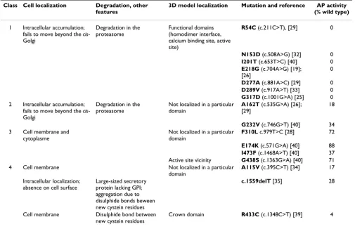
Table 2:
Class Cell localization Degradation, other features
3D model localization Mutation and reference AP activity (% wild type)
1 Intracellular accumulation; fails to move beyond the cis -Golgi
Degradation in the proteasome
Functional domains (homodimer interface, calcium binding site, active site)
R54C (c.211C>T), [29] 0
N153D (c.508A>G) [32] 0
I201T (c.653T>C) [40] 0
E218G (c.704A>G) [19]; [26]
0
D277A (c.881A>C) [29] 0
D289V (c.917A>T) [33] 0
G317D (c.1001G>A) [25] 0 2 Intracellular accumulation;
fails to move beyond the cis -Golgi
Degradation in the proteasome
Not localized in a particular domain
A162T (c.535G>A) [26]; [29]
18
G232V (c.746G>T) [40] 34 3 Cell membrane and
cytoplasme
Not localized in a particular domain
F310L c.979T>C [28] 72
E174K (c.571G>A) [40] 88
I473F (c.1468A>T) [40] 37 Active site vicinity G438S (c.1363G>A) [40] 71 4 Cell membrane Not localized in a particular
domain
A115V (c.395C>T) [34] 17
Intracellular localization; absence on cell surface
Large-sized secretory protein lacking GPI; aggregation due to disulphide bonds beween new cystein residues
c.1559delT [35] 28
Cell membrane Disulphide bond between new cystein residues
Crown domain R433C (c.1348C>T) [39] 4
Attempt to classify the ALPL gene mutations according to site-directed mutagenesis, in vitro alkaline phosphatase activity assays, and cell localization by immunofluorescence. Only mutations studied for all these parameters are shown. Class 1 represents the most severe mutations resulting in mutant proteins accumulated in the cytoplasm, subsequently degraded, and therefore producing no in vitro residual activity. These mutations affect residues of functional domains of the enzyme and were mostly found in patients with severe hypophosphatasia. Mutations of class 2 are also accumulated in the cell but exhibit low but significant in vitro residual activity and could be therefore degraded with delay. These mutations, that do not affect particular functional domains of the protein, must be also considered as severe alleles. Mutations of class 3 are in part accumulated in the cytoplasm but also in part reach the cell membrane. They exhibit high in vitro residual activity and except G438S, do not affect residues of functional domains. These mutations are observed in patients with mild forms of hypophosphatasia. Class 4 regroups particular mutations not assignable to the above classes.
symptoms of the disease [2,4]. This may be attributable both to the progressive improvement of affected patients from infancy to adulthood [13,36,51,52] and to epige-netic factors involved in the variable expression of the dis-ease. It is possible that in particular stages of development alkaline phosphatase requirements are beyond the capac-ity of the heterozygous cell, resulting in hypophosphata-sia symptoms. Then, AP requirements may be less important and filled by the heterozygous cell, which may explain the improvement in adult patients. It is also pos-sible that the maternal alkaline phosphatase plays a role
via fetal-maternal exchanges, as suggested by the prenatal benign form that seems to be observed only when the mutation is inherited from the mother [4-6].
Diagnostic methods
In addition to clinical and radiographic examinations (see clinical description), hypophosphatasia diagnosis is based on laboratory assays, and since 1990s, molecular biology which appears to be very effective.
Laboratory assays
Total serum AP activity is markedly reduced in hypophos-phatasia. Thus, the diagnosis can be suggested in individ-uals in whom serum AP activity is clearly and consistently subnormal. In general, the more severe the disease, the lower the serum AP activity level appropriate for age [2]. However, AP activity is only a helpful diagnostic indicator as other conditions may also show this finding: early preg-nancy, drug administration, hypothyroidism, anemia, celiac disease etc. It must be also noticed that serum AP dramatically varies with age and sex.
Increased urinary phosphoethanolamine (PEA) levels supports a diagnosis of hypophosphatasia but is not pathognomonic. It is also observed in a variety of other conditions, including several metabolic bone diseases, and some hypophosphatasia patients may have normal PEA excretion. In fact, the demonstration that PEA is also a natural substrate of TNAP in vivo remains to be con-firmed [53].
Increased pyridoxal 5'-phosphate (PLP) may be a sensitive marker for hypophosphatasia. [2].
Heterozygous carriers of the severe forms are usually clin-ically normal but often show modestly reduced serum AP activity and increased urinary PEA [54].
Molecular biology
Screening for mutations in the TNAP gene is essential to confirm the hypophosphatasia diagnosis when biochem-ical and clinbiochem-ical data are not clear enough, to offer genetic counseling or to offer molecular prenatal diagnosis to families affected by severe forms of the disease (see
below). Clinical and biochemical data may not always distinguish hypophosphatasia from other skeletal dis-eases such as osteogenesis imperfecta. Mutation screening may be performed by single-stranded conformation poly-morphism (SSCP) or denaturing gradient gel electro-phoresis (DGGE) followed by sequencing of exons exhibiting variants [55-62], by direct sequencing of the cDNA [36,46,63] or by direct sequencing of genomic sequences [30,64-67]. The exons are small and few in number, making relatively easy the analyze. However, the fact that the mutations are spread over all the exons often means that the whole coding sequence has to be analyzed. In addition, some mutations remain undetectable despite of exhaustive sequencing of the coding sequence, intron-exon borders and untranslated intron-exons. This may be due to mutations lying in intronic or regulatory sequences, but also to the expression of heterozygous mutations, espe-cially in moderate (childhood, adult and odonto-) hypo-phosphatasia. By using sequencing, approximately 95% of mutations are detected in severe (perinatal and infan-tile) hypophosphatasia, while patients with mild forms often carry only one detected mutated allele. This may be due to expression of the disease at the heterozygous state in some of these patients.
Differential diagnosis
• Osteogenesis imperfecta• Rickets
• Achondrogenesis
Antenatal diagnosis
Prenatal assessment of severe hypophosphatasia may be performed in couples with a previous affected child or a previous affected pregnancy. Mutation analysis of chori-onic villus DNAs is now well documented [68-71] and is routinely performed in few laboratories. It seems that mutation analysis is more reliable than AP assay of chori-onic villus sampling at least for heterozygote detection where low AP values may be misinterpreted [71]. Prenatal and postnatal diagnoses were also reported by using linked or intragenic polymorphisms [20,72]. In pregnan-cies with clinical symptoms detected by ultrasound but no familial history of hypophosphatasia, the prenatal diag-nosis by mutation analysis remains possible. However, such analyze is difficult, due to the time needed for the
ALPL gene sequencing, and may not always lead to a result.
Genetic counseling
dis-ease in heterozygotes, the probable effect of ALPL gene polymorphisms, and the possible effect of mutations and polymorphisms of other genes that may modulate the hypophosphatasia phenotype (modifier genes).
Severe forms of the disease (perinatal and infantile) are transmitted as an autosomal recessive trait, while both autosomal recessive and autosomal dominant transmis-sion have been shown in clinically milder forms [13,43-45]. Therefore, the risk of recurrence of severe forms is 25%. In moderate forms, it may be 25% (recessive trans-mission), 50% (dominant transmission) or still different (less than 50%) due to the variable expressivity of domi-nant forms [49,50]. The mutations detected in domidomi-nant forms and responsible for moderate hypophosphatasia are also found in severe recessive hypophosphatasia, asso-ciated to other mutations [48-50]. These mutations have a dominant negative effect due to the inhibition of AP activ-ity of the wild-type/mutant heterodimer [47,49], or due to intracytoplasmic sequestration of the heterodimer [Lia-baldini et al., in preparation]. Testing patient's relatives is useful since heterozygotes may express a mild form of the disease. In regard to the frequency of the disease, testing spouses of carriers is not primordial unless there is an his-tory of consanguinity.
Management including treatment
There is no curative treatment of hypophosphatasia, but symptomatic treatments are starting to be used in addi-tion to orthopedic management. Treatments with zinc and magnesium (catalytic ions of the enzyme), and pyri-doxal 5'-phosphate were reported to not significantly improve the patient's condition. However, the high clini-cal heterogeneity and the fact that the disease is rare make almost impossible controlled clinical trials. Preliminary results suggest that dietary phosphate restriction could be helpful in hypophosphatasia [73]. Non-steroidal anti-inflammatory drugs were shown to significantly improve the clinical features of childhood hypophosphatasia, especially in regard to pain [12,74] and to the secondary metabolic inflammation resulting from the disease [11]. Teriparatide (the recombinant human parathyroide hor-mone PTH 1–34) was successfully used to improve and resolve metatarsal stress fractures in adult hypophos-phatasia [62].
In 1997, MP Whyte's group (Saint-Louis, MI, USA) attempted to treat an 8-month-old girl affected with highly severe hypophosphatasia by bone marrow cell transplantation [75]. The patient was given T-cell-depleted, haplo-identical marrow from her healthy sister, and significant and prolonged clinical and radiographic improvement were observed. Another 9-month-old girl suffering from similar course of infantile hypophosphata-sia was treated by using bone fragments and cultured
oste-oblasts [76]. Seven years after transplantation, the patient was reported to be active and growing, and having the clinical phenotype of the more mild childhood form of hypophosphatasia [76]. These results suggest that donor bone fragments and marrow may provide precursor cells to form TNAP replete osteoblasts that can improve miner-alization [75,76]. Another interesting way of treatment would be to act onto the expression of the plasma cell membrane glycoprotein-1 (PC-1) gene, an antagonist of the TNAP gene [17]. Indeed, it has been shown in mice that inactivation of the Pc-1 gene in TNAP-knock-out mice allows to restore the normal bone phenotype [17]. Finally, enzyme replacement therapy by using a substitu-tive enzyme targeting mineralized tissue should be the most promising challenge of the next few years.
Prognosis
The perinatal form is almost always lethal within days or weeks, and around one half of patients with the infantile form dye from respiratory complications. Longevity stud-ies were not reported in the infantile and childhood forms. Patients affected with adult or odontohypophos-phatasia are believed to have normal lifespan.
References
1. Fraser D: Hypophosphatasia. Am J Med 1957, 22:730-46. 2. Whyte MP: Hypophosphatasia and the role of alkaline
phos-phatase in skeletal mineralization. Endocr Rev 1994, 15:439-61. 3. Shohat M, Rimoin DL, Gruber HE, Lachman RS: Perinatal lethal hypophosphatasia; clinical, radiologic and morphologic find-ings. Pediatr Radiol 1991, 21:421-7.
4. Pauli RM, Modaff P, Sipes SL, Whyte MP: Mild hypophosphatasia mimicking severe osteogenesis imperfecta in utero: bent but not broken. Am J Med Genet 1999, 86:434-8.
5. Moore CA, Curry CJ, Henthorn PS, Smith JA, Smith JC, O'Lague P, Coburn SP, Weaver DD, Whyte MP: Mild autosomal dominant hypophosphatasia: in utero presentation in two families. Am J Med Genet 1999, 86:410-5.
6. Wenkert D, McAlister WH, Coburn S, Ryan L, Hersh JH, Zerega J, Mumm S, MP W: Non-lethal hypophosphatasia interpreted as severe skeletal dysplasia in utero. Fifth International Alkaline Phos-phatase Symposium: "Understanding alkaline phosPhos-phatase function – Pathophysiology and treatment of Hypophosphatasia and other AP-related diseases", Huningue, France 2007.
7. Whyte MP, Magill HL, Fallon MD, Herrod HG: Infantile hypophatasia: normalization of circulating bone alkaline phos-phatase activity followed by skeletal remineralization. Evidence for an intact structural gene for tissue nonspecific alkaline phosphatase. J Pediatr 1986, 108:82-8.
8. Cole D: Hypophosphatasia. Amsterdam: Academic Press; 2003. 9. Fallon MD, Teitelbaum SL, Weinstein RS, Goldfischer S, Brown DM,
Whyte MP: Hypophosphatasia: clinicopathologic comparison of the infantile, childhood, and adult forms. Medicine (Baltimore) 1984, 63:12-24.
10. Kozlowski K, Sutcliffe J, Barylak A, Harrington G, Kemperdick H, Nolte K, Rheinwein H, Thomas PS, Uniecka W: Hypophosphatasia. Review of 24 cases. Pediatr Radiol 1976, 5:103-17.
11. Girschick HJ, Mornet E, Beer M, Warmuth-Metz M, Schneider P:
Chronic multifocal non-bacterial osteomyelitis in hypophos-phatasia mimicking malignancy. BMC Pediatr 2007, 7:3. 12. Girschick HJ, Schneider P, Haubitz I, Hiort O, Collmann H, Beer M,
Shin JS, Seyberth HW: Effective NSAID treatment indicates that hyperprostaglandinism is affecting the clinical severity of childhood hypophosphatasia. Orphanet J Rare Dis 2006, 1:24. 13. Whyte MP, Teitelbaum SL, Murphy WA, Bergfeld MA, Avioli LV:
investigation of a large kindred with review of the literature.
Medicine (Baltimore) 1979, 58:329-47.
14. Whyte MP, Murphy WA, Fallon MD: Adult hypophosphatasia with chondrocalcinosis and arthropathy. Variable pene-trance of hypophosphatasemia in a large Oklahoma kindred.
Am J Med 1982, 72:631-41.
15. Beumer J 3rd, Trowbridge HO, Silverman S Jr, Eisenberg E: Child-hood hypophosphatasia and the premature loss of teeth. A clinical and laboratory study of seven cases. Oral Surg Oral Med Oral Pathol 1973, 35:631-40.
16. Jemmerson R, Low MG: Phosphatidylinositol anchor of HeLa cell alkaline phosphatase. Biochemistry 1987, 26:5703-9. 17. Hessle L, Johnson KA, Anderson HC, Narisawa S, Sali A, Goding JW,
Terkeltaub R, Millan JL: Tissue-nonspecific alkaline phosphatase and plasma cell membrane glycoprotein-1 are central antag-onistic regulators of bone mineralization. Proc Natl Acad Sci USA 2002, 99:9445-9.
18. Hoylaerts MF, Millan JL: Site-directed mutagenesis and epitope-mapped monoclonal antibodies define a catalytically impor-tant conformational difference between human placental and germ cell alkaline phosphatase. Eur J Biochem 1991,
202:605-16.
19. Mornet E, Stura E, Lia-Baldini AS, Stigbrand T, Menez A, Le Du MH:
Structural evidence for a functional role of human tissue nonspecific alkaline phosphatase in bone mineralization. J Biol Chem 2001, 276:31171-8.
20. Greenberg CR, Evans JA, McKendry-Smith S, Redekopp S, Haworth JC, Mulivor R, Chodirker BN: Infantile hypophosphatasia: local-ization within chromosome region 1p36.1-34 and prenatal diagnosis using linked DNA markers. Am J Hum Genet 1990,
46:286-92.
21. Weiss MJRK, Henthorn PS, Lamb B, Kadesch T, Harris H: Structure of the human liver/bone/kidney alkaline phosphatase gene. J Biol Chem 1988, 263:12002-12010.
22. Mornet E: Hypophosphatasia: the mutations in the tissue-non-specific alkaline phosphatase gene. Hum Mutat 2000, 15:309-15. 23. Mornet E: The Tissue Nonspecific Alkaline Phosphatase Gene Mutations Database. 2007 [http://www.sesep.uvsq.fr/Data base.html].
24. Sugimoto N, Iwamoto S, Hoshino Y, Kajii E: A novel missense mutation of the tissue-nonspecific alkaline phosphatase gene detected in a patient with hypophosphatasia. J Hum Genet 1998, 43:160-4.
25. Fukushi M, Amizuka N, Hoshi K, Ozawa H, Kumagai H, Omura S, Misumi Y, Ikehara Y, Oda K: Intracellular retention and degra-dation of tissue-nonspecific alkaline phosphatase with a Gly317-->Asp substitution associated with lethal hypophos-phatasia. Biochem Biophys Res Commun 1998, 246:613-8.
26. Shibata H, Fukushi M, Igarashi A, Misumi Y, Ikehara Y, Ohashi Y, Oda K: Defective intracellular transport of tissue-nonspecific alkaline phosphatase with an Ala162-->Thr mutation associ-ated with lethal hypophosphatasia. J Biochem (Tokyo) 1998,
123:968-77.
27. Zurutuza L, Muller F, Gibrat JF, Taillandier A, Simon-Bouy B, Serre JL, Mornet E: Correlations of genotype and phenotype in hypo-phosphatasia. Hum Mol Genet 1999, 8:1039-46.
28. Cai G, Michigami T, Yamamoto T, Yasui N, Satomura K, Yamagata M, Shima M, Nakajima S, Mushiake S, Okada S, Ozono K: Analysis of localization of mutated tissue-nonspecific alkaline phos-phatase proteins associated with neonatal hypophosphatasia using green fluorescent protein chimeras. J Clin Endocrinol Metab 1998, 83:3936-42.
29. Fukushi-Irie M, Ito M, Amaya Y, Amizuka N, Ozawa H, Omura S, Ike-hara Y, Oda K: Possible interference between tissue-non-spe-cific alkaline phosphatase with an Arg54-->Cys substitution and acounterpart with an Asp277-->Ala substitution found in a compound heterozygote associated with severe hypophos-phatasia. Biochem J 2000, 348(Pt 3):633-42.
30. Taillandier A, Cozien E, Muller F, Merrien Y, Bonnin E, Fribourg C, Simon-Bouy B, Serre JL, Bieth E, Brenner R, Cordier MP, De Bie S, Fellmann F, Freisinger P, Hesse V, Hennekam RC, Josifova D, Kerzin-Storrar L, Leporrier N, Zabot MT, Mornet E: Fifteen new muta-tions (-195C>T, L-12X, 298-2A>G, T117N, A159T, R229S, 997+2T>A, E274X, A331T, H364R, D389G, 1256delC, R433H, N461I, C472S) in the tissue-nonspecific alkaline
phosphatase (TNSALP) gene in patients with hypophos-phatasia. Hum Mutat 2000, 15:293.
31. Watanabe H, Goseki-Sone M, Orimo H, Hamatani R, Takinami H, Ishikawa I: Function of mutant (G1144A) tissue-nonspecific ALP gene from hypophosphatasia. J Bone Miner Res 2002,
17:1945-8.
32. Ito M, Amizuka N, Ozawa H, Oda K: Retention at the cis-Golgi and delayed degradation of tissue-non-specific alkaline phos-phatase with an Asn153-->Asp substitution, a cause of peri-natal hypophosphatasia. Biochem J 2002, 361:473-80.
33. Ishida Y, Komaru K, Ito M, Amaya Y, Kohno S, Oda K: Tissue-non-specific alkaline phosphatase with an Asp(289)-->Val muta-tion fails to reach the cell surface and undergoes proteasome-mediated degradation. J Biochem (Tokyo) 2003,
134:63-70.
34. Watanabe H, Takinami H, Goseki-Sone M, Orimo H, Hamatani R, Ishikawa I: Characterization of the mutant (A115V) tissue-nonspecific alkaline phosphatase gene from adult-type hypo-phosphatasia. Biochem Biophys Res Commun 2005, 327:124-9. 35. Komaru K, Ishida Y, Amaya Y, Goseki-Sone M, Orimo H, Oda K:
Novel aggregate formation of a frame-shift mutant protein of tissue-nonspecific alkaline phosphatase is ascribed to three cysteine residues in the C-terminal extension. Retarded secretion and proteasomal degradation. Febs J 2005, 272:1704-17.
36. Fedde KN, Michell MP, Henthorn PS, Whyte MP: Aberrant proper-ties of alkaline phosphatase in patient fibroblasts correlate with clinical expressivity in severe forms of hypophosphata-sia. J Clin Endocrinol Metab 1996, 81:2587-94.
37. Di Mauro S, Manes T, Hessle L, Kozlenkov A, Pizauro JM, Hoylaerts MF, Millan JL: Kinetic characterization of hypophosphatasia mutations with physiological substrates. J Bone Miner Res 2002,
17:1383-91.
38. Orimo H, Girschick HJ, Goseki-Sone M, Ito M, Oda K, Shimada T:
Mutational analysis and functional correlation with pheno-type in German patients with childhood-pheno-type hypophos-phatasia. J Bone Miner Res 2001, 16:2313-9.
39. Nasu M, Ito M, Ishida Y, Numa N, Komaru K, Nomura S, Oda K:
Aberrant interchain disulfide bridge of tissue-nonspecific alkaline phosphatase with an Arg433-->Cys substitution associated with severe hypophosphatasia. Febs J 2006,
273:5612-24.
40. Brun-Heath I, Lia-Baldini A, Maillard S, Taillandier A, Utsch B, Nunes ME, Serre JL, Mornet E: Delayed transport of tissue-nonspecific alkaline phosphatase with missense mutations causing hypo-phosphatasia. Eur J Med Genet 2007, 50(5):367-378.
41. Kim EE, Wyckoff HW: Reaction mechanism of alkaline phos-phatase based on crystal structures. Two-metal ion catalysis.
J Mol Biol 1991, 218:449-64.
42. Le Du MH, Stigbrand T, Taussig MJ, Menez A, Stura EA: Crystal structure of alkaline phosphatase from human placenta at 1.8 A resolution. Implication for a substrate specificity. J Biol Chem 2001, 276:9158-65.
43. Whyte MP, Vrabel LA, Schwartz TD: Adult hypophosphatasia: generalized deficiency of alkaline phosphatase activity dem-onstrated with cultured skin fibroblasts. Trans Assoc Am Physi-cians 1982, 95:253-63.
44. Eastman JR, Bixler D: Clinical, laboratory, and genetic investi-gations of hypophosphatasia: support for autosomal domi-nant inheritance with homozygous lethality. J Craniofac Genet Dev Biol 1983, 3:213-34.
45. Eberic FHS, Pralle H, Kabish A: Adult hypophosphatasia without apparent skeletal disease: "ondotohypophosphatasia" in four heterozygote members of a family. Klin Wochenschr 1984,
62:371.
46. Henthorn PS, Raducha M, Fedde KN, Lafferty MA, Whyte MP: Differ-ent missense mutations at the tissue-nonspecific alkaline phosphatase gene locus in autosomal recessively inherited forms of mild and severe hypophosphatasia. Proc Natl Acad Sci USA 1992, 89:9924-8.
Publish with BioMed Central and every scientist can read your work free of charge "BioMed Central will be the most significant development for disseminating the results of biomedical researc h in our lifetime."
Sir Paul Nurse, Cancer Research UK
Your research papers will be:
available free of charge to the entire biomedical community
peer reviewed and published immediately upon acceptance
cited in PubMed and archived on PubMed Central
yours — you keep the copyright
Submit your manuscript here:
http://www.biomedcentral.com/info/publishing_adv.asp
BioMedcentral
48. Hu JC, Plaetke R, Mornet E, Zhang C, Sun X, Thomas HF, Simmer JP:
Characterization of a family with dominant hypophosphata-sia. Eur J Oral Sci 2000, 108:189-94.
49. Lia-Baldini AS, Muller F, Taillandier A, Gibrat JF, Mouchard M, Robin B, Simon-Bouy B, Serre JL, Aylsworth AS, Bieth E, Delanote S, Freis-inger P, Hu JC, Krohn HP, Nunes ME, Mornet E: A molecular approach to dominance in hypophosphatasia. Hum Genet 2001, 109:99-108.
50. Herasse M, Spentchian M, Taillandier A, Keppler-Noreuil K, Fliorito AN, Bergoffen J, Wallerstein R, Muti C, Simon-Bouy B, Mornet E:
Molecular study of three cases of odontohypophosphatasia resulting from heterozygosity for mutations in the tissue non-specific alkaline phosphatase gene. J Med Genet 2003,
40:605-9.
51. Robinow M: Twenty-year follow-up of a case of hypophos-phatasia. Birth Defects Orig Artic Ser 1971, 7:86-93.
52. Lepe X, Rothwell BR, Banich S, Page RC: Absence of adult dental anomalies in familial hypophosphatasia. J Periodontal Res 1997,
32:375-80.
53. Millan J: Mammalian alkaline phosphatases: from biology to applications in medicine and biotechnology. Weinheim: Wiley-VCH Verlag GmbH; 2006.
54. Rasmussen H: Hypophosphatasia. McGraw-Hill, New York; 1983. 55. Orimo H, Hayashi Z, Watanabe A, Hirayama T, Hirayama T, Shimada T: Novel missense and frameshift mutations in the tissue-nonspecific alkaline phosphatase gene in a Japanese patient with hypophosphatasia. Hum Mol Genet 1994, 3:1683-4. 56. Orimo H, Goseki-Sone M, Sato S, Shimada T: Detection of
dele-tion 1154–1156 hypophosphatasia mutadele-tion using TNSALP exon amplification. Genomics 1997, 42:364-6.
57. Mornet E, Taillandier A, Peyramaure S, Kaper F, Muller F, Brenner R, Bussiere P, Freisinger P, Godard J, Le Merrer M, Oury JF, Plauchu H, Puddu R, Rival JM, Superti-Furga A, Touraine RL, Serre JL, Simon-Bouy B: Identification of fifteen novel mutations in the tissue-nonspecific alkaline phosphatase (TNSALP) gene in Euro-pean patients with severe hypophosphatasia. Eur J Hum Genet 1998, 6:308-14.
58. Goseki-Sone M, Orimo H, Iimura T, Takagi Y, Watanabe H, Taketa K, Sato S, Mayanagi H, Shimada T, Oida S: Hypophosphatasia: identi-fication of five novel missense mutations (G507A, G705A, A748G, T1155C, G1320A) in the tissue-nonspecific alkaline phosphatase gene among Japanese patients. Hum Mutat 1998:S263-7.
59. Mumm S, Jones J, Finnegan P, Henthorn PS, Podgornik MN, Whyte MP: Denaturing gradient gel electrophoresis analysis of the tissue nonspecific alkaline phosphatase isoenzyme gene in hypophosphatasia. Mol Genet Metab 2002, 75:143-53.
60. Watanabe H, Hashimoto-Uoshima M, Goseki-Sone M, Orimo H, Ishikawa I: A novel point mutation (C571T) in the tissue-non-specific alkaline phosphatase gene in a case of adult-type hypophosphatasia. Oral Dis 2001, 7:331-5.
61. Watanabe H, Goseki-Sone M, Iimura T, Oida S, Orimo H, Ishikawa I:
Molecular diagnosis of hypophosphatasia with severe perio-dontitis. J Periodontol 1999, 70:688-91.
62. Whyte MP, Mumm S, Deal C: Adult hypophosphatasia treated with teriparatide. J Clin Endocrinol Metab 2007, 92:1203-8. 63. Greenberg CR, Taylor CL, Haworth JC, Seargeant LE, Philipps S,
Triggs-Raine B, Chodirker BN: A homoallelic Gly317-->Asp mutation in ALPL causes the perinatal (lethal) form of hypo-phosphatasia in Canadian mennonites. Genomics 1993,
17:215-7.
64. Taillandier A, Zurutuza L, Muller F, Simon-Bouy B, Serre JL, Bird L, Brenner R, Boute O, Cousin J, Gaillard D, Heidemann PH, Steinmann B, Wallot M, Mornet E: Characterization of eleven novel muta-tions (M45L, R119H, 544delG, G145V, H154Y, C184Y, D289V, 862+5A, 1172delC, R411X, E459K) in the tissue-non-specific alkaline phosphatase (TNSALP) gene in patients with severe hypophosphatasia. Mutations in brief no. 217. Online. Hum Mutat 1999, 13:171-2.
65. Spentchian M, Merrien Y, Herasse M, Dobbie Z, Glaser D, Holder SE, Ivarsson SA, Kostiner D, Mansour S, Norman A, Roth J, Stipoljev F, Taillemite JL, van der Smagt JJ, Serre JL, Simon-Bouy B, Taillandier A, Mornet E: Severe hypophosphatasia: characterization of fif-teen novel mutations in the ALPL gene. Hum Mutat 2003,
22:105-6.
66. Brun-Heath I, Taillandier A, Serre JL, Mornet E: Characterization of 11 novel mutations in the tissue non-specific alkaline phos-phatase gene responsible for hypophosphatasia and geno-type-phenotype correlations. Mol Genet Metab 2005, 84:273-7. 67. Spentchian M, Brun-Heath I, Taillandier A, Fauvert D, Serre JL,
Simon-Bouy B, Carvalho F, Grochova I, Mehta SG, Muller G, Oberstein SL, Ogur G, Sharif S, Mornet E: Characterization of Missense Muta-tions and Large DeleMuta-tions in the ALPL Gene by Sequencing and Quantitative Multiplex PCR of Short Fragments. Genet Test 2006, 10:252-7.
68. Watanabe A, Yamamasu S, Shinagawa T, Suzuki Y, Miyake H, Takes-hita T, Orimo H, Shimada T: Prenatal genetic diagnosis of severe perinatal (lethal) hypophosphatasia. J Nippon Med Sch 2007,
74:65-9.
69. Henthorn PS, Whyte MP: Infantile hypophosphatasia: successful prenatal assessment by testing for tissue-non-specific alka-line phosphatase isoenzyme gene mutations. Prenat Diagn 1995, 15:1001-6.
70. Orimo H, Nakajima E, Hayashi Z, Kijima K, Watanabe A, Tenjin H, Araki T, Shimada T: First-trimester prenatal molecular diagno-sis of infantile hypophosphatasia in a Japanese family. Prenat Diagn 1996, 16:559-63.
71. Mornet E, Muller F, Ngo S, Taillandier A, Simon-Bouy B, Maire I, Oury JF: Correlation of alkaline phosphatase (ALP) determination and analysis of the tissue non-specific ALP gene in prenatal diagnosis of severe hypophosphatasia. Prenat Diagn 1999,
19:755-7.
72. Iqbal SJ, Plaha DS, Linforth GH, Dalgleish R: Hypophosphatasia: diagnostic application of linked DNA markers in the domi-nantly inherited adult form. Clin Sci (Lond) 1999, 97:73-8. 73. Wenkert D, Podgornik MN, Coburn SP, Ryan LM, Mumm S, Whyte
MP: Dietary phosphate restriction therapy for hypophos-phatasia: preliminary observations. Fifth International Alkaline Phosphatase Symposium: "Understanding alkaline phosphatase function – Pathophysiology and treatment of Hypophosphatasia and other AP-related diseases" 2007, Huningue, France 2007.
74. Girschick HJ, Seyberth HW, Huppertz HI: Treatment of child-hood hypophosphatasia with nonsteroidal antiinflammatory drugs. Bone 1999, 25:603-7.
75. Whyte MP, Kurtzberg J, McAlister WH, Mumm S, Podgornik MN, Coburn SP, Ryan LM, Miller CR, Gottesman GS, Smith AK, Douville J, Waters-Pick B, Armstrong RD, Martin PL: Marrow cell trans-plantation for infantile hypophosphatasia. J Bone Miner Res 2003, 18:624-36.
76. Cahill RA, Wenkert D, Perlman SA, Steele A, Coburn SP, McAlister WH, Mumm S, Whyte MP: Infantile Hypophosphatasia: Trans-plantation Therapy Trial Using Bone Fragments and Cul-tured Osteoblasts. J Clin Endocrinol Metab 2007.
77. Weiss MJ, Ray K, Henthorn PS, Lamb B, Kadesch T, Harris H: Struc-ture of the human liver/bone/kidney alkaline phosphatase gene. J Biol Chem 1988, 263:12002-10.
78. Antonarakis SE: Recommendations for a nomenclature system for human gene mutations. Nomenclature Working Group.