1.1
34
0
0
Full text
(2) Start-Up Activities. LAUNCH Lab. Biological Molecules Make the following Foldable to help you organize information about biological molecules.. How do you test for simple sugars? Many different food sources supply the energy that your body uses constantly. This energy is stored in the bonds of molecules called simple sugars.. STEP 1 Fold a sheet of notebook paper lengthwise, keeping the margin visible on the left side.. STEP 2 Cut the top flap into four tabs.. Analysis 1. Describe the color changes you observed. 2. Classify Which foods contained a simple sugar? Inquiry Think about what you had for dinner last night. Which foods likely contained simple sugars? How could you test the food to find out?. STEP 3 Label as follows: Biological Molecules, Proteins, Carbohydrates, Lipids, and Nucleic Acids.. Proteins. Biological Molecules. Procedure 1. Read and complete the lab safety form. 2. Fill a 400-mL beaker until it is one-third full of water. Place it on a hot plate and heat it to boiling. 3. Use a graduated cylinder to measure 5.0 mL 10% glucose solution. Pour it into a test tube. 4. Add 3.0 mL Benedict’s solution to the test tube. Mix the two solutions using a stirring rod. Add a boiling chip to the test tube. WARNING: Benedict’s solution is an eye and skin irritant. 5. Using tongs, place the test tube in the boiling water bath for 5 min. 6. A color change to yellow or orange indicates the presence of a simple sugar. Record your observations. 7. Repeat the procedure using a 10% starch solution, a 10% gelatin suspension, and a few drops of honey suspended in water.. tes. Carbohydra. Lipids s. Nucleic Acid. &/,$!",%3 Use this Foldable with Sections 23.1, 23.2, 23.3, and 23.4. As you read, summarize the general structure and function of the biological molecules, and give examples of each.. Visit glencoe.com to: ▶ study the entire chapter online ▶. explore. ▶. take Self-Check Quizzes. ▶. use the Personal Tutor to work Example Problems step-by-step. ▶. access Web Links for more information, projects, and activities. ▶. find the Try at Home Lab, Modeling Sugars. Chapter 23 • The Chemistry of Life. 825. Matt Meadows.
(3) Section 23.1 Objectives ◗ Describe the structures of amino acids and proteins. ◗ Explain the roles of proteins in cells.. Review Vocabulary polymer: large molecules composed of many repeating units called monomers. New Vocabulary protein amino acid peptide bond peptide denaturation enzyme substrate active site. Proteins MAIN Idea Proteins perform essential functions, including regulation of chemical reactions, structural support, transport of materials, and muscle contractions. Real-World Reading Link Some cleaning products, such as contact lens. cleaning solution, contain enzymes. Did you ever wonder what an enzyme was?. Protein Structure Enzymes form a class of proteins. Proteins are organic polymers made of amino acids linked together in a specific order. Proteins are not just large, randomly arranged chains of amino acids. To function properly, each protein must be folded into a specific three-dimensional structure. All living organisms, including the mountain goat and the plants shown in Figure 23.1, are composed of proteins. In this section, you will read about how proteins are made from their amino-acid building blocks and how different types of proteins function. Amino acids As you read in Chapter 22, many different functional groups are found in organic compounds. Amino acids, as their name implies, are organic molecules that have both an amino group and an acidic carboxyl group. The general structure of an amino acid is shown below. —. R Variable side chain. Hydrogen atom. —. H2N — C — C — OH Carboxyl group —. Amino group. H. O. Each amino acid has a central carbon atom around which four groups are arranged: an amino group (—NH 2), a carboxyl group (—COOH), a hydrogen atom, and a variable side chain, R. The side chains range from a single hydrogen atom to a complex double-ring structure. Figure 23.1 All living organisms contain proteins. A goat’s hair, hooves, and muscles are made up of structural proteins, as are the roots and leaves of plants.. ■. 826. Chapter 23 • The Chemistry of Life. (l)© John Conrad/CORBIS, (r)©Ron Niebrugge/Alamy.
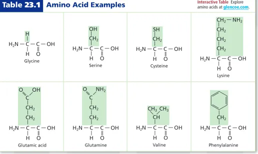
(4) Interactive Table Explore amino acids at glencoe.com.. Table 23.1 Amino Acid Examples. O. Serine. —. H. CH2. H2N — C — C — OH —. Glycine. CH2. H. O. H2N — C — C — OH. Cysteine. —. H2N — C — C — OH —. —. CH2. —. —. O. CH2. — —. — —. —. CH2. H2N — C — C — OH H. SH. —. OH. H. — — — —. CH2 — NH2. H. O. Lysine. ——. C. CH3 CH3. O. H. O. H2N — C — C — OH. Valine. —. H. Glutamine. Glutamic acid. — —. — —. — O. —. — H. H2N — C — C — OH. H2N — C — C — OH. H2N — C — C — OH. CH2. —. —. — —. CH. CH2. CH2. —. — CH2. — —. CH2. —. —. C. —. ——. NH2. —. O. OH. —. O. H. O. Phenylalanine. Examine the different side chains of the amino acids shown in Table 23.1. Identify the nonpolar alkanes, polar hydroxyl groups, acidic and basic groups such as carboxyl and amino groups, aromatic rings, and sulfur-containing groups. This wide range of side chains gives the different amino acids a large variety of chemical and physical properties and is an important reason why proteins can perform so many different functions. The peptide bond The amino and carboxyl groups provide convenient bonding sites for linking amino acids together. Because an amino acid is both an amine and a carboxylic acid, two amino acids can combine to form an amide, releasing water in the process. This reaction is a condensation reaction. As Figure 23.2 shows, the carboxyl group of one amino acid reacts with the amino group of another amino acid to form an amide functional group. Reading Check Explain how an amide functional group forms.. Figure 23.2 The amino group of one amino acid bonds to the carboxyl group of another amino acid to form a dipeptide and water. The organic functional group formed is an amide linkage called a peptide bond.. ■. Peptide bond. Amino acid. Amino acid. —. O. H. O. H. O. —. H. R2. — —. H. →. H. —. O. N — C — C — OH. R1. — —. H. +. H —. H. N — C — C — OH. R2. — —. H —. R1. — —. H. N — C — C — N — C — C — OH H. Dipeptide. +. H 2O Water. Section 23.1 • Proteins 827.
(5) The amide bond that joins two amino acids, shown in Figure 23.3, is referred to by biochemists as a peptide bond. A chain of two or more amino acids linked together by peptide bonds is called a peptide. A molecule that consists of two amino acids bound together by a peptide bond is called a dipeptide. Figure 23.4a shows the structure of a dipeptide that is formed from the amino acids glycine (Gly) and phenylalanine (Phe). Figure 23.4b shows a different dipeptide, also formed by linking together glycine and phenylalanine. Is Gly-Phe the same compound as Phe-Gly? No, they’re different. Examine these two dipeptides to see that the order in which amino acids are linked in a dipeptide is important. Each end of the two-amino-acid unit in a dipeptide still has a free group—one end has a free amino group and the other end has a free carboxyl group. Each of those groups can be linked to the opposite end of yet another amino acid, forming more peptide bonds. Living cells always build peptides by adding amino acids to the carboxyl end of a growing chain.. —. H. —. — C — N — Peptide bond O Figure 23.3 A peptide bond joins two amino acids to form a dipeptide.. ■. Reading Check Explain the difference between a peptide and a. dipeptide. Polypeptides As peptide chains increase in length, other ways of referring to them become necessary. A chain of ten or more amino acids joined by peptide bonds is referred to as a polypeptide. An example of a polypeptide is shown in Figure 23.5. When a chain reaches a length of about 50 amino acids, it is called a protein. Because there are only 20 different amino acids that form proteins, it might seem reasonable to think that only a limited number of different protein structures are possible. However, a protein can have as few as 50 or more than a 1000 amino acids, arranged in any possible sequence. To calculate the number of possible sequences these amino acids can have, consider that each position on the chain can have any of 20 possible amino acids. For a peptide that contains n amino acids, there are 20 n possible sequences of the amino acids. So a dipeptide, with only two amino acids, can have 20 2, or 400, different possible amino acid sequences. Even the smallest protein, containing only 50 amino acids, has 20 50, or more than 1 × 10 65, possible arrangements of amino acids! It is estimated that human cells make between 80,000 and 100,000 different proteins. You can see that this is only a small fraction of the total number of proteins possible. Reading Check Calculate the possible number of sequences for a. peptide chain comprised of four amino acids. a. H. O. Gly. Phe. Glycylphenylalanine (Gly-Phe). 828. Chapter 23 • The Chemistry of Life. H. —. H. H. — —. O. —. H. N — C — C — N — C — C — OH. CH2. —. H —. CH2. — —. H. —. H. —. H. — —. —. —. b. — —. Figure 23.4 Glycine (Gly) and phenylalanine (Phe) can combine in two configurations. Explain Why are these two structures different substances? ■. H. O. H. O. N — C — C — N — C — C — OH H. Phe. Gly. Phenylalanylglycine (Phe-Gly).
(6) C C H O N. C C NH O C C H C H C N O C N O C H C H O N C N O C. O H O H O H O H C C N C C C N C C C N C C C N N N N C N C C C C H O H O H O H O. Hydrogen bonds. O C H O C H O C H O C H C N C N C N C N C N C N C N C N C C C C H O H O H O H O. Helix. Pleated sheet. Figure 23.5 The folding of polypeptide chains into both helices and sheets involves amino acids in the chain held in position by hydrogen bonds. Other interactions among the various side chains are not shown here but play an important role in determining the three-dimensional shape of a polypeptide.. ■. Three-dimensional protein structure Long chains of amino acids start to fold into unique three-dimensional shapes before they are fully synthesized. The three-dimensional shape is determined by the interactions among the amino acids. Some areas of a polypeptide might twirl into helices, which are similar to the coils on a telephone cord. Other areas might bend back and forth repeatedly into a pleated sheet structure, like the folds of an accordion. A polypeptide chain might also fold back on itself and change direction. A given protein might have several helices, sheets, and turns, or none at all. Figure 23.5 shows the folding patterns of a typical helix and a sheet. The overall three-dimensional shape of many proteins is globular—shaped like an irregular sphere. Other proteins have a long, fibrous shape. The shape is important to the function of the protein. If the shape of the protein changes, it might not be able to carry out its function in the cell.. Real-World Chemistry Enzymes. Denaturation Changes in temperature, ionic strength, pH, and other. factors result in the unfolding and uncoiling of a protein. Denaturation is the process in which a protein’s natural three-dimensional structure is disrupted. Cooking often denatures the proteins in foods. When an egg is hard-boiled, the protein-rich egg white solidifies due to the denaturation of its protein. Because proteins function properly only when folded, denatured proteins are generally inactive.. The Many Functions of Proteins Proteins play many roles in living cells. They are involved in speeding up chemical reactions, transport of substances, regulation of cellular processes, structural support of cells, communication within cells and among cells, cellular motion, and even serving as an energy source when other sources are scarce. Speeding up reactions In most organisms, the largest number of proteins function as enzymes, catalyzing the many reactions that occur in living cells. An enzyme is a biological catalyst. In Chapter 16, you read that a catalyst speeds up a chemical reaction without being consumed in the reaction. A catalyst usually lowers the activation energy of a reaction by stabilizing the transition state.. Papain An example of an enzyme you might have used is papain, found in papayas, pineapples, and other plant sources. This enzyme catalyzes a reaction that breaks down protein molecules into free amino acids. Papain is the active ingredient in many meat tenderizers. When you sprinkle the dried form of papain onto moist meat, the papain forms a solution that breaks down the tough protein fibers in the meat, making the meat more tender.. Section 23.1 • Proteins 829 ©Janet Horton.
(7) Complex sugar. Enzymes act on specific substrates, such as a complex sugar. Active sites. Each substrate fits into the active site. The enzyme changes shape slightly to fit with the substrate.. Enzyme (protein). Induced fit Products +H2O. Simple sugar. Simple sugar Active sites The products are released; in this case the complex sugar is divided into less complex sugars.. After the reaction, the enzyme released is in its original shape and can carry out the same reaction repeatedly. Enzyme. Figure 23.6 Enzymes lower the activation energy needed for a reaction to occur. Enzymes change the speed at which chemical reactions occur without being altered themselves in the reaction.. ■. Figure 23.7 Hemoglobin is a globular protein with four polypeptide chains, each containing an iron group (called a heme) to which oxygen binds.. ■. Heme. How do enzymes function? The term substrate refers to a reactant in an enzyme-catalyzed reaction, as shown in Figure 23.6. Substrates bind to specific sites on enzyme molecules, usually pockets or crevices. The spot to which the substrates bind is called the active site of the enzyme. After the substrates bind to the active site, the active site changes shape slightly to fit more tightly around the substrates. This recognition process is called induced fit. The shapes of the substrates must fit the shape of the active site, in the same way that puzzle pieces or a lock and key fit together. A molecule that is only slightly different in shape from an enzyme’s normal substrate will not bind as well to the active site and might not undergo the catalyzed reaction. The structure that forms when substrates are bound to an enzyme is called an enzyme-substrate complex. The large size of enzyme molecules allows them to form multiple bonds with their substrates, and the large variety of amino acid side chains in the enzyme allows a number of different intermolecular forces to form. These intermolecular forces lower the activation energy needed for the reaction in which bonds are broken and the substrates are converted to product. Reading Check Describe in your own words how an enzyme works.. Transport proteins Some proteins are involved in transporting smaller particles throughout the body. Figure 23.7 shows the protein hemoglobin, which carries oxygen in the blood from the lungs to the rest of the body. Other proteins combine with biological molecules called lipids to transport them from one part of the body to another through the bloodstream. You will learn about lipids later in this chapter. 830. Chapter 23 • The Chemistry of Life.
(8) Figure 23.8 Human hair is made up of a fibrous structural protein called keratin.. ■. SEM magnification: 500×. Structural support The sole function of certain proteins is to form structures vital to organisms. These molecules are known as structural proteins. The most abundant structural protein in most animals is collagen, which is part of skin, ligaments, tendons, and bones. Other structural proteins make up feathers, fur, wool, hooves, fingernails, cocoons, and hair, as shown in Figure 23.8. Communication Hormones are messenger molecules that carry signals from one part of the body to another. Some hormones are proteins. Insulin, a familiar example, is a small (51 amino acids) protein hormone made by pancreas cells. When insulin is released into the bloodstream, it signals body cells that blood sugar is abundant and should be stored. A lack of insulin often results in diabetes, a disease that results when there is too much sugar in the bloodstream. Because modern technology has made possible the laboratory synthesis of proteins, some protein hormones are being synthetically produced for use as medicines. Insulin, thyroid hormones, and growth hormones are some examples. Both natural and synthetic proteins are used in a variety of products—from meat tenderizer to cleaning solutions to health and beauty aids.. Section 23.1. &/,$!",%3. Incorporate information from this section into your Foldable.. Assessment. Section Summary. 1.. ◗ Proteins are biological polymers made of amino acids that are linked by peptide bonds.. 2. Compare the structures of amino acids, dipeptides, polypeptides, and proteins. Which has the largest molecular mass? The smallest?. ◗ Protein chains fold into intricate three-dimensional structures.. 4. Evaluate How do the properties of proteins make them such useful catalysts? How do they differ from other catalysts you have studied?. ◗ Proteins have many functions in the human body, including functions within cells, functions between cells, and functions of structural support.. 5. Explain three roles of proteins in cells, and give an example of each role.. MAIN Idea. Describe three proteins and identify their functions.. 3. Draw the structure of the dipeptide Gly-Ser, circling the peptide bond.. 6. Categorize Identify an amino acid from Table 23.1 that can be classified into each of the categories in the following pairs. a. nonpolar v. polar b. aromatic v. aliphatic c. acidic v. basic. Self-Check Quiz glencoe.com. Section 23.1 • Proteins 831 (l)©Royalty-Free/Corbis, (r)©Medical-on-Line/Alamy.
(9) Section 23.2 Objectives ◗ Describe the structures of monosaccharides, disaccharides, and polysaccharides. ◗ Explain the functions of carbohydrates in living things.. Review Vocabulary stereoisomers: a class of isomers whose atoms are bonded in the same order but are arranged differently in space. New Vocabulary carbohydrate monosaccharide disaccharide polysaccharide. Carbohydrates MAIN Idea Carbohydrates provide energy and structural material for living things. Real-World Reading Link A lot of media attention has been focused on carbohydrates. Low-carb diets have become a popular way of controlling weight. However, carbohydrates are an important energy source for the body.. Kinds of Carbohydrates Analyzing the term carbohydrate offers a hint about the structure of this group of molecules. Early observations that these compounds have the general chemical formula C n(H 2O) n and appear to be hydrates of carbon led to their being called carbohydrates. Although scientists now know that there are no full water molecules attached to carbohydrates, the name has stayed. The main function of carbohydrates in living organisms is as a source of energy, both immediate and stored. Foods rich in carbohydrates include pasta, milk, fruit, bread, and potatoes. Carbohydrates are compounds that contain multiple hydroxyl groups (—OH) as well as a carbonyl functional group (C=O). These molecules range in size from single monomers to polymers made of hundreds or even thousands of monomer units. Monosaccharides The simplest carbohydrates, often called simple sugars, are monosaccharides. The most common monosaccharides have either five or six carbon atoms. Examples of monosaccharides are shown in Figure 23.9. Notice that they have a carbonyl group on one carbon and hydroxyl groups on most of the other carbons. The presence of a carbonyl group makes these compounds either aldehydes or ketones, depending on the location of the carbonyl group. Multiple polar groups make monosaccharides water-soluble and give them high melting points.. Figure 23.9 Glucose, galactose, and fructose are monosaccharides. In aqueous solutions, they exist in an equilibrium between their open-chain and cyclic forms.. ■. H C— O H—C— OH. CH2OH H C H C OH HO C H. HO— C— H. O H. H— C—OH. C ⇌ H C OH. Cyclic form. HO C. O H. H—C— OH. H C. H—C— OH. H C. C OH. H Open-chain form. Glucose 832. H—C— OH HO— C— H. CH2OH. C OH. OH. Chapter 23 • The Chemistry of Life. H. H C— O. H. HO— C— H ⇌. H— C— OH H—C— OH. OH. Cyclic form. H Open-chain form. Galactose. H— C — OH —O C— HO— C— H. CH2OH O C H HO C OH. H. H— C— OH. HO C ⇌ C CH2OH. H— C— OH H— C— OH. H. Cyclic form. H Open-chain form. Fructose.
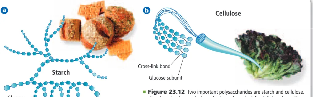
(10) CH2OH OH HO. CH2OH. CH2OH O. O. HO. + OH HO OH OH. Glucose. OH. → CH2OH. CH2OH O. O. HO O. HO OH. Fructose. + CH2OH. H2O. OH Sucrose. Water. Figure 23.10 When glucose and fructose bond, the disaccharide sucrose forms. Note that water is also a product of this condensation reaction. Remember that each ring structure is made of carbon atoms, which are not shown for simplicity.. ■. Glucose is a six-carbon sugar that has an aldehyde structure. Glucose is present in high concentration in blood because it serves as the major source of immediate energy for the body. For this reason, glucose is often called blood sugar. Closely related to glucose is galactose, which differs only in how a hydrogen and a hydroxyl group are oriented in space around one of the six carbon atoms. Recall from Chapter 21 that this relationship makes glucose and galactose stereoisomers. Fructose, also known as fruit sugar because it is the major carbohydrate in most fruits, is a six-carbon monosaccharide that has a ketone structure. Fructose is a structural isomer of glucose. When monosaccharides are in aqueous solution, they exist in both open-chain and cyclic structures, but they rapidly interconvert forms. The cyclic structures are more stable and are the predominant form of monosaccharides at equilibrium. Note in Figure 23.9 that the carbonyl groups are present only in the open-chain structures. In the cyclic structures, they are converted to hydroxyl groups. Disaccharides Like amino acids, monosaccharides can be linked together by a condensation reaction in which water is released. When two monosaccharides bond together, a disaccharide is formed, as shown in Figure 23.10. The new bond formed is an ether functional group (C–O–C). One common disaccharide is sucrose, also known as table sugar because sucrose is used mainly as a sweetener. Sucrose is formed by the linking of glucose and fructose. Another common disaccharide is lactose, the most important carbohydrate in milk. It is often called milk sugar. Lactose is formed when glucose and galactose bond. Polysaccharides Complex carbohydrate is a term used in some nutrition books and journal articles. Another name for a complex carbohydrate is polysaccharide, which is a polymer of simple sugars that contains 12 or more monomers, or subunits. The same type of bond that joins two monosaccharides in a disaccharide also links the monomers in a polysaccharide. Glycogen, shown in Figure 23.11, is a polysaccharide. It is composed of glucose subunits. It stores energy and is found mostly in the liver and muscles of humans and other animals. It is also found in some species of microorganisms including bacteria and fungi.. VOCABULARY WORD ORIGIN Polysaccharide comes from the Greek word polys, which means many and the ancient Sanskrit word śarkarā, which means sugar. Figure 23.11 The glycogen found in the muscle and liver of animals is a polysaccharide made of glucose.. ■. Glycogen Glucose subunit. Reading Check Explain the differences among a monosaccharide, a disaccharide, and a polysaccharide. Section 23.2 • Carbohydrates 833 ©Royalty Free/IndexStock.
(11) a. b. Cellulose. Cross-link bond. Starch. Glucose subunit. Figure 23.12 Two important polysaccharides are starch and cellulose. a. Starch molecules can be branched or unbranched. b. Cellulose has a linear, unbranched structure that resembles a chain-link fence.. ■. Glucose subunit. &/,$!",%3. Incorporate information from this section into your Foldable.. Section 23.2. Two other important polysaccharides are starch and cellulose, shown in Figure 12.12. Starch and cellulose are also composed solely of glucose subunits. However, that is the only similarity among the three polysaccharides, as all three have different properties and functions. Plants make both starch and cellulose. Starch is a soft, water-insoluble molecule used to store energy, whereas cellulose is a water-insoluble polymer that forms rigid plant-cell walls, such as those found in wood. Glycogen, starch, and cellulose are composed of glucose subunits, but they have different properties. The bonds that link the subunits together are oriented differently in space. Because of this difference in bond shape, humans can digest glycogen and starch but not cellulose. Digestive enzymes cannot fit cellulose into their active sites. The cellulose in the fruits, vegetables, and grains that we eat is called dietary fiber because it passes through the digestive system largely unchanged.. Assessment. Section Summary. 7.. ◗ Carbohydrates are compounds that contain multiple hydroxyl groups (–OH) and a carbonyl functional group (C=O).. 8. Describe the structures of monosaccharides, disaccharides, and polysaccharides. Which has the largest molecular mass? The smallest?. ◗ Carbohydrates range in size from single monomers to polymers composed of hundreds or thousands of monomers. ◗ Monosaccharides in aqueous solution exist in both open-chain and cyclic structures.. MAIN Idea. Explain the functions of carbohydrates in living things.. 9. Compare and contrast the structures of starch and cellulose. How do the structural differences affect our ability to digest these two polysaccharides? 10. Calculate If a carbohydrate has 2 n possible isomers, where n is equal to the number of chiral carbon atoms in the structure, calculate the number of possible isomers for the following monosaccharides: galactose, glucose, and fructose. 11. Interpret Scientific Illustrations Copy the illustration of sucrose on a separate sheet of paper, and circle the ether functional group that bonds the monomer sugars together.. CH2OH O OH O. HO OH. 834. Chapter 23 • The Chemistry of Life. (l)©Foodcollection.com/Alamy, (r)©Brand X Pictures/Alamy. CH2OH O HO OH. CH2OH. Self-Check Quiz glencoe.com.
(12) Section 23.3. Lipids. Objectives ◗ Describe the structures of fatty acids, triglycerides, phospholipids, and steroids. ◗ Explain the functions of lipids in living organisms. ◗ Identify some reactions that fatty acids undergo. ◗ Relate the structure and function of cell membranes.. MAIN Idea Lipids make cell membranes, store energy, and regulate cellular processes. Real-World Reading Link The wax used to polish cars, the fat that drips out of hamburgers, and the vitamin D that fortifies the milk people drink—what do these things have in common? They are all lipids.. What is a lipid? A lipid is a large, nonpolar biological molecule. Because lipids are nonpolar, they are insoluble in water. Lipids have two major functions in living organisms. They store energy efficiently, and they make up most of the structure of cell membranes. Unlike proteins and carbohydrates, lipids are not polymers with repeated monomer subunits.. Review Vocabulary nonpolar: without separate positive and negative areas or dipoles. New Vocabulary. Fatty acids Although lipids are not polymers, many lipids have a major building block in common. This building block is the fatty acid, a long-chain carboxylic acid. Most naturally occurring fatty acids contain between 12 and 24 carbon atoms. Their structure can be represented by the following formula.. lipid fatty acid triglyceride saponification phospholipid wax steroid. CH 3(CH 2) nCOOH Most fatty acids have an even number of carbon atoms, which is a result of being constructed two carbons at a time in enzymatic reactions. Fatty acids can be grouped into two main categories, depending on the presence or absence of double bonds between carbon atoms. Fatty acids that contain no double bonds are referred to as saturated. Those that have one or more double bonds are called unsaturated. The structures of two common fatty acids are shown in Figure 23.13. Reading Check Explain why oleic acid is described as unsaturated.. Figure 23.13 Two fatty acids, which are found in many foods, including butter, are the 18-carbon unsaturated oleic acid and the 18-carbon saturated stearic acid. Explain how the structure of the molecule is affected by the presence of a double bond. ■. Oleic acid. O HO. CCH2CH2CH2CH2CH2CH2CH2CH — CHCH2CH2CH2CH2CH2CH2CH2CH3. Stearic acid. O HO. CCH2CH2CH2CH2CH2CH2CH2CH2CH2CH2CH2CH2CH2CH2CH2CH2CH3. Section 23.3 • Lipids 835 ©D. Hurst/Alamy.
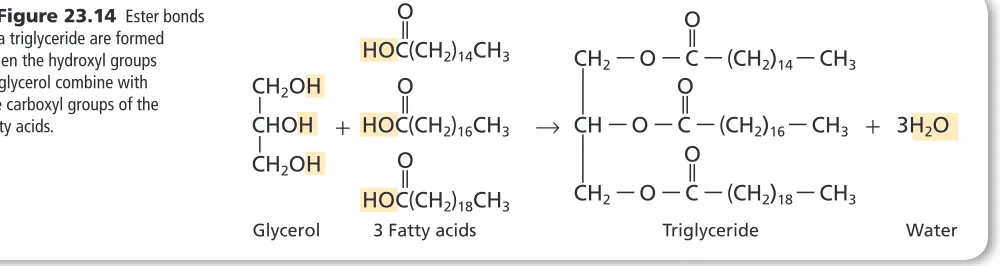
(13) O. CH2OH. —. O. CHOH + HOC(CH2)16CH3 O. —. CH2OH. HOC(CH2)18CH3 Glycerol. CH2 — O — C — (CH2)14 — CH3 O —. HOC(CH2)14CH3. —. —. O. → CH — O — C — (CH2)16 — CH3 + 3H2O O —. Figure 23.14 Ester bonds in a triglyceride are formed when the hydroxyl groups of glycerol combine with the carboxyl groups of the fatty acids.. ■. CH2 — O — C — (CH2)18 — CH3. 3 Fatty acids. Triglyceride. Water. An unsaturated fatty acid can become saturated if it reacts with hydrogen. As you read in Chapter 22, hydrogenation is an addition reaction in which hydrogen gas reacts with carbon atoms that are linked by multiple bonds. Each unsaturated carbon atom can pick up one hydrogen atom to become saturated. For example, oleic acid, shown in Figure 23.13, can be hydrogenated to form stearic acid. The double bonds in naturally occurring fatty acids are almost all in the cis geometric isomer form. Recall from Chapter 21 that the cis isomer has identical groups oriented on the same side of the molecule around a double bond. Because of the cis orientation, unsaturated fatty acids have a kink, or bend, in their structure that prevents them from packing together. They do not form as many intermolecular attractions as saturated fatty acid molecules. As a result, unsaturated fatty acids have lower melting points.. VOCABULARY SCIENCE USAGE V. COMMON USAGE Saturate Science usage: to add something to the point that no more can be absorbed, dissolved, or retained The salt water solution was saturated with salt. Common usage: to furnish a market with a product or products to its full purchasing capacity The shops in the coastal town are saturated with sea shell craft items.. 836. Chapter 23 • The Chemistry of Life. ©Michael Newman/PhotoEdit. Triglycerides Although fatty acids are abundant in living organisms, they are rarely found alone. They are most often found bonded to glycerol, a molecule with three carbons, each containing a hydroxyl group. When three fatty acids are bonded to a glycerol backbone through ester bonds, a triglyceride is formed. The formation of a triglyceride is shown in Figure 23.14. Triglycerides can be either solids or liquids at room temperature, as shown in Figure 23.15. If liquid, they are usually called oils. If solid at room temperature, they are called fats. Reading Check Identify two plant oils and two animal fats. Figure 23.15 Most mixtures of triglycerides from plant sources are liquids because the triglycerides contain unsaturated fatty acids. Animal fats contain a larger proportion of saturated fatty acids. They are usually solids at room temperature.. ■.
(14) Figure 23.16 Soap forms by the reaction of a triglyceride and a strong base.. O. —. ■. CH2 — O — C — (CH2)14CH3. CH2OH —. O. —. O. CH — O — C — (CH2)14CH3 + 3NaOH → CHOH + 3CH3(CH2)14 — C — O-Na+ —. O. CH2 — O — C — (CH2)14CH3 Triglyceride. CH2OH Base. Glycerol. Soap. Fatty acids are stored in the fat cells of your body as triglycerides. When energy is abundant, fat cells store the excess energy in the fatty acids of triglycerides. When energy is scarce, the cells break down the triglycerides, releasing the energy used to form them. Although enzymes break down triglycerides in living cells, the reaction can be duplicated outside of cells by using a strong base, such as sodium hydroxide. This reaction—the hydrolysis of a triglyceride using an aqueous solution of a strong base to form carboxylate salts and glycerol—is saponification, as shown in Figure 23.16. Saponification is used to make soaps, which are usually the sodium salts of fatty acids. A soap molecule has both a polar end and a nonpolar end. Soaps are used with water to clean nonpolar dirt and oil because the nonpolar dirt and oil bond to the nonpolar end of the soap molecules, and the polar end of the soap molecules is soluble in water. Thus, the dirt-laden soap molecules can be rinsed away with the water.. Observe a Saponification Reaction How is soap made? The reaction between a triglyceride and a strong base is called saponification. A sample chemical reaction is shown in Figure 23.16. Procedure 1. Read and complete the lab safety form. 2. Place a 250-mL beaker on a hot plate. Add 25 g solid vegetable shortening to the beaker. Turn on the hot plate to a medium setting. 3. As the vegetable shortening melts, use a 25-mL graduated cylinder to slowly add 12 mL ethanol and then 5 mL 6.0M NaOH to the beaker. WARNING: Ethanol is flammable. NaOH causes skin burns. Wear gloves. 4. Heat the mixture for about 15 min. Use a stirring rod to occasionally stir the mixture. Do not allow it to boil.. 5. When the mixture begins to thicken, use tongs to remove the beaker from the heat. Allow the beaker to cool for 5 min, then place it in a cold water bath in a 600-mL beaker. 6. Add 25 mL saturated NaCl solution to the mixture in the beaker. The soap is not very soluble and will appear as small clumps. 7. Collect the solid soap clumps by filtering them through a cheesecloth-lined funnel. 8. Using gloved hands, press the soap into an evaporating dish. Remove your gloves and wash your hands. Analysis. 1. Explain What type of bonds present in the triglycerides are broken during the saponification reaction? 2. Identify the type of salt formed in this chemical reaction. 3. Determine which is the polar end and which is the nonpolar end of the soap molecule.. Section 23.3 • Lipids 837.
(15) Figure 23.17 A phospholipid has a polar head and two nonpolar tails. The membranes of living cells are formed by a double layer of lipids, called a bilayer. The polar heads are on the outer and inner perimeter of the membrane and the tails are on the inside of the bilayer. ■. Outside the cell. Phospholipid bilayer. Polar heads. Nonpolar tails. Inside the cell. Phospholipids Another important type of triglyceride, a phospholipid, is found in greatest abundance in cellular membranes. A phospholipid is a triglyceride in which one of the fatty acids is replaced by a polar phosphate group. As shown in Figure 23.17, the polar part of the molecule forms a head and the nonpolar fatty acids look like tails. A typical cell membrane has two layers of phospholipids, which are arranged with their nonpolar tails pointing inward and their polar heads pointing outward. This arrangement is called a lipid bilayer. Because the lipid bilayer structure acts as a barrier, the cell is able to regulate the materials that enter and leave through the membrane. Connection. Figure 23.18 Plants produce a wax that coats their leaves. The wax protects the leaves from drying out.. ■. Biology. The venom of poisonous snakes contains a class of enzymes known as phospholipases. These enzymes catalyze the breakdown of phospholipids—triglycerides in which one fatty acid has been replaced by a phosphate group. The venom of the eastern diamondback rattlesnake contains a phospholipase that hydrolyzes the ester bond at the middle carbon of phospholipids. If the larger of the two breakdown products of this reaction gets into the bloodstream, it dissolves the membranes of red blood cells, causing them to rupture. A bite from the eastern diamondback can lead to death if it is not treated immediately.. —. Waxes Another type of lipid, wax, also contains fatty acids. A wax is a lipid that is formed by combining a fatty acid with a long-chain alcohol. The general structure of these soft, solid fats with low melting points is shown below, with x and y representing variable numbers of CH 2 groups. O CH3(CH2)x — C — O — (CH2)yCH3. Both plants and animals make waxes. Plant leaves are often coated with wax, which prevents water loss. Notice in Figure 23.18 how raindrops bead up on the leaves of a plant, indicating the presence of the waxy layer. The honeycombs that bees make are also made of a wax, commonly called beeswax. Combining the 16-carbon fatty acid palmitic acid and a 30-carbon alcohol chain makes a common form of beeswax. Candles are sometimes made of beeswax because it tends to burn slowly and evenly. 838. Chapter 23 • The Chemistry of Life. ©Pat O’Hara/CORBIS.
(16) Figure 23.19 This Giant Marine toad uses a steroid toxin called bufotoxin as a defense mechanism. The toxin is fatal to some animals, including dogs and cats.. ■. Steroids Not all lipids contain fatty acid chains. Steroids are lipids that have multiple cyclic rings in their structures. All steroids are built from the basic four-ring steroid structure shown below.. &/,$!",%3. Incorporate information from this section into your Foldable.. Some hormones, such as many sex hormones, are steroids that function to regulate metabolic processes. Cholesterol, another steroid, is an important structural component of cell membranes. Vitamin D also contains the four-ring steroid structure and plays a role in the formation of bones. The Giant Marine toad, Bufo marinus, shown in Figure 23.19 uses a steroid called bufotoxin as a defense mechanism. The toad secretes the toxin from warts on its back and from glands just behind the eye. The toxin is only an irritant for humans, but in small animals the toxin causes drooling, loss of coordination, convulsions, and death.. Section 23.3. Assessment. ◗ Fatty acids are long-chain carboxylic acids that usually have between 12 and 24 carbon atoms. ◗ Saturated fatty acids have no double bonds; unsaturated fatty acids have one or more double bonds.. 12.. MAIN Idea. Describe the function of lipids.. 13. Describe the structures of fatty acids, triglycerides, phospholipids, and steroids. 14. List an important function of each of these types of lipids. a. triglycerides c. waxes b. phospholipids d. steroids. ◗ Fatty acids can be linked to glycerol backbones to form triglycerides.. 15. Identify two reactions that fatty acids undergo.. ◗ Steroids are lipids that have multiplering structures.. 17. Compare and contrast the structures of a steroid, a phospholipid, and a wax.. 16. Describe the structure and function of cell membranes. 18. Write the equation for the complete hydrogenation of the polyunsaturated fatty acid linoleic acid, CH 3(CH 2) 4CH=CHCH 2CH=CH(CH 2) 7COOH. 19. Interpret Scientific Illustrations Draw the general structure of a phospholipid. Label the polar and nonpolar portions of the structure.. Self-Check Quiz glencoe.com. Section 23.3 • Lipids 839 ©MC DONALD, JOE/Animals Animals/Earth Scene.
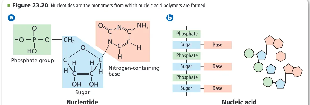
(17) Section 23.4. Nucleic Acids. Objectives ◗ Identify the structural components of nucleic acids. ◗ Relate the function of DNA to its structure. ◗ Describe the structure and function of RNA.. MAIN Idea Nucleic acids store and transmit genetic information. Real-World Reading Link DNA testing is becoming more routine in medicine, forensic science, genealogy, and identification of victims in disasters. Modern techniques have made it possible to get a useful DNA sample from surprising sources, such as a strand of hair or dried saliva on a postage stamp.. Review Vocabulary. Structure of Nucleic Acids. genetic information: an inherited sequence of RNA or DNA that causes traits or characteristics to pass from one generation to the next. Nucleic acids comprise a fourth class of biological molecules. They are the information-storage molecules of the cell. This group of molecules got its name from the cellular location in which the molecules are primarily found—the nucleus. It is from this control center of cells that nucleic acids carry out their major functions. A nucleic acid is a nitrogencontaining biological polymer that is involved in the storage and transmission of genetic information. The monomer that makes up a nucleic acid is called a nucleotide. Each nucleotide has three parts: an inorganic phosphate group, a five-carbon monosaccharide sugar, and a nitrogencontaining structure called a nitrogenous base. Examine each part of Figure 23.20a. Although the phosphate group is the same in all nucleotides, the sugar and the nitrogen base vary. In a nucleic acid, the sugar of one nucleotide is bonded to the phosphate of another nucleotide, as shown in Figure 23.20b. Thus, the nucleotides are strung together in a chain, or strand, containing alternating sugar and phosphate groups. Each sugar is also bonded to a nitrogen base that sticks out from the chain. The nitrogen bases on adjoining nucleotide units are stacked one above the other in a slightly askew position, much like the steps in a staircase. This orientation is shown in Figure 23.20b. Intermolecular forces hold each nitrogen base close to the nitrogen bases above and below it.. New Vocabulary nucleic acid nucleotide. ■. Figure 23.20 Nucleotides are the monomers from which nucleic acid polymers are formed.. a. — —. Phosphate group. N. O. HO. C H H C OH. C. N. —. HO — P — O —— CH2. —. O. —. O. C. C. C. NH2. Phosphate. H. H H C Nitrogen-containing C H base OH. Sugar. Nucleotide Each nucleotide contains a nitrogen-containing base, a five-carbon sugar, and a phosphate group.. 840. Chapter 23 • The Chemistry of Life. b. Sugar. Base. Phosphate Sugar. Base. Phosphate Sugar. Base. Nucleic acid Nucleic acids are linear chains of alternating sugars and phosphates. Attached to every sugar is a nitrogen base. Because the nucleotides are offset, the chains resemble steps in a staircase..
(18) DNA: The Double Helix You might have heard of DNA (deoxyribonucleic acid), one of the two kinds of nucleic acids found in living cells. DNA contains the master plans for building all the proteins in an organism’s body.. Interactive Figure To see an animation of the structure of DNA, visit glencoe.com.. The structure of DNA DNA consists of two long chains of nucleotides wound together to form a spiral structure, as shown in Figure 23.21. Each nucleotide in DNA contains a phosphate group, the five-carbon sugar deoxyribose, and a nitrogenous base. The alternating sugar and phosphate groups in each chain make up the outside, or backbone, of the spiral structure, The nitrogen bases are on the inside of the structure. Because the spiral structure is composed of two chains, it is known as a double helix.. G. C T. A T. A G. C. A T T. A G. Reading Check Describe what forms the teeth of the DNA zipper.. T. A. DNA contains four different nitrogenous bases: adenine (A), thymine (T), cytosine (C), and guanine (G). As Figure 23.21 shows, both adenine and guanine contain a double ring. Thymine and cytosine are single-ring structures. Looking again at Figure 23.21, notice that each nitrogen base on one strand of the helix is oriented next to a nitrogen base on the opposite strand, in the same way that the teeth of a zipper are oriented. The side-by-side base pairs are close enough so that hydrogen bonds form between them. Because each nitrogen base has a unique arrangement of organic functional groups that can form hydrogen bonds, the nitrogen bases always pair in a specific way so that the optimum number of hydrogen bonds form. As Figure 23.22 shows, guanine always binds to cytosine, and adenine always binds to thymine. The G–C and A–T pairs are called complementary base pairs. Because of complementary base pairing, the amount of adenine in a molecule of DNA always equals the amount of thymine, and the amount of cytosine always equals the amount of guanine. In 1953, James Watson and Francis Crick used this observation to make one of the greatest scientific discoveries of the twentieth century when they determined the double-helix structure of DNA. They accomplished this feat without performing many laboratory experiments themselves. Instead, they analyzed and synthesized the work of numerous scientists who had carefully carried out studies on DNA.. G C. C. G. Figure 23.21 The structure of DNA is a double helix that resembles a twisted zipper. The two sugar-phosphate backbones form the outsides of the zipper.. ■. Figure 23.22 In DNA, base pairing exists between a double-ringed base and a singleringed base. Adenine and thymine always pair, forming two hydrogen bonds between them. Guanine and cytosine always form three hydrogen bonds when they pair.. ■. O ...... H— N. N. —. CH3. H. H. N. N — H ..... N. H—. O. Thymine. N. Deoxyribose. H Adenine. H. N — H ...... O. N Deoxyribose. O ....... H— N. Cytosine. H. N N. N ...... H— N. H—. —. Deoxyribose. —. N. H. —. Hydrogen bond. N. Deoxyribose. H Guanine Section 23.4 • Nucleic Acids 841.
(19) The function of DNA Watson and Crick used their model to predict how DNA’s chemical structure enables it to function. DNA stores the genetic information of a cell in the cell’s nucleus. Before the cell divides, the DNA is copied so that the new generation of cells gets the same genetic information. Having determined that the two chains of the DNA helix are complementary, Watson and Crick realized that complementary base pairing provides a mechanism by which the genetic material of a cell is copied. The four nitrogenous bases of DNA serve as the letters of the alphabet in the information-storage language of living cells. The specific sequence of these letters represents an organism’s master instructions, just as the sequence of letters in the words of this sentence convey special meaning. The sequence of bases is different in every species of organism, allowing for an enormous diversity of life-forms—all from a language that uses only four letters. It is estimated that the DNA in a human cell has about three billion complementary base pairs, arranged in a sequence unique to humans.. Problem-solving lab Formulate a Model How does DNA replicate? DNA replicates, before a cell divides so that each of the two newly formed cells has a complete set of genetic instructions. When DNA begins to replicate, the two nucleotide strands start to unzip. An enzyme breaks the hydrogen bonds between the nitrogenous bases, and the strands separate. Other enzymes deliver free nucleotides from the surrounding medium to the exposed nitrogenous bases, adenine hydrogen-bonding with thymine, and cytosine bonding with guanine. Thus, each strand builds a complementary strand by basepairing with free nucleotides. This process is shown in the top diagram at the right. When the free nucleotides have been hydrogen-bonded into place, their sugars and phosphates bond covalently to those on adjacent nucleotides to form the new backbone. Each strand of the original DNA molecule is now bonded to a new strand. Analysis The bottom diagram shows a small segment of a DNA molecule. Copy the base sequence onto a clean sheet of paper, being careful not to make copying errors. Show the steps of replication to produce two segments of the DNA. Think Critically 1. Describe how the base sequence of a newly synthesized strand compares with the original strand to which it is bonded.. 842. Chapter 23 • The Chemistry of Life. A T G C C G T A A T T G. A C T A. A T C A T G C C G T. G A T G C C A A T A. T. G T. A A T A. T. A T C G G T T A A A C G T A G C C A A T T T G C 2. Explain If the original DNA segment is colored red and the free nucleotides are colored blue, what pattern of colors will the newly replicated DNA segments have? Will all new segments have the same color pattern? 3. Explain how an organism might be affected if an error occurs during replication of its DNA. Are the affects permanent? Explain..
(20) a. DNA. b. RNA. OH. H. O. H3C. H. H. H. N. N —. HOCH2 O. H. H. O. OH. H. Deoxyribose. H. Thymine. OH H. H. O. H. —. H. H. OH. —. HOCH2 O. H. N. OH. N —. H O. H. Ribose. Uracil. Figure 23.23 DNA and RNA differ in their components. The two structures on the left are found in DNA. The two structures on the right are found in RNA. Identify two differences in the structures of RNA and DNA. ■. RNA RNA (ribonucleic acid) is also a nucleic acid. Its general structure differs from that of DNA in three important ways, as shown in Figure 23.23. First, as you have read, DNA contains the nitrogen bases adenine, cytosine, guanine, and thymine. RNA contains adenine, cytosine, guanine, and uracil. Thymine is never found in RNA. Second, RNA contains the sugar ribose. DNA contains the sugar deoxyribose, which has a hydrogen atom in place of a hydroxyl group at one position. The third difference between DNA and RNA is a result of these structural differences. DNA is normally arranged in a double helix in which hydrogen bonding links the two chains together through their bases. RNA is usually single-stranded, with no such hydrogen bonds forming among the bases. Whereas DNA functions to store genetic information, RNA allows cells to use the information found in DNA. You have read that the genetic information of a cell is contained in the sequence of nitrogen bases in the DNA molecule. Cells use this base sequence to make RNA with a corresponding sequence. The RNA is then used to make proteins, each with an amino-acid sequence that is determined by the order of nitrogen bases in RNA. The sequences of bases are referred to as the genetic code. Because proteins are the molecular tools that carry out most activities in a cell, the DNA double helix is ultimately responsible for controlling the thousands of chemical reactions that take place in cells.. Section 23.4. &/,$!",%3. Incorporate information from this section into your Foldable.. Assessment. Section Summary. 20.. ◗ Nucleic acids are polymers of nucleotides, which consist of a nitrogen base, a phosphate group, and a sugar.. 21. Identify the specific structural components of both RNA and DNA.. ◗ DNA and RNA are the informationstorage molecules of a cell. ◗ DNA is double stranded, and RNA is single stranded.. MAIN Idea. Explain the primary function of RNA and DNA.. 22. Relate the function of DNA to its structure. 23. Relate the function of RNA to its structure. 24. Analyze the structure of nucleic acids to determine what structural feature makes them acidic. 25. Predict what might happen if the DNA that coded for a protein contained the wrong base sequence.. Self-Check Quiz glencoe.com. Section 23.4 • Nucleic Acids 843.
(21) Section 23.5 Objectives. Metabolism. ◗ Distinguish between anabolism and catabolism. ◗ Describe the role of ATP in metabolism. ◗ Compare and contrast the processes of photosynthesis, cellular respiration, and fermentation.. Real-World Reading Link You have studied the four major kinds of biological molecules and learned that they are all present in the food you eat. What happens to these molecules after they enter your body?. Review Vocabulary. Anabolism and Catabolism. redox process: a chemical reaction in which electrons are transferred from one atom to another. Many thousands of chemical reactions take place in the cells of a living organism. The set of chemical reactions that occur within an organism is its metabolism. Why are so many reactions involved in metabolism? Living organisms must accomplish two major functions in order to survive. They have to extract energy from nutrients in forms that they can use immediately as well as store for future use. In addition, they have to use nutrients to make building blocks for synthesizing all of the molecules needed to perform their life functions. These processes are summarized in Figure 23.24. The term catabolism refers to the metabolic reactions that break down complex biological molecules such as proteins, polysaccharides, triglycerides, and nucleic acids for the purposes of forming smaller building blocks and extracting energy. After you eat a meal of spaghetti and meatballs, your body immediately begins to break down the starch polymer in the pasta into glucose. The glucose is then broken down into smaller molecules in a series of energy-releasing catabolic reactions. Meanwhile, the protein polymers in the meatballs are catabolized into amino acids. The term anabolism refers to the metabolic reactions that use energy and small building blocks to synthesize the complex molecules needed by an organism. After your body has extracted the energy from the starch in the pasta, it uses that energy and the amino-acid building blocks produced from the meat proteins to synthesize the specific proteins that allow your muscles to contract, catalyze metabolic reactions, and perform many other functions in your body.. New Vocabulary metabolism catabolism anabolism ATP photosynthesis cellular respiration fermentation. MAIN Idea Metabolism involves many thousands of reactions in living cells.. Figure 23.24 A large number of different metabolic reactions take place in living cells. Some involve breaking down nutrients to extract energy; these are catabolic processes. Others involve using energy to build large biological molecules; these reactions are anabolic processes. Describe Choose one food that you ate recently, and describe how it was metabolized. ■. Nutrients ingested Carbohydrates Fats Proteins. Catabolism (nutrients broken down). 844 Chapter 23 • The Chemistry of Life. Intermediate products Amino acids Simple sugars Fatty acids Nucleotides ATP. Anabolism (new molecules synthesized). Complex cellular molecules Proteins Polysaccharides Triglycerides Nucleic acids ADP + P.
(22) Adenine Triphosphate group P. P. P. +. H2O. Ribose Anabolism. Catabolism. ATP. Adenine Diphosphate group P Ribose. P. +. + P Phosphate. Energy. ADP. Figure 23.25 The breakdown of ATP provides energy for cellular processes in living organisms. Explain where the energy is stored in ATP. ■. Figure 23.24 shows the relationship between catabolism and anabolism. The nutrients listed on the left side of the diagram are broken down into intermediate products. These intermediate products are used as building blocks for the products listed on the right side of the diagram. Another way of conceptualizing this process is to view the nutrients ingested as the raw materials for the complex cellular molecules formed in a living organism.. VOCABULARY ACADEMIC VOCABULARY Conceptualize visualizing or conceiving an abstract idea in the mind The atomic cloud model is hard to conceptualize.. Reading Check Explain how the terms metabolism, catabolism, and. anabolism are related. ATP Catabolism and anabolism are linked by common building blocks that catabolic reactions produce and anabolic reactions use. A common form of potential chemical energy also links the two processes, as shown in Figure 23.25. ATP (adenosine triphosphate) is a nucleotide that functions as the universal energy-storage molecule in living cells. During catabolic reactions, cells harness the chemical energy of foods and store it in the bonds of ATP. When these bonds are broken, the chemical energy is released and used by cells to drive anabolic reactions that might not otherwise occur. Most cellular reactions have an efficiency of only about 40% at best; the remaining 60% of the energy in food is lost as heat, which your body uses to keep warm. During catabolic reactions, cells produce ATP by adding an inorganic phosphate group to the nucleotide adenosine diphosphate (ADP) in an endothermic reaction. One mole of ATP stores approximately 30.5 kJ of energy under normal cellular conditions. During anabolism, the reverse reaction occurs. ATP is broken down to form ADP and inorganic phosphate in an exothermic reaction. Approximately 30.5 kJ of energy is released from each mole of ATP. Reading Check Describe what occurs when ATP becomes ADP. Section 23.5 • Metabolism 845.
(23) Photosynthesis What is the source of energy that fuels metabolism? For most living things, including the grass and cows shown in Figure 23.26, certain wavelengths of sunlight provide this energy. Some bacteria and the cells of all plants and algae are able to capture light energy and convert some of it into chemical energy. Animals cannot capture light energy, so they get energy by eating plants or by eating other animals that eat plants. The process that converts energy from sunlight to chemical energy in the bonds of carbohydrates is called photosynthesis. During the complex process of photosynthesis, carbon dioxide and water yield a carbohydrate (glucose) and oxygen gas. The following net reaction takes place during photosynthesis. 6CO 2 + 6H 2O Carbon Water dioxide. + light energy. →. C 6H 12O 6 Glucose. +. 6O 2 Oxygen. Photosynthesis results in the reduction of the carbon atoms in carbon dioxide as glucose is formed. During this redox process, oxygen atoms in water are oxidized to oxygen gas. Figure 23.26 Grass and other green plants use certain wavelengths of sunlight as an energy source. Other living organisms, such as cows, obtain energy by eating plants or eating other organisms that eat plants.. ■. Cellular Respiration Most organisms need oxygen to live. Oxygen that is produced during photosynthesis is used by living things during cellular respiration, the process in which glucose is broken down to form carbon dioxide, water, and large amounts of energy. Cellular respiration is the major energyproducing process in living organisms. Figure 23.27 shows one use of energy in the body. This energy is stored in the bonds of ATP. Cellular respiration is a redox process; the carbon atoms in glucose are oxidized while oxygen atoms in oxygen gas are reduced to the oxygen in water. The net reaction that takes place during cellular respiration is as follows. C 6H 12O 6 Glucose. Personal Tutor For an online tutorial on photosynthesis and respiration, visit glencoe.com.. +. 6O 2 → 6CO 2 + Oxygen Carbon dioxide. 6H 2O Water. +. energy. Figure 23.27 Swimmers need large amounts of energy when they compete in a race. This energy is stored in the bonds of ATP in their cells.. ■. 846. Chapter 23 • The Chemistry of Life. (t)©Royalty-Free/Corbis, (b)©AP Photo/Joe Cavaretta.
(24) Figure 23.28 Carbon dioxide formed during fermentation, leaving holes in the bread. These holes give bread a light, lessdense texture.. ■. Fermentation During cellular respiration, glucose is completely oxidized, and oxygen gas is required to act as the oxidizing agent. Cells extract energy from glucose in the absence of oxygen, but not nearly as efficiently. Without oxygen, only a fraction of the chemical energy of glucose can be released. Whereas cellular respiration produces 38 mol of ATP for every 1 mol of glucose catabolized in the presence of oxygen, only 2 mol of ATP are produced per mole of glucose that is catabolized in the absence of oxygen. This provides enough energy for oxygen-deprived cells so that they do not die. The process by which glucose is broken down in the absence of oxygen is known as fermentation. There are two common kinds of fermentation. In one, ethanol and carbon dioxide are produced. In the other, lactic acid is produced.. Careers In chemistry Baker Using a variety of chemical processes to create tasty and often beautiful creations is the job of a baker. Not only do breads and doughnuts undergo fermentation processes, but cakes and other pastries are often acid-base reactions. For more information on chemistry careers, visit glencoe.com.. Alcoholic fermentation Yeast and some bacteria can ferment glucose to produce the alcohol ethanol.. C 6H 12O 6 Glucose. →. 2CH 3CH 2OH Ethanol. + 2CO 2 + Carbon dioxide. energy. This reaction, called alcoholic fermentation, is important in producing some foods, as shown in Figure 23.28. Alcoholic fermentation is needed to make bread dough rise, form tofu from soybeans, and produce the ethanol in alcoholic beverages. Another use of the ethanol is as an additive to gasoline, as shown in Figure 23.29. Figure 23.29 Ethanol is often added to gasoline and used as a fuel in some cars and trucks. Ethanol is made from grain. Explain how the use of ethanol can reduce the dependence on fossil fuels. ■. Section 23.5 • Metabolism 847 (t)©David Young-Wolff/PhotoEdit, (b)©Alex Farnsworth/The Image Works.
(25) Figure 23.30 During strenuous activity, oxygen can be depleted in cells. Then, energy is produced without oxygen and lactic acid is produced. Soreness in muscles a day or two after the activity is a sign of lactic acid formation.. ■. Lactic acid fermentation Have you ever experienced muscle fatigue while running a race, like the person shown in Figure 23.30? During strenuous activity, muscle cells often use oxygen faster than it can be supplied by the blood. When the supply of oxygen is depleted, cellular respiration stops. Although animal cells cannot undergo alcoholic fermentation, they can produce lactic acid and a small amount of energy from glucose through lactic acid fermentation.. C 6H 12O 6 Glucose. →. 2CH 3CH(OH)COOH Lactic acid. +. energy. The lactic acid that is produced is moved from the muscles through the blood to the liver. There, it is converted back into glucose that can be used in catabolic processes to yield more energy once oxygen becomes available. However, if lactic acid builds up in muscle cells at a faster rate than the blood can remove it, muscle fatigue results. An immediate burning sensation and soreness a few days later is an indication that lactic acid was produced in the muscles during exercise.. Section 23.5. Assessment. Section Summary. 26.. ◗ Living organisms undergo catabolism and anabolism.. 27. Compare and contrast the processes of anabolism and catabolism.. ◗ Photosynthesis directly or indirectly provides all living things with energy.. 29. Compare and contrast the processes of photosynthesis, cellular respiration, and fermentation.. ◗ The net equation for cellular respiration is the reverse of the net equation for photosynthesis.. 30. Determine whether each process is anabolic or catabolic. a. photosynthesis b. cellular respiration c. fermentation. MAIN Idea. Explain why metabolism is important to living cells.. 28. Explain the role of ATP in the metabolism of living organisms.. 31. Evaluate Why is it necessary to use sealed casks when making wine? 32. Calculate How many moles of ATP would a yeast cell produce if 6 mol of glucose were oxidized completely in the presence of oxygen? How many moles of ATP would the yeast cell produce from 6 mol of glucose if the cell were deprived of oxygen?. 848. Chapter 23 • The Chemistry of Life. ©Wally McNamee/CORBIS. Self-Check Quiz glencoe.com.
(26) Career: Molecular Paleontologist Acid Test Reveals Surprise “No right-thinking paleontologist would do what Mary did. We don’t go to all this effort to dig this stuff out of the ground and then destroy it in acid.” So says a colleague about Mary Schweitzer, the scientist who used the techniques of molecular biology to discover soft tissue where none should be—in the thighbone of a 68-million-year-old Tyrannosaurus rex. Mother Bob When the fossilized T. rex, nicknamed Bob, was recovered in 2003 from a remote section of Montana by paleontologists, the bones were encased in plaster for protection during transport. However, the bones and plaster weighed more than the helicopter could lift. So the paleontologists were forced to break the intact thighbone to move the dinosaur out of the remote area. Schweitzer took small fragments from the broken thighbone for further study. The first surprise came quickly. “Bob” was a female, and she had been producing eggs at the time of her death. The bone Schweitzer studied is called medullary bone. Previously, this bone tissue was known only in birds, as shown in Figure 1. Ovulating hens produce medullary bone, then later use the calcium stored in the bone to make eggshells. After egg production, the bone disappears. Figure 1 shows the medullary bone found in the T. rex thighbone.. Figure 2 Scientists also found blood vessels and individual cells in the soft tissue of the T. rex. The acid test To study the medullary bone more closely, Schweitzer dissolved fragments of the bone in dilute acid to remove calcium phosphate—a technique normally used to examine fresh tissue. Because a fossilized bone has usually mineralized, it was assumed that the bone would completely dissolve in dilute acid. Yet this step yielded astonishing results—within the bone was soft tissue. Under the microscope, the tissue showed what looked like preserved blood vessels and even individual cells, as shown in Figure 2. But how could soft tissue have survived 68 million years in the ground? More work Schweitzer has since subjected other bones to the same acid test, and found similar soft tissue and fine structures. No one knows yet just what these fine structures are showing, but, says a colleague, “there may be a lot of things out there that we’ve missed because of our assumption of how preservation works.” Clearly, more research is needed.. Figure 1 The hen bone and T. rex bone both have a hard outer bone called cortical bone (CB) and softer medullary bone (MB).. Chemistry. that dino-. ely riting It is unlik Persuasive W found in these soft tissues. : the question ur DNA will be. sa gs up discovery brin recovEven so, the cloned from be s al im an t nc say expressing Should exti persuasive es a te ri W ? A ered DN rmation on For more info your opinion. t animals, nc ti NA from ex D ng ri ve co re com. visit glencoe.. Hen bone. T. rex thighbone. In the Field 849 (t)©epa/Corbis, (bl)©Mary Schweitzer, (br)©Mary Schweitzer.
(27) Probeware Alternate CBL instructions can be found at glencoe.com.. OBSERVE TEMPERATURE AND ENZYME ACTION Background: Enzymes are natural catalysts used by living things to speed reactions. These proteins have specialized structures that enable them to interact with specific substances.. Question: How does temperature affect the action of enzymes?. Water Bath. Temperature (°C). Height of Foam (cm). Potato Ice water Room-temperature water Body-temperature water. Materials red-skin potato pulp hydrogen peroxide (3% H 2O 2) water 250-mL beaker (4) test tubes (4) test-tube rack test-tube clamp. Data Table. 25-mL graduated cylinder thermometer ice ruler clock hot plate raw fresh liver pulp. Safety Precautions. Boiling water (near 100°C) Liver Ice water Room-temperature water Body-temperature water Boiling water (near 100°C). 14. Cleanup and Disposal Dispose of the remaining solutions as directed by your teacher. Wash and return all lab equipment to its designated location.. Procedure 1. Read and complete the lab safety form. 2. Write a hypothesis that identifies the temperature at which the enzymes are the most active. 3. Copy the data table on a separate sheet of paper. 4. Place the four test tubes in the test-tube rack. 5. Measure and place 2.0 mL of red-skin potato pulp into each test tube. 6. Using the hot plate and ice, prepare water baths in the beakers at four different temperatures: ice water, room-temperature water, body-temperature water, and gently boiling water at or near 100°C. 7. Place one test tube in each water bath using a test-tube clamp. 8. Measure and record the temperature of each water bath. 9. After 5 min in the water baths, measure and place 5.0 mL of 3% H 2O 2 in each test tube. 10. Allow the reaction to proceed for 5 min. 11. Measure the height of the foam produced in each test tube. 12. Dispose of the contents of the test tubes are directed by your teacher and wash the test tubes. 13. Repeat Steps 4–12 using 2.0 mL of beef liver pulp instead of potato pulp. 850. Chapter 23 • The Chemistry of Life. Analyze and Conclude 1. Make and Use Graphs Make a line graph with temperature on the x-axis and height of foam on the y-axis. Use a different color for the potato and liver data points and lines. 2. Summarize How does temperature affect the action of enzymes? Infer why the maximum reaction occurred at the temperature in which it did for the potato and liver. 3. Recognize Cause and Effect Which water bath produced the least amount of foam for each material? Propose explanations for why this happened. 4. Compare and Contrast Did the experimental data support your hypothesis in Step 2? Explain. 5. Model Write a balanced reaction for the decomposition of hydrogen peroxide for each reaction. How are the reactions similar and infer why? 6. Error Analysis Identify potential sources of errors for this investigation and suggest methods to correct them.. INQUIRY EXTENSION Design an Experiment Would a change in pH affect the results? Design an experiment to find out..
(28) Download quizzes, key terms, and flash cards from glencoe.com.. BIG Idea Biological molecules—proteins, carbohydrates, lipids, and nucleic acids—interact to carry out activities necessary to living cells. Section 23.1 Proteins MAIN Idea Proteins perform essential functions, including regulation of chemical reactions, structural support, transport of materials, and muscle contractions.. Vocabulary • active site (p. 830) • amino acid (p. 826) • denaturation (p. 829) • enzyme (p. 829). • peptide (p. 828) • peptide bond (p. 828) • protein (p. 826) • substrate (p. 830). Key Concepts • Proteins are biological polymers made of amino acids that are linked by peptide bonds. • Protein chains fold into intricate three-dimensional structures. • Proteins have many functions in the human body, including functions within cells, functions between cells, and functions of structural support.. Section 23.2 Carbohydrates MAIN Idea Carbohydrates provide energy and. structural material for living things.. Vocabulary • carbohydrate (p. 832) • disaccharide (p. 833). • monosaccharide (p. 832) • polysaccharide (p. 833). Key Concepts • Carbohydrates are compounds that contain multiple hydroxyl groups (–OH) and a carbonyl functional group (C=O). • Carbohydrates range in size from single monomers to polymers composed of hundreds or thousands of monomers. • Monosaccharides in aqueous solution exist in both open-chain and cyclic structures.. Section 23.3 Lipids MAIN Idea Lipids make cell membranes, store energy, and regulate cellular processes.. Vocabulary • fatty acid (p. 835) • lipid (p. 835) • phospholipid (p. 838) • saponification (p. 837). • steroid (p. 839) • triglyceride (p. 836) • wax (p. 838). Key Concepts • Fatty acids are long-chain carboxylic acids that usually have between 12 and 24 carbon atoms. • Saturated fatty acids have no double bonds; unsaturated fatty acids have one or more double bonds. • Fatty acids can be linked to glycerol backbones to form triglycerides. • Steroids are lipids that have multiple-ring structures.. Section 23.4 Nucleic Acids MAIN Idea Nucleic acids store and transmit genetic. information.. Vocabulary • nucleic acid (p. 840). • nucleotide (p. 840). Key Concepts • Nucleic acids are polymers of nucleotides, which consist of a nitrogen base, a phosphate group, and a sugar. • DNA and RNA are the information-storage molecules of a cell. • DNA is double stranded, and RNA is single stranded.. Section 23.5 Metabolism MAIN Idea Metabolism involves many thousands of. reactions in living cells.. Vocabulary • anabolism (p. 844) • fermentation (p. 847) • ATP (p. 845) • metabolism (p. 844) • catabolism (p. 844) • photosynthesis (p. 846) • cellular respiration (p. 846). Key Concepts • Living organisms undergo catabolism and anabolism. • Photosynthesis directly or indirectly provides all living things with energy. • The net equation for cellular respiration is the reverse of the net equation for photosynthesis.. Vocabulary PuzzleMaker glencoe.com. Chapter 23 • Study Guide 851.
Figure

+6


Related documents
Because of the draw- backs in using the above references, standards and indices, we used the DHS maternal weight and height data to define a de facto reference for use in
a) A Test CAM package capable of reading in IPC-D-356x net lists and performing CAD to Gerber (RS-274X or ODB++) checks. b) This Test CAM suite must also extract compliant
■ Before using, test the Ground Fault Circuit Interrupter (GFCI) provided with the power cord to ensure it is operating properly.. GFCI reduces the risk of
Presently, wireless sensor network (WSN) has been used in home monitoring to optimally manage the „well-being‟ of power distribution in home in order to minimize
Our Institute spans six campus locations across the northern Sydney region – Crows Nest, Hornsby, Meadowbank, Northern Beaches, Ryde and St Leonards.. We offer nationally
O halde, mitik geleneklerin ele§tirisi sorunu yanlı§ olarak ortaya konulmu§tur; bir Pausanias'ın, doğruyu yanlı§tan ayırmak bir yana, efsanelerdeki her §eyi yanlı§ sayanz6
Seeking a new recipe for managing grain procurement Executive overview Company objectives Resolution Business transformation Future plans.. Allied
Tal como intentó demostrar en la serie de artículos Le Celluloïd et le marbre (2010), Rohmer consideraba que el cine iba a contramano de las otras artes; no solo porque era
