Volume 2013, Article ID 502451,5pages http://dx.doi.org/10.1155/2013/502451
Case Report
Gastric Endocrine Cell Carcinoma
Coexistent with Adenocarcinoma
Nobuhiro Takeuchi,
1Nomura Yusuke,
1Tetsuo Maeda,
1and Kazuyoshi Naba
21Department of Gastroenterology, Kawasaki Hospital, Kobe, Hyogo 652-0042, Japan 2Department of Laboratory Medicine, Kawasaki Hospital, Kobe, Hyogo 652-0042, Japan
Correspondence should be addressed to Nobuhiro Takeuchi; takeuchi nobuhiro@kawasaki-hospital-kobe.or.jp
Received 16 October 2013; Revised 20 November 2013; Accepted 4 December 2013
Academic Editor: Martin G. Mack
Copyright © 2013 Nobuhiro Takeuchi et al. This is an open access article distributed under the Creative Commons Attribution License, which permits unrestricted use, distribution, and reproduction in any medium, provided the original work is properly cited.
A 69-year-old female presented to our institution with epigastralgia and abdominal distension. Upper gastrointestinal series revealed a 5 cm ulcerative lesion with irregular margins and elevated distinct borders from the angle to the pyloric ring. Gastroendoscopy revealed a Borrmann type 2 tumor. Several biopsied specimens revealed proliferation of small and heterogeneous cancer cells with rich chromatin and fibrous septum with rich vessels at connective tissues, which was confirmed as gastric endocrine cell carcinoma (ECC) on immunostaining with chromogranin and synaptophysin. Furthermore, other specimens revealed atypical cells forming glandular structures, which were confirmed as well-differentiated tubular adenocarcinomas. Distal gastrectomy with D2 lymph node dissection and Billroth I reconstruction was performed. Pathological examination of the gross specimen revealed that adenocarcinoma comprised<10% of all cancer cells. Close analysis of ECC revealed a mixture of small and large cells. According to the WHO 2010 classification of gastrointestinal neuroendocrine tumors, this gastric tumor was diagnosed as neuroendocrine carcinoma. The patient was administered adjuvant chemotherapy with cisplatin and etoposide. One year following surgery, follow-up abdominal CT revealed multiple liver metastases. The patient received the best sfollow-upportive care but eventually died 18 months after surgery. Here we present this case of gastric ECC coexistent with adenocarcinoma.
1. Introduction
Gastric endocrine cell carcinoma (ECC), characterized by endocrine differentiation and aggressive biological behavior, is occasionally accompanied by the presence of adenocarci-noma cells. On the basis of the analysis of p53 gene alteration, the hypothesis that was derived proved that adenocarcinoma cells have the potential to develop into gastric ECC [1]. Here we present a case of gastric ECC coexistent with adenocar-cinoma as well as p53 gene analyses of ECC and adenocarci-noma.
2. Case Presentation
A 69-year-old female presented to our institution with epi-gastralgia and abdominal distension. Her past medical his-tory included hypertension and dyslipidemia. On physical examination, her cognitive consciousness was alert. Her height, weight, and body mass index were 144.2 cm, 53.3 kg,
and 25.6 kg/m2, respectively. Mild anemia was evident in her
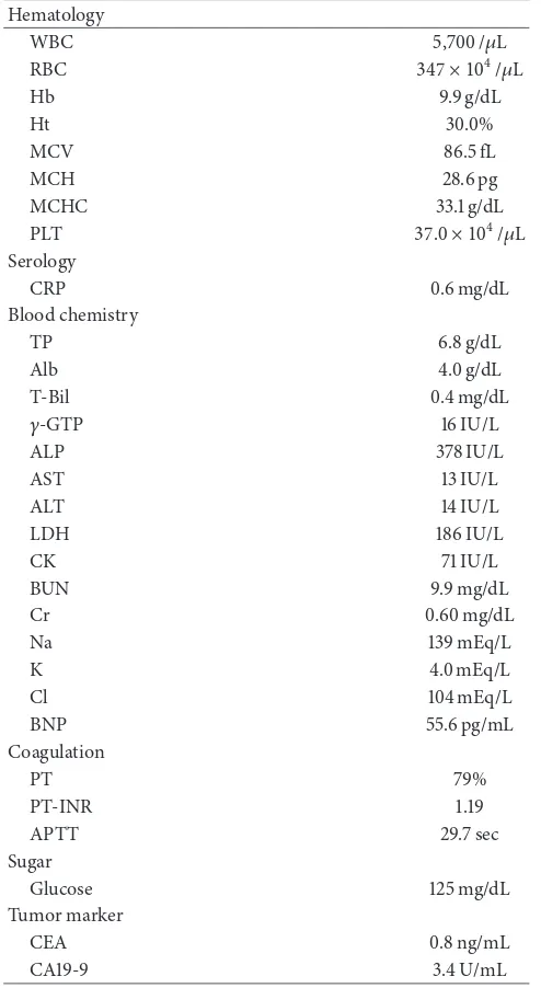
palpebral conjunctiva. Chest auscultation revealed no abnor-mal findings. The abdomen was soft and flat with norabnor-mal bowel sounds. No lymph nodes were palpable, and no tumors were palpable on rectal examination. Blood chemistry analyses (Table 1) revealed mild anemia (red blood cell count,
347×104/𝜇L; hemoglobin levels, 9.9 mg/dL), mildly increased
Hb 9.9 g/dL
Ht 30.0%
MCV 86.5 fL
MCH 28.6 pg
MCHC 33.1 g/dL
PLT 37.0 × 10⁴/𝜇L
Serology
CRP 0.6 mg/dL
Blood chemistry
TP 6.8 g/dL
Alb 4.0 g/dL
T-Bil 0.4 mg/dL
𝛾-GTP 16 IU/L
ALP 378 IU/L
AST 13 IU/L
ALT 14 IU/L
LDH 186 IU/L
CK 71 IU/L
BUN 9.9 mg/dL
Cr 0.60 mg/dL
Na 139 mEq/L
K 4.0 mEq/L
Cl 104 mEq/L
BNP 55.6 pg/mL
Coagulation
PT 79%
PT-INR 1.19
APTT 29.7 sec
Sugar
Glucose 125 mg/dL
Tumor marker
CEA 0.8 ng/mL
CA19-9 3.4 U/mL
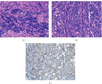
(Figure 1(b)) revealed a Borrmann type 2 tumor, extending from the angle to the antrum of the lesser curvature. The tumor comprised two different parts: ulcers with severe inva-siveness and smooth protrusion from the endocrine cells (Figure 2(a)) and well-differentiated adenocarcinoma cells (Figure 2(b)) as exhibited by specimens biopsied under gastroendoscopy. ECC was confirmed by immunostaining examinations of cells, exhibiting positive results of chro-mogranin, synaptophysin, cytokeratin (CK) 7, and EMA
(Figures2(c)–2(f)). Other specimens revealed atypical cells
forming glandular structures, leading to the diagnosis of well-differentiated tubular adenocarcinoma (tub1) (Figure 2(b)). Preoperative enhanced whole-body computed tomography (CT) revealed no distant metastases, signs of peritoneal dis-semination, and regional lymph nodes swelling. Therefore,
(a)
ECC
Tub1
(b)
Figure 1: Upper gastrointestinal series revealed a 5 cm ulcerative lesion with irregular margins and elevated distinct borders from the angle to the pyloric ring (a). Gastroendoscopy revealed a Borrmann type 2 tumor, extending from the angle to the antrum of the lesser curvature (b). The tumor comprised two different parts: ulcers with severe invasiveness and smooth protrusion from the endocrine cells and well-differentiated adenocarcinoma cells, as revealed by specimens biopsied under gastroendoscopy.
our gastric tumor was diagnosed as stage IIA tumor pre-operatively. Subsequently, distal gastrectomy with D2 lymph node dissection and Billroth I reconstruction was performed. Pathological examination of the gross specimen (Figure 3(a))
revealed that adenocarcinoma cells comprised <10% of all
cancer cells. Close analysis of ECC revealed a mixture of small and large cells, and 58% of Ki-67 labeling index (Figures 4(a)–4(c)). According to the WHO 2010 classification of gastrointestinal neuroendocrine tumors (NETs), ECC that coexists with adenocarcinoma (>30%) should be classified as “mixed adenoneuroendocrine carcinoma”. However,
adeno-carcinoma comprised<10% of all cancer cells; therefore, this
gastric tumor was classified as NEC.
Postoperatively, this gastric cancer was diagnosed as M,
(a) (b)
(c) (d)
(e) (f)
Figure 2: Proliferation of small and heterogeneous cancer cells with rich chromatin and fibrous septum with rich vessels at connective tissues was evident (hematoxylin and eosin) (a). Atypical cells forming glandular structures were evident (hematoxylin and eosin) (b). Immunostaining of small and heterogeneous cancer cells by chromogranin (c), synaptophysin (d), cytokeratin 7 (e), and EMA (f) was positive.
Tub1
ECC
(a) (b)
(c)
Figure 4: Microscopic examination of the resected specimen revealed the mixture of small (a) and large cells (b). Immunostaining by Ki-67 antibody revealed 58% of positive cells (c).
A
C C C A T G A
(a)
A
C C C C T G A
(b)
Figure 5: p53 gene sequences in exon 5–8 were analyzed. No alteration was identified in adenocarcinoma cells (a); in contrast, a transitional mutation (A to C) was identified in codon 179 of exon 5 in endocrine cells (b).
(4/38) of Stage IIIB. Immunostaining of endocrine and ade-nocarcinoma cells with p53 antibody revealed positive results for both components. Analysis indicated that transformation of pre-existing adenocarcinoma cells into endocrine cells is influenced by p53 gene alteration. In the present case, p53 gene sequences of exon 5–8, wherein most p53 gene muta-tions have been reported to occur, were analyzed, and
transitional mutation (A to C) was identified in codon 179 of exon 5 in ECC (Figure 5). Although two types of cancers could develop separately, there might be the possibility that endocrine cells transformed from adenocarcinoma cells by p53 gene alteration.
etoposide. One year following surgery, follow-up abdominal CT revealed multiple liver metastases. The patient received the best supportive care but eventually died 18 months after surgery.
3. Discussion
ECC composes highly atypical neoplastic endocrine cells and is a poor highly malignant cancer with rapid progression, early vessel invasion, early metastasis, and poor prognosis [2]. ECC is opposite of carcinoid, which is low atypical and low malignant. The clinical presentation of ECC is similar to that of adenocarcinoma except that it has a more biologi-cally aggressive character. The mean survival time of ECC is 7 months [2]. Morphologically, most ECCs develop into Borrmann type 2 tumors; however, few predominantly develop into submucosal layers similar to carcinoid tumors and form ulcerated lesions along with severe invasion into submucosal layers, thereby exhibiting Borrmann type 3 tumors. ECC is usually treated using a combination of sur-gery and chemotherapy. Because the response rate of chem-otherapy widely varies, no established regimen exists to date. ECC is treated using the treatment regimen for small cell lung carcinoma with cisplatin and etoposide, irinotecan, or
paclitaxel [3,4]. The gastric cancer regimen, S-1 with cisplatin,
is also reportedly effective [5].
ECC comprises either small or large or both types of cells. In addition, the prevalence of coexistence of ECC with adenocarcinoma can be as high as 72.7% [1]. ECC is con-sidered to develop as follows: differentiated glandular cancer cells arise initially, followed by the emergence of neoplastic endocrine cells (with high proliferative ability) through the differentiation of glandular cancer cells, and subsequently, endocrine cancer cells rapidly proliferate from the deep gland to submucosal layers, thereby forming glandular endocrine cancer.
Gastric ECC usually arises under submucosal layers, coexisting with mucosal adenocarcinoma. Normal mucosa covers over ECC; therefore, ECC is usually diagnosed as differentiated adenocarcinoma by specimens biopsied under endoscopy. With regard to the growth of ECC, the following four types of pathways have been hypothesized: (i) ECCs arise from pre-existing adenocarcinomas; (ii) ECCs arise from pre-existing carcinoid tumors: (iii) ECCs arise from nonneoplastic multipotent stem cells; (iv) ECCs arise from nonneoplastic endocrine cells. Of these, most gastrointestinal ECCs are considered to follow the first pathway [6].
4. Conclusions
When biopsied specimens reveal proliferation of small nuclear cancer cells without glandular structure, immunos-taining should be performed to confirm ECC. Moreover, ECC occasionally comprised various types of cancer cells; therefore, endoscopic morphology of gastric ECC may vary according to the proportion of endocrine cancer cells. In the present case, during adjuvant chemotherapy following R0 resection, multiple liver metastases developed. Thus, future
studies must focus on the emergence of promising anticancer agents or chemotherapy protocols.
References
[1] K. Nishikura, H. Watanabe, M. Iwafuchi, T. Fujiwara, K. Kojima, and Y. Ajioka, “Carcinogenesis of gastric endocrine cell carcinoma: analysis of histopathology and p53 gene alteration,” Gastric Cancer, vol. 6, no. 4, pp. 203–209, 2003.
[2] K. Matsui, M. Kitagawa, A. Miwa, Y. Kuroda, and M. Tsuji, “Small cell carcinoma of the stomach: a clinicopathologic study of 17 cases,”American Journal of Gastroenterology, vol. 86, no. 9, pp. 1167–1175, 1991.
[3] K. Hasegawa, M. Maruyama, I. Takashima et al., “A case of gastric endocrine cell carcinoma with neck lymph node metas-tasis resected after neoadjuvant chemotherapy,”Gan to Kagaku Ryoho, vol. 31, no. 11, pp. 1935–1938, 2004.
[4] T. Ohnishi, S. Kinami, E. Morioka et al., “A case of gastric endo-crine cell carcinoma that responded to chemotherapy with CPT-11+CDDP.,”Gan to Kagaku Ryoho, vol. 39, pp. 2384–2386, 2012.
[5] R. Ohhinata, Y. Iwasaki, M. Ohashi et al., “A case of gastric endocrine cell carcinoma with liver metastases treated with S-1/CDDP,”Gan to Kagaku Ryoho, vol. 37, no. 12, pp. 2508–2510, 2010.
Submit your manuscripts at
http://www.hindawi.com
Stem Cells
International
Hindawi Publishing Corporation
http://www.hindawi.com Volume 2014
Hindawi Publishing Corporation
http://www.hindawi.com Volume 2014
Hindawi Publishing Corporation
http://www.hindawi.com Volume 2014
Behavioural
Neurology
Endocrinology
International Journal ofHindawi Publishing Corporation
http://www.hindawi.com Volume 2014 Hindawi Publishing Corporation
http://www.hindawi.com Volume 2014
Disease Markers
Hindawi Publishing Corporation
http://www.hindawi.com Volume 2014 BioMed
Research International
Oncology
Journal of Hindawi Publishing Corporationhttp://www.hindawi.com Volume 2014
Hindawi Publishing Corporation
http://www.hindawi.com Volume 2014
Oxidative Medicine and Cellular Longevity
Hindawi Publishing Corporation
http://www.hindawi.com Volume 2014
PPAR Research
The Scientific
World Journal
Hindawi Publishing Corporationhttp://www.hindawi.com Volume 2014
Immunology Research Hindawi Publishing Corporation
http://www.hindawi.com Volume 2014
Journal of
Obesity
Journal ofHindawi Publishing Corporation
http://www.hindawi.com Volume 2014
Hindawi Publishing Corporation
http://www.hindawi.com Volume 2014 Computational and Mathematical Methods in Medicine
Ophthalmology
Journal ofHindawi Publishing Corporation
http://www.hindawi.com Volume 2014
Diabetes Research
Journal ofHindawi Publishing Corporation
http://www.hindawi.com Volume 2014
Hindawi Publishing Corporation
http://www.hindawi.com Volume 2014
Research and Treatment
AIDS
Hindawi Publishing Corporation
http://www.hindawi.com Volume 2014
Gastroenterology Research and Practice
Hindawi Publishing Corporation
http://www.hindawi.com Volume 2014
Parkinson’s
Disease
Evidence-Based Complementary and Alternative Medicine
Volume 2014