R E S E A R C H A R T I C L E
Open Access
Determinants of exercise capacity in cystic
fibrosis patients with mild-to-moderate lung
disease
Jean Pastré
1, Anne Prévotat
1, Catherine Tardif
2, Carole Langlois
3, Alain Duhamel
3and Benoit Wallaert
1*Abstract
Background:Adult patients with cystic fibrosis (CF) frequently have reduced exercise tolerance, which is
multifactorial but mainly due to bronchial obstruction. The aim of this retrospective analysis was to determine the mechanisms responsible for exercise intolerance in patients with mild-to-moderate or severe disease.
Methods:Cardiopulmonary exercise testing with blood gas analysis at peak exercise was performed in 102 patients aged 28 ± 11 years: 48 patients had severe lung disease (FEV1< 50%, group 1) and 54 had mild-to-moderate lung disease (FEV1≥50%, group 2). VO2peak was measured and correlated with clinical, biological, and functional parameters.
Results:VO2peak for all patients was 25 ± 9 mL/kg/min (65 ± 21% of the predicted value) and was < 84% of predicted in 82% of patients (100% of group 1, 65% of group 2). VO2peak was correlated with body mass index, C-reactive protein, FEV1, FVC, RV, DLCO, VE/VCO2peak, VD/VT, PaO2, PaCO2, P(A-a)O2, and breathing reserve. In multivariate analysis, FEV1and overall hyperventilation during exercise were independent determinants of exercise capacity (R2= 0.67). FEV1was the major significant predictor of VO2peak impairment in group 1, accounting for 31% of VO2peak alteration, whereas excessive overall hyperventilation (reduced or absent breathing reserve and VE/VCO2) accounted for 41% of VO2alteration in group 2.
Conclusion:Exercise limitation in adult patients with CF is largely dependent on FEV1in patients with severe lung disease and on the magnitude of the ventilatory response to exercise in patients with mild-to-moderate lung disease.
Keywords:Cystic fibrosis, Cardiopulmonary exercise testing, Pulmonary function, Exercise
Background
Cystic fibrosis (CF) is characterized by deterioration of nutritional status and irreversible loss of lung function [1-3]. Patients with CF often experience exertional dys-pnea and have reduced maximal exercise capacity, which is an important predictor of mortality [4-7]. Regular exercise in these patients has been associated with improved aerobic exercise endurance and quality of life [4,8]. Physical exercise requires the cardiopulmonary system to deliver oxygen to muscles in sufficient quan-tity to generate energy through aerobic glycolysis. There
are conflicting data on the precise mechanisms
underlying exercise intolerance in CF, and a number of factors have been implicated [9], including poor nutri-tional status, peripheral muscle dysfunction [10,11], and especially, ventilatory limitation [12,13]. In other stud-ies, dysfunctional gas exchange has been shown to play a crucial role in limiting exercise performance [14-17].
Only a third of the variability in exercise capacity of
CF patients can be explained by FEV1, demonstrating
that resting pulmonary function tests (PFTs) alone are insufficient to explain the exercise limitation [1,9,13]. By comparison, cardiopulmonary exercise testing (CPET) offers a sensitive evaluation of potential physiological disturbances in cardiovascular, respiratory, peripheral, or neurosensory responses to a standardized exercise protocol [18]. Although it remains underutilized in CF [19], CPET could provide important exercise-related * Correspondence:benoit.wallaert@chru-lille.fr
1Université Lille 2 et Clinique des Maladies Respiratoires, CRCM Hôpital
Calmette, CHRU Lille, France
Full list of author information is available at the end of the article
measures that might explain the reduced exercise per-formance and thus assist in CF patient management aimed at improving exercise capacity.
With this in mind, we initiated a study to determine the mechanisms responsible for exercise limitation in 102 adult CF patients with mild-to moderate or severe lung disease. The patients were subjected to CPET with blood gas analysis during exercise and the results were correlated with clinical and functional characteristics.
Methods
Patients
A total of 102 adult patients (sex ratio M:F 0.52) with CF were enrolled at four CF centers in France: Lille (75 patients), Rouen (15 patients), Dijon (5 patients), and
Grenoble (7 patients). Written informed consent for
participation in the study was obtained from partici-pants. Use of the patient data was approved by the local ethics committee, and the study was considered obser-vational and approved as such by the Institutional Review Board of the French Learned Society for
Pulmo-nology (Société de Pneumologie de Langue Française,
CEPRO 2012 009).
Clinical, nutritional, biological, PFT, and CPET data were obtained on the same day, either at diagnosis or at the routine annual evaluation, and were retrospectively collected. When patients were seen at several follow-up visits, only the data from the first visit were recorded. Hypoxemic patients did not perform CPET and were ex-cluded from the analysis. A diagnosis of CF was obtained by sweat chloride > 60 mmol/L and the presence of CFTR gene mutations by molecular analysis. Additional characteristics recorded at the time of testing were dis-eases usually associated with CF, bacterial colonization, treatments, and nutritional status, including height and weight measurements and impedance analysis.
Cardiopulmonary testing
Forced vital capacity (FVC), forced expiratory volume in 1 s (FEV1), FEV1to FVC ratio, total lung capacity (TLC), and residual volume (RV) were measured by plethysmog-raphy (Jaeger-Masterlab® cabin). Diffusing capacity of the lung for carbon monoxide (DLCO: mL CO/min/mm Hg) was adjusted for hemoglobin concentration in g/dL
according to Cotes’ equation: corrected (Hb) DLCO =
DLCO × (10.2 + Hb)/(1.7 × Hb). Following ATS/ERS 2005 guidelines, the lower limits of normal were set at the 5th percentile (or predicted minus 1.64 SD) of each reference population. The results are expressed as percentages of the predicted values. Predicted normal values were derived from standard equations [20-22].
PFTs and CPET were performed in an air-conditioned laboratory (22°C constant temperature), using a stan-dardized protocol as previously described [23,24]. The
CPET protocol was the same at each center. Each patient underwent a symptom-limited incremental exer-cise test on an ergometric bicycle (Ergoline-Ergometrics 800®). The protocol included a warm-up period of 3 min at 20 W followed by a progressively increasing work rate (WR) in a ramp fashion and then 3 min recovery. The ramped WR increment was individualized (range,
8–30 W/min). During exercise, heart rate (HR) was
monitored continuously by 12-lead ECG, and arterial
oxygen saturation (SpO2) was measured by pulse
oxim-etry (Nellcor N-395). The expired gases were analyzed with an Ergocard®, focusing on oxygen consumption (VO2), carbon dioxide production (VCO2), minute ven-tilation (VE), and tidal volume (VT). The oxygen pulse
(VO2/HR) was calculated. Measurements of PaO2 and
PaCO2were performed on room air at rest and at peak
exercise. Normal values for PaO2 were derived from
[25]. Lactatemia was determined at maximal exercise. Breathing reserve (BR) was calculated as BR = (predicted
maximum minute ventilation [MMV] –VE peak)/MMV,
with MMV estimated from MMV = FEV1× 40. HR peak
was expressed as a percentage of maximum predicted
HR, calculated as HR max = 210 – (0.65 × age). Dead
space (VD/VT) was calculated according to Bohr’s equa-tion corrected for the addiequa-tional instrument dead space: VD/VT= (PaCO2–PECO2mean)/PaCO2–(VD[machine]/ VT) where PECO2 is the partial pressure of expired CO2.
Predicted values for VO2 max were calculated from
reference equations [26]. Poor motivation appeared not to be an interfering factor in our analysis, as all patients had at least one of the following: BR < 15%, peak HR > 90% of predicted, peak lactate > 7 mmol/L, peak exercise PaO2< 55 mm Hg, and peak VE/VO2> 35 or peak RER > 1.15 [23]. Immediately after exercise, subjects were asked to score their sense of breathlessness and muscle fatigue at peak exercise using Borg scales.
Statistical analysis
The continuous variables are reported as mean ± SD. Normal distribution of quantitative variables was tested by the Shapiro–Wilk test. Differences in FEV1between the groups were determined with the Student’s t-test or
Mann–Whitney test. Bivariate analyses were performed
following strategy: first, variables with p < 0.2 were selected and included in a Principal Component Analysis (PCA) in order to study their correlations. Then, the vari-ables included in the multivariable regression model were selected by the results of PCA (graphic correlation circle) on the basis of their clinical pertinence. The stability of the model was assessed by a bootstrap method [27]. The bootstrap resampling method was based on 1000 repli-cates of the initial dataset. Multivariable regression with a stepwise selection at the level 0.2 was performed on each of these replicates. The inclusion of the variable in the final model was confirmed if this candidate variable was selected in at least 70% of these 1000 analyses. In the final model, for each variable we computed the par-tial R-square, coefficient, 95% confidence intervals and adjusted p-value. Final variables from the multivariate
analysis were applied to each group of FEV1. All
ana-lyses were achieved with SAS software version 9.2 (SAS Institute Inc., Cary, NC). All tests were performed at the significant level at 0.05.
Results
Subjects
The demographic and clinical characteristics are shown in Table 1, and the resting PFT results are presented in Table 2. The cohort consisted of 102 CF patients with a mean age of 28 ± 11 years (range 17–67). The time from diagnosis to evaluation was 16 ± 10 years. For data ana-lysis, the cohort was divided into two groups according to
their FEV1: group 1 patients with severe lung disease
(FEV1< 50%, 48 patients) and group 2 patients with mild-to-moderate lung disease (FEV1≥50%, 54 patients). Group 1 had a significantly higher frequency of homozygosity for the CFTR ΔF 508 mutation, pancreatic insufficiency, bronchial colonization withPseudomonas aeruginosa, and biological inflammatory syndrome, and significantly lower body mass index (BMI) and longer disease duration (data not shown).
The patients had a range of disease severities and were recruited from four centers in France. Patients from dif-ferent centers all had characteristics consistent with the French CF Registry 2009 Report [28] and showed equiva-lent frequencies of key CF characteristics (ΔF508 muta-tion, exocrine pancreatic insufficiency, and colonization with P. aeruginosa), supporting the reproducibility of the results and the potential for extrapolation to other patient populations.
Resting PFT values (Table 2) were significantly more altered in group 1 than in group 2 (Table 2). As expected, three major functional abnormalities were found: ob-structive syndrome (FEV1/FVC = 65 ± 15% of predicted), altered distension (RV = 176 ± 65% of predicted), and altered DLCO (68 ± 18% of predicted).
Exercise responses
Exercise cessation was mainly due to leg fatigue in com-bination with dyspnea (62%), whereas leg fatigue alone or dyspnea alone was observed in 17% and 21% of pa-tients, respectively. The VO2peak value (weight-adjusted
VO2) was decreased to < 84% of predicted in 83/102
(82%) of patients (48/48 [100%] in group 1 and 35/54 [65%] in group 2) and was significantly lower in group 1 than in group 2 (Table 3).
Analysis of the ventilatory response (VE peak, BR,
respiratory rate (RR), VT/FVC peak) highlighted the
dif-ferences according to FEV1impairment (Table 3). Group
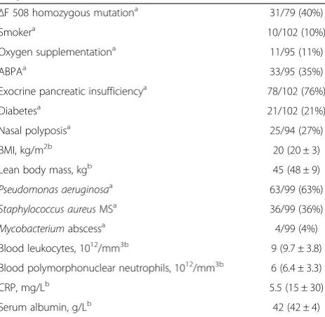
Table 1 Demographic and clinical characteristics of the CF patients
ΔF 508 homozygous mutationa 31/79 (40%)
Smokera 10/102 (10%)
Oxygen supplementationa 11/95 (11%)
ABPAa 33/95 (35%)
Exocrine pancreatic insufficiencya 78/102 (76%)
Diabetesa 21/102 (21%)
Nasal polyposisa 25/94 (27%)
BMI, kg/m2b 20 (20 ± 3)
Lean body mass, kgb 45 (48 ± 9)
Pseudomonas aeruginosaa 63/99 (63%)
Staphylococcus aureusMSa 36/99 (36%)
Mycobacteriumabscessa 4/99 (4%)
Blood leukocytes, 1012/mm3b 9 (9.7 ± 3.8)
Blood polymorphonuclear neutrophils, 1012/mm3b 6 (6.4 ± 3.3)
CRP, mg/Lb 5.5 (15 ± 30)
Serum albumin, g/Lb 42 (42 ± 4)
ABPA = allergic bronchopulmonary aspergillosis; BMI = body mass index; CRP = C-reactive protein.
Qualitative variables are given as frequency and percentage. a
Results are expressed as number (percentage) of patients. b
Results are expressed as median values (mean ± SD).
Table 2 Resting pulmonary function tests in CF patients classified according to FEV1
All patients (n = 102)
Group 1 (n = 48)
Group 2 (n = 54)
FEV1 < 50% ≥50%
FEV1a 60 ± 28 35 ± 9b 82 ± 18
FVCa 75 ± 24 56 ± 14b 93 ± 16
FEV1/FVC 65 ± 15 54 ± 11b 75 ± 10
RVa 176 ± 65 220 ± 50b 135 ± 48
DLCOa 68 ± 18 56 ± 13b 78 ± 14
PaO2, mm Hg 80 ± 14 71 ± 10
b
87 ± 12
PaCO2, mm Hg 38 ± 4 38 ± 5 37 ± 4
P(A-a)O2, mm Hg 29 ± 13 38 ± 8
b
21 ± 12
a
Results are expressed as mean ± SD percentage of predicted values. b
1 had a lower absolute value of VE at peak exercise, and a depletion of BR. Hyperventilation was due to simultan-eous increases in RR and tidal volume. Impairment in pulmonary gas exchange was more severe in group 1, as shown by higher values of P(A-a)O2, VD/VT peak, and
PaCO2, and lower values of PaO2. Cardiocirculatory
responses were normal in group 2, but patients in group
1 showed low VO2/HR values and a significant decrease
in peak HR. Four patients experienced ECG abnormal-ities but continued with the exercise test.
Determinants of exercise capacity
Significant correlations were observed between VO2peak and nutritional status (BMI, lean body mass), inflamma-tion markers (C-reactive protein [CRP], leukocytosis),
resting PFT (FVC, FEV1, RV, DLCO, P(A-a)O2), and
quantifiable parameters of CPET (VE peak, VE/VO2
peak, VE/VCO2peak, BR, VD/VTpeak, PaO2peak, P(A-a) O2peak, and HR (Table 4 and Figure 1).
The results of the stepwise multiple regression analysis for determinants of exercise capacity are shown in Table 5. Of the variables entered into the model (BMI, FEV1, FVC,
DLCO, PaO2, VE/VCO2 peak, BR, VD/VT peak, PaO2
peak, PaCO2peak, P(A-a)O2peak, and lactatemia peak),
only FEV1,VE/VCO2peak, and BR were found to be inde-pendent predictors of exercise capacity (r2= 0.67). Ana-lysis of these three variables showed that, for group 1, 31%
of the VO2 peak was explained by FEV1, whereas the
major determinants of the VO2peak in group 2 were BR , FEV1and VE/VCO2peak (Table 5).
Separate analysis in the cohort of Lille (75 out of the
102 patients) showed the same results: FEV1, BR and
VE/CO2 were independent predictors of exercise
cap-acity (r2= 0.65) (data not shown).
Discussion
Our study focused on a population of 102 adults with CF who underwent CPET with blood gas analysis at peak exercise. Maximal oxygen uptake was impaired in 82% of patients and was more pronounced in patients
with low FEV1. We noted a high prevalence of abnormal
exercise responses in our population, including abnor-mal gas exchange, ventilatory and cardiocirculatory Table 3 Cardiopulmonary exercise tests in CF patients
classified according to FEV1
All patients (n = 102)
Group 1 (n = 48)
Group 2 (n = 54)
FEV1 < 50% ≥50%
VO2peak, mL/kg/min 25 ± 9 20 ± 5d 30 ± 8.5
VO2peaka 65 ± 21 51 ± 13d 77 ± 20
Borg dyspnea 4.7 ± 1.9 5.6 ± 2b 4.1 ± 1.5
Borg Leg fatigue 4.5 ± 1.8 5.2 ± 1.8b 4.4 ± 1.7
VE peak, L/min 58 ± 22 44 ± 11d 70 ± 21
RR peak, min 41 ± 9 43 ± 9b 39 ± 8
VT/FVC peak, % 48 ± 11 47 ± 13 48.6 ± 8
VE/VO2peak 41 ± 7 42 ± 7 41 ± 7
VE/VCO2peak 36 ± 6 37 ± 6 35 ± 5
BR, % 24 ± 20 8 ± 11d 37.5 ± 16
VD/VTpeak 0.32 ± 0.11 0.39 ± 0.07 d
0.25 ± 0.10
pH peak 7.34 ± 0.04 7.34 ± 0.04 7.35 ± 0.04
PaO2peak, mm Hg 76 ± 16 63 ± 10
d
89 ± 12
PaCO2peak, mm Hg 40 ± 7 44 ± 6
d
36 ± 4
P(A-a)O2peak, mm Hg 37 ± 13 46 ± 8 d
28 ± 10
Lactatemia peak, mmol/L 7 ± 2.6 6 ± 2d 7.9 ± 3
HR peaka 82 ± 10 79 ± 8d 86 ± 9
VO2/HR peaka 79 ± 22 67 ± 16d 90 ± 21
a
Results are expressed as mean ± SD percentage of predicted values. b
P< .01 compared with group 2. c
P< .001 compared with group 2. d
P< .0001 compared with group 2.
Table 4 Correlation of clinical and functional variables with VO2peak in CF patients
Qualitative variables n Pvalue
Female 102 .22
ΔF508 homozygous mutation 79 .02
Exocrine pancreatic insufficiency 102 .55
Pseudomonas aeruginosa 99 .36
Quantitative variables n Correlation (r)
Age, years 102 −0.11 .29
BMI, kg/m2* 102 0.26 .009
Leukocytosis, 109/mm3 70 −0.42 .0003
CRP, mg/L 64 −0.34 .006
Serum albumin, g/L 43 0.34 .02
FEV1a* 102 0.71 < .0001
FVCa* 102 0.69 < .0001
RVa 79 −0.58 < .0001
DLCOa 68 0.56 < .0001
PaO2, mm Hg 98 0.43 < .0001
PaCO2, mm Hg 98 0.11 .29
Cardiopulmonary exercise parameters n Correlation (r)
VEpeak, L 102 0.64 < .0001
VE/VCO2peak* 98 −0.35 < .0001
BR, %* 102 0.37 .0001
VD/VTpeak* 96 −0.64 < .0001
PaCO2peak, mm Hg* 97 −0.45 < .0001
P(A-a)O2peak, mm Hg* 97 −0.54 < .0001
Lactatemia peak, mmol/L* 86 0.59 < .0001
HR peaka 102 0.40 < .0001
a
responses, and peripheral limitation. The main findings from this study are that exercise intolerance in CF is multifactorial and is correlated mainly with resting pul-monary function, nutritional status, and inflammatory status, but is also affected by the magnitude of the over-all ventilatory response during exercise. Multivariate analysis revealed that bronchial obstruction plays a dom-inant role in patients with severe disease, whereas
excessive hyperventilation during exercise was the major determinant of exercise limitation in patients with mild-to-moderate disease.
CF can be associated with abnormal gas exchange, ventilatory, cardiocirculatory, and muscular responses to exercise [3,9,13,29]. In our study, these abnormalities were responsible for limiting the aerobic capacity of 82% of patients, a proportion consistent with previous studies 0
20 40 60 80 100 120
20 40 60 80 100 120 140
-10 0 10 20 30 40 50 60 70
20 40 60 80 100 120 140 VO2 peak %
0 10 20 30 40 50 60 70
20 40 60 80 100 120 140 0 0.1 0.2 0.3 0.4 0.5 0.6
20 40 60 80 100 120 140 FEV1%
VE/VCO2 peak BR %
P(A-a)O2mmHg peak
VO2 peak % VD/VTpeak
VO2 peak %
20 40 60 80 100 120 140
20 40 60 80 100 120 140 FVC %
VO2 peak %
VO2 peak % VO2 peak % 20
25 30 35 40 45 50 55
20 40 60 80 100 120 140
R =.71 R =.69
R =.37
R = -.64 R = -.35
R = -.54
Figure 1Correlation between VO2peak and FEV1, FVC, VE/CO2peak, BR, P(A-a)O2peak, and VD/VTpeak in CF patients.VO2peak, FEV1,
FVC, and BR are expressed as percentage of predicted values. P(A-a)O2peak is expressed as mm Hg.
Table 5 Determinants of VO2peak in CF patients
Variable All patients
(n = 102)
Group 1 (n = 48)
Group 2 (n = 54)
FEV1 50 (0.84 [0.70;0.98])
a
31 (1.08 [0.70;1.45])a 18 (0.67 [0.46;0.88])a
BR 12 (−0.62 [−0.82;-0.42])a 6 (−0.36 [−0.68;-0.04])b 35 (−0.85 [−1.10;-0.59])a
VE/VCO2peak 5 (−1.13 [−1.56;-0.69]) a
15 (−0.95 [−1.41;-0.48])c 6 (−1.52 [−2.22;-0.82])a
Results are expressed as the partial r-square (r2), i.e. the percentage of VO2alteration explained by the variable. Coefficient and 95% confidence intervals are shown between parentheses. FEV1,VE/VCO2peak, and BR were independent predictors explaining 67% of exercise capacity (r2
= 0.67). In group 1, 31% of the VO2 peak was explained by FEV1, whereas, in group 2, BR was the major determinant explaining 35% of the VO2peak.
Results are derived from multivariable analysis. a
of adult CF patients [5,12,30]. We did not observe a single exercise profile common to all patients, reflecting the complexity of mechanisms involved in exercise limita-tion in CF patients. Some patients showed abnormalities predominantly in gas exchange, others in the ventilatory response. Still others experienced exercise intolerance despite the absence of ventilatory limitation. The rela-tive contribution of these factors differed between the two groups.
In our study, BMI and CRP levels were strongly corre-lated with exercise limitation, which is consistent with several studies indicating the importance of inflamma-tory and nutritional status in exercise limitation. Nutri-tional status plays a well-established role in CF exercise intolerance [31] and prognosis [32], and may be linked to the chronic inflammation observed in CF patients, which is mainly due to respiratory colonization [33]. Inflammatory markers such as CRP are also negatively associated with exercise capacity in patients with CF [7]. Moreover, inflammation is experimentally correlated with loss of muscle mass [34] and skeletal muscle weak-ness [10] and could explain the association observed here between CRP, lean body mass, and reduced max-imal oxygen uptake.
Multivariate analysis showed that FEV1 was the most
significant predictor of VO2peak in patients with severe lung disease. This result is consistent with data from earlier studies [3,35] and demonstrates the predominant role of ventilatory disorders in exercise limitation in severe CF patients. Additional functional parameters, such as distension, obstruction, and CO diffusion also
correlated with VO2 peak, but were not independent
predictors. The low BR exhibited by our population is another characteristic of the exercise response in severe
CF patients. Tantisira et al. showed that the BR index
(VE/maximal voluntary ventilation calculated at ventila-tory threshold) was the most powerful predictor of mor-tality in CF patients awaiting lung transplantation [36]. This has also been observed in COPD [37] but is not common to all obstructive lung diseases. For example, McNichollet al.reported that only 18% of severe asthma patients had ventilatory limitation due to obstructive lung function [38].
In contrast, the VO2 peak was not fully explained by
FEV1 in patients with mild-to-moderate lung disease,
and some patients exhibited impaired aerobic capacity despite having normal resting lung function (Figure 1). Indeed, multivariate analysis showed that two CPET parameters were the major independent determinants
of VO2peak in group 2: hyperventilation due to
abnor-mal ventilatory control, resulting in high ventilatory equivalents (as demonstrated by VE/VO2and VE/VCO2 peaks), and BR depletion. Exercise ventilation is regu-lated by numerous mechanisms, most of which remain
incompletely understood [39]. Hyperventilation during exercise reflects a nonspecific response to one or more dysfunctional links in the respiratory chain, but the main cause is not known [40]. In some diseases, such as heart failure, hyperventilation is recognized as a more relevant
prognostic factor than VO2 peak. The hyperventilatory
response may be due to several factors, including
ineffi-cient gas exchange as reflected by P(A-a)O2 and the
VD/VT ratio. Although hyperventilation is difficult to relate to other abnormalities, the strong correlation of hyperventilation with oxygen pulse and peak lactatemia suggests that central (cardiovascular) and peripheral (muscle) determinants play a role [10].
In our study, all patients underwent blood gas analysis at peak effort and we noted a high prevalence of gas exchange abnormalities during exercise. It is interesting to note that patients with identical lung function did not all show gas exchange abnormalities. This could be explained by an inadequate ventilatory response in some patients or by a high degree of ventilation-perfusion mismatch. Exercise-induced hypoxemia was common in
our study and correlated with VO2peak, workload, peak
VD/VT, and dyspnea assessed by the Borg scale (results
not shown). We found that P(A-a)O2 correlated well
with peak VO2, highlighting the relevance of this par-ameter in gas exchange analysis. Other studies have examined impairment of gas exchange during exercise
in CF patients. Nixon et al. showed that PETCO2>
41 mm Hg at peak exercise is associated with a twofold higher relative risk of mortality [4]. However, PETCO2is
not a reliable marker for PaCO2 during exercise and
does not allow accurate calculation of dead space [41]. Compared with PFT, CPET with blood gas analysis at peak exercise is better able to assess gas exchange ab-normalities and highlight exercise hypoxemia, a recog-nized prognosis marker, and thus gauge the need for oxygen supplementation.
The primary limitation of our study is its retrospective nature and the possibility of missing data. Peripheral muscle strength was not assessed and might be a signifi-cant contributing factor [10]. These results should be confirmed by a prospective study.
Conclusion
Competing interests
For each author, no significant competing interest exists with any companies or organisations whose products or services are mentioned in this article. The authors declare that they have no competing interests.
Authors’contributions
Conception and design: BW, JP and AP; Analysis and interpretation: BW, JP, AP, CT, CL and AD; Drafting the manuscript for important intellectual content: BW, JP, AP, and CL. All authors read and approved the final manuscript.
Acknowledgments
The authors wish to thank Anne M. O’Rourke for editing of the manuscript. Collaborators (to be referenced in PubMed): T. Perez (Lille), L. Wémeau-Stervinou (Lille), Abderrahmane Mammar (Grenoble), J.M. Perruchini (Dijon).
Author details 1
Université Lille 2 et Clinique des Maladies Respiratoires, CRCM Hôpital Calmette, CHRU Lille, France.2Service de Physiologie Respiratoire, CHRU Rouen, France.3Unité de Biostatistiques, CHRU Lille, France.
Received: 28 June 2013 Accepted: 23 April 2014 Published: 30 April 2014
References
1. Marcotte JE, Grisdale RK, Levison H, Coates AL, Canny GJ:Multiple factors limit exercise capacity in cystic fibrosis.Pediatr Pulmonol1986,2:274–281. 2. Boucher GP, Lands LC, Hay JA, Hornby L:Activity levels and the
relationship to lung function and nutritional status in children with cystic fibrosis.Am J Phys Med Rehabil1997,76:311–315.
3. Lands LC, Heigenhauser GJ, Jones NL:Analysis of factors limiting maximal exercise performance in cystic fibrosis.Clin Sci1992,83:391–397. 4. Nixon PA, Orenstein DM, Kelsey SF, Doershuk CF:The prognostic value of
exercise testing in patients with cystic fibrosis.N Engl J Med1992, 327:1785–1788.
5. Moorcroft AJ, Dodd ME, Webb AK:Exercise testing and prognosis in adult cystic fibrosis.Thorax1997,52:291–293.
6. Pianosi P, Leblanc J, Almudevar A:Peak oxygen uptake and mortality in children with cystic fibrosis.Thorax2005,60:50–54.
7. Van de Weert-van Leeuwen PB, Slieker MG, Hulzebos HJ, Kruitwagen CLJJ, van der Ent CK, Arets HGM:Chronic infection and inflammation affect exercise capacity in cystic fibrosis.Eur Respir J2012,39:893–898. 8. Bilton D, Dodd ME, Abbot JV, Webb AK:The benefits of exercise combined
with physiotherapy in the treatment of adults with cystic fibrosis.Respir Med 1992,86:507–511.
9. Almajed A, Lands LC:The evolution of exercise capacity and its limiting factors in Cystic Fibrosis.Paediatr Respir Rev2012,13:195–199. 10. Troosters T, Langer D, Vrijsen B, Segers J, Wouters K, Janssens W, Gosselink
R, Decramer M, Dupont L:Skeletal muscle weakness, exercise tolerance and physical activity in adults with cystic fibrosis.Eur Respir J2009, 33:99–106.
11. Selvadurai HC, Allen J, Sachinwalla T, Macauley J, Blimkie CJ, Van Asperen PP: Muscle function and resting energy expenditure in female athletes with cystic fibrosis.Am J Respir Crit Care Med2003,168:1476–1480.
12. Cerny FJ, Pullano TP, Cropp GJ:Cardiorespiratory adaptations to exercise in cystic fibrosis.Am Rev Respir Dis1982,126:217–220.
13. Shah AR, Gozal D, Keens TG:Determinants of aerobic and anaerobic exercise performance in cystic fibrosis.Am J Respir Crit Care Med1998, 157:1145–1150.
14. Lebecque P, Lapierre JG, Lamarre A, Coates AL:Diffusion capacity and oxygen desaturation effects on exercise in patients with cystic fibrosis. Chest1987,91:693–697.
15. Bradley S, Solin P, Wilson J, Johns D, Walters EH, Naughton MT:Hypoxemia and hypercapnia during exercise and sleep in patients with cystic fibrosis. Chest1999,116:647–654.
16. McKone EF, Barry SC, Fitzgerald MX, Gallagher CG:Role of arterial hypoxemia and pulmonary mechanics in exercise limitation in adults with cystic fibrosis.J Appl Physiol2005,99:1012–1018.
17. Marcus CL, Bader D, Stabile MW, Wang CI, Osher AB, Keens TG: Supplemental oxygen and exercise performance in patients with cystic fibrosis with severe pulmonary disease.Chest1992,101:52–57.
18. ATS/ACCP:Statement on cardiopulmonary exercise testing.Am J Respir Crit Care Med2003,167:211–277.
19. Stevens D, Oades PJ, Armstrong N, Williams CA:A survey of exercise testing and training in UK cystic fibrosis clinics.J Cyst Fibros2010, 9:302–306.
20. Miller MR, Hankinson J, Brusasco V, Burgos F, Casaburi R, Coates A, Crapo R, Enright P, van der Grinten CPM, Gustafsson P, Jensen R, Johnson DC, MacIntyre N, McKay R, Navajas D, Pedersen OF, Pellegrino R, Viegi G, Wanger J:Standardisation of spirometry.Eur Respir J2005, 26:319–338.
21. Macintyre N, Crapo RO, Viegi G, Johnson DC, van der Grinten CPM, Brusasco V, Burgos F, Casaburi R, Coates A, Enright P, Gustafsson P, Hankinson J, Jensen R, McKay R, Miller MR, Navajas D, Pedersen OF, Pellegrino R, Wanger J:Standardisation of the single-breath determin-ation of carbon monoxide uptake in the lung.Eur Respir J2005, 26:720–735.
22. Wanger J, Clausen JL, Coates A, Pedersen OF, Brusasco V, Burgos F, Casaburi R, Crapo R, Enright P, van der Grinten CPM, Gustafsson P, Hankinson J, Jensen R, Johnson D, Macintyre N, McKay R, Miller MR, Navajas D, Pellegrino R, Viegi G:Standardisation of the measurement of lung volumes.Eur Respir J 2005,26:511–522.
23. Aguilaniu B, Richard R, Costes F, Bart F, Martinat Y, Stach B, Aguilaniu B, Richard R, Costes F, Bart F, Martinat Y, Stach B, Denjean A, Scientific Council of the French Lung Society:[Cardiopulmonary exercise testing].Rev Mal Respir2007,24:2S111–2S160.
24. Wallaert B, Talleu C, Wemeau-Stervinou L, Duhamel A, Robin S, Aguilaniu B: Reduction of maximal oxygen uptake in sarcoidosis: relationship with disease severity.Respiration2011,82:501–508.
25. Sorbini CA, Grassi V, Solinas E, Muiesan G:Arterial oxygen tension in relation to age in healthy subjects.Respiration1968,25:3–13. 26. Hansen JE, Sue DY, Wasserman K:Predicted values for clinical exercise
testing.Am Rev Respir Dis1984,129:S49–S55.
27. Sauerbrei W:The Use of Resampling Methods to Simplify Regression Models in Medical Statistics.Journal of the Royal Statistical Society: Series C (Applied Statistics)1999,48:313–329.
28. Vaincre La Mucoviscidose.http://www.vaincrelamuco.org/. 2011 Rapport annuel. 2012.
29. Leroy S, Perez T, Neviere R, Aguilaniu B, Wallaert B:Determinants of dyspnea and alveolar hypoventilation during exercise in cystic fibrosis: impact of inspiratory muscle endurance.J Cyst Fibros2011, 10:159–165.
30. Godfrey S, Mearns M:Pulmonary function and response to exercise in cystic fibrosis.Arch Dis Child1971,46:144–151.
31. Gulmans VA, de Meer K, Brackel HJ, Helders PJ:Maximal work capacity in relation to nutritional status in children with cystic fibrosis.Eur Respir J 1997,10:2014–2017.
32. Nguyen S, Leroy S, Cracowski C, Perez T, Valette M, Neviere R, Aguilaniu B, Wallaert B:Prognostic value of clinical exercise testing in adult patients with cystic fibrosis.Rev Mal Respir2010,27:219–225.
33. Van de Weert-van Leeuwen PB, Arets HGM, van der Ent CK, Beekman JM: Infection, inflammation and exercise in cystic fibrosis.Respir Res2013, 14:32.
34. Van Heeckeren AM, Tscheikuna J, Walenga RW, Konstan MW, Davis PB, Erokwu B, Haxhiu MA, Ferkol TW:Effect of Pseudomonas infection on weight loss, lung mechanics, and cytokines in mice.Am J Respir Crit Care Med2000,161:271–279.
35. Klijn PHC, van der Net J, Kimpen JL, Helders PJM, van der Ent CK: Longitudinal determinants of peak aerobic performance in children with cystic fibrosis.Chest2003,124:2215–2219.
36. Tantisira KG, Systrom DM, Ginns LC:An elevated breathing reserve index at the lactate threshold is a predictor of mortality in patients with cystic fibrosis awaiting lung transplantation.Am J Respir Crit Care Med2002, 165:1629–1633.
37. Medoff BD, Oelberg DA, Kanarek DJ, Systrom DM:Breathing reserve at the lactate threshold to differentiate a pulmonary mechanical from cardiovascular limit to exercise.Chest1998,113:913–918.
38. McNicholl DM, Megarry J, McGarvey LP, Riley MS, Heaney LG:The utility of cardiopulmonary exercise testing in difficult asthma.Chest2011, 139:1117–1123.
40. Péronnet F, Aguilaniu B:Lactic acid buffering, nonmetabolic CO2 and exercise hyperventilation: a critical reappraisal.Respir Physiol Neurobiol 2006,150:4–18.
41. Lewis DA, Sietsema KE, Casaburi R, Sue DY:Inaccuracy of noninvasive estimates of VD/VT in clinical exercise testing.Chest1994, 106:1476–1480.
doi:10.1186/1471-2466-14-74
Cite this article as:Pastréet al.:Determinants of exercise capacity in cystic fibrosis patients with mild-to-moderate lung disease.BMC Pulmonary Medicine201414:74.
Submit your next manuscript to BioMed Central and take full advantage of:
• Convenient online submission
• Thorough peer review
• No space constraints or color figure charges
• Immediate publication on acceptance
• Inclusion in PubMed, CAS, Scopus and Google Scholar
• Research which is freely available for redistribution