RNA Polymerase I Assembly, Localization, and Function
Jan-Peter Daniels,aKeith Gull,aand Bill Wicksteada,b
Sir William Dunn School of Pathology, University of Oxford, Oxford, United Kingdom,aand Centre for Genetics and Genomics, University of Nottingham, Nottingham, United Kingdomb
African trypanosomes are the only organisms known to use RNA polymerase I (pol I) to transcribe protein-coding genes. These genes includeVSG, which is essential for immune evasion and is transcribed from an extranucleolar expression site body (ESB). Several trypanosome pol I subunits vary compared to their homologues elsewhere, and the question arises as to how these varia-tions relate to pol I function. A clear example is the N-terminal extension found on the second-largest subunit of pol I, RPA2. Here, we identify an essential role for this region. RPA2 truncation leads to nuclear exclusion and a growth defect which pheno-copies single-allele knockout. The N terminus is not a general nuclear localization signal (NLS), however, and it fails to accumu-late unreaccumu-lated proteins in the nucleus. An ectopic NLS is sufficient to reinstate nuclear localization of truncated RPA2, but it does not restore function. Moreover, NLS-tagged, truncated RPA2 has a different subnuclear distribution to full-length protein and is unable to build stable pol I complexes. We conclude that the RPA2 N-terminal extension does not have a role exclusive to the expression of protein-coding genes, but it is essential for all pol I functions in trypanosomes because it directs trypanosomatid-specific interactions with RPA1.
A
frican trypanosomes of the speciesTrypanosoma bruceiare unicellular eukaryotic parasites that are responsible for hu-man African trypanosomiasis (HAT, or sleeping sickness) and the wasting disease nagana in livestock in sub-Saharan Africa. The annual burden of HAT has been estimated at⬃70,000 cases, and more than a million disability-adjusted life years (DALYs) are lost (12)T. bruceiis transmitted by fly bite to the host bloodstream where it proliferates extracellularly, evading the host immune re-sponse by the antigenic variation of surface protein (for a review, see reference 4). The cell surface of the bloodstream-form try-panosome is covered with a dense coat of monotypic variant sur-face glycoprotein (VSG). TheT.bruceigenome contains hundreds ofVSGgenes, many of which are pseudogenes (3). In addition, there are⬃20 telomeric bloodstream expression sites (BESs), each of which contains a singleVSGgene toward its 3=end (16, 49). Only one of these BESs is transcriptionally active at any time, such that no more than oneVSGgene is ever expressed by any cell. By switching the expressed VSG, the parasite can escape the host im-mune response against previously exposed VSGs and hence main-tain the waves of parasitemia that are characteristic of the disease (for a review, see reference 36).The active BES, containing the expressedVSGin bloodstream-formT. brucei, is transcribed by RNA polymerase I (pol I) (14, 28, 37). This is also the case for the metacyclic ESs, which encode VSGs expressed during the initial transmission to the bloodstream and the genes coding for the major surface proteins of the procy-clic (midgut) stage of the tsetse fly. This transcription of endoge-nous protein-coding genes by pol I apparently makes African try-panosomes unique. In all other eukaryotic organisms analyzed, pol I is restricted to the transcription of the 45S rRNA precursor. In trypanosomes, the transcription of the active BES is spatially distinct from the nucleolar transcription of rRNA, occurring in-stead from an extranucleolar transcription focus called the expres-sion site body (ESB) (33). Alongside the VSG, BESs contain a number of expression site-associated genes (ESAGs). A BES is transcribed as a single long polycistron from which mature mRNA
is produced by the trans-splicing of a spliced-leader mini-exon to the 5=end and polyadenylation at the 3=end. It has been postu-lated that the ability of pol I to transcribe through the BES requires the association of the polymerase with the transcription elonga-tion and processing factors necessary for mRNA generaelonga-tion (36, 45), although no components that might facilitate this association have been identified thus far (43). This raises the question of whether there are specific differences in the composition of pol I compared to other organisms which might allow such interactions to occur, or which function solely in the transcription of mRNA and not rRNA.
Eukaryotic RNA polymerases are multisubunit enzymes. Trypanosomatids differ from other model systems in the compo-sition and form of their polymerase subunits. In trypanosomes, there are two paralogues for RNA polymerase subunits RPB5, RPB6, and RPB10, which in other eukaryotes are single paralogues shared between all three RNA polymerases (20, 24, 34, 47). The canonical RPB5, RPB6, and RPB10 paralogues are associated with pol II and pol III but are not part of the pol I complex (8, 9, 32). Instead, variant RPB5z, RPB6z, and RPB10z paralogues, which contain sequence insertions relative to the sequences of the canon-ical set, are specifcanon-ically substituted for their canoncanon-ical counter-parts in pol I (10, 34, 47). The RNA interference (RNAi)-mediated knockdown of RPB5 or RPB5z abolished either pol II/pol III or pol I transcription, respectively, demonstrating that the function of these two subunits is class specific (10). Trypanosomal pol I also contains a novel subunit, p31, which is essential for the
transcrip-Received6 February 2012Accepted23 February 2012
Published ahead of print2 March 2012
Address correspondence to Bill Wickstead, [email protected].
Supplemental material for this article may be found athttp://ec.asm.org/.
Copyright © 2012, American Society for Microbiology. All Rights Reserved.
doi:10.1128/EC.00036-12
on September 8, 2020 by guest
http://ec.asm.org/
tion of rRNA (35, 47), but the complex lacks the pol I-specific RPA43/RPA14 heterodimer, which is not encoded in the genome (24). Hence, several trypanosomatid-specific features of pol I have been demonstrated, but the functional relevance, if any, of the insertions in the pol I-specific subunits is not yet understood.
The most striking difference between the pol I complexes of trypanosomes and other eukaryotes is the sequence of the N ter-minus of the second-largest subunit, RPA2 (RPA135). In a pro-teomic study of theT.bruceipol I complex, Schimanski et al. showed that the RPA2 protein is⬃50 kDa larger than orthologues in most other eukaryotes (38) and contains an apparently trypanosomatid-specific N-terminal domain of⬃250 amino ac-ids. This RPA2 N-terminal extension contains a serine-rich region but is probably not a site for phosphorylation, since RPA2 was found not to be phosphorylated in bloodstream-form or procyclic T.brucei(47).
Given its conservation among trypanosomatids, it has been hypothesized that the RPA2 N terminus provides a crucial func-tion for these parasites (38), but this funcfunc-tion and its biological relevance have remained elusive to date. One interesting scenario is that the RPA2 N terminus is necessary for the residency of pol I in different parts of the nucleus, in particular endowing pol I the capability to occupy the ESB in addition to its canonical location in the nucleolus. In this work, we address this hypothesis by per-forming a molecular dissection of the N-terminal region of RPA2 inT. bruceicells. In so doing, we characterize the influence of the N-terminal region on the cell and subnuclear compartmentaliza-tion of RPA2, as well as on the stable assembly of the pol I complex and polymerase function.
MATERIALS AND METHODS
Bioinformatic analyses.Using BLASTp (1), a data set of 28 eukaryotic RPA2 proteins was generated by querying 28 predicted eukaryotic pro-teomes of evolutionarily diverse organisms with proteins annotated as RPA2 in the NCBI databases. Multiple-sequence alignments were done with MAFFT (23). The profile-hidden Markov model of the trypanosome N terminus was built from a seed alignment of RPA2 amino acids 1 to 374 ofT.bruceiand 1 to 381 ofLeishmania majorwith hmmer 2.3.2 (10, 11). As a control model, a seed alignment ofT.bruceiRPA2 amino acids 375 to 750 withL.majorRPA2 amino acids 382 to 767 was generated. These models were then used to search the predicted proteomes of 28 eukary-otic, evolutionarily diverse organisms (see Table S1 in the supplemental material).
Cell lines and cell culture.To facilitate the transfection of procyclic-form trypanosomes with RNAi and endogenous locus-tagging constructs, we made a single-marker procyclic line (SiMP1) expressing Tet repressor protein from the endogenous EP1 locus. Briefly, the T7 promoter was removed from the plasmid pBluescript II SK(⫹) (X52328; Stratagene) by digestion with NaeI and EcoRV, followed by self ligation to create the plasmid pSPR0. From genomic DNA (gDNA) purified from the single-marker bloodstream line of Wirtz et al. (48), we generated the following by PCR: a fragment of DNA corresponding to the region immediately upstream of the EP1 promoter (Ups), a fragment of DNA corresponding to the region downstream of the EP1 promoter (Downs), the EP1 pro-moter itself (EP1-prom), and a polycistron containing sequence encoding the Tet repressor protein and the neomycin resistance marker (Tet-Neo; see Table S2 in the supplemental material for all primer sequences). To create pSPR1, the Ups fragment was digested with AvaI/BglII, Downs was digested with SphI/SacI, and both were ligated, together with a linker made by annealing linker_F and linker_R, into pSPR0-cut AvaI/SacI. To create pSPR2, the EP1-prom fragment was digested with HindIII/BamHI, Tet-Neo was digested with BglII/SphI (partial, with selection for not cut-ting the internal SphI site), and both were ligated with pSPR1-cut HindIII/
SphI. The identity of pSPR2 was confirmed by sequencing, and the insert was removed by SacI digestion and transfected into Lister 427 strain pro-cyclic cells using standard electroporation protocols (48). Sequences and maps for all vectors are available atwww.wicksteadlab.co.uk/.
SiMP1 cells were grown at 28°C in SDM79 medium (5) supplemented with 10% fetal bovine serum. Bloodstream-form Lister 427 and 90-13 (48) cells were grown in HMI-9 medium supplemented with 15% fetal bovine serum at 37°C and 5% CO2(17). SiMP1 cells were cloned by limiting dilution on 96-well plates. Bloodstream-form growth was assessed by di-luting the cells to 5⫻104cells ml⫺1and counting and rediluting them 24 h later during a period of 120 h. Procyclic cells were diluted to 2⫻105 cells ml⫺1, counted after 24 h, recounted after another 24 h, and rediluted during a period of 144 h. Tests were performed using the statistical pro-gramming package R (The R Project for Statistical Computing;www .r-project.org).
Endogenous locus tagging/truncation and ectopic expression of chi-meric proteins.The following strategy was used for the generation of cells with the N-terminal sequence of one endogenous allele of RPA2 replaced with a tag of the TY epitope (2) and enhanced yellow fluorescent protein (YFP). Fragments of 320 to 430 bp of the 5=end of the targeted coding region and the 3=end of the upstream intergenic region of theRPA2gene (Tb11.03.0450) were amplified by PCR, and the fragments were ligated together into the endogenous locus-tagging vector pEnT5-Y (25) using the XbaI and BamHI restriction sites. Thus, the vectors pEnT5-Y:RPA2, pEnT5-Y:rpa2⌬1-113, pEnT5-Y:rpa2⌬1-172, and pEnT5-Y:rpa2⌬1-182were generated. To add theT.bruceiLa protein NLS (RGHKRSRE) (31) to the N terminus of these chimeric proteins, DNA oligonucleotides with the relevant restriction sites, a start codon, codons coding for the La NLS optimal forT.bruceicodon usage (18), and the TY epitope codons were designed. The oligonucleotides were annealed, phosphorylated, and li-gated between the HindIII and SpeI sites of pEnT5-Y:rpa2⌬1-172, replac-ing the TY codreplac-ing sequence withNLS-TYand generating pEnT5-Y:NLS: rpa2⌬1-172. pEnT5-Y:RPA2 and pEnT5-Y:NLS:rpa2⌬1-172were then cut with SpeI and XhoI. The resulting fragment containing the YFP open reading frame (ORF) and the RPA2 targeting sequence was ligated be-tween the SpeI and XhoI sites of the pEnT5-Y:NLS:rpa2⌬1-172backbone fragment, thus generating pEnT5-Y:NLS:RPA2. These vectors were lin-earized at the XhoI restriction site and transfected into SiMP1 or blood-stream-form Lister 427 cells by following standard electroporation proto-cols (48). Stable transformants were selected for with 10 g ml⫺1 (bloodstream form) or 50g ml⫺1(procyclic form) hygromycin (Sigma). For the generation of 90-13 bloodstream-form cell lines expressing the TY-YFP-RPA2N1-172fusion protein, a DNA fragment coding for amino acids 2 to 172 of RPA2 followed by a stop codon was generated by PCR, and the product was ligated into pDEX577-Y (25) utilizing the XbaI and BamHI restriction sites and generating pDEX577-Y:RPA2N1-172. This vector and pDEX577-Y were linearized using NotI and transfected into 90-13 bloodstream-form cells by following standard procedures. Cells were selected for with 1g ml⫺1phleomycin (Melford Laboratories). The expression of TY-YFP-RPA2N1-172and TY-YFP-TY was induced by add-ing doxycycline (Sigma) to a final concentration of 1g ml⫺1.
Generation of single-allele knockouts and multiplex PCR. Ampli-cons encompassing 306 bp of the intergenic sequence directly upstream of theRPA2coding sequence and 570 bp directly downstream were gener-ated by PCR using the primer pairs GGAATTCCAGTTGTGAGGTTGTT GCGA and CTGGATCCGTTTGCACGCTTCACGC and also GCTC TAGA TAAGGAGGCAACTCAGA and TGGAGCTC TTTATTATATTT TATTTGCATTTTCCGG. These were digested with EcoRI/BamHI and XbaI/SacI, respectively, and ligated directly to BamHI/XbaI-cut puromy-cin resistance marker coding sequence amplified from pEnT6P-G (25) with CAGGATCCATGACCGAGTACAAGCC and CATCTAGATTAGG CACCGGGCTT. This DNA fragment was cloned into pBluescriptII SK(⫹) (Stratagene) and digested with EcoRI/SacI. The resultant vector was cut with EcoRI/SacI to release the puromycin resistance marker flanked by theRPA2untranslated region (UTR), and this was transfected
on September 8, 2020 by guest
http://ec.asm.org/
into Lister 427 bloodstream-form cells as described above. Multiplex PCR used the primers CAGTTGTGAGGTTGTTGCGA, AAGCCGAACCTAC AACGG, GTCTGGATCGACGGTGT, and TCCGCCCTGAGCAAAGA, all at an equimolar nonsaturating concentration (100M).
Fluorescence microscopy and radial pixel intensity profiles.T. bru-ceicells expressing fluorescent fusion proteins and control cells were washed in phosphate-buffered saline (PBS; 137 mM NaCl, 3 mM KCl, 10 mM Na2HPO4, 1.8 mM KH2PO4) and allowed to settle onto glass (procyclic cells) or glutaraldehyde-derivatized silanized slides (blood-stream-form cells). Subsequently, cells were fixed for 10 min in 3% (wt/vol) formaldehyde. Cells then were permeabilized in⫺20°C cold methanol for at least 30 s. The slides were rehydrated in 0.25% (wt/vol) glycine in PBS and washed in PBS. Slides were then mounted in 1,4-diazabicyclo[2.2.2]octane (DABCO) medium containing 4= ,6-di-amidino-2-phenylindole (DAPI).
Radial pixel intensity profiles were extracted from images using the publicly available ImageJ software (National Institutes of Health; rsb .info.nih.gov) with the Radial Profile Plot plugin (rsb.info.nih.gov/ij /plugins/radial-profile.html). All profiles start from a pixel manually de-fined as the center of the nucleolus as assessed by eye from the DAPI signal. Plots shown in Fig. 8 show the mean intensities from several cells at increasing distances from the nucleolar center normalized to the total mean intensity for that cell line. The significance of divergence between curves was assessed by the Monte Carlo permutation (randomization) of the data sets using the sum square difference between the data as a test statistic.Pvalues represent the proportion of 10,000 permutations that resulted in a sum square difference equal to or larger than that for the observed data. All tests were performed using the statistical pro-gramming package R.
Immunopurification of pol I complex.For the purification of pol I complexes containing TY-YFP-tagged RPA2 derivatives,⬃8⫻109 pro-cyclic-form cells expressing either RPA2, NLS-TY-YFP-rpa2⌬1-172, or untagged RPA2 were harvested by centrifugation from mid-log-phase cultures. Cells were washed twice in ice-cold 300 mM glu-cose, 10 mM HEPES, pH 7.8, and then lysed by the addition of 2.4 ml of ice-cold HKMEG (150 mM KCl, 150 mM glucose, 25 mM HEPES, pH 7.8, 4 mM MgCl2, 1 mM EGTA) containing 1% (vol/vol) Nonidet P-40, 1 mM dithiothreitol, and protease inhibitors (2 mM 1,10-phenanthroline, 0.5 mM phenylmethanesulfonyl fluoride, 50M leupeptin, 7.5M pepstatin A, 5M E64-d), followed by vortexing for 2 min. Lysates were cleared by centrifugation at 20,000⫻g for 30 min and then allowed to bind to affinity-purified rabbit anti-green fluorescent protein (GFP) polyclonal antibodies attached to paramagnetic beads (Dynabeads protein G; Invit-rogen) for 2 h on ice. Beads were washed extensively in HKMEG contain-ing 0.1% (vol/vol) Nonidet P-40, 0.5 mM dithiothreitol, and bound com-plex was subsequently eluted by incubating the beads in 100 mM glycine, pH 2.7. Eluates were separated by conventional SDS-PAGE and either stained with SYPRO ruby (Invitrogen) or transferred to nitrocellulose membranes for immunoblotting as described above. Immunoblottings were performed using either a mixture of mouse anti-GFP monoclonal antibodies 7.1 and 13.1 (Roche) or mouse monoclonal antibody 16B1a, which recognizes the largest subunit of pol I (33).
Mass spectrometry.For the identification of isolated peptides, indi-vidual bands were excised from SYPRO ruby-stained gels. Gel fragments were washed with 50% acetonitrile in 50 mM NH4HCO3, pH 8.5, dehy-drated in 100% acetonitrile, and air dried. Proteins were digested for 16 h with trypsin (Promega) at 20g ml⫺1in 25 mM NH4HCO3, pH 8.5, at 37°C. Matrix-assisted laser desorption ionization tandem mass spectrom-etry (MALDI MS/MS) was performed on an ABI 4800 mass spectrometer at the University of Oxford Central Proteomics Facility (www.proteomics .ox.ac.uk). Masses were analyzed by the MASCOT search engine (Matrix Science) against a database ofT. bruceipredicted protein sequences (www .tritrypdb.org).
RESULTS
No homology between the trypanosome RPA2 N terminus and other proteins.To test whether the extension of the trypanosome RPA2 N terminus is truly unique to these organisms, we built a multiple-sequence alignment from a data set of 28 eukaryotic RPA2 proteins. The alignment of N-terminal residues from a se-lection of organisms is shown in Fig. 1 (for a wider cross-section of organisms, see Fig. S1 in the supplemental material). The se-quence N terminal to the conserved RPA2 domain A (marked with a green bar in Fig. 1) inT. bruceiandLeishmania major contains⬎200 amino acids more than any of the other RPA2 proteins. Our alignment suggests that there follows a region of limited but identifiable similarity (marked with red bar in Fig. 1), implying that the sequence of the trypanosome RPA2 N terminus is the result of (i) an extension event (N terminal of the region of limited similarity) and (ii) an insertion event (between the region of limited similarity and domain A). Homologous RPA2 N-termi-nal extensions were found in the sequences of related trypanoso-matids from publicly available sources (www.genedb.org; also see Fig. S2 and Table S3 in the supplemental material).
To test whether sequence homologous to the trypanosome N terminus can be found elsewhere in the trypanosome genome or in nontrypanosomatid organisms, a profile-hidden Markov model was built from theT. bruceiandL. majorN termini and used to search 28 predicted eukaryotic proteomes. This recovered only RPA2 orthologues in trypanosomatids. In contrast, a profile-hidden Markov model of similar length built from sequence else-where in RPA2 unambiguously identified 16 of the 28 homologues without iteration. These data suggest that the RPA2 protein N-ter-minal extension is specific to trypanosomatids and is not found in other sequences, including in trypanosomal RPB2 and RPC128, which are the pol II- and pol III-specific paralogues of RPA2.
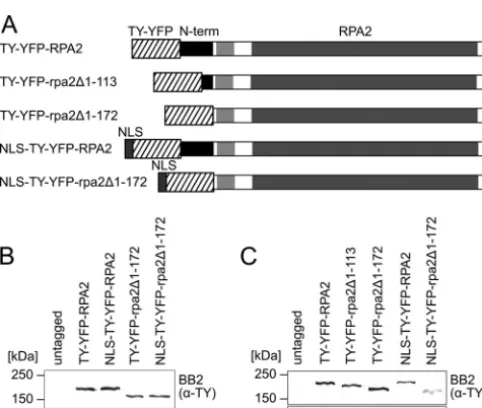
Homology models built by aligning trypanosome RPA2 to the second-largest subunit of budding yeast RNA polymerases with published structures (7, 29) predict that the trypanosome RPA2 sequence N terminal to the region of limited similarity is likely to protrude from the pol I core structure (data not shown) and there-fore is likely to be available for interactions with other complexes. The RPA2 N terminus is required for nuclear localization.To test whether theT.bruceiRPA2 N terminus is required for either nuclear or subnuclear localization, we designed a strategy to re-place the N terminus of one endogenousRPA2allele with a tag. We generated bloodstream-form cells expressing either full-length RPA2 or rpa2 missing residues 1 to 172 with an N-terminal TY epitope (2) and YFP tag (Fig. 1). Residues 1 to 172 were chosen because they are just N terminal of the region of limited similarity and therefore correspond to the first trypanosome-specific exten-sion. The tagging of a similar position ofSaccharomyces cerevisiae RPA2 (RPA135) has been used previously to isolate functional pol I complex (39, 41). A schematic representation of fusion proteins TY-YFP-RPA2 and TY-YFP-rpa2⌬1-172is shown in Fig. 2A. Suc-cessful integration and expression was established by genomic PCR (see Fig. S2 in the supplemental material) and immunoblot-ting (Fig. 2B).
In bloodstream-form trypanosomes, TY-YFP-RPA2 localized to the nucleus with the major signal from the nucleolus, which can be clearly seen as a region of low DNA density of⬃0.5m in diameter in the nucleus (Fig. 3). A majority of cells (67%⫾5%; 68/102 nonmitotic cells) additionally displayed a single site of
on September 8, 2020 by guest
http://ec.asm.org/
strong extranucleolar signal corresponding to the pol I-containing VSG ESB (Fig. 4A). This site often colocalized with a small nuclear region that was low in DAPI signal, most likely reflecting the transcriptionally permissive chromatin structure of the active
ES (13, 42). As previously described (33), in a minority of cells it is not possible to unambiguously distinguish the ESB from the greater nucleolar pool of pol I because of the proximity of the two.
The fusion protein produced by the N-terminal TY-YFP tag-ging of full-length RPA2 is still competent for incorporation and function in the pol I complex, both in its nucleolar context and in the ESB. This was demonstrated by the knockout of the wild-type allele to produce cells containing only tagged protein (Fig. 4B and 5). The tagging of one allele ofRPA2has no detectable effect on the growth of bloodstream-form cells (Fig. 5A). However, heterozy-gous⌬rpa2/RPA2bloodstream-form cells show slowed growth relative to that of wild-type or tagged cells (Fig. 5A) (P⬍0.01 by Student’sttest), which was unchanged on the tagging of the re-maining allele (⌬rpa2/TY-YFP-RPA2) (P⫽0.81 by Student’st test). The correct integration of constructs and the absence of extra copies ofRPA2was confirmed by diagnostic PCR from the genomic DNA of transformants (Fig. 5B and C). Importantly, the localization of TY-YFP-RPA2 is unchanged by the absence of un-tagged RPA2 (Fig. 4B), showing ESB structures in 72%⫾4% (81/113) of nonmitotic cells. Since trypanosomes cannot grow without a functional pol I enzyme and bloodstream-form cells requireVSGmRNA (40), these data show that TY-YFP-RPA2 is a functional pol I subunit and that the localization seen for this tagged protein is representative of the wild-type complex.
In contrast to functional RPA2 protein, TY-YFP-rpa2⌬1-172 was distributed throughout the cytoplasm and ap-peared to be completely excluded from the nucleus (Fig. 3). Equiv-alent results were obtained with cells expressing TY-YFP-rpa2⌬1-182, which lacks 10 more amino acids than TY-YFP-rpa2⌬1-172(Fig.
FIG 1Multiple-sequence alignment of the RPA2 N terminus. A multiple-sequence alignment was generated from a data set of 16 eukaryotic RPA2 proteins, 7 of which were selected for display in this figure. Residues are shown with a blue or cyan background indicating either identity or similarity to the majority consensus, respectively. Yellow flags on theT.bruceisequence indicate the residues tagged in this study. Blue bars mark stretches rich in basic residues in the trypanosomatid homologues. The red bar marks the region of limited similarity; the green bar marks RPA2 domain A.
FIG 2Expression from endogenous loci of N-terminally truncated RPA2 tagged with TY-YFP or NLS-TY-YFP. (A) Schemes of chimeric proteins ex-pressed. The region of limited similarity to nontrypanosomatid RPA2 homo-logues is indicated by a light gray box. Also shown are Western blottings of lysates from bloodstream-form (B) and procyclic (C) cells expressing RPA2 fusion proteins using monoclonal antibody BB2 (anti-TY-tag). Monoclonal antibody AB10 was used as a loading control.
on September 8, 2020 by guest
http://ec.asm.org/
3). These results showed that amino acids 1 to 172 of RPA2 are required for nuclear localization inT.brucei. To assess if this was an effect specific to bloodstream-form parasites, we also generated procyclic cell lines expressing tagged and truncated RPA2 fusion proteins from one of their endogenous alleles. We also generated a cell line expressing a fusion protein that had only 113 amino acids removed from the RPA2 N terminus, TY-YFP-rpa2⌬1-113(Fig. 1). Successful integration was tested by genomic sequencing and
im-munoblotting (Fig. 2C; also see Fig. S3 in the supplemental mate-rial). In procyclic-form cells, as in bloodstream-form cells, TY-YFP-RPA2 localized predominantly to the nucleolus, whereas truncated forms TY-YFP-rpa2⌬1-113 and TY-YFP-rpa2⌬1-172 were entirely excluded from the nucleus (see Fig. S4 in the supple-mental material). Hence, the N-terminal extension of RPA2 is required for the nuclear localization of this subunit in both blood-stream-form and procyclic cells.
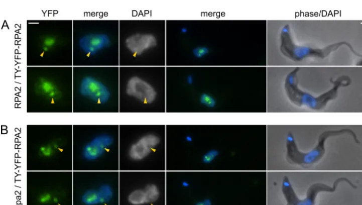
FIG 3Localization of N-terminal RPA2 truncations in bloodstream-form cells. While TY-YFP-RPA2 localizes to the nucleus, TY-YFP-rpa2⌬1-172is excluded.
NLS-TY-YFP-RPA2 is nuclear but has a subnuclear localization different from that of NLS-TY-YFP-rpa2⌬1-172. YFP fluorescence is pseudocolored in green,
DAPI in blue. Bars, 1m.
FIG 4Localization of TY-YFP-RPA2 in bloodstream-form cells. Cells containing one tagged allele and a second which is either wild type (A) or a knockout (B) are shown. Localization is unaffected by the absence of untagged protein, showing incorporation into both the nucleolus and the ESB (arrows). YFP fluorescence is pseudocolored in green, DAPI in blue. Bars, 1m.
on September 8, 2020 by guest
http://ec.asm.org/
The RPA2 N terminus is not sufficient for nuclear accumula-tion of a fusion protein.One obvious explanation for the mislo-calization of truncated RPA2 is that the N-terminal sequence con-tains a nuclear localization signal. Two small regions rich in basic residues are present within the N-terminal sequence ofT.brucei RPA2 (blue bar in Fig. 1), potentially indicating an NLS in this region. However, these residues are poorly conserved in RPA2 sequences from other trypanosomatid species (see Fig. S2 in the supplemental material), which would not be expected if they form a necessary NLS.
As a direct test of whether this sequence acts as a general NLS, we generated bloodstream-form cell lines conditionally express-ing TY-YFP-RPA2N1-172or TY-YFP-TY. Cells were induced for 48 h and then analyzed by fluorescence microscopy. TY-YFP-TY accumulated in cells to more than 10-fold the level of TY-YFP-RPA2N1-172. TY-YFP-TY was found throughout the cell body and also displayed a certain amount of accumulation in the nucleus (Fig. 6). The cellular distribution of TY-YFP-RPA2N1-172was sim-ilar, but TY-YFP-RPA2N1-172did not accumulate in the nucleus to any greater extent than TY-YFP-TY lacking the putative RPA2 NLS. Therefore, the N terminus of RPA2 is not an autonomous signal for nuclear localization.
Growth of bloodstream-form cells is slowed by N-terminal truncation of RPA2. We showed above that the haploinsuffi-ciency of RPA2 created by single-allele knockout caused slowed growth in bloodstream-form trypanosomes (Fig. 5). To assess the effect of the truncation of RPA2, we assessed the growth rate of
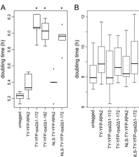
bloodstream-form and procyclic cells expressing truncated pro-teins. While bloodstream-form cells expressing TY-YFP-RPA2 did not have a significantly different doubling time compared to that of wild-type cells (Fig. 5A and 6A), cells expressing either TY-YFP-rpa2⌬1-172 or TY-YFP-rpa2⌬1-182 grew with a signifi-cantly longer doubling time (⬃0.8-h increase in doubling time; P⬍0.01 by Student’sttest) (Fig. 7A). Hence, the truncation of a single allele ofRPA2phenocopies haploinsufficiency created by complete gene knockout in bloodstream-form cells. In procyclic cells, no significant differences were seen between cells expressing wild-type, tagged, or truncated RPA2 from one of the twoRPA2 loci (Fig. 7). This most likely reflects the different dependencies on pol I levels of these two life cycle stages (see Discussion).
Nuclear localization of truncated RPA2 does not restore function.To test if the RPA2 N terminus is only required for nuclear import or provides additional functions, we created bloodstream-form and procyclic cell lines which expressed TY-YFP-RPA2 and TY-YFP-rpa2⌬1-172from their endogenous loci with an ectopic NLS at their N termini (NLS-TY-YFP-RPA2 and NLS-TY-YFP-rpa2⌬1-172). As an NLS, we chose theT.bruceiLa protein sequence RGHKRSRE, which has been shown previously to be sufficient for the accumulation of exogenous protein in the trypanosome nucleus (31). Correct integration was confirmed by genomic PCR and immunoblotting (Fig. 2A and B; also see Fig. S3 in the supplemental material).
In both bloodstream-form and procyclic cells, the La protein NLS was sufficient for the accumulation of truncated RPA2 in the
FIG 5Growth rates ofT.bruceicells expressing only N-terminally tagged RPA2 protein. (A) Tukey boxplot of doubling times for bloodstream-form cells expressing TY-YFP-RPA2 from one locus in cells which are either wild type (RPA2) or knocked out (⌬rpa2) at the other allele. Cells with haploinsufficiency caused by single-allele knockout grow more slowly (*,P⬍0.01 byttest), but there are no significant differences between tagged and untagged cell lines;n⫽6 for each boxplot. Bars indicate medians, boxes indicate interquartile ranges (IQR), and whiskers indicate ranges of measurements within 1.5⫻IQR. (B) Diagnostic multiplex PCR to confirm genotypes of cell lines. The presence ofRPA2,TY-YFP-RPA2, or⌬rpa2results in a specific band of the size shown. Lanes labeled⌬rpa2/TY-YFP-RPA2#1and⌬rpa2/TY-YFP-RPA2#2are independent clones of these cell lines. (C) Schematic showing the amplicons generated from the
multiplex PCR around the N terminus of the genes of interest.
on September 8, 2020 by guest
http://ec.asm.org/
nucleus (Fig. 3; also see Fig. S4 in the supplemental material). As for TY-YFP-RPA2, in a majority of bloodstream-form cells, NLS-TY-YFP-RPA2 forms ESB structures which are visibly distinct from the nucleolus (see Fig. S5 in the supplemental material).
There were, however, consistent differences between the nuclear distribution of the full-length and truncated NLS-TY-YFP-tagged proteins, with NLS-TY-YFP-rpa2⌬1-172 apparently being less present in the center of the nucleolus than NLS-TY-YFP-RPA2 (Fig. 3; also see Fig. S4 in the supplemental material). To investi-gate these differences, we measured the signal intensities of the YFP fusion proteins and also DAPI signal in a number of cells radially from the center of the nucleolus outwards (Fig. 8). The nucleolus forms a local minimum in DAPI signal, and its border can be seen from the position of the maximum in the radial DAPI distribution. The distribution of full-length NLS-TY-YFP-RPA2 is relatively homogeneous throughout the nucleolus, reflecting its incorporation into nucleolar pol I complexes. However, in both bloodstream-form and procyclic cells, truncated NLS-TY-YFP-rpa2⌬1-172is significantly less present in the nucleolus (P⬍0.001 in permutation test), instead having maximum intensity at the nucleolar periphery (Fig. 8). Moreover, while bloodstream-form cells expressing NLS-TY-YFP-RPA2 grew at rates similar to those of untagged controls, the doubling time of NLS-TY-YFP-rpa2⌬1-172 -expressing cells was significantly longer for bloodstream-form para-sites (P⬍0.01 by Student’sttest) (Fig. 7) but indistinguishable from cells in which RPA2 was excluded from the nucleus. These results show that the RPA2 N terminus not only is necessary for nuclear import but also is required for full pol I function and subnuclear distribution of the protein once inside the nucleus.
The RPA2 N-terminal extension is required for assembly of stable pol I complex.Since the N-terminal truncation of RPA2 phenocopies the effect of single-gene knockout (Fig. 5) with or without nuclear localization, we asked if this was a consequence of disruption in subnuclear localization or an inability to form func-tional polymerase complexes. To test this, we used the exogenous tag to immunoprecipitate the complexes formed by either full-length NLS-TY-YFP-RPA2 or NLS-TY-YFP-rpa2⌬1-172and iden-tified components by tandem mass spectrometry (Fig. 9). From cells expressing NLS-TY-YFP-RPA2, we isolated the 8-subunit core pol I complex isolated by Nguyen et al. by the tandem affinity purification of tagged RPA1 (34). This complex lacks RPB6z, RPB12, and p31, which are labile under the conditions used, but did show a strong interaction with the protein encoded by gene Tb11.02.1040. This protein is a predicted GTPase, and our
analy-FIG 6Cellular localization of ectopically expressed TY-YFP-RPA2N1-172in procyclic cells. TY-YFP-RPA2N1-172is not accumulated in the nucleus more than the
control protein, TY-YFP-TY. Note that the YFP fluorescence of TY-YFP-TY was more than a magnitude higher than that for TY-YFP-RPA2N1-172, so they had
to be processed differently for this figure. YFP fluorescence is pseudocolored in green, DAPI in blue. Bars, 1m.
FIG 7Growth rates ofT.bruceicells expressing truncated RPA2 proteins. (A) Tukey boxplot of doubling times for bloodstream-form cells expressing trun-cated RPA2 proteins during 120 h. Cells expressing the truntrun-cated proteins grow at lower rates independently of the nuclear localization of the protein; n⫽5 for each boxplot. Asterisks label cell lines with a doubling time signifi-cantly different from that of wild-type cells (P⬍0.01 byttest). Bars, medians; boxes, interquartile ranges (IQR); whiskers, ranges of measurements within 1.5⫻IQR; dots, measurements outside the range of whiskers. (B) Tukey box-plot of doubling times for procyclic cell clones expressing tagged RPA2 pro-teins during 144 h. No significant differences between the growth rates of the cell lines were found (n⫽6 for SiMP1 and TY-YFP-rpa2⌬1-172;n⫽18 for
others;ttest was used to determine significance).
on September 8, 2020 by guest
http://ec.asm.org/
sis (not shown) suggests it is the trypanosome homologue of hu-man GPN-loop GTPase 1 (GPN1/XAB1) and budding yeast NPA3, which have been found to interact with RNA pol II in these organisms (22, 44).
In contrast to the full-length protein, complexes formed by truncated NLS-TY-YFP-rpa2⌬1-172contain only 3 other pol I sub-units, RPC40, RPC19, and RPB10z (Fig. 9A). RPA1, RPB5z, RPA12, and RPB8, which are found associated with NLS-TY-YFP-RPA2, are absent from NLS-TY-YFP-rpa2⌬1-172complexes. Lev-els of RPA1 in these truncation mutant complexes were so low that they were undetectable by Western blotting (Fig. 9B). Moreover, the missing subunits RPB5z, RPA12, and RPB8 all are predicted to interact directly with RPA1 based on the structure of yeast RNA polymerases (7, 29), while those still present interact directly with the RPA2 subunit. This suggests that the loss of the N-terminal region of RPA2 specifically disrupts the interaction between the largest and second-largest subunits of trypanosomal pol I.
DISCUSSION
Transcription by pol I inT.bruceiis critical for the evasion of the mammalian immune response. Research during the last decade has described several trypanosome pol I-specific subunits which differ from their homologues in other systems by sequence inser-tions. Most dramatically, the second-largest subunit of pol I, RPA2, bears⬎200 additional residues at its N terminus, forming a region that is specific to trypanosomatids (38) and also to this polymerase complex (being absent from RPB2 and RPC128). The
work described here tests whether this sequence is necessary for pol I function, either generally or in an ESB-specific context.
Our comparative analysis of trypanosome RPA2 sequences with a broad data set of eukaryotic RPA2 proteins suggests that the additional sequence is the result of both an insertion and an ex-tension event near the N terminus. Homology modeling using the structural information available for theS. cerevisiaepol I and pol II complexes (7, 29) suggests that the RPA2 N terminus protrudes out of the pol I complex. It thus could provide a platform for interactions with other molecules, such as those involved in pol I biogenesis or transcription activation.
The trypanosomatid-specific RPA2 extension is required for the import of the protein into the nucleus. When we replaced the N-terminal 113, 172, or 183 residues of endogenous RPA2 with a tag, the fusion protein was excluded from the nucleus. This mis-localization was not an artifact of tagging, as tagged full-length RPA2 accumulated in the nucleus, localized to the nucleolus and ESB, and could fully replace the function of wild-type RPA2 with no detectable loss of fitness. Moreover, tags placed at the N termi-nus of RPA2 in budding yeast, which lacks the N-terminal exten-sion found in trypanosomes, have been used to isolate functional pol I complex (39, 41).
In bloodstream-form cells, when oneRPA2allele is trun-cated, growth is significantly reduced. It is likely that TY-YFP-rpa2⌬1-172-expressing cells have only half the amount of func-tional RPA2 protein available in the nucleus that wild-type cells (or cells expressing tagged full-length RPA2) have. Studies of
FIG 8Analysis of radial fluorescence signal intensities of NLS-TY-YFP-RPA2 and NLS-TY-YFP-rpa2⌬1-172. Normalized radial intensities of YFP signal (left)
and DAPI (right) measured from the center of the nucleolus in bloodstream-form (A) and procyclic (B) cells. Graphs represent means from 11 and 12 cells (A) and 10 and 11 cells (B) for NLS-TY-YFP-RPA2 and NLS-TY-YFP-rpa2⌬1-172, respectively. NLS-TY-YFP-rpa2⌬1-172(red) is concentrated at the nucleolar
periphery, while NLS-TY-YFP-RPA2 (black) is more homogeneously distributed in the nucleolus. Bars represent standard errors of the mean estimates.Pvalues represent Monte-Carlo estimates from 10,000 random permutations. Dotted lines show the position of maximum intensity for the NLS-TY-YFP-rpa2⌬1-172
profiles.
on September 8, 2020 by guest
http://ec.asm.org/
Schizosaccharomyces pombepol II have shown that the three larg-est subunits are the least abundant pol II subunits (27). It seems plausible, therefore, that a 50% reduction of nuclear RPA2 cru-cially limits the number of pol I complexes and thus interferes with rRNA and/orVSGtranscription. In agreement with this hy-pothesis, haploinsufficiency resulting from a clean knockout of a single allele ofRPA2resulted in the same growth rate decrease as N-terminal truncations. Interestingly, inS.cerevisiaethe deletion of a short sequence (residues 5 to 15) in the N-terminal region of the second-largest subunit of pol III, RPC128, resulted in the ex-clusion of this protein from the nucleus and slow growth. As for trypanosomal RPA2 truncations, this functional impairment could not be cured by the addition of an ectopic NLS to the trun-cated sequence (15).
In contrast to the situation for bloodstream-form cells, no ef-fect on growth was observed when RPA2 was truncated in procy-clic cells, even though the resultant proteins still failed to access the nucleus. This difference might be explained by a lower require-ment on pol I in procyclic cells, perhaps due to the higher levels of VSG required by bloodstream-form cells (21) compared to that for procyclin in insect-form cells (6). Bloodstream-form cells in which VSG production is depleted by RNAi are nonviable and very quickly arrest in their cell cycle (40), while culture-adapted procyclin-null procyclic mutants grow at wild-type rates (46). However, it may also be that the faster growth of bloodstream-form cells makes this life cycle stage more sensitive to reduced ribosome biosynthesis as a result of the depletion of nucleolar pol I.
Despite being required for nuclear localization, the RPA2 N-terminal sequence does not bear a classical NLS and does not act solely in nuclear import. The N-terminal 172 residues of RPA2 are not sufficient to confer nuclear localization on exogenous
pro-teins. Our results cannot exclude the possibility of a complex nuclear localization signal that is only partially provided by RPA2N1-172. However, the nuclear import of the truncated protein by the ad-dition of an ectopic NLS did not restore function or subnuclear localization, and the truncated protein was unable to form stable pol I complexes. This defect in complex formation suggests an alternative cause for mislocalization upon the removal of the N-terminal extension. If RPA2 accesses the nucleus not by an NLS in the N-terminal region but instead by virtue of an NLS on another pol I subunit with which it interacts, then the disruption of this interaction would also disrupt the nuclear import of RPA2. These two alternative scenarios are not separated by our data. It is clear, however, that the RPA2 N terminus does not act merely (if at all) as an NLS but is explicitly required to form functional pol I com-plexes in trypanosomes. This requirement identifies a trypanoso-matid-specific component to either the assembly or stability of pol I in these organisms, as nontrypanosomatid organisms form pol I complexes without the need for an N-terminal extension to RPA2. Since truncation disrupts the normal nucleolar localization of RPA2, it is difficult to unambiguously determine in these cells if truncated RPA2 is still recruited to the ESB. However, the trunca-tion of RPA2 results in a complete failure to assemble stable pol I complexes with no detectable interaction with RPA1. This will disrupt the function of both nucleolar and ESB pol I regardless of potential recruitment to a particular subnuclear domain, and it is clear that the N-terminal extension does not function exclusively in ESB transcription.
Our data do not distinguish between defects in pol I assembly and defects in the stability of the assembled complex. In bacteria, the assembly of DNA-dependent RNA polymerase starts with the two␣subunits and continues with the addition of first theand then the=subunit (19). The stability of the subassembly of the
FIG 9Immunoprecipitation of complexes formed by tagged RPA2. (A) Purified complexes from cells expressing NLS-TY-YFP-RPA2, NLS-TY-YFP-rpa2⌬1-172,
or untagged RPA2. Peptides have been separated by SDS-PAGE and stained with SYPRO ruby. The identity of the major component(s) of individual gel bands as determined by mass spectrometry is indicated next to each band. Colors indicate tagged subunit (green), pol I subunit interacting with tagged RPA2 and rpa2⌬1-172(orange) or RPA2 only (red), or proteins that are not known components of pol I complex (gray). (B) Immunoblot of pol I precipitation showing the
reaction of isolated peptides to antibodies raised against GFP or RPA1. (C) Schematic of the positions of the identified components of pol I complex on theS. cerevisiaecomplex (29). Colors are as described for panel A.
on September 8, 2020 by guest
http://ec.asm.org/
pol II homologues of␣2inS. cerevisiaesupported the view that the core assembly of the eukaryotic DNA-dependent RNA poly-merases follows the same sequence (26). By extension, the equiv-alent assembly process for pol I would involve first RPC40 and RPC19 (the two␣homologues), followed by RPA2 () and then the largest subunit RPA1 (=) (30). Significantly, our RPA2 trun-cation mutants assemble complexes containing both RPC40 and RPC19, forming a pol I homologue of␣2. These complexes also contain RPB10z, suggesting that this subunit can also be incorpo-rated directly into this subcomplex. Missing from the complexes are RPA1 and all subunits that interact directly with RPA1, indi-cating that it is the specific interaction between RPA2 and RPA1 that has a requirement for the RPA2 N-terminal extension in try-panosomes.
Our results with the N terminus of RPA2 provide the first evi-dence of functional significance for one of the sequence insertions seen in several pol I-specific subunits of trypanosomatids. They show that this protein region is required for nuclear import and subnuclear localization, either directly or through interaction with other subunits. They also demonstrate that this sequence is required for pol I function and for the stable interaction between RPA1 and RPA2 via a trypanosomatid-specific mechanism. How-ever, the function of this region is not exclusive to the ESB but globally affects pol I complexes in trypanosomes.
ACKNOWLEDGMENTS
This work was supported by the Wellcome Trust. J.-P.D. was supported by the EP Abraham Trust and the Studienstiftung des deutschen Volkes (German National Merit Foundation).
Predicted protein data sets were obtained from the sources specified in Table S4 in the supplemental material. We thank each of these organiza-tions and the respective genome-sequencing projects for making se-quence, gene model, and annotation data publicly available.
REFERENCES
1.Altschul SF, et al.1997. Gapped BLAST and PSI-BLAST: a new genera-tion of protein database search programs. Nucleic Acids Res.25:3389 – 3402.
2.Bastin P, Bagherzadeh Z, Matthews KR, Gull K.1996. A novel epitope tag system to study protein targeting and organelle biogenesis in Trypano-soma brucei. Mol. Biochem. Parasitol.77:235–239.
3. Berriman M, et al.2005. The genome of the African trypanosome Trypanosoma brucei. Science309:416 – 422.
4.Borst P.2002. Antigenic variation and allelic exclusion. Cell109:5– 8. 5.Brun R, Jenni L.1977. A new semi-defined medium forTrypanosoma
bruceisspp. Acta Trop.34:21–33.
6.Clayton CE, Mowatt MR.1989. The procyclic acidic repetitive proteins of Trypanosoma brucei. Purification and post-translational modification. J. Biol. Chem.264:15088 –15093.
7.Cramer P, Bushnell DA, Kornberg RD.2001. Structural basis of tran-scription: RNA polymerase II at 2.8 angstrom resolution. Science292: 1863–1876.
8.Das A, Li H, Liu T, Bellofatto V.2006. Biochemical characterization of Trypanosoma bruceiRNA polymerase II. Mol. Biochem. Parasitol.150: 201–210.
9.Devaux S, et al.2006. Characterization of RNA polymerase II subunits of Trypanosoma brucei. Mol. Biochem. Parasitol.148:60 – 68.
10. Devaux S, et al.2007. Diversification of function by different isoforms of conventionally shared RNA polymerase subunits. Mol. Biol. Cell18:1293– 1301.
11. Eddy SR.1998. Profile hidden Markov models. Bioinformatics14:755–763. 12. Fèvre EM, Wissmann BV, Welburn SC, Lutumba P.2008. The burden
of human African trypanosomiasis. PLoS Negl. Trop. Dis.2:e333. 13. Figueiredo LM, Cross GAM.2010. Nucleosomes are depleted at the VSG
expression site transcribed by RNA polymerase I in African trypanosomes. Eukaryot. Cell9:148 –154.
14. Günzl A, et al.2003. RNA polymerase I transcribes procyclin genes and variant surface glycoprotein gene expression sites inTrypanosoma brucei. Eukaryot. Cell2:542–551.
15. Hardeland U, Hurt E.2006. Coordinated nuclear import of RNA poly-merase III subunits. Traffic7:465– 473.
16. Hertz-Fowler C, et al.2008. Telomeric expression sites are highly con-served inTrypanosoma brucei. PLoS One3:e3527.
17. Hirumi H, Hirumi K.1989. Continuous cultivation of Trypanosoma bruceiblood stream forms in a medium containing a low concentration of serum protein without feeder cell layers. J. Parasitol.75:985–989. 18. Horn D.2008. Codon usage suggests that translational selection has a
major impact on protein expression in trypanosomatids. BMC Genomics
9:2.
19. Ishihama A.1981. Subunit of assembly ofEscherichia coliRNA polymer-ase. Adv. Biophys.14:1–35.
20. Ivens AC, et al.2005. The genome of the kinetoplastid parasite, Leishma-nia major. Science309:436 – 442.
21. Jackson DG, Owen MJ, Voorheis HP.1985. A new method for the rapid purification of both the membrane-bound and released forms of the vari-ant surface glycoprotein fromTrypanosoma brucei. Biochem. J.230:195– 202.
22. Jeronimo C, et al.2007. Systematic analysis of the protein interaction network for the human transcription machinery reveals the identity of the 7SK capping enzyme. Mol. Cell27:262–274.
23. Katoh K, Misawa K, Kuma K, Miyata T.2002. MAFFT: a novel method for rapid multiple sequence alignment based on fast Fourier transform. Nucleic Acids Res.30:3059 –3066.
24. Kelly S, Wickstead B, Gull K.2005. An in silico analysis of trypanoso-matid RNA polymerases: insights into their unusual transcription. Biochem. Soc. Trans.33:1435–1437.
25. Kelly S, et al.2007. Functional genomics inTrypanosoma brucei: a col-lection of vectors for the expression of tagged proteins from endogenous and ectopic gene loci. Mol. Biochem. Parasitol.154:103–109.
26. Kimura M, Ishiguro A, Ishihama A.1997. RNA polymerase II subunits 2, 3, and 11 form a core subassembly with DNA binding activity. J. Biol. Chem.272:25851–25855.
27. Kimura M, Sakurai H, Ishihama A. 2001. Intracellular contents and assembly states of all 12 subunits of the RNA polymerase II in the fission yeastSchizosaccharomyces pombe. Eur. J. Biochem.268:612– 619. 28. Kooter JM, Borst P.1984. Alpha-amanitin-insensitive transcription of
variant surface glycoprotein genes provides further evidence for discon-tinuous transcription in trypanosomes. Nucleic Acids Res.12:9457–9472. 29. Kuhn C, et al.2007. Functional architecture of RNA polymerase I. Cell
131:1260 –1272.
30. Kwapisz M, Beckouët F, Thuriaux P.2008. Early evolution of eukaryotic DNA-dependent RNA polymerases. Trends Genet.24:211–215. 31. Marchetti MA, Tschudi C, Kwon H, Wolin SL, Ullu E.2000. Import of
proteins into the trypanosome nucleus and their distribution at karyoki-nesis. J. Cell Sci.113:899 –906.
32. Martínez-Calvillo S, Saxena A, Green A, Leland A, Myler PJ. 2007. Characterization of the RNA polymerase II and III complexes in Leishma-nia major. Int. J. Parasitol.37:491–502.
33. Navarro M, Gull K.2001. A pol I transcriptional body associated with VSG mono-allelic expression inTrypanosoma brucei. Nature414:759 – 763.
34. Nguyen TN, Schimanski B, Zahn A, Klumpp B, Günzl A.2006. Purifi-cation of an eight subunit RNA polymerase I complex inTrypanosoma brucei. Mol. Biochem. Parasitol.149:27–37.
35. Nguyen TN, Schimanski B, Günzl A.2007. Active RNA polymerase I of Trypanosoma bruceiharbors a novel subunit essential for transcription. Mol. Cell. Biol.27:6254 – 6263.
36. Pays E.2005. Regulation of antigen gene expression in Trypanosoma brucei. Trends Parasitol.21:517–520.
37. Rudenko G, Bishop D, Gottesdiener K, Van der Ploeg LH. 1989. Alpha-amanitin resistant transcription of protein coding genes in insect and bloodstream formTrypanosoma brucei. EMBO J.8:4259 – 4263. 38. Schimanski B, et al.2003. The second largest subunit ofTrypanosoma
brucei’s multifunctional RNA polymerase I has a unique N-terminal ex-tension domain. Mol. Biochem. Parasitol.126:193–200.
39. Schneider DA, Nomura M.2004. RNA polymerase I remains intact with-out subunit exchange through multiple rounds of transcription in Saccha-romyces cerevisiae. Proc. Natl. Acad. Sci. U. S. A.101:15112–15117. 40. Sheader K, et al.2005. Variant surface glycoprotein RNA interference
on September 8, 2020 by guest
http://ec.asm.org/
triggers a precytokinesis cell cycle arrest in African trypanosomes. Proc. Natl. Acad. Sci. U. S. A.102:8716 – 8721.
41. Shou W, et al.2001. Net1 stimulates RNA polymerase I transcription and regulates nucleolar structure independently of controlling mitotic exit. Mol. Cell8:45–55.
42. Stanne TM, Rudenko G.2010. Active VSG expression sites in Trypano-soma bruceiare depleted of nucleosomes. Eukaryot. Cell9:136 –147. 43. Stewart M, et al.2010. Processing of a phosphoglycerate kinase reporter
mRNA inTrypanosoma bruceiis not coupled to transcription by RNA polymerase II. Mol. Biochem. Parasitol.172:99 –106.
44. Tarassov K, et al.2008. An in vivo map of the yeast protein interactome. Science320:1465–1470.
45. Vanhamme L, Pays E, McCulloch R, Barry JD.2001. An update on anti-genic variation in African trypanosomes. Trends Parasitol.17:338 –343.
46. Vassella E, Bütikofer P, Engstler M, Jelk J, Roditi I.2003. Procyclin null mutants ofTrypanosoma bruceiexpress free glycosylphosphatidylinositols on their surface. Mol. Biol. Cell14:1308 –1318.
47. Walgraffe D, et al.2005. Characterization of subunits of the RNA poly-merase I complex inTrypanosoma brucei. Mol. Biochem. Parasitol.139: 249 –260.
48. Wirtz E, Leal S, Ochatt C, Cross GA.1999. A tightly regulated inducible expression system for conditional gene knock-outs and dominant-negative genetics inTrypanosoma brucei. Mol. Biochem. Parasitol.99:89 – 101.
49. Young R, et al.2008. Isolation and analysis of the genetic diversity of repertoires of VSG expression site containing telomeres from Trypano-soma brucei gambiense,T. b. bruceiandT. equiperdum. BMC Genomics
9:385.