Lymphomas Indicates Subtype-Specific Viral Gene Expression
Patterns and Immune Cell Microenvironments
Hani Nakhoul,
aZhen Lin,
aXia Wang,
aClaire Roberts,
aYan Dong,
bErik Flemington
aaDepartment of Pathology, Tulane Cancer Center, Tulane University School of Medicine, New Orleans, Louisiana, USA
bDepartment of Structural and Cellular Biology, Tulane Cancer Center, Tulane University School of Medicine, New Orleans, Louisiana, USA
ABSTRACT
Certain peripheral T-cell lymphomas (PTCLs) have been associated with
viral infection, particularly infection with Epstein-Barr virus (EBV). However, a
com-prehensive virome analysis across PTCLs has not previously been reported. Here we
utilized published whole-transcriptome RNA sequencing (RNA-seq) data sets from
seven different PTCL studies and new RNA-seq data from our laboratory to screen
for virus association, to analyze viral gene expression, and to assess B- and T-cell
re-ceptor diversity paradigms across PTCL subtypes. In addition to identifying EBV in
angioimmunoblastic T-cell lymphoma (AITL) and extranodal NK/T-cell lymphoma
(EN-KTL), two PTCL subtypes with well-established EBV associations, we also detected
EBV in several cases of anaplastic large-cell lymphoma (ALCL), and we found
evi-dence of infection by the oncogenic viruses Kaposi’s sarcoma-associated herpesvirus
and human T-cell leukemia virus type 1 in isolated PTCL cases. In AITLs, EBV gene
expression analysis showed expression of immediate early, early, and late lytic
genes, suggesting either low-level lytic gene expression or productive infection in a
subset of EBV-infected B-lymphocyte stromal cells. Deconvolution of immune cell
subpopulations demonstrated a greater B-cell signal in AITLs than in other PTCL
subtypes, consistent with a larger role for B-cell support in the pathogenesis of
AITL. Reconstructed T-cell receptor (TCR) and B-cell receptor (BCR) repertoires
demonstrated increased BCR diversity in AITLs, consistent with a possible
EBV-driven polyclonal response. These findings indicate potential alternative roles for
EBV in PTCLs, in addition to the canonical oncogenic mechanisms associated
with EBV latent infection. Our findings also suggest the involvement of other
vi-ruses in PTCL pathogenesis and demonstrate immunological alterations
associ-ated with these cancers.
IMPORTANCE
In this study, we utilized next-generation sequencing data from 7
dif-ferent studies of peripheral T-cell lymphoma (PTCL) patient samples to globally
as-sess viral associations, provide insights into the contributions of EBV gene
expres-sion to the tumor phenotype, and assess the unique roles of EBV in modulating the
immune cell tumor microenvironment. These studies revealed potential roles for
EBV replication genes in some PTCL subtypes, the possible role of additional
hu-man tumor viruses in rare cases of PTCLs, and a role for EBV in providing a
unique immune microenvironmental niche in one subtype of PTCLs. Together,
these studies provide new insights into the understudied role of tumor viruses in
PTCLs.
KEYWORDS
B-cell receptor diversity, EBV, Epstein-Barr virus, Kaposi’s
sarcoma-associated herpesvirus, T cell, human T-cell leukemia virus, immune
microenvironment, lymphoma, murine leukemia virus, sequencing
CitationNakhoul H, Lin Z, Wang X, Roberts C, Dong Y, Flemington E. 2019. High-throughput sequence analysis of peripheral T-cell lymphomas indicates subtype-specific viral gene expression patterns and immune cell microenvironments. mSphere 4:e00248-19.
https://doi.org/10.1128/mSphere.00248-19. EditorBlossom Damania, University of North Carolina, Chapel Hill
Copyright© 2019 Nakhoul et al. This is an open-access article distributed under the terms of theCreative Commons Attribution 4.0 International license.
Address correspondence to Erik Flemington, [email protected].
Received2 April 2019 Accepted24 June 2019 Published10 July 2019
on September 8, 2020 by guest
http://msphere.asm.org/
phoma (ENKTL), EBV directly infects tumor cells of T- or NK-cell lineage and is
consis-tently detected in episomal form in these cells. On the other hand, in
angioimmuno-blastic T-cell lymphoma (AITL), EBV-infected B cells are found adjacent to tumor cells of
the T follicular helper (Tfh) phenotype. It has been proposed that interaction between
the tumor cells and the B lymphocytes provides oncogenic support for the tumor,
although the mechanism for this process is not clear. In still other PTCL subtypes,
including anaplastic large-cell lymphoma (ALCL) and peripheral T-cell lymphoma not
otherwise specified (PTCL-NOS), reports of cases involving EBV and other viruses have
been limited or controversial. Studies of EBV in NK- and T-cell neoplasms have been
complicated by a relative lack of cell lines and animal models that replicate key features
of these diseases. Although some high-throughput sequencing studies have confirmed
the presence of EBV in particular PTCL subtypes, a comprehensive virome analysis
across PTCL subtypes has not, to our knowledge, been previously reported.
Latent EBV infection is associated with the constitutive expression of a subset of viral
genes without the production of infectious virions. The particular viral genes expressed,
which depend on immune surveillance and the characteristics of the host cell, define
the latency type of the EBV infection (9). EBV-associated cancers are each typically
associated with a specific latency type, although even within a tumor type,
patient-to-patient and intratumoral variations exist. Endemic Burkitt’s lymphoma typically exhibits
latency type I, in which EBV expresses only the episomal maintenance factor EBNA1, the
noncoding RNAs EBER1 and EBER2 (10), the BART long noncoding RNAs (lncRNAs)
(referred to here as RPMS1 and A73) (11–14), the BART microRNAs (15–18), and the
circular RNAs circRPMS1_E4_E3a and circRPMS1_E4_E2 (19–24). Other lymphomas in
immunocompetent patients, gastric carcinoma, and nasopharyngeal carcinoma usually
exhibit type II latency, in which LMP1 and LMP2A are expressed, in addition to type I
genes (25, 26). Lymphomas in the setting of immunosuppression often exhibit type III
latency, in which all EBV latent genes are expressed (27, 28). The lytic or productive
phase of EBV infection is not classically associated with malignancy, although lytic cycle
proteins have been shown to play a role in the pathogenesis of certain tumors (29–32).
Both lytic and latent EBV infections are subject to surveillance by the host immune
system through antigen-specific T- and B-cell responses. EBV-associated cancers in
immunocompetent patients demonstrate a variety of strategies for immune evasion,
including suppression of antigen presentation through downregulation of the major
histocompatibility complex (MHC) and upregulation of the immunomodulatory host
genes PD-L1 and IDO1 (6). In the case of lymphoid malignancies, immune dysfunction
is also caused by derangements of B-cell receptor (BCR) and T-cell receptor (TCR)
signaling in neoplastic cells themselves (33, 34). Functional studies of BCR and TCR
repertoires and their interactions with abnormalities in particular cancers are now
possible due to both efficient high-throughput methods for targeted amplification of
BCRs and TCRs and computational techniques that permit reconstruction of these
repertoires from untargeted whole-transcriptome sequencing data (35–37).
on September 8, 2020 by guest
found isolated cases of primary tumors that were positive for the oncogenic viruses
Kaposi’s sarcoma-associated herpesvirus (KSHV) and human T-cell leukemia virus type
1 (HTLV-1), raising the possibility that these are sporadically involved in the
pathogen-esis of T-cell malignancies. In AITLs, we observed the expected EBV latency gene
expression profile but also substantial expression of lytic genes, representing evidence
of abortive lytic replication and/or productive replication in a subset of infected cells.
Deconvolution of immune cell subpopulations from RNA-seq data showed greater
B-cell signals and increased BCR repertoire diversity in AITLs, consistent with a possible
EBV-driven polyclonal response.
RESULTS
Virus read detection.
EBV contributes to the etiology of a number of PTCL subtypes
(1, 7, 49) with a high prevalence in AITLs (50) and ENKTLs (51). While the cellular
transcriptomes of AITL, ALCL, ENKTL, and PTCL-NOS patient samples have been
previ-ously analyzed by RNA sequencing (38–43), the viral compositions of these
transcrip-tomes were not considered in these analyses. As potential drivers of tumor progression
in PTCLs, the viral contributions to the overall transcriptomes are germane to
under-standing the mechanisms driving the tumor phenotype.
Our initial approach to assessing the viral impact on these tumors was to take a
macroscopic view of viral etiology by analyzing previously published clinical data sets
(38–43) for reads mapping to a panel of mammalian viruses (52, 53). Reads from each
sample were aligned to a genome index containing all human chromosomes plus 740
genomes for viruses known to infect human cells (listed in Table S1 in the supplemental
material), as we have done for previous studies (52, 53). To limit false-positive calls
related to contamination (54) or low-level virus presence in a small percentage of
stromal cells, we utilized a threshold frequency of 0.2 read per million mapped human
reads (RPMHRs). This cutoff is in rough agreement with that used in previous
investi-gations by our group and others assessing exogenous viruses in sequencing data (52,
55–57).
In line with previous reports (50, 51), EBV was the most prevalent virus detected
across all PTCL subtypes, with 18 of 21 AITL and 15 of 17 ENKTL patient samples being
positive for EBV (Fig. 1 and 2). While infiltrating EBV-positive B lymphocytes are
characteristic of AITL tumors (reviewed in reference 58), the etiology of ENKTLs involves
direct EBV infection of the tumor cells of NK- or T-cell lineage (40, 59). Consistent with
direct infection of ENKTL tumor cells, 8 of 10 ENKTL cell lines that we analyzed,
including 5 sequenced by our lab, were found to be positive for EBV. Eleven of 35 ALCL
samples and 7 of 21 PTCL-NOS samples were also positive for EBV, although only 2
ALCLs showed high levels of EBV reads (451 and 1,158 RPMHRs) and only 3 PTCL-NOS
samples showed at least moderate levels of EBV reads (2, 6, and 9 RPMHRs). These
results are consistent with the established role of EBV in the pathogenesis of AITL and
ENKTL, while at the same time they raise the possibility of the involvement of EBV in a
subset of ALCL and PTCL-NOS cases (60–62).
In addition to pervasive evidence of EBV in these PTCL patient and cell line samples,
one AITL patient sample contained high read numbers (5,767 RPMHRs) for another
oncogenic gammaherpesvirus, KSHV, with a second AITL sample containing lower but
potentially meaningful numbers of KSHV reads (3 RPMHRs) (Fig. 2). Viral transcriptome
coverage from the AITL sample with high KSHV detection was similar to that observed
from the KSHV-positive primary effusion lymphoma cell line BCP-1, with expression of
the classic latency genes LANA and Kaposin (63, 64), as well as expression of the viral
on September 8, 2020 by guest
http://msphere.asm.org/
interleukin-6 (IL-6) and IL-8 homologues (65, 66) and the viral E3 ubiquitin ligase (67)
(Fig. 3). These findings indicate a predominantly latent infection that may contribute to
the tumor phenotype in these patients.
Although betaherpesviruses, such as the ubiquitous human cytomegalovirus (CMV)
and the human herpesvirus 6 (HHV-6), are not classically considered tumor viruses,
there are a number of sometimes controversial reports of their unconventional roles in
cancer (68–71). Whether it is etiological or detected due to local immune suppression
in the tumor microenvironment, we have previously detected HHV-6 in occasional
B-cell lymphomas (53). Here, we similarly detected moderate to low levels of HHV-6 in
an ALCL and a PTCL-NOS, and we detected high levels of CMV in one AITL and lower
levels in a PTCL-NOS (Fig. 2 and Fig. S1). As we suggested for our previous HHV-6 B-cell
lymphoma findings, the results here could simply represent tolerance for replication of
these ubiquitous viruses in a local immune-suppressed microenvironment.
HIV reads were detected at low levels in one ENKTL cell line and one PTCL-NOS
patient (Fig. 2 and Fig. S1). With little previous evidence of a direct oncogenic role of
HIV, similar to the findings of betaherpesviruses in some tumor samples, this could
represent replication in an immune-compromised microenvironment in the PTCL-NOS
sample and possible carryover or tissue culture contamination with HIV in the ENKTL
cell line. On the other hand, HTLV-1 is a known oncogenic T-cell tumor virus, and the
detection of HTLV-1 in one ALCL (Fig. 2) could represent a pathological infection.
Lastly, the murine leukemia virus (MuLV) and its relative, the mouse mammary
tumor virus (MMTV), were detected in two ENKTL cell lines (Fig. 2). While initially
thought to be pathological infections in prostate cancers, cell line findings for MuLV
FIG 1 Distribution of total EBV gene expression (transcripts per million [TPM]) for each PTCL subtype. For each sample, theyaxis indicates the sum of TPMs of all EBV genes. Red bars indicate 95% bootstrap confidence intervals about the mean.
FIG 2 Viruses detected in RNA-seq data from PTCLs. Only those samples with at least 0.2 read per million human mapped reads (RPMHR) are shown.
on September 8, 2020 by guest
were later found to be due to propagation of the respective cell lines in MuLV-infected
mice, where they picked up the virus (72, 73). The presence of MuLV/MMTV reads in
multiple cell lines is consistent with cross-contamination or infection during culture
(74). What is particularly striking here, however, is the unusually high levels of MuLV/
MMTV in two ALCL patient samples (Fig. 2). We investigated the metadata for this study
and could not identify any potential artifactual evidence to explain these findings.
Clearly, these murine viruses can infect human cells, and there is the formal possibility
that MuLV could play an etiological role in these patients. However, given the
perva-siveness of MuLV in laboratories and previous erroneous reports, such conclusions must
be made in an extremely guarded fashion.
EBV gene expression analysis.
The results of our screen for viruses in PTCLs
confirm EBV as the viral pathogen most consistently associated with PTCLs. EBV gene
expression patterns vary across malignancies and, to some degree, between patients
with a specific tumor type. The unique pattern of viral genes expressed in a particular
tumor setting is directly pertinent to viral oncogenesis in the respective tumor type
and/or patient (9, 75). Although EBV latency types have been previously defined in AITL
and ENKTL on the basis of immunohistochemistry, comprehensive sequencing-based
analyses of EBV gene expression have not been performed to date for AITL and have
only recently been performed for ENKTL (76). We therefore studied the distribution and
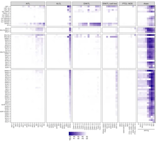
variation of EBV gene expression across PTCL subtypes. Figure 4 shows the expression
of individual EBV genes in EBV-positive PTCL samples, as well as in the EBV-positive
Burkitt lymphoma-derived Akata cell line, before and after induction of the lytic cycle
with anti-human IgG (77, 78). In AITLs, where EBV is present in the stromal B cells,
variable expression of LMP1 and LMP2A/B was detected, consistent with previous
reports of type II latency infection in AITLs (60, 79) (Fig. 4 and Fig. S2). More consistently
expressed were the EBV noncoding BART lncRNAs RPMS1 and A73, whose expression
in vivo
can exert effector signaling to the host cell regulatory circuitry without eliciting
an adaptive immune response. Also notable is detection of immediate early BZLF1 and
BRLF1 transcripts as well as some early and late lytic genes in AITLs but not ENKTL
patient or cell lines (Fig. 4). While this may represent a small percentage of cells
undergoing lytic replication, their expression levels rival those of the latency genes
(Fig. 4). These results are consistent with previous reports detecting expression of the
immediate early Zta gene by immunohistochemistry in AITL patients (80). They are also
consistent with our findings of relatively high lytic gene expression in B-cell lymphoma
patient samples (53), suggesting the common observation of lytic gene expression in
B cells
in vivo
. Since early genes, such as BZLF1, are expressed transiently during initial
infection of naive B cells (81, 82), where they provide an initial growth stimulation (83),
BZLF1 and other lytic genes may similarly support the proliferation of the stromal B-cell
population in AITLs.
The antigenic type III latency EBNA proteins were generally not detected in AITLs,
with the exception of EBNA2 expression in patient 3 (Fig. 4 and Fig. S2). EBNA2
expression in this patient coincides with KSHV infection. In the setting of EBV and KSHV
coinfection, EBV has recently been shown to interact with KSHV and enhance the
stability of KSHV infection (84). However, whether KSHV regulates EBNA2 expression in
FIG 3 KSHV exhibits an expression profile consistent with latent infection in one AITL sample (PAT3). KSHV coverage is also shown for BCP-1, a cell line derived from a primary effusion lymphoma. Transcripts were detected from the primary latency transcript region located between the K12 and ORF74 open reading frames, K2/viral IL-6 (vIL-6), the noncoding RNA PAN, and the viral IRF (vIRF) family of transcription factors.
on September 8, 2020 by guest
http://msphere.asm.org/
this case, whether this tumor displays generally higher local immune suppression to
tolerate two viral infections as well as EBNA2 expression, or whether this is a
coinci-dental observation remains to be determined.
In ENKTLs, we similarly observed primarily evidence of type II latency, consistent
with previous studies (9), and robust expression of the BART lncRNAs RPMS1 and A73
(Fig. 4 and Fig. S2). ALCLs generally exhibited very low expression of EBV genes in most
EBV-positive samples. However, 2 of 15 EBV-positive samples showed high levels of EBV
gene expression, and 1 of these showed high levels of lytic gene expression (Fig. 4).
Extensive B-cell receptor repertoire diversity in AITLs.
In most EBV-associated
tumors, EBV is present within the tumor cell, where the expressed viral genes
contrib-ute directly to the tumor phenotype. AITLs are unusual in that EBV infects the stromal
B cells, which appear to provide essential support to the T-cell tumor (reviewed in
reference 7). To explore the tumor and stromal cell composition across these PTCL
FIG 4 Expression of latent (LT), immediate early lytic (IE), early lytic (E), and late lytic (L) EBV genes in PTCL primary tumors and cell lines. Columns indicate PTCL samples, and each row indicates an EBV gene. In the rightmost panel, expression data from the EBV-positive Burkitt lymphoma cell line Akata is shown at several time points, as lytic reactivation was induced with human IgG.
on September 8, 2020 by guest
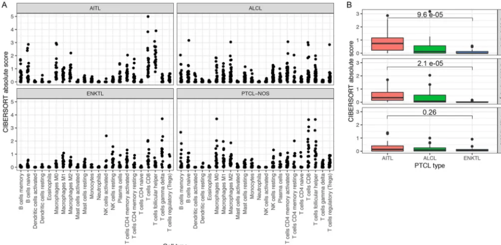
subtypes, we performed deconvolution of gene expression profiles to estimate the
immune cell composition in each tumor sample (85) (Fig. 5A). Consistent with the
proposed requirement of a tumor-supporting B-cell population in AITLs, there is
comparatively high B-cell evidence in AITLs relative to ALCLs and ENKTLs, with ENKTLs
showing low B-cell signals and ALCLs showing intermediate B-cell signals (Fig. 5B).
These results also indicated enrichment of the T follicular helper subtype of immune
cells in AITLs, consistent with the characterization of AITL itself as a neoplasm of the T
follicular helper phenotype (1, 86–88). This analysis also showed enrichment of other
immune cell subpopulations, such as macrophages, consistent with the known
heter-ogeneous tumor microenvironment of AITL (89). As an indication of the validity of the
deconvolution method, enrichment of B cells and macrophages was not detected in
cell lines and purified T-cell samples (Fig. S3). B-cell and macrophage scores were
decreased in ENKTL relative to AITL. These results indicate that the pathological
characterization of the heterogeneity of the tumor microenvironment in PTCLs is
broadly reproducible from RNA-seq data.
T- and B-cell receptor analyses.
Since enrichment of EBV-positive B lymphocytes
in the tumor microenvironment is characteristic of AITLs (45, 46, 90), we hypothesized
that T-cell receptor (TCR) and B-cell receptor (BCR) repertoire diversity would vary
across PTCL subtypes, with greater BCR diversity in AITLs due to polyclonal
EBV-mediated B-cell expansion. To test this hypothesis, we reconstructed BCR sequences
from the RNA-seq data and assessed the relationship between total assembled
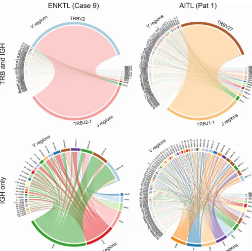
clono-types and unique clonoclono-types. We first assessed this by constructing circle plots
dis-playing TCR (T-cell receptor

[TRB]) and BCR (immunoglobulin heavy chain [IGH]) V(D)J
recombinations for each AITL and ENKTL. This analysis showed considerable variation
across samples but generally displayed monoclonal TCR expansions likely of tumor cell
origin, with typically lower T- and B-cell subpopulations that likely represent infiltrating
immune cells (representative plots are shown in Fig. 6, top; note the major TRB J2-7
joining region-to-V2 variable region clone in the ENKTL patient and the major TRB J1-1
joining region-to-V27 variable region clone in the AITL patient). Assessing the BCR
repertoire alone, variation was observed across samples, but in general, greater
diver-FIG 5 (A) Enrichment scores for 22 immune cell subpopulations based on deconvolution by CIBERSORT. Each panel indicates a subtype of primary tumor. (B) Box plots of enrichment scores for three B-cell subpopulations in AITL, ALCL, and ENKTL primary tumor samples. For naive and memory B cells, scores are significantly greater in AITLs than in ENKTLs.
on September 8, 2020 by guest
http://msphere.asm.org/
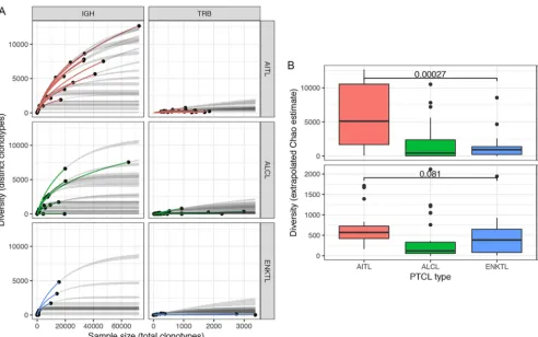
sity was seen in AITLs (a representative plot is shown in Fig. , bottom; note the higher
number of J regions and the greater number of V region subclones in the AITL sample).
This greater B-cell diversity in AITLs may be driven by EBV-mediated polyclonal
expan-sion. To investigate the relationship between the size of the reconstructed BCR
reper-toires and their diversity, we generated rarefaction plots, an approach borrowed from
the ecological literature that is now commonly used in computational immunology
(35–37). We computed extrapolated Chao estimates of the diversity of B- and T-cell
receptor clonotypes in each sample, permitting comparison of these repertoires across
different sampling depths (91). Using this approach for the AITL, ALCL, and ENKTL PTCL
subtypes, a greater number of total and unique BCR clonotypes were observed in AITLs
than in ALCLs or ENKTLs, indicating a greater average BCR diversity for AITLs (Fig. 7A
FIG 6 Circle plots indicating V(D)J recombination in an AITL sample and an ENKTL sample.
on September 8, 2020 by guest
and B). This suggests that EBV facilitates polyclonal B-cell expansion in AITLs, whereas
in infiltrating B cells in ALCLs and ENKTLs, less diversity is observed due to expansion
of a relatively small number of antigen-reactive clones.
DISCUSSION
Frequent detection of viruses in PTCL samples.
Viruses exert characteristic effects
on cancer progression and define unique subtypes of malignancies (28, 92). Although
the association between EBV and certain PTCL subtypes has been firmly established on
the basis of immunochemistry, our study is, to our knowledge, the first unbiased virome
analysis across PTCL subtypes. The sporadic detection of oncogenic viruses, such as
KSHV and HTLV-1, in this study raises the possibility that, in addition to EBV, these
viruses may similarly contribute to the pathogenesis of PTCL in rare cases.
KSHV infection is critical to the pathogenesis of several lymphoproliferative
disor-ders, including multicentric Castleman disease, immunodeficiency-related diffuse large
B-cell lymphoma, and primary effusion lymphoma (93). These disorders are
heteroge-neous in clinical presentation, histology, immunophenotype, and EBV involvement; for
example, cases of primary effusion lymphoma with the T-cell phenotype have been
reported, although an indeterminate immunophenotype is more common (94). There
have been previous reports of occasional KSHV-positive cases of AITL, as determined by
PCR (95), although this finding was not reproduced in subsequent studies by either PCR
or immunohistochemistry (96–98). The quantitative nature of our approach revealed a
strong presence of KSHV in 1 of 22 AITLs, supporting an etiological role for KSHV in a
minor percentage of cases of this tumor type. Although rare in the AITL cases assessed
here, it will be important to determine whether KSHV displays a more substantial
penetrance in areas of high KSHV seroprevalence (e.g., sub-Saharan Africa) (99).
FIG 7 (A) Rarefaction plots for AITL, ALCL, and ENKTL samples, based on multinomial models of diversity as a function of sample size (91, 128). Thexaxis indicates the number of clonotypes detected in each sample (sample size), and theyaxis indicates the number of distinct clonotypes (diversity). Solid dots indicate the observed sample size and diversity, colored lines indicate interpolation of a curve to this value, and gray lines indicate extrapolation of the curve to the largest sample size in the data set. (B) Box plots showing extrapolated Chao estimates, a diversity measure that permits comparisons of clonotype diversity across samples with distinct numbers of total clonotypes (91). IGH diversity is significantly greater in AITLs than in ENKTLs (Wilcoxon rank-sum test).
on September 8, 2020 by guest
http://msphere.asm.org/
recurrent mutations in AITL are cell type specific, with RHOA and IDH2 mutations
occurring exclusively in neoplastic PD-1-positive T cells, NOTCH1 mutations occurring
exclusively in B cells, and TET2 mutations occurring in both T and B cells (90, 103). EBV
is believed to contribute to the neoplastic transformation of B cells in the progression
from AITL to B-cell lymphoma, although EBV is not uniformly observed in these cases
(104).
Interactions between EBV-infected cells and other cells in the tumor
microenviron-ment, representing a subversion of normal mechanisms of immune signaling, are a
common feature in lymphoid malignancies (6) and represent a possible mechanism for
immune evasion or modulation in these settings (105). In the case of AITL, previous
studies of B-cell receptor repertoires have suggested that EBV supports the survival of
clonal expansions of B cells with unfavorable BCR mutations, permitting them to escape
B-cell selection (46). We present support for this mechanism in our demonstration of
expanded B-cell repertoires in AITL, although current technology precludes a complete
study of somatic mutations in BCR repertoires from RNA-seq data (36). In our study, the
finding of substantive expression of a subset of lytic EBV genes is consistent with a
possible decrease in the immune control of viral infection in AITLs, perhaps allowing for
the growth-stimulatory functions of these lytic genes in contributing to tumor cell
growth. Indeed, a recent study of chronic active EBV infection, a lymphoproliferative
disorder characterized by EBV-infected T/NK cells, has shown recurrent intragenic
deletions in the EBV genome that upregulate lytic genes and promote
lymphomagen-esis in a xenograft model (106). Together, these findings affirm that immune
modula-tion, possibly directly mediated by EBV, is a significant feature of the pathogenesis of
AITL.
EBV in ENKTL.
Although EBV infection is consistently associated with the
patho-genesis of ENKTL, the fact that the virus infects neoplastic NK and T cells implicates risk
factors and oncogenic pathways that are distinct from those involved in EBV-associated
B-cell malignancies (reviewed in references 7 and 8). These include recurrent
loss-of-function mutations in the RNA helicase DDX3X, which has been conjectured to play a
role in the interaction between viral and cellular proteins (59), and polymorphisms in
HLA alleles that play a role in antigen processing to T lymphocytes (107). As in other
EBV-associated malignancies, the viral oncogene LMP1 has been shown to promote
immune evasion by upregulating PD-L1, which is a poor prognostic factor for ENKTL
(108). A recent study has analyzed EBV genomes, transcriptomes, and T-cell epitopes in
an independent cohort of ENKTL patients recruited from two centers in China and
Singapore (76). Findings from this study include recurrent long-fragment deletions at
the viral BART locus, integration of short EBV genomic sequences at the host NHEJ1
gene, and overexpression of lytic genes relative to EBV-associated gastric carcinoma
and nasopharyngeal carcinoma. However, in our data set, less lytic expression was
observed in ENKTL than in AITL, and expression of the BNLF2a and BNLF2b genes is
on September 8, 2020 by guest
EBV-positive CD30
⫹ALCLs have been reported in case series of South Korean patients
(62), and recent case reports have highlighted cases of EBV-positive ALCLs, particularly
in the setting of immunosuppression (115, 116). Our data suggest that low-level EBV
expression can occasionally be detected in ALCLs and that these cases likely represent
prototypical ubiquitous infection rather than a tumorigenic event. Nevertheless, two
samples showing high EBV levels and a lytic expression pattern were found in this
cohort, suggesting that EBV might play a more active tumorigenic role in some
patients.
Diversity of T-cell and B-cell repertoires.
Previous studies have reconstructed TCR
repertoires from RNA-seq data for PTCLs in order to assess T-cell clonality, differential
patterns of V(D)J recombination, and abnormalities in TCR expression, using both
multiplex PCR (61) and RNA-seq (43). Although analogous studies of BCR repertoires
from RNA-seq are limited by insufficient resolution to detect phenomena such as
somatic hypermutation and class switching, it is possible to assess BCR clonality,
diversity, and recombination patterns from RNA-seq (36, 37). Although EBV infection
has been associated with decreased TCR diversity in some EBV-positive cancers,
re-flecting a possible antigen-driven proliferative response (117), another study has tied
EBV infection to increases in B-cell receptor diversity and abundance, likely due to
increased infiltration of B lymphocytes (57). The abnormalities of TCR repertoires that
are associated with T-cell neoplasms, as well as an incomplete understanding of
CDR3-epitope relationships for EBV, preclude analysis of the functional characteristics
of the T-cell response to EBV in PTCLs. However, the increased immunoglobulin heavy
chain (IGH) abundance and diversity noted in AITLs relative to other PTCL subtypes is
consistent with a proliferative response driven by both the direct effects of EBV and
abnormalities of T-cell signaling.
MATERIALS AND METHODS
Data sets.RNA-seq data from PTCL patient tumors and cell lines and from isolated subpopulations of T lymphocytes were obtained from the NCBI Sequence Read Archive (SRA) and Database of Genotypes and Phenotypes (dbGaP) (38–44) (accession numbersSRP029591,SRP040799,SRP049695,SRP044708, SRP039591,SRP099016, andSRP043339). We also performed RNA sequencing of five EBV-positive ENKTL tumor cell lines (SNK6, SNK9, SNK10, SNT15, and SNT16) characterized previously (118, 119). For ENKTL cell line sequencing, total RNA was extracted from cultured cells with the TRIzol reagent (catalog no. 15596-018; Life Technologies) according to the manufacturer’s protocol. Library preparations of polyad-enylated RNAs were generated using an Illumina TruSeq stranded mRNA library preparation kit, and 100-base single-end sequencing was performed using an Illumina HiSeq 2000 sequencer (University of Wisconsin Biotechnology Center).
Virus read detection.Virus read detection was performed as previously described (52) with minor modifications. Reads were aligned to a reference genome containing the GRCh37 assembly of the human genome and 740 mammalian virus genomes (NCBI) (see Table S1 in the supplemental material) using the short-read aligner STAR (options chimOutType WithinBAM, and outFilterMultimapNmax 50) (120). To reduce the number of false-positive alignments to regions of viral genomes that share homology with the human genome (for example, see reference 121 for human herpesvirus-6 and -7/human telomere homology artifacts), only primary alignments in which both reads of a read pair were properly mapped to the reference were considered hits. Furthermore, reads aligning to five viruses in the panel (Table S2) were judged to be likely of human origin, based on manual assessment, and were excluded from further analysis (52, 53). Specifically, as in previous studies by our group, low numbers of reads aligning to three strains of the hepatitis C virus genome mapped to a poly(T) tract in the viral genome and were considered to be likely derived from poly(A) tracts of cellular mRNAs (52). Shamonda and Simbu orthobunyavirus mapped reads were found to align to short sections of these viral genomes with strong homology to human rRNA, and these reads were similarly assumed to be of human origin. Lastly, some low-level viral read counts were found to be consistent with the possibility of cross-sample contamina-tion or other artifacts (54). For example, adenovirus C reads in one ALCL sample aligned to a porcontamina-tion of the adenovirus genome that was also included in multiple cloning vectors, indicating a possible artifact of library preparation (data not shown).
on September 8, 2020 by guest
http://msphere.asm.org/
vdjtools (128), was used to compare the diversity of the IGH and TRB repertoires across samples of different sizes. Briefly, this is an abundance-based coverage estimator in which frequency counts for rare clonotypes are accounted for in the estimate (see reference 91 for details).
Data availability.The RNA sequencing data generated for this study have been submitted to the NCBI GEO repository (accession numberGSE131261).
SUPPLEMENTAL MATERIAL
Supplemental material for this article may be found at
https://doi.org/10.1128/
mSphere.00248-19
.
FIG S1
, PDF file, 1.5 MB.
FIG S2
, PDF file, 0.01 MB.
FIG S3
, PDF file, 0.04 MB.
TABLE S1
, XLSX file, 0.1 MB.
TABLE S2
, TXT file, 0.01 MB.
ACKNOWLEDGMENTS
This work was supported by National Institutes of Health grants R01AI106676,
P01CA214091, and R21CA236549 (to E.F.) and R01CA188609 (to Y.D.) and U.S.
Depart-ment of Defense grants W81XWH-16-1-0317 (to Y.D.) and W81XWH-16-1-0318 (to E.F.).
REFERENCES
1. Swerdlow SH, Campo E, Pileri SA, Harris NL, Stein H, Siebert R, Advani R, Ghielmini M, Salles GA, Zelenetz AD, Jaffe ES. 2016. The 2016 revision of the World Health Organization classification of lymphoid neoplasms. Blood 127:2375–2390.https://doi.org/10.1182/blood-2016-01-643569. 2. Lemonnier F, Gaulard P, de Leval L. 2018. New insights in the
patho-genesis of T-cell lymphomas. Curr Opin Oncol 30:277–284.https://doi .org/10.1097/CCO.0000000000000474.
3. Arnam JSV, Lim MS, Elenitoba-Johnson K. 2018. Novel insights into the pathogenesis of T-cell lymphomas. Blood 131:2320 –2330.https://doi .org/10.1182/blood-2017-11-764357.
4. Pizzi M, Margolskee E, Inghirami G. 2018. Pathogenesis of peripheral T cell lymphoma. Annu Rev Pathol 13:293–320.https://doi.org/10.1146/ annurev-pathol-020117-043821.
5. Wilcox RA. 2016. A three-signal model of T-cell lymphoma pathogen-esis. Am J Hematol 91:113–122.https://doi.org/10.1002/ajh.24203. 6. Farrell PJ. 2019. Epstein-Barr virus, and cancer. Annu Rev Pathol 14:
29 –53.https://doi.org/10.1146/annurev-pathmechdis-012418-013023. 7. Gru AA, Haverkos BH, Freud AG, Hastings J, Nowacki NB, Barrionuevo C,
Vigil CE, Rochford R, Natkunam Y, Baiocchi RA, Porcu P. 2015. The Epstein-Barr virus (EBV) in T cell and NK cell lymphomas: time for a reassessment. Curr Hematol Malig Rep 10:456 – 467.https://doi.org/10 .1007/s11899-015-0292-z.
8. Kimura H. 2018. EBV in T-/NK-cell tumorigenesis. Adv Exp Med Biol 1045:459 – 475.https://doi.org/10.1007/978-981-10-7230-7_21. 9. Kang M-S, Kieff E. 2015. Epstein-Barr virus latent genes. Exp Mol Med
47:e131.https://doi.org/10.1038/emm.2014.84.
10. Rosa MD, Gottlieb E, Lerner MR, Steitz JA. 1981. Striking similarities are exhibited by two small Epstein-Barr virus-encoded ribonucleic acids
and the adenovirus-associated ribonucleic acids VAI and VAII. Mol Cell Biol 1:785–796.https://doi.org/10.1128/MCB.1.9.785.
11. Tao Q, Robertson KD, Manns A, Hildesheim A, Ambinder RF. 1998. Epstein-Barr virus (EBV) in endemic Burkitt’s lymphoma: molecular analysis of primary tumor tissue. Blood 91:1373–1381.
12. Chen H, Smith P, Ambinder RF, Hayward SD. 1999. Expression of Epstein-Barr virus BamHI-A rightward transcripts in latently infected B cells from peripheral blood. Blood 93:3026 –3032.
13. Smith PR, de Jesus O, Turner D, Hollyoake M, Karstegl CE, Griffin BE, Karran L, Wang Y, Hayward SD, Farrell PJ. 2000. Structure and coding content of CST (BART) family RNAs of Epstein-Barr virus. J Virol 74: 3082–3092.https://doi.org/10.1128/jvi.74.7.3082-3092.2000.
14. Marquitz AR, Mathur A, Edwards RH, Raab-Traub N. 2015. Host gene expression is regulated by two types of noncoding RNAs transcribed from the Epstein-Barr virus BamHI A rightward transcript region. J Virol 89:11256 –11268.https://doi.org/10.1128/JVI.01492-15.
15. Pfeffer S, Sewer A, Lagos-Quintana M, Sheridan R, Sander C, Grässer FA, van Dyk LF, Ho CK, Shuman S, Chien M, Russo JJ, Ju J, Randall G, Lindenbach BD, Rice CM, Simon V, Ho DD, Zavolan M, Tuschl T. 2005. Identification of microRNAs of the herpesvirus family. Nat Methods 2:269 –276.https://doi.org/10.1038/nmeth746.
16. Cai X, Schäfer A, Lu S, Bilello JP, Desrosiers RC, Edwards R, Raab-Traub N, Cullen BR. 2006. Epstein-Barr virus microRNAs are evolutionarily conserved and differentially expressed. PLoS Pathog 2:e23.https://doi .org/10.1371/journal.ppat.0020023.
17. Dong M, Chen J, Huang J, Gong L, Shao C. 2019. The roles of EBV-encoded microRNAs in EBV-associated tumors. Crit Rev Oncol Hematol 135:30 –38.https://doi.org/10.1016/j.critrevonc.2019.01.014.
on September 8, 2020 by guest
Lehman T, Ma T, Dong Y, Renne R, Tibbetts SA, Flemington EK. 2018. Comparative analysis of gammaherpesvirus circular RNA repertoires: conserved and unique viral circular RNAs. J Virol 93:e01952-18.https:// doi.org/10.1128/JVI.01952-18.
21. Toptan T, Abere B, Nalesnik MA, Swerdlow SH, Ranganathan S, Lee N, Shair KH, Moore PS, Chang Y. 2018. Circular DNA tumor viruses make circular RNAs. Proc Natl Acad Sci U S A 115:E8737–E8745.https://doi .org/10.1073/pnas.1811728115.
22. Tagawa T, Gao S, Koparde VN, Gonzalez M, Spouge JL, Serquiña AP, Lurain K, Ramaswami R, Uldrick TS, Yarchoan R, Ziegelbauer JM. 2018. Discovery of Kaposi’s sarcoma herpesvirus-encoded circular RNAs and a human antiviral circular RNA. Proc Natl Acad Sci U S A 115: 12805–12810.https://doi.org/10.1073/pnas.1816183115.
23. Huang J-T, Chen J-N, Gong L-P, Bi Y-H, Liang J, Zhou L, He D, Shao C-K. 2019. Identification of virus-encoded circular RNA. Virology 529: 144 –151.https://doi.org/10.1016/j.virol.2019.01.014.
24. Ungerleider N, Tibbetts SA, Renne R, Flemington EK. 2019. Gammaher-pesvirus RNAs come full circle. mBio 10:e00071-19.https://doi.org/10 .1128/mBio.00071-19.
25. Kieser A, Sterz KR. 2015. The latent membrane protein 1 (LMP1). Curr Top Microbiol Immunol 391:119 –149. https://doi.org/10.1007/978-3 -319-22834-1_4.
26. Cen O, Longnecker R. 2015. Latent membrane protein 2 (LMP2). Curr Top Microbiol Immunol 391:151–180. https://doi.org/10.1007/978-3 -319-22834-1_5.
27. Gottschalk S, Rooney CM, Heslop HE. 2005. Post-transplant lymphopro-liferative disorders. Annu Rev Med 56:29 – 44.https://doi.org/10.1146/ annurev.med.56.082103.104727.
28. Shannon-Lowe C, Rickinson AB, Bell AI. 2017. Epstein-Barr virus-associated lymphomas. Philos Trans R Soc Lond B Biol Sci 372: 20160271.https://doi.org/10.1098/rstb.2016.0271.
29. Kelly GL, Long HM, Stylianou J, Thomas WA, Leese A, Bell AI, Bornkamm GW, Mautner J, Rickinson AB, Rowe M. 2009. An Epstein-Barr virus anti-apoptotic protein constitutively expressed in transformed cells and implicated in Burkitt lymphomagenesis: the Wp/BHRF1 link. PLoS Pathog 5:e1000341.https://doi.org/10.1371/journal.ppat.1000341. 30. Ma S-D, Hegde S, Young KH, Sullivan R, Rajesh D, Zhou Y,
Jankowska-Gan E, Burlingham WJ, Sun X, Gulley ML, Tang W, Gumperz JE, Kenney SC. 2011. A new model of Epstein-Barr virus infection reveals an important role for early lytic viral protein expression in the develop-ment of lymphomas. J Virol 85:165–177. https://doi.org/10.1128/JVI .01512-10.
31. Ma S-D, Yu X, Mertz JE, Gumperz JE, Reinheim E, Zhou Y, Tang W, Burlingham WJ, Gulley ML, Kenney SC. 2012. An Epstein-Barr virus (EBV) mutant with enhanced BZLF1 expression causes lymphomas with abor-tive lytic EBV infection in a humanized mouse model. J Virol 86: 7976 –7987.https://doi.org/10.1128/JVI.00770-12.
32. Takada K. 2012. Role of EBER and BARF1 in nasopharyngeal carcinoma (NPC) tumorigenesis. Semin Cancer Biol 22:162–165.https://doi.org/10 .1016/j.semcancer.2011.12.007.
33. Küppers R. 2005. Mechanisms of B-cell lymphoma pathogenesis. Nat Rev Cancer 5:251–262.https://doi.org/10.1038/nrc1589.
34. Mathas S, Hartmann S, Küppers R. 2016. Hodgkin lymphoma: pathology and biology. Semin Hematol 53:139 –147. https://doi.org/10.1053/j .seminhematol.2016.05.007.
35. Heather JM, Ismail M, Oakes T, Chain B. 2018. High-throughput se-quencing of the T-cell receptor repertoire: pitfalls and opportunities. Brief Bioinform 19:554 –565.https://doi.org/10.1093/bib/bbw138. 36. Miho E, Yermanos A, Weber CR, Berger CT, Reddy ST, Greiff V. 2018.
Computational strategies for dissecting the high-dimensional complex-ity of adaptive immune repertoires. Front Immunol 9:224.https://doi .org/10.3389/fimmu.2018.00224.
37. Chaudhary N, Wesemann DR. 2018. Analyzing immunoglobulin rep-ertoires. Front Immunol 9:462.https://doi.org/10.3389/fimmu.2018 .00462.
blastic T cell lymphoma. Nat Genet 46:371–375. https://doi.org/10 .1038/ng.2916.
40. Küçük C, Jiang B, Hu X, Zhang W, Chan JKC, Xiao W, Lack N, Alkan C, Williams JC, Avery KN, Kavak P, Scuto A, Sen E, Gaulard P, Staudt L, Iqbal J, Zhang W, Cornish A, Gong Q, Yang Q, Sun H, d’Amore F, Leppä S, Liu W, Fu K, de Leval L, McKeithan T, Chan WC. 2015. Activating mutations of STAT5B and STAT3 in lymphomas derived from gd-T or NK cells. Nat Commun 6:6025.https://doi.org/10.1038/ncomms7025.
41. Crescenzo R, Abate F, Lasorsa E, Tabbo’ F, Gaudiano M, Chiesa N, Di Giacomo F, Spaccarotella E, Barbarossa L, Ercole E, Todaro M, Boi M, Acquaviva A, Ficarra E, Novero D, Rinaldi A, Tousseyn T, Rosenwald A, Kenner L, Cerroni L, Tzankov A, Ponzoni M, Paulli M, Weisenburger D, Chan WC, Iqbal J, Piris MA, Zamo’ A, Ciardullo C, Rossi D, Gaidano G, Pileri S, Tiacci E, Falini B, Shultz LD, Mevellec L, Vialard JE, Piva R, Bertoni F, Rabadan R, Inghirami G, European T-Cell Lymphoma Study Group, T-Cell Project: Prospective Collection of Data in Patients with Peripheral T-Cell Lymphoma and the AIRC 5xMille Consortium “Genetics-Driven Targeted Management of Lymphoid Malignancies.” 2015. Convergent mutations and kinase fusions lead to oncogenic STAT3 activation in anaplastic large cell lymphoma. Cancer Cell 27:516 –532. https://doi .org/10.1016/j.ccell.2015.03.006.
42. Finalet Ferreiro J, Rouhigharabaei L, Urbankova H, van der Krogt J-A, Michaux L, Shetty S, Krenacs L, Tousseyn T, De Paepe P, Uyttebroeck A, Verhoef G, Taghon T, Vandenberghe P, Cools J, Wlodarska I. 2014. Integrative genomic and transcriptomic analysis identified candidate genes implicated in the pathogenesis of hepatosplenic T-cell lym-phoma. PLoS One 9:e102977. https://doi.org/10.1371/journal.pone .0102977.
43. Gong Q, Wang C, Zhang W, Iqbal J, Hu Y, Greiner TC, Cornish A, Kim J-H, Rabadan R, Abate F, Wang X, Inghirami GG, McKeithan TW, Chan WC. 2017. Assessment of T-cell receptor repertoire and clonal expansion in peripheral T-cell lymphoma using RNA-seq data. Sci Rep 7:11301. https://doi.org/10.1038/s41598-017-11310-0.
44. Weinstein JS, Lezon-Geyda K, Maksimova Y, Craft S, Zhang Y, Su M, Schulz VP, Craft J, Gallagher PG. 2014. Global transcriptome analysis and enhancer landscape of human primary T follicular helper and T effector lymphocytes. Blood 124:3719 –3729.https://doi.org/10.1182/ blood-2014-06-582700.
45. Ho JW, Ho FC, Chan AC, Liang RH, Srivastava G. 1998. Frequent detec-tion of Epstein-Barr virus-infected B cells in peripheral T-cell lympho-mas. J Pathol 185:79 – 85. https://doi.org/10.1002/(SICI)1096-9896 (199805)185:1<79::AID-PATH52>3.0.CO;2-3.
46. Bräuninger A, Spieker T, Willenbrock K, Gaulard P, Wacker HH, Rajewsky K, Hansmann ML, Küppers R. 2001. Survival and clonal expansion of mutating “forbidden” (immunoglobulin receptor-deficient) Epstein-Barr virus-infected B cells in angioimmunoblastic T cell lymphoma. J Exp Med 194:927–940.https://doi.org/10.1084/jem.194.7.927. 47. Harabuchi Y, Yamanaka N, Kataura A, Imai S, Kinoshita T, Mizuno F,
Osato T. 1990. Epstein-Barr virus in nasal T-cell lymphomas in patients with lethal midline granuloma. Lancet 335:128 –130.https://doi.org/10 .1016/0140-6736(90)90002-M.
48. Hart DN, Baker BW, Inglis MJ, Nimmo JC, Starling GC, Deacon E, Rowe M, Beard ME. 1992. Epstein-Barr viral DNA in acute large granular lymphocyte (natural killer) leukemic cells. Blood 79:2116 –2123. 49. Siaghani PJ, Song JY. 2018. Updates of peripheral T cell lymphomas
based on the 2017 WHO classification. Curr Hematol Malig Rep 13: 25–36.https://doi.org/10.1007/s11899-018-0429-y.
50. Federico M, Rudiger T, Bellei M, Nathwani BN, Luminari S, Coiffier B, Harris NL, Jaffe ES, Pileri SA, Savage KJ, Weisenburger DD, Armitage JO, Mounier N, Vose JM. 2013. Clinicopathologic characteristics of angio-immunoblastic T-cell lymphoma: analysis of the international periph-eral T-cell lymphoma project. J Clin Oncol 31:240 –246.https://doi.org/ 10.1200/JCO.2011.37.3647.
51. Kim H-J, Ko YH, Kim JE, Lee S-S, Lee H, Park G, Paik JH, Cha HJ, Choi Y-D,
on September 8, 2020 by guest
http://msphere.asm.org/
55. Strong MJ, Xu G, Coco J, Baribault C, Vinay DS, Lacey MR, Strong AL, Lehman TA, Seddon MB, Lin Z, Concha M, Baddoo M, Ferris M, Swan KF, Sullivan DE, Burow ME, Taylor CM, Flemington EK. 2013. Differences in gastric carcinoma microenvironment stratify according to EBV infection intensity: implications for possible immune adjuvant therapy. PLoS Pathog 9:e1003341.https://doi.org/10.1371/journal.ppat.1003341. 56. Arvey A, Ojesina A, Pedamallu CS, Ballon G, Jung J, Duke F, Leoncini L,
De Falco G, Bressman E, Tam W, Chadburn A, Meyerson M, Cesarman E. 2015. The tumor virus landscape of AIDS-related lymphomas. Blood 125:e14.https://doi.org/10.1182/blood-2014-11-599951.
57. Selitsky SR, Marron D, Mose LE, Parker JS, Dittmer DP. 2018. Epstein-Barr virus-positive cancers show altered B-cell clonality. mSystems 3:e00081-18.https://doi.org/10.1128/mSystems.00081-18.
58. Gaulard P, de Leval L. 2014. The microenvironment in T-cell lymphomas: emerging themes. Semin Cancer Biol 24:49 – 60.https://doi.org/10.1016/ j.semcancer.2013.11.004.
59. Jiang L, Gu Z-H, Yan Z-X, Zhao X, Xie Y-Y, Zhang Z-G, Pan C-M, Hu Y, Cai C-P, Dong Y, Huang J-Y, Wang L, Shen Y, Meng G, Zhou J-F, Hu J-D, Wang J-F, Liu Y-H, Yang L-H, Zhang F, Wang J-M, Wang Z, Peng Z-G, Chen F-Y, Sun Z-M, Ding H, Shi J-M, Hou J, Yan J-S, Shi J-Y, Xu L, Li Y, Lu J, Zheng Z, Xue W, Zhao W-L, Chen Z, Chen S-J. 2015. Exome sequencing identifies somatic mutations of DDX3X in natural killer/T-cell lymphoma. Nat Genet 47:1061–1066. https://doi.org/10.1038/ng .3358.
60. Zettl A, Lee S-S, Rüdiger T, Starostik P, Marino M, Kirchner T, Ott M, Müller-Hermelink HK, Ott G. 2002. Epstein-Barr virus-associated B-cell lymphoproliferative disorders in angioimmunoblastic T-cell lymphoma and peripheral T-cell lymphoma, unspecified. Am J Clin Pathol 117: 368 –379.https://doi.org/10.1309/6UTX-GVC0-12ND-JJEU.
61. Tan BT, Warnke RA, Arber DA. 2006. The frequency of B- and T-cell gene rearrangements and Epstein-Barr virus in T-cell lymphomas: a compar-ison between angioimmunoblastic T-cell lymphoma and peripheral T-cell lymphoma, unspecified with and without associated B-cell pro-liferations. J Mol Diagn 8:466 – 475. https://doi.org/10.2353/jmoldx .2006.060016.
62. Kim YC, Yang WI, Lee M-G, Kim SN, Cho KH, Lee SJ, Lee MW, Koh JK. 2006. Epstein-Barr virus in CD30⫹anaplastic large cell lymphoma
involving the skin and lymphomatoid papulosis in South Korea. Int J Dermatol 45:1312–1316.https://doi.org/10.1111/j.1365-4632.2006 .02951.x.
63. Dittmer DP, Damania B. 2016. Kaposi sarcoma-associated herpesvirus: immunobiology, oncogenesis, and therapy. J Clin Invest 126: 3165–3175.https://doi.org/10.1172/JCI84418.
64. Manners O, Murphy JC, Coleman A, Hughes DJ, Whitehouse A. 2018. Contribution of the KSHV and EBV lytic cycles to tumourigenesis. Curr Opin Virol 32:60 –70.https://doi.org/10.1016/j.coviro.2018.08.014. 65. Moore PS, Boshoff C, Weiss RA, Chang Y. 1996. Molecular mimicry of
human cytokine and cytokine response pathway genes by KSHV. Sci-ence 274:1739 –1744.https://doi.org/10.1126/science.274.5293.1739. 66. Osborne J, Moore PS, Chang Y. 1999. KSHV-encoded viral IL-6 activates
multiple human IL-6 signaling pathways. Hum Immunol 60:921–927. https://doi.org/10.1016/S0198-8859(99)00083-X.
67. Ashizawa A, Higashi C, Masuda K, Ohga R, Taira T, Fujimuro M. 2012. The ubiquitin system and Kaposi’s sarcoma-associated herpesvirus. Front Microbiol 3:66.https://doi.org/10.3389/fmicb.2012.00066.
73. Cingöz O, Paprotka T, Delviks-Frankenberry KA, Wildt S, Hu W-S, Pathak VK, Coffin JM. 2012. Characterization, mapping, and distribution of the two XMRV parental proviruses. J Virol 86:328 –338.https://doi.org/10 .1128/JVI.06022-11.
74. Lin Z, Puetter A, Coco J, Xu G, Strong MJ, Wang X, Fewell C, Baddoo M, Taylor C, Flemington EK. 2012. Detection of murine leukemia virus in the Epstein-Barr virus-positive human B-cell line JY, using a computa-tional RNA-Seq-based exogenous agent detection pipeline, PARSES. J Virol 86:2970 –2977.https://doi.org/10.1128/JVI.06717-11.
75. Cesarman E. 2013. Gammaherpesviruses and lymphoproliferative dis-orders. Annu Rev Pathol 9:349 –372. https://doi.org/10.1146/annurev -pathol-012513-104656.
76. Peng R-J, Han B-W, Cai Q-Q, Zuo X-Y, Xia T, Chen J-R, Feng L-N, Lim JQ, Chen S-W, Zeng M-S, Guo Y-M, Li B, Xia X-J, Xia Y, Laurensia Y, Chia BKH, Huang H-Q, Young KH, Lim ST, Ong CK, Zeng Y-X, Bei J-X. 2018. Genomic and transcriptomic landscapes of Epstein-Barr virus in extra-nodal natural killer T-cell lymphoma. Leukemia 33:1451–1462.https:// doi.org/10.1038/s41375-018-0324-5.
77. O’Grady T, Cao S, Strong MJ, Concha M, Wang X, Splinter Bondurant S, Adams M, Baddoo M, Srivastav SK, Lin Z, Fewell C, Yin Q, Flemington EK. 2014. Global bidirectional transcription of the Epstein-Barr virus ge-nome during reactivation. J Virol 88:1604 –1616. https://doi.org/10 .1128/JVI.02989-13.
78. Concha M, Wang X, Cao S, Baddoo M, Fewell C, Lin Z, Hulme W, Hedges D, McBride J, Flemington EK. 2012. Identification of new viral genes and transcript isoforms during Epstein-Barr virus reactivation using RNA-Seq. J Virol 86:1458 –1467.https://doi.org/10.1128/JVI.06537-11. 79. Anagnostopoulos I, Hummel M, Finn T, Tiemann M, Korbjuhn P,
Dim-mler C, Gatter K, Dallenbach F, Parwaresch MR, Stein H. 1992. Heter-ogeneous Epstein-Barr virus infection patterns in peripheral T-cell lymphoma of angioimmunoblastic lymphadenopathy type. Blood 80:1804 –1812.
80. Anagnostopoulos I, Jöhrens K. 2014. Frequent expression of the Epstein-Barr virus encoded BZLF1 protein in angioimmunoblastic T-cell lymphoma. Leuk Lymphoma 55:677– 679. https://doi.org/10 .3109/10428194.2013.809075.
81. Wen W, Iwakiri D, Yamamoto K, Maruo S, Kanda T, Takada K. 2007. Epstein-Barr virus BZLF1 gene, a switch from latency to lytic infection, is expressed as an immediate-early gene after primary infection of B lymphocytes. J Virol 81:1037–1042. https://doi.org/10.1128/JVI.01416 -06.
82. Kalla M, Hammerschmidt W. 2012. Human B cells on their route to latent infection— early but transient expression of lytic genes of Epstein-Barr virus. Eur J Cell Biol 91:65– 69. https://doi.org/10.1016/j .ejcb.2011.01.014.
83. Kalla M, Schmeinck A, Bergbauer M, Pich D, Hammerschmidt W. 2010. AP-1 homolog BZLF1 of Epstein-Barr virus has two essential functions dependent on the epigenetic state of the viral genome. Proc Natl Acad Sci U S A 107:850 – 855.https://doi.org/10.1073/pnas.0911948107. 84. Bigi R, Landis JT, An H, Caro-Vegas C, Raab-Traub N, Dittmer DP. 2018.
Epstein-Barr virus enhances genome maintenance of Kaposi sarcoma-associated herpesvirus. Proc Natl Acad Sci U S A 115:E11379 –E11387. https://doi.org/10.1073/pnas.1810128115.
85. Newman AM, Liu CL, Green MR, Gentles AJ, Feng W, Xu Y, Hoang CD, Diehn M, Alizadeh AA. 2015. Robust enumeration of cell subsets from
on September 8, 2020 by guest
46:171–175.https://doi.org/10.1038/ng.2872.
87. Lemonnier F, Couronné L, Parrens M, Jaïs J-P, Travert M, Lamant L, Tournillac O, Rousset T, Fabiani B, Cairns RA, Mak T, Bastard C, Bernard OA, de Leval L, Gaulard P. 2012. Recurrent TET2 mutations in peripheral T-cell lymphomas correlate with TFH-like features and adverse clinical parameters. Blood 120:1466 –1469.https://doi.org/10.1182/blood-2012 -02-408542.
88. Cairns RA, Iqbal J, Lemonnier F, Kucuk C, de Leval L, Jais J-P, Parrens M, Martin A, Xerri L, Brousset P, Chan LC, Chan W-C, Gaulard P, Mak TW. 2012. IDH2 mutations are frequent in angioimmunoblastic T-cell lym-phoma. Blood 119:1901–1903.https://doi.org/10.1182/blood-2011-11 -391748.
89. Lemonnier F, Mak TW. 2017. Angioimmunoblastic T cell lymphoma: more than a disease of T follicular helper cells. J Pathol 242:387–390. https://doi.org/10.1002/path.4920.
90. Schwartz FH, Cai Q, Fellmann E, Hartmann S, Mäyränpää MI, Karjalainen-Lindsberg M-L, Sundström C, Scholtysik R, Hansmann M-L, Küppers R. 2017. TET2 mutations in B cells of patients affected by angioimmunoblastic T-cell lymphoma. J Pathol 242:129 –133.https:// doi.org/10.1002/path.4898.
91. Colwell RK, Chao A, Gotelli NJ, Lin S-Y, Mao CX, Chazdon RL, Longino JT. 2012. Models and estimators linking individual-based and sample-based rarefaction, extrapolation and comparison of assemblages. J Plant Ecol 5:3–21.https://doi.org/10.1093/jpe/rtr044.
92. The Cancer Genome Atlas Research Network. 2014. Comprehensive molecular characterization of gastric adenocarcinoma. Nature 513: 202–209.https://doi.org/10.1038/nature13480.
93. Chadburn A, Said J, Gratzinger D, Chan JKC, de Jong D, Jaffe ES, Natkunam Y, Goodlad JR. 2017. HHV8/KSHV-positive lymphoprolifera-tive disorders and the spectrum of plasmablastic and plasma cell neoplasms. Am J Clin Pathol 147:171–187. https://doi.org/10.1093/ ajcp/aqw218.
94. Carbone A, Gloghini A. 2008. KSHV/HHV8-associated lymphomas. Br J Haematol 140:13–24. https://doi.org/10.1111/j.1365-2141.2007 .06879.x.
95. Luppi M, Barozzi P, Maiorana A, Artusi T, Trovato R, Marasca R, Savarino M, Ceccherini-Nelli L, Torelli G. 1996. Human herpesvirus-8 DNA se-quences in human immunodeficiency virus-negative angioimmuno-blastic lymphadenopathy and benign lymphadenopathy with giant germinal center hyperplasia and increased vascularity. Blood 87: 3903–3909.
96. Zhou Y, Attygalle AD, Chuang S-S, Diss T, Ye H, Liu H, Hamoudi RA, Munson P, Bacon CM, Dogan A, Du M-Q. 2007. Angioimmunoblastic T-cell lymphoma: histological progression associates with EBV and HHV6B viral load. Br J Haematol 138:44 –53.https://doi.org/10.1111/j .1365-2141.2007.06620.x.
97. Chadburn A, Cesarman E, Nador RG, Liu YF, Knowles DM. 1997. Kaposi’s sarcoma-associated herpesvirus sequences in benign lymphoid prolif-erations not associated with human immunodeficiency virus. Cancer 80:788 –797. https://doi.org/10.1002/(SICI)1097-0142(19970815)80 :4⬍788::AID-CNCR18⬎3.3.CO;2-2.
98. Vrsalovic MM, Korac P, Dominis M, Ostojic S, Mannhalter C, Kusec R. 2004. T- and B-cell clonality and frequency of human herpes viruses-6, -8 and Epstein Barr virus in angioimmunoblastic T-cell lymphoma. Hematol Oncol 22:169 –177.https://doi.org/10.1002/hon.740. 99. Cesarman E, Damania B, Krown SE, Martin J, Bower M, Whitby D. 2019.
Kaposi sarcoma. Nat Rev Dis Primers 5:9. https://doi.org/10.1038/ s41572-019-0060-9.
100. Luppi M, Marasca R, Barozzi P, Artusi T, Torelli G. 1993. Frequent detection of human herpesvirus-6 sequences by polymerase chain reaction in paraffin-embedded lymph nodes from patients with angioimmunoblastic lymphadenopathy and angioimmunoblastic lymphadenopathy-like lym-phoma. Leuk Res 17:1003–1011.https://doi.org/10.1016/0145-2126(93) 90049-Q.
101. Matsue K, Itoh M, Tsukuda K, Kokubo T, Hirose Y. 1998. Development of
103. Nguyen TB, Sakata-Yanagimoto M, Asabe Y, Matsubara D, Kano J, Yoshida K, Shiraishi Y, Chiba K, Tanaka H, Miyano S, Izutsu K, Nakamura N, Takeuchi K, Miyoshi H, Ohshima K, Minowa T, Ogawa S, Noguchi M, Chiba S. 2017. Identification of cell-type-specific mutations in nodal T-cell lymphomas. Blood Cancer J 7:e516.https://doi.org/10.1038/bcj .2016.122.
104. Suefuji N, Niino D, Arakawa F, Karube K, Kimura Y, Kiyasu J, Takeuchi M, Miyoshi H, Yoshida M, Ichikawa A, Sugita Y, Ohshima K. 2012. Clinico-pathological analysis of a composite lymphoma containing both T- and B-cell lymphomas. Pathol Int 62:690 – 698. https://doi.org/10.1111/j .1440-1827.2012.02858.x.
105. Menter T, Tzankov A. 2018. Mechanisms of immune evasion and im-mune modulation by lymphoma cells. Front Oncol 8:54.https://doi.org/ 10.3389/fonc.2018.00054.
106. Okuno Y, Murata T, Sato Y, Muramatsu H, Ito Y, Watanabe T, Okuno T, Murakami N, Yoshida K, Sawada A, Inoue M, Kawa K, Seto M, Ohshima K, Shiraishi Y, Chiba K, Tanaka H, Miyano S, Narita Y, Yoshida M, Goshima F, Kawada J, Nishida T, Kiyoi H, Kato S, Nakamura S, Morishima S, Yoshikawa T, Fujiwara S, Shimizu N, Isobe Y, Noguchi M, Kikuta A, Iwatsuki K, Takahashi Y, Kojima S, Ogawa S, Kimura H. 2019. Defective Epstein-Barr virus in chronic active infection and haematological ma-lignancy. Nat Microbiol 4:404 – 413.https://doi.org/10.1038/s41564-018 -0334-0.
107. Li Z, Xia Y, Feng L-N, Chen J-R, Li H-M, Cui J, Cai Q-Q, Sim KS, Nairismägi M-L, Laurensia Y, Meah WY, Liu W-S, Guo Y-M, Chen L-Z, Feng Q-S, Pang CP, Chen LJ, Chew SH, Ebstein RP, Foo JN, Liu J, Ha J, Khoo LP, Chin ST, Zeng Y-X, Aung T, Chowbay B, Diong CP, Zhang F, Liu Y-H, Tang T, Tao M, Quek R, Mohamad F, Tan SY, Teh BT, Ng SB, Chng WJ, Ong CK, Okada Y, Raychaudhuri S, Lim ST, Tan W, Peng R-J, Khor CC, Bei J-X. 2016. Genetic risk of extranodal natural killer T-cell lymphoma: a genome-wide association study. Lancet Oncol 17:1240 –1247.https://doi.org/10 .1016/S1470-2045(16)30148-6.
108. Bi X, Wang H, Zhang W, Wang J, Liu W, Xia Z, Huang H, Jiang W, Zhang Y, Wang L. 2016. PD-L1 is upregulated by EBV-driven LMP1 through NF-B pathway and correlates with poor prognosis in natural killer/T-cell lymphoma. J Hematol Oncol 9:109.https://doi.org/10.1186/s13045 -016-0341-7.
109. Strong MJ, Laskow T, Nakhoul H, Blanchard E, Liu Y, Wang X, Baddoo M, Lin Z, Yin Q, Flemington EK. 2015. Latent expression of the Epstein-Barr virus (EBV)-encoded major histocompatibility complex class I TAP in-hibitor, BNLF2a, in EBV-positive gastric carcinomas. J Virol 89: 10110 –10114.https://doi.org/10.1128/JVI.01110-15.
110. Delecluse H-J, Feederle R, O’Sullivan B, Taniere P. 2006. Epstein-Barr virus-associated tumours: an update for the attention of the working pathologist. J Clin Pathol 60:1358 –1364.https://doi.org/10.1136/jcp .2006.044586.
111. Kinney MC, Higgins RA, Medina EA. 2011. Anaplastic large cell lymphoma: twenty-five years of discovery. Arch Pathol Lab Med 135: 19 – 43.https://doi.org/10.1043/2010-0507-RAR.1.
112. Anagnostopoulos I, Herbst H, Niedobitek G, Stein H. 1989. Demonstra-tion of monoclonal EBV genomes in Hodgkin’s disease and Ki-1-positive anaplastic large cell lymphoma by combined Southern blot and in situ hybridization. Blood 74:810 – 816.
113. Herbst H, Dallenbach F, Hummel M, Niedobitek G, Finn T, Young LS, Rowe M, Muller-Lantzsch N, Stein H. 1991. Epstein-Barr virus DNA and latent gene products in Ki-1 (CD30)-positive anaplastic large cell lym-phomas. Blood 78:2666 –2673.
114. Herling M, Rassidakis GZ, Jones D, Schmitt-Graeff A, Sarris AH, Medeiros LJ. 2004. Absence of Epstein-Barr virus in anaplastic large cell lymphoma: a study of 64 cases classified according to World Health Organization criteria. Hum Pathol 35:455– 459.https://doi.org/10.1016/ j.humpath.2003.10.013.
115. Gru A, Williams E, Junkins-Hopkins J. 2018. An iatrogenic EBV-positive cutaneous anaplastic large cell lymphoma with BRAF V600E mutations. Eur J Cancer 101:S26.https://doi.org/10.1016/j.ejca.2018.07.275.
on September 8, 2020 by guest
http://msphere.asm.org/
120. Dobin A, Davis CA, Schlesinger F, Drenkow J, Zaleski C, Jha S, Batut P, Chaisson M, Gingeras TR. 2013. STAR: ultrafast universal RNA-seq aligner. Bioinformatics 29:15–21. https://doi.org/10.1093/bio informatics/bts635.
121. Strong MJ, Blanchard E, Lin Z, Morris CA, Baddoo M, Taylor CM, Ware ML, Flemington EK. 2016. A comprehensive next generation sequencing-based virome assessment in brain tissue suggests no
adaptive immunity profiling. Nat Methods 12:380 –381.https://doi.org/ 10.1038/nmeth.3364.
128. Shugay M, Bagaev DV, Turchaninova MA, Bolotin DA, Britanova OV, Putintseva EV, Pogorelyy MV, Nazarov VI, Zvyagin IV, Kirgizova VI, Kirgizov KI, Skorobogatova EV, Chudakov DM. 2015. VDJtools: unifying post-analysis of T cell receptor repertoires. PLoS Comput Biol 11: e1004503.https://doi.org/10.1371/journal.pcbi.1004503.
![FIG 1 Distribution of total EBV gene expression (transcripts per million [TPM]) for each PTCL subtype](https://thumb-us.123doks.com/thumbv2/123dok_us/8664739.1729280/4.594.49.543.599.723/fig-distribution-total-expression-transcripts-million-ptcl-subtype.webp)