RESEARCH ARTICLE
IBOPAMINE EYE DROPS IN OPHTHALMOLOGY
*Italo Giuffrè MD PhD
Department of Ophthalmology (Head: Prof. A. Caporossi), Catholic University, Rome, Italy, EU
ARTICLE INFO
ABSTRACT
Ibopamine eyedrops are used in Ophthalmology. This molecule is characterized by a dual mechanism of action, since it acts on both adrenergic and dopaminergic receptors. The adrenergic action is responsible for a marked mydriasis without accommodative paralysis, while the dopaminergic action increases the production of aqueous humor. Given this dual action, ibopamine may be useful for diagnostic purposes in Ophthalmology. This paper aims at presenting the most intriguing evidence on ibopamine and discusses the profile of patients who may be best suitable to this molecule.
Copyright © 2018,Xiao-Fei Gao et al. This is an open access article distributed under the Creative Commons Attribution License, which permits unrestricted use,
distribution, and reproduction in any medium, provided the original work is properly cited.
INTRODUCTION
Ibopamine eyedrops are used in Ophthalmology since 1986
(Virno et al., 1986). Ibopamine induces a marked mydriasis
without accommodative paralysis and increases the production of aqueous humor: given this dual effect, ibopamine may be useful for diagnosis and therapy in multiple clinical pictures in
Ophthalmology (McLaren et al., 2003). This paper aims at
presenting the most intriguing evidence on ibopamine and discusses the profile of patients who may be best suitable to this molecule.
Pharmacology of ibopamine
Ibopamine is a dopaminergic prodrug. Intraocular pressure (IOP) is modulated by dopaminergic system, mostly about the
inflow of aqueous humor (Platania et al, 2013; Leggio et al.,
2016). After the adsorption, it is rapidly transformed into the active metabolite epinine, a catecholamine with multiple agonist activity on D1 and α2-adrenergic receptors (Figure 1)
(Marchini et al, 2001, Virno et al., 2003). After entering the
conjunctival sac through the cornea, ibopamine is hydrolyzed – with a half-life of about two minutes - into epinine by the
esterases of the aqueous humor and ocular tissues (Soldati et
al. 1993, Ugahary et al., 2006). In its active form, ibopamine stimulates the α2-adrenergic and D1 dopaminergic receptors. The interaction with the α2-adrenergic receptors of the dilating muscle of the pupil causes the mydriatic effect of ibopamine (Giuffre', 2007).
*Corresponding author:Italo Giuffrè
Department of Ophthalmology (Head: Prof. A. Caporossi), Catholic University, Rome, Italy, EU
Of note, since ibopamine has no effect on the ciliary muscle, the mydriasis is not associated with cycloplegia. The
adrenergic stimulation can be antagonized by the
administration of α-blockers such as timoxamine or dapiprazole. On the other hand, the D1 dopaminergic activity increases the stimulation of aqueous humor production (Giuffre', 2007;Giuffre' et al., 2004; Giuffre' et al., 2013). The administration of ibopamine 2%, i.e. the dose currently used in clinical practice, is not associated with any clinically-relevant local or systemic adverse effect: a single drop contains 1 mg of ibopamine, while the oral dose is >200 mg/day. This favorable toxicity profile may lend support to the use of ibopamine also in the paediatric field. In particular, electrophysiological assessment showed that ibopamine is not retinotoxic, and the administration of this molecule is not associated with any
change in laboratory parameters (Giuffre' et al., 2013). After
local instillation, normal refraction is preserved (Marchini et
al., 2001; Giuffre', 2007).
Diagnostic mydriasis
Ibopamine causes a strong mydriasis. In a study on 22 healthy volunteers, mydriasis induced by different dosages of ibopamine was superior to the mydriasis associated with tropicamide; no effect on accommodation or refraction was found with ibopamine, while a cycloplegic effect was
constantly reported with tropicamide (Gelmi et al., 1989). In
another study on 15 patients with open-angle glaucoma and 15 healthy volunteers, 2% ibopamine induced a marked mydriatic effect, greater than those associated with 10% phenylephrine or 1% tropicamide, with no concomitant changes in refraction
or visual acuity (Marchini et al., 2003). Noteworthy,
ISSN: 0976-3376
Vol. 09, Issue, 02, pp.7508-7512, February,Asian Journal of Science and Technology 2018Available Online at http://www.journalajst.com
ASIAN JOURNAL OF
SCIENCE AND TECHNOLOGY
Article History:
Received 11th November, 2017
Received in revised form 26th December, 2017 Accepted 04th January, 2018
Published online 28th February, 2018
ibopamine induced some biometric changes: the anterior chamber angle (ACA) showed a mean 5 degrees widening with an increase in scleral-iris angle (SIA) and sclera-ciliary process angle. In 37% of cases, separation of the pupil border and lens surface occurred, whereas contact was maintained only with the zonule in the remaining 63% of cases. These changes were quantitatively greater than those induced by 10% phenylephrine and 1% tropicamide, with this advantage likely
due to the greater mydriatic effect of ibopamine (Marchini et
al., 2003). The mydriatic effect of ibopamine lasts <1 hour, but it can be prolonged every 30 minutes by rechallenge and it can be reversed by the instillation of dapiprazole 0.5% (Marchini et al., 2001). In addition, the favorable toxicity profile of ibopamine compares well with those of other mydriatic agents such as phenylephrine and tropicamide, which might be sometimes associated with some adverse events such as contact dermatitis and with a temporary reduction of visual
acuity (Haroun-Diaz et al., 2014). In more details,
phenylephrine has been associated with blood pressure increase in premature newborns and elderly patients, as well as with other major cardiovascular events including syncope, myocardial infarction, tachycardia, arrhythmia, and fatal subarachnoid hemorrhage (Shiwey et al., 1996). Based on this evidence, in Italy, phenylephrine at concentration >2.5% is not indicated in children <12 years. Collectively these findings suggest the use of ibopamine to induce a safe and reversible mydriasis, in line with the landmark “safe mydriasis” principle by Mapstone (Mapstone, 1970). Actually, the use of ibopamine in this setting can be extended also to the pediatric
population, thanks to its excellent safety profile (Virno et al.,
1998).
Pre-surgical mydriasis
In eye surgery – especially in cataract and in posterior segment surgery – pre-operatory mydriasis has a crucial role in achieving best clinical outcome and safety. Some factors including change in pupillary diameter and stability should be given particular attention. The stability of mydriasis depends on pre-existing factors. Among these, the so-called intraoperative floppy iris syndrome (IFIS), described for the first time by Chang et al in 2005 (Chang et al., 2005) and then observed in other studies (Chang et al., 2008a; Chang, 2008b; Chang et al., 2014; Yuksel et al., 2015). Special concerns exist regarding the risk of IFIS development following cataract surgery in patients on the α-blocker tamsulosine (or with previous exposure to this molecule) or, although to a modest
extent, risperidone and paliperidone (Zaman et al., 2012;
Zygoura et al., 2014). A retrospective analysis, conducted in
the USA, showed that 1254/4923 patients (25.5%) undergoing cataract surgery were in treatment with α-blockers; of them, 428 (8.7% of the total population) developed IFIS and 125 (2.5%) reported intra-operatory complications. Noteworthy, 75.2% of patients have taken α-blockers before the intervention, thus confirming the correlation between α-blocker therapy and IFIS development. Data on IFIS prophylaxis and treatment are poor. An interruption of tamsulosine therapy – or appropriate therapeutic switching – is recommended, but this approach is not supported by robust evidence given the irreversible antagonism of α1-receptors exerted by this molecule [Tamsulosine SpC]. Local injection of adrenaline/atropine or specific surgical procedures potentially associated with further adverse events are recommended in cases of overt IFIS (Chang et al., 2008a). The
efficacy and safety of ibopamine in this setting have been
shown in different studies. Gelmi et al. reported a mean
pupillary diameter of 9.1 mm with ibopamine, as compared
with 8.6 mm with tropicamide (Giuffre' et al., 2004;Gelmi et
al., 1989). These findings were corroborated in another study, in which ibopamine was superior over phenylephrine while
showing a similar safety profile (Giuffre' et al., 2004,Corbett
et al., 1994). In another study performed in paediatric patients, phenylephrine has been associated with hemodynamic complications (Sbaraglia et al., 2014). Noteworthy, at the time of the drafting of this chapter no cases of IFIS have been reported in association with ibopamine. In addition, this molecule can have a role in the maintenance of mydriasis in eye surgery patients given its efficacy in promoting pupillary dilation also in poorly-pigmented iris or in patients with peudoexfoliation syndrome.
Provocative test
It has been consistently shown that the instillation of ibopamine 2% results in a marked (up to 92%) increase of IOP in eyes with open-angle glaucoma, while no such effect is reported in healthy eyes (Marchini et al., 2001;Giuffre' et al.,
2013; Virno et al., 1987;Boles-Carenini et al., 1992;Ulrich et
al., 2014,Dominguez-Duenas et al., 2016). This
dose-dependent effect reaches its peak within 45 minutes from the first instillation and lasts about 180 minutes, without any
painful symptom or ocular discomfort (Virno et al., 1987).
This increase in IOP is not dependent upon the mydriatic effect of the drug, since it cannot be inhibited by the administration of α-blockers (Virno et al., 2003). Conversely, the increase in IOP can be attributed to D1-dopaminergic
activity (Virno et al., 1998). In more details, the increase in
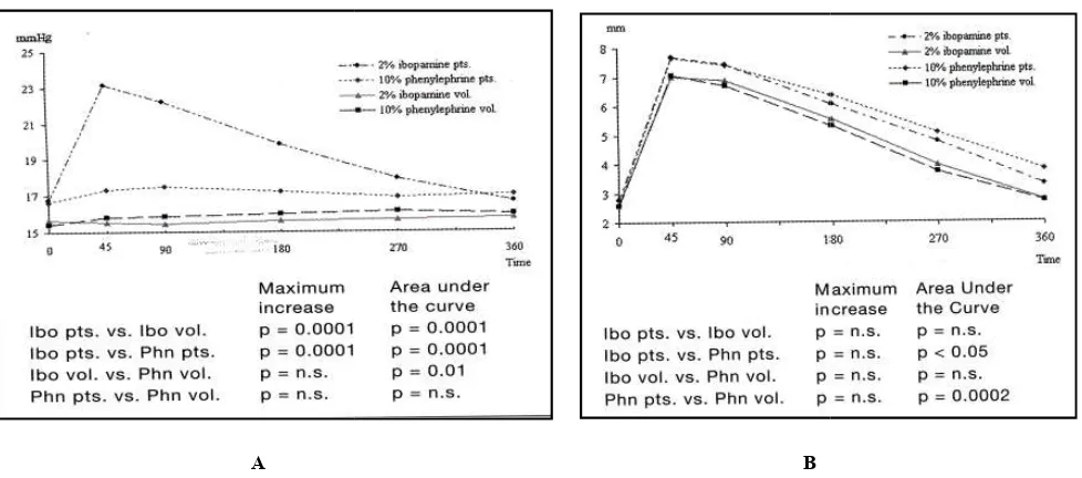
aqueous humor induced by the action of ibopamine on dopaminergic receptors is compensated by a corresponding outflow in the healthy eye, but not in the glaucoma eye (Virno et al., 1992). In a study on 20 open-angle glaucoma patients and 20 healthy subjects who were treated with ibopamine 2% eye drops and phenylephrine 10% eye drops, ibopamine was able to induce a significant increase in IOP only in glaucomatous eyes (p<0.001). No hypertensive effect was observed in normal eyes, and a similar mydriatic activity as that exerted by phenylephrine was shown (Figure 2) (Giuffre' et al., 2004). Given this peculiar effect, ibopamine has a role in
the provocative test for glaucoma detection (Giuffre' et al.,
2013, Ullrich et al., 2014, Domingues-Duenas et al., 2016).
This test is performed by two consecutive administrations of ibopamine 2%, with a 5-minute interval. IOP is measured at baseline and 45 minutes after the first instillation: the test is considered positive if a ≥3 mmHg rise in IOP is reported (Giuffre' et al., 2013).
The diagnostic performance of this test has been shown in different studies. In the analysis of 18 glaucoma suspects and 21 patients with glaucoma, ibopamine showed the potential to
differentiate between them (Ullrich et al., 2014). In fact, an
increase of IOP of 1.9±1.6 mmHg (12.5±10.3%) was observed for glaucoma suspects, whereas the corresponding figure for
glaucoma patients was 7.4±4.0 mmHg (52.4±26.5%;
p<0.0001). Four glaucoma suspects (22%) and 19 glaucoma patients (90%) were tested as positive (p<0.0001). In a recent study on 44 patients and 37 controls, early glaucoma was diagnosed in 26 patients, and the sensitivity and specificity of the ibopamine test to identify patients with early glaucoma
were 78.7% and 71.6%, respectively (Domingues
al., 2016; Virno et al., 1992). At multivariable analyses,
subjects with a positive ibopamine test at baseline had an 8 fold higher risk of glaucoma compared with those with a negative test. The main application for the provo
currently represented by pseudoexfoliation glaucoma (Scuderi et al., 2010). On the other hand, in corticosteroid
glaucoma the frequency of test-positive patients rises with the increase of steroid potency (Virno et al., 2013).
In another study, conducted in 163 patients aged 8
who were born from at least one glaucoma parent, ibopamine provocative test indicated that offspring of at least 1 parent with primary open-angle glaucoma - offspring without glaucomatous damages - shows an increase of IOP after
ibopamine administration in 44.3% of patients (Virno
2013). This finding suggests the impaired function of outflow structures and, therefore, a predisposition to intraocular hypertension and possible glaucoma. However, a glaucoma patient with a positive ibopamine challenge will likely show a negative challenge result when re-challenged following trabeculectomy surgery, as suggested by a small study in 13 patients (Landers et al., 2016).
Table 1. Clinical features of the most important and widespread used mydriatic drugs in Ophthalmology (modified from Paliaga GP. I vizi di refrazione. I
Drug Mydriasis
Maximal (m)
Atropine 30-40
Omatropine 50-70
Cyclopentolate 20-40
Tropicamide 15-25
Ibopamine 40-50
Legenda: d: day; h: hour; m: minute
Table 2. Recommendations for the use of ibopamine in the ophthalmological setting
Setting
Diagnostic mydriasis
Pre-surgical mydriasis
Other
Figure 1.
7510 Asian Journal of Science and Technology
pectively (Domingues-Duenas et
At multivariable analyses, subjects with a positive ibopamine test at baseline had an 8-fold higher risk of glaucoma compared with those with a
The main application for the provocative test is currently represented by pseudoexfoliation glaucoma (Scuderi 2010). On the other hand, in corticosteroid-associated positive patients rises with the
2013).
patients aged 8-40 years who were born from at least one glaucoma parent, ibopamine provocative test indicated that offspring of at least 1 parent offspring without shows an increase of IOP after
in 44.3% of patients (Virno et al.,
2013). This finding suggests the impaired function of outflow structures and, therefore, a predisposition to intraocular However, a glaucoma ive ibopamine challenge will likely show a challenged following trabeculectomy surgery, as suggested by a small study in 13
Post-surgical hypotony
Ocular hypotony is a condition of low IOP
dependent upon impaired production or excretion of aqueous humor. It can ultimately lead to permanent blindness and excruciating bulbar pain (Fine
secondary to metabolic alterations, loss of intraocular fluids, post-surgical or inflammatory blockade of aqueous humor synthesis.
The incidence of this condition has increased after the introduction of antimetabolites for filtration surgery (Rahman et al., 2000). Therefore, interventions aimed at a rapid resolution of ocular hypotony are eagerly awaited.
standard medical treatment of ocular hypotony is represented by mydriatic cycloplegic drugs or corticosteroids together with appropriate supportive procedures (Blok
its marked effect on the increase
by stimulating the D1 dopaminergic receptor (Virno 1996), ibopamine can have a key role in the treatment of ocular hypotony in eyes with undamaged ciliary body. preliminary study on 14 patients with ocular hypotony treated for 30-60 days with ibopamine 2% and corticosteroids
Table 1. Clinical features of the most important and widespread used mydriatic drugs in Ophthalmology (modified from Paliaga GP. I vizi di refrazione. II Edition, Minerva Medica 1985)
Paralysis of Accomodation
Maximal (m) Recovery
(d)
Maximal (m)
7-10d 60-180
1-2d 20-40
11-13h 30-40
0.25-0.50d 15-20
0.25-0.30 -
ecommendations for the use of ibopamine in the ophthalmological setting
Patients with
Need of extensive mydriasis
Need to avoid cycloplegic effect (e.g. young patients) Need of reduced duration of mydriasis
Need of avoiding adverse events Angle-closure
surgical mydriasis Cardiovascular disease
Diabetes Exfoliation
Glaucoma (overt or suspected) Risk of ocular hypotony
Risk of post-surgical hypo/athalamia
Need of corticosteroid therapy (also as pre-procedure test) All patients in whom cycloplegic effect is not required.
Figure 1. Chemical formulas of dopamine, epinine and ibopamine
Asian Journal of Science and Technology, Vol. 09, Issue, 02, pp.7508-7512, February
Ocular hypotony is a condition of low IOP (<6 mmHg) dependent upon impaired production or excretion of aqueous humor. It can ultimately lead to permanent blindness and excruciating bulbar pain (Fine et al., 2007). This condition is secondary to metabolic alterations, loss of intraocular fluids, surgical or inflammatory blockade of aqueous humor
The incidence of this condition has increased after the introduction of antimetabolites for filtration surgery (Rahman 2000). Therefore, interventions aimed at a rapid resolution of ocular hypotony are eagerly awaited. At present, standard medical treatment of ocular hypotony is represented by mydriatic cycloplegic drugs or corticosteroids together with
appropriate supportive procedures (Blok et al., 1990). Given
its marked effect on the increase of aqueous humor production
by stimulating the D1 dopaminergic receptor (Virno et al.,
, ibopamine can have a key role in the treatment of ocular hypotony in eyes with undamaged ciliary body. In a preliminary study on 14 patients with ocular hypotony treated
ith ibopamine 2% and corticosteroids
Table 1. Clinical features of the most important and widespread used mydriatic drugs in Ophthalmology I Edition, Minerva Medica 1985)
Paralysis of Accomodation
Recovery (d) 6-12d
1-2d 1-3d 0.50-0.75
-
ecommendations for the use of ibopamine in the ophthalmological setting
procedure test)
following vitreoretinal surgical intervention, Virno showed a significant increase in mean IOP versus baseline values, with IOP being increased in most patients (mean value
+3.64 mmHg; p=0.03) (Virno et al., 1996). One month after
ibopamine-treatment discontinuation, mean IOP returned to pre-treatment values. Similar findings were reported in studies on patients with ocular hypotony associated with hypo athalamia: the administration of ibopamine 2% eyedrops resulted in improved clinical outcomes comp
cycloplegic drugs and corticosteroids (Di Giulio, 2000).
Conclusions
Ibopamine has both an α2-adrenergic and a D1 dopaminergic action. This peculiar mechanism of action makes ibopamine able to induce a mydriatic effect without cycloplegia as we an increase in the production of aqueous humor. These effects are not associated with the onset of serious adverse events or alterations of visual acuity, as shown by extensive clinical experience collected on this molecule. The time of ibopamine action is short, with a peak at about 45 minutes since administration, and usually lasts no more six hours, thus comparing favorably with other molecules used in Ophthalmology (Table 1). Given the above
characteristics, and beyond its established r
provocative test, ibopamine may find a role in Ophthalmology, in almost all patients who do not need a cycloplegic effect (Table 2). In particular, ibopamine can be particularly suitable in the diagnostic mydriasis in some patients such as those need an extensive mydriasis or must avoid cycloplegic effect. Moreover, the short duration of action of ibopamine and the favorable safety profile of this molecule lend support to its use in the diagnostic mydriasis in patients at particular risk of adverse events or with angle closure. In the pre
setting, ibopamine may be considered for patients with concomitant systemic conditions including cardiovascular disease, benign prostatic hyperplasia or diabetes, those with glaucoma, and subjects at risk of ocular hypotony of hypothalamia. In the future, it will be interesting to further explore the efficacy and safety of ibopamine in the above mentioned indications.
A
Figure 2. The effect of 2% ibopamine and 10% phenylephrine eye drops on IOP (Panel A) and patients compared with healthy volunteers [Giuffrè
7511 Asian Journal of Science and Technology
following vitreoretinal surgical intervention, Virno et al.
showed a significant increase in mean IOP versus baseline values, with IOP being increased in most patients (mean value 1996). One month after t discontinuation, mean IOP returned to treatment values. Similar findings were reported in studies on patients with ocular hypotony associated with hypo-athalamia: the administration of ibopamine 2% eyedrops resulted in improved clinical outcomes compared with cycloplegic drugs and corticosteroids (Di Giulio, 2000).
adrenergic and a D1 dopaminergic action. This peculiar mechanism of action makes ibopamine able to induce a mydriatic effect without cycloplegia as well as an increase in the production of aqueous humor. These effects are not associated with the onset of serious adverse events or alterations of visual acuity, as shown by extensive clinical experience collected on this molecule. The time of ibopamine ion is short, with a peak at about 45 minutes since administration, and usually lasts no more six hours, thus comparing favorably with other molecules used in Given the above-mentioned characteristics, and beyond its established role in the provocative test, ibopamine may find a role in Ophthalmology, in almost all patients who do not need a cycloplegic effect (Table 2). In particular, ibopamine can be particularly suitable in the diagnostic mydriasis in some patients such as those who need an extensive mydriasis or must avoid cycloplegic effect. Moreover, the short duration of action of ibopamine and the favorable safety profile of this molecule lend support to its use in the diagnostic mydriasis in patients at particular risk of dverse events or with angle closure. In the pre-surgical setting, ibopamine may be considered for patients with concomitant systemic conditions including cardiovascular disease, benign prostatic hyperplasia or diabetes, those with risk of ocular hypotony of In the future, it will be interesting to further explore the efficacy and safety of ibopamine in the
above-Such studies will help expand knowledge on the use of this dual-acting molecule in the ophthalmological setting.
REFERENCES
Blok, M.D.V., Kok, J.H.C., V
Kijltra, A. 1990. Use of the megasoft bandage lens for treatment of complications after trabeculectomy. Ophthalmol., 110: 264-268.
Boles Carenini, B., Brogliatti,
1992. Ibopamine: a new drug and its use as provocative test for the diagnosis of glaucoma.
7: 3.
Chang, D.F. and Campbell, J
iris syndrome associated with tamsulosin. Refract Surg., 31: 664-673.
Chang, D.F. 2008b. Use of Malyugin pupil expansion device for intraoperative
floppy-consecutive cases. J Cataract Refract Surg Chang, D.F., Braga-Mele, R., Mamalis
K.M., Nichamin, L.D., Packard
2008a. ASCRS Cataract Clinical Committee (2008a):
Clinical experience with intraoperative floppy
syndrome. Results of the 2008 ASCRS member survey. Cataract Refract Surg., 34: 1201
Chang, D.F., Campbell, J.R., Colin
Study Surgeon Group Prospective masked comparison of intraoperative floppy iris syndrome severity with tamsulosin versus alfuzosin.
Corbett, M.C., Buckley, S.A
Ibopamine: a new preoperative mydriatic for cataract surgery. Eur J Ophthalmol.,
Di Giulio, S., De Stefano, B., Boccassini
G. 2000. Post-trabeculectomy hypotension and
hypoathalamia: efficacy of treatment with ibopamine eyedrops. Acta Ophthalmol Scand Suppl
Dominguez-Duenas, F., Plaza
Fernandez, E.E., Jimenez-Reynoso E. and Barrientos-Gutierrez
screening using the ibopamine provocative test. Glaucoma, May; 25 (5): e441
A B
The effect of 2% ibopamine and 10% phenylephrine eye drops on IOP (Panel A) and mydriasis (Panel B) in glaucomatous patients compared with healthy volunteers [Giuffrè et al. 2004]
Asian Journal of Science and Technology, Vol. 09, Issue, 02, pp.7508-7512, February
Such studies will help expand knowledge on the use of this acting molecule in the ophthalmological setting.
, Van Mil, C., Greve, E.L. & Use of the megasoft bandage lens for
treatment of complications after trabeculectomy. Am J
268.
, B., Boles, Carenini, A., et al
Ibopamine: a new drug and its use as provocative test for the diagnosis of glaucoma. New Trends Ophthalmol
J.R. 2005. Intraoperative floppy
associated with tamsulosin. J Cataract
Use of Malyugin pupil expansion device -iris syndrome: results in 30 J Cataract Refract Surg 34: 835-841.
, Mamalis, N., Masket, S., Miller, , Packard, R.B. and Packer, M. ASCRS Cataract Clinical Committee (2008a):
Clinical experience with intraoperative floppy-iris
syndrome. Results of the 2008 ASCRS member survey. J
34: 1201-1209.
, Colin, J. & Schweitzer, C. 2014. Prospective masked comparison of intraoperative floppy iris syndrome severity with tamsulosin versus alfuzosin. Ophthalmology 121: 829-834.
A. and Richards, A.B. 1994. Ibopamine: a new preoperative mydriatic for cataract
., 4: 29-34.
, Boccassini, B. and Boccassini,
trabeculectomy hypotension and
hypoathalamia: efficacy of treatment with ibopamine Acta Ophthalmol Scand Suppl., 232: 65-66.
, Plaza-Espinosa, L.,
Mundo-Reynoso, C.A., Barojas-Weber, Gutierrez, T. 2016. Early glaucoma
screening using the ibopamine provocative test. J
May; 25 (5): e441-445.
mydriasis (Panel B) in glaucomatous
Fine, H.F., Biscette, O., Chang, S. & Schiff, W.M. 2007.
Ocular hypotony: a review. Comp Ophthalmol Update 8:
29-37.
Gelmi, C., Palazzuolo, A., Lucchetti, M. and Trimarchi, F. 1989. Pupillographic evaluation of the mydriatic effect of ibopamine solution. Int J Clin Pharmacol Ther Toxicol 27: 346-351.
Giuffré, I. 2007. Ibopamine stimulates α-adrenergic receptors and D1-dopaminergic receptors in the eye. Curr Drug Ther 2: 127-132.
Giuffrè, I., Falsini, B., Gari, M.A. and Balestrazzi, E. 2013. Pattern electroretinogram assessment during ibopamine test in ocular hypertension. Eur J Ophthalmol., 23: 819-822. Giuffre', I., Taverniti, L., Di Staso, S. 2004. The effects of 2%
ibopamine eye drops on the intraocular pressure and pupil
motility of patients with open-angle glaucoma. Eur J
Ophthalmol, 14: 508-513.
Haroun-Díaz, E., Ruíz-García, M., De Luxán, de la Lastra, S., Pastor-Vargas, C., De las Heras, M., Sastre Domínguez, J. and Cuesta-Herranz, J. 2014. Contact dermatitis to both tropicamide and phenylephrine eye drops. Dermatitis 25: 149-150.
Landers, J., Ulrich, K. and Craig, J.E. 2016. Ibopamine challenge testing becomes negative following successful
trabeculectomy surgery. Clin Experiment Ophthalmol,
44(3):166-169 Epub ahead of print 2016 Feb. 12.
Leggio, G.M., Bucolo, C., Platania, C.B., Salomone, S., Drago, F. 2016. Current drug treatments targeting
dopamine D3 receptor. Pharmacol Ther Sep; 165:
164-177..
Mapstone, R. 1970. Safe mydriasis. Br J Ophthalmol 54: 690-692.
Marchini, G., Babighian, S., Tosi, R., Perfetti, S. and Bonomi, L, 2001. Effects of 2% ibopamine on pupil, refraction, anterior segment anatomy and intraocular pressure. J Ocul Pharmacol Ther 17: 215-223.
Marchini, G., Babighian, S., Tosi, R., Perfetti, S. and Bonomi, L. 2003. Comparative study of the effects of 2% ibopamine, 10% phenylephrine, and 1% tropicamide on the anterior segment. Invest Ophthalmol Vis Sci 44: 281-289. McLaren, J.W., Herman, D.C., Brubaker, R.F., Nau, C.B.,
Wayman, L.L., Ciarniello, M.G., Rosignoli, M.T., Dionisio, P. 2003. Effect of ibopamine on aqueous humor production in normotensive humans. Invest Ophthalmol Vis Sci 44: 4853-4858.
Platania, G.B., Leggio, G.M., Drago, F., Salomone, S., Bucolo, C. 2013. Regulation of intraocular pressure in mice: structural analysis of dopaminergic and serotonergic systems in response to cabergoline. Biochem Pharmacol Nov; 1,86 (9): 1347-1356.
Rahman, A., Mendonca, M., Simmons, R.B. and Simmons, R.J. 2000. Hypotony after glaucoma filtration surgery. Int Ophthalmol Clin 40: 127-136.
Sbaraglia, F., Mores, N., Garra, R., Giuratrabocchetta, G., Lepore, D., Molle, F., Savino, G., Piastra, M., Pulitano, S., Sammartino, M. 2014. Phenylephrine eye drops in
pediatric patients undergoing ophthalmic surgery:
incidence, presentation, and management of complications during general anesthesia. Paediatr Anaesth 24: 400-405.
Scuderi, G.L., Regine, F., Perdicchi, A., Turtoro, A. & Contestabile, M.T. 2010. Efficacy of 2% ibopamine on the dilation of patients with pseudoexfoliation syndrome. Eur J Ophthalmol 20: 120-123.
Shiuey, Y., Eisenberg, M.J. 1996. Cardiovascular effects of commonly used ophthalmic medications. Clin Cardiol 19: 5-8.
Soldati, L., Gianesello, V., Galbiati, I., Gazzaniga, A. & Virno, M. 1993. Ocular pharmacokinetics and pharmacodynamics
in rabbits of ibopamine, a new mydriatic agent. Exp Eye
Res 56: 247-254.
Ugahary, L.C., Ganteris, E., Veckeneer, M., Cohen, A.C., Jansen, J., Mulder, P.G. & van Meurs, J.C. 2006. Topical ibopamine in the treatment of chronic ocular hypotony attributable to vitreoretinal surgery, uveitis, or penetrating trauma. Am J Ophthalmol 141: 571-573.
Ullrich, K., Craig, J.E. and Landers, J. 2014. Ibopamine challenge test can be used to differentiate glaucoma suspects from glaucoma patients. Clin Experiment Ophthalmol 42: 342-346.
Virno, M., De Gregorio, F., Pannarale, L. and Arrico, L. 1996-1997. Topical ibopamine and corticosteroids in the treatment of post-surgery ocular hypotony. Int Ophthalmol 20: 147-150.
Virno, M., Gazzaniga, A., Taverniti, L. et al 1992. Dopamine,
dopaminergic drugs and ocular hypertension. Intern
Ophthalmol., 16: 349-353.
Virno, M., Pecori Giraldi, J., Taverniti, L., De Gregorio, F. and Sedran, L. 1998. L’ibopamina in oftalmologia. I.N.C. Editor, Rome (Italy).
Virno, M., Pecori Giraldi, J., Taverniti, L., et al 1987. Effetti ipertensivi oculari dell’ibopamina somministrata per via locale in soggetti con turbe idrodinamiche endoculari (nuovo test di provocazione). Boll Ocul 66: 5.
Virno, M., Pecori Giraldi, J., Taverniti, L. et al 2003.
Ibopamine. D-1 dopaminergic agonist in the
physiopathology of intraocular pressure. I.N.C.Editor,
Rome (Italy).
Virno M, Sampaolesi R, Pecori Giraldi J, De Gregorio F, Taloni M, Brusini P, Di Staso S & Stecchi G (2013): Ibopamine: D1-dopaminergic agonist in the diagnosis of glaucoma. J Glaucoma 22: 5-9.
Virno, M., Taverniti, L., Motolese, E. et al. 1986. Ibopamina:
nuovo midriatico non cicloplegico (nota preliminare). Boll
Ocul 65: 11.
Yuksel, N., Ozer, M.D., Takmaz, T., Ozen, U., Metin, M. and Akcay, E. 2015. Anterior segment morphologic changes
related to ɑ-1 adrenergic receptor antagonists use. Eur J
Ophthalmol 25: 512-515.
Zaman, F., Bach, C., Junaid, I., Papatsoris, A.G., Pati, J., Masood, J. and Buchholz, N. 2012. The floppy iris syndrome - what urologists and ophthalmologists need to know. Curr Urol 6: 1-7.
Zygoura, V., Kopsachilis, N. and Carifi, G. 2014. Intraoperative floppy iris and prevalence of intraoperative complications: results from ophthalmic surgery outcomes database. Am J Ophthalmol 158: 846-847.