Protein transport
and the
reassembly of
the Goig!
apparatus
during telophase
in HeLa ceils
A thesis submitted for the degree of Ph.D.
University Coiiege London
June 1993
W. Ewen L. Bouter
Ceii Biology Laboratory,
Imperial Cancer Research Fund,
ProQuest Number: 10046014
All rights reserved
INFORMATION TO ALL USERS
The quality of this reproduction is dependent upon the quality of the copy submitted.
In the unlikely event that the author did not send a complete manuscript and there are missing pages, these will be noted. Also, if material had to be removed,
a note will indicate the deletion.
uest.
ProQuest 10046014
Published by ProQuest LLC(2016). Copyright of the Dissertation is held by the Author.
All rights reserved.
This work is protected against unauthorized copying under Title 17, United States Code. Microform Edition © ProQuest LLC.
ProQuest LLC
789 East Eisenhower Parkway P.O. Box 1346
Abstract
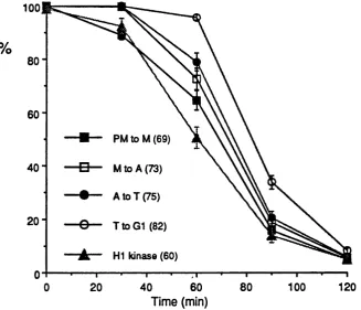
At the onset of M phase in animal cells, the Golgi apparatus undergoes disassembly. The pathway of disassembly involves the generation of several hundred discrete Golgi stacks which break down further to yield vesicles. These vesicles are dispersed throughout the mitotic cytoplasm which subsequently provide the building blocks for the reassembly of two daughter organelles during telophase. Mitosis is also characterised by the arrest of a number of membrane- mediated transport processes including transfer of newly synthesised protein from the endoplasmic reticulum to the Golgi apparatus. The kinetic relationship
between the resumption of this protein transport with the reassembly of the Golgi stack during telophase has been investigated in HeLa cells. Prometaphase- arrested cells were pulse-labelled with ^^s-methionine and chased in the absence of nocodazole to allow passage through mitosis and into G1. Resumption of transport of histocompatibility antigen (HLA) molecules to the medialan6 trans
Golgi cisternae was measured by monitoring the resistance to endoglycosidase H and the acquisition of sialic acid residues respectively. Transport to the plasma membrane was measured using neuraminidase to remove sialic acid residues on surface HLA molecules. The half-time for transport to each of these compartments was about 70 min longer in cells progressing out of mitosis than in G1 cells. The half-time for reassembly of the Golgi stack, quantified using stereological
Table of Contents
Chapter 1 Introduction
1.1 Overview...8
1.2 intraceliufar transport... 9
1.2.1 Gompartmentalisation...9
1.2 . 2 The anatomy of the secretory pathway... 10
1.2.3 Maintaining structure within the secretory pathway.... 1 2 1.2.4 The nature of transport signals...12
1.2.5 Golgi retention signals ... 13
1.2.6 Lipid traffic... 14
1.2.7 Molecular mechanisms of vesicle-mediated transport 15 1.2.8 Molecular dissection of intra-Golgi transport... 16
1.2.9 Proteins required for fusion of transport vesicles 18 1.2.10 Molecular dissection of ER to Golgi transport...20
1.2 . 1 1 Glycosylation and the kinetics of transport... 21
1.2.12 Analysis of the glycosylation pathway...22
1.2.13 Assembly and transport of the MHO Class I proteins 23 1.2.13.1 The structure and function of the MHC Class I proteins... 24
1.2.13.2 MHC Class I assembly....25
1.2.14 Protein folding and chaperonins...28
1.2.15 Kinetics of protein transport out of the ER... 28
1.2.16 Glycosylation of secretory proteins... 30
1.2.16.1 The types of glycosylation... 30
1.2.16.2 The sequence of oligosaccharide processing 31 1.2.17 Using oligosaccharide processing to monitor glycoprotein transport...31
1.3 M itosis...33
1.3.1 Mitotic control...33
1.3.2 p3 4cdc2 substrates... 37
1.3.3 Calcium and cell cycle control... 37
1.3.4 Features of the animal cell during M phase... 38
1.3.4.1 Functional changes during M phase...38
1.3.4.2 Species-specific differences in intracellular transport during M phase... 40
1.3.4.3 Molecular mechanisms of transport inhibition... 41
1.3.5 Morphological changes...42
1.3.5.1 The nuclear envelope and ER....42
1.4 The Golgi apparatus...43
1.4.1 The Golgi apparatus during interphase...44
1.4.1.1 An historical perspective... 44
1.4.1.2 Interphase organisation of the Golgi apparatus.... 45
1.4.1.3 Protein interactions.... 45
1.4.2 The Golgi apparatus during M phase...48
1.4.2.1 Disassembly of the Golgi apparatus... 48
1.4.2.2 Reassembly of the Golgi apparatus...49
1.4.2.3 Models of Golgi disassembly in M phase... 51
1.4.2.4 Models of Golgi reassembly in M phase...52
1.4.3 The role of fragmentation in animal cells... 52
Chapter 2 Materials and Methods
2.1 Overview...55
2.2 Media and reagents... 55
2.3 Cell culture...55
2.4 Production of populations of G1 and prometaphase HeLa cells... 55
2.5 Labelling mitotic cells...56
2.6 Labelling G1 cells... 56
2.7 Nocodazole controls... 57
2 . 8 Cell surface antibody binding... 57
2.9 Pre-condensation of Triton X-114...57
2.10 Preparation of cell lysates... 58
2 . 1 1 Cloud-point separation... 58
2.12 Pre-clearing lysates prior to immunoprécipitation 58 2.13 Antibodies... 58
2.14 Immunoprécipitation... 59
2.15 Washing the immunoprecipitates... 59
2.16 Enzyme digestions...59
2.16.1 Cell surface neuraminidase digestion...59
2.16.2 Endoglycosidase H digest of immunoprecipitated HLA...59
2.16.3 Endoglycosidase D digest of immunoprecipitated HLA...60
2.16.4 Neuraminidase digest of immunoprecipitated HLA... 60
2.17 Analysis of immunoprecipitated protein... 60
2.18 Laser densitometry... 61
2.19 Determination of protein synthesis levels...61
2.20 Determination of cell latency...62
2 . 2 1 Mitotic index scoring... 62
2 . 2 2 Histone HI kinase assay... 62
2.23 Electron microscopy... 63
2.24 Golgi fractionation from tissue culture cells... 64
2.25 Chloroform-methanol precipitation of TX-114 lysates 65 2.26 Two-dimensional gel electrophoresis...65
2.26.1 Preparation of the 1st. dimensional gel... 65
2.26.2 Pre-focusing the 1st. dimensional gel...6 6 2.26.3 Running the sample on the 1st. dimensional gel 6 6 2.26.4 Running the 2nd. dimension...6 6 2.27 Western blotting... 67
Chapter 3 3.1 3.2 3.3 3.4 3.5 3.6 3.7 3.8 3.9 Establishing a system to monitor intracellular protein transport Aims and objectives...6 8 Choice of marker protein...6 8 Monitoring transport from the ER to the mecf/a/Golgi cistemae... 69
Use of G1 rather than interphase cells... 69
Cell synchronisation; production of a G1 population 69 Labelling and isolating HLA molecules...70
Transport of HLA to the med/a/Golgi... 71
Quantitation of enzymatic digests...73
3.9.1 Isoelectric focusing of HLA... 74
3.10 Transport of HLA to the cell surface...77
3.10.1 Exogenous neuraminidase method... 77
3.10.2 Normalisation of transport profiles... 77
3.10.3 Cell surface antibody method...79
3.11 Control experiments... 80
3.11.1 Kinetics of transport of HLA from the ER to the Golgi in interphase HeLa cells... 80
3.11.2 Nocodazole controls...80
3.12 Summary and conclusions... 82
Chapter 4 Investigation of transport in mitotic ceiis 4.1 Aims... 83
4.2 Characterising mitotic progression... 83
4.2.1 Mitotic index... 84
4.2.2 Mitotic anomalies... 86
4.2.3 Quantitation of progression through M phase 86 4.2.4 Cell density and mitotic progression... 89
4.2.5 HI kinase activity... 89
4.2.5.1 Characterisation of the H1 kinase assay,... 89
4.2.5 2 Quantitation of the H I kinase assay.... 91
4.2.6 Conclusions about mitotic parameters...92
4.3 Monitoring transport in mitotic HeLa cells...92
4.3.1 Protein synthesis during M phase...92
4.3.2 Monitoring transport from the ER to the medial Golgi apparatus... 94
4.3.3 Monitoring transport from the ER to the trans Golgi apparatus and cell surface... 94
4.3.4 Quantitation of HLA transport in mitotic cells...96
4.3.5 Resumption of protein transport is independent of protein synthesis... 96
4.4 Control experiments...98
4.4.1 Independence of the timing of the pulse... 98
4.4.2 Establishing transport-competence of immunoprecipitated HLA... 98
4.4.3 Transport of the transferrin receptor... 99
4.4.4 Endo D digestion... 101
4.4.5 Preservation of cell integrity during the chase period 102 4.5 Timing of the resumption of protein transport... 102
4.6 Summary and conclusions... 102
Chapter 5 5.1 5.2 5.3 5.3.1 5.3.2 5.3.3 5.3.4 Kinetic relationship between protein transport and Golgi reassembly In telophase Overview... 105
Visualisation of morphology... 105
Experimental strategy for comparing the kinetics of protein transport and Golgi reassembly... 107
Characterisation of the mitotic progression in preliminary experiments...107
Quantitation of HLA transport... 107
EM analysis of Golgi reassembly... 107
5.4 5.4.1 5.4.2
5.4.3 5.5
5.6
Refinement of the analysis of Golgi reassembly kinetics 11 2
Stacked cisternal membrane analysis...1 1 2
Analysis of the number of cistemae within each
stack... 1 1 2
Analysis of cisternal length... 114
Relative rates of transport resumption, reassembly and HI kinase activity... 114
Summary and conclusions...116
Chapter 6 Discussion 6.1 6.2 6.3 6.4 6.5 Investigation of intracellular protein transport... 117
Reassembly of the Golgi apparatus during telophase 119
Conclusions from the correlation of transport and reassembly data...1 2 0 The purpose of Golgi fragmentation... 124
Future prospects... 124
Appendix A investigation of cell-cycle dependent post-translational modification of resident Golgi proteins A.1 Overview... 126
A.2 Relaxation of intracistemal protein interactions within the Golgi apparatus during M phase...126
A.3 Characterising antibody detection of NAGTI with 9E10 antibody...127
A.4 Analysis of NAGT I in synchronised cells... 127
Appendix B Kinetics of HLA transport In transfected HeLa cells with disturbed Golgi morphology B.1 Overview... 130
B.2 Description of cell lines... 130
B.3 Kinetics of HLA transport in the stable transfectants 131 B.3.1 HLA transport in the 10:40 cell line... 131
B.3.2 HLA transport in the 448 cell line... 133
ACKNOWLEDGEMENTS...136
Table of Figures
Chapter 1 introduction
1.1 The secretory pathway in animal cells... 11
1 . 2 Retention of Golgi enzymes by "kin recognition" 1 1 1.3 The ARP cycle during a single round of Intra-Golgl transport... 19
1.4 Model Illustrating SNAP function during Intra-Golgl transport... 19
1.5 MHC Class I assembly In the endoplasmic reticulum 27 1. 6 N-llnked oligosaccharide processing of HLA during secretion... 27
1.7 The cyclln-cdc2 cycle... ... 35
1. 8 Inhibition of membrane traffic during M phase... 35
1.9 Behaviour of the Golgi apparatus during M phase...50
1 . 1 0 Three models of Golgi reassembly...50
Chapter 3 Establishing a system to monitor Intracellular protein transport 3.1 Transport of HLA In G1 cells: endo H digestion...72
3.2 Quantitation of kinetics of HLA transport In G1 cells...72
3.3 1ER analysis of HLA Immunoprecipitated from G1 cells... 75
3.4 1ER analysis of HLA-A treated with neuraminidase... 75
3.5 1ER analysis of HLA-A treated with exogenous neuraminidase... 78
3.6 Immunoprécipitation of cell surface HLA from G1 cells... 78
3.7 Comparison of transport kinetics In Interphase and G1 cells... 81
3.8 Effect of nocodazole on Incorporation of methionine label... 81
Chapter 4 Investigation of transport In mitotic cells Table 4.1 Distinguishing features of telophase and G1 cells 84 4.1 Visualisation of mitotic phases... 85
4.2 Dependence of mitotic progression on release from nocodazole...8 8 4.3 Mitotic phase transitions and HI kinase activity...90
4.4 Mitotic phase peak plot... 90
4.5 Transport of HLA In mitotic cells: endo H digestion 93 4.6 1ER analysis of HLA-A Immunoprecipitated from mitotic cells and treated with neuraminidase... 93
4.7 1ER analysis of HLA-A Immunoprecipitated from mitotic cells treated with exogenous neuraminidase...95
4.8 Immunoprécipitation of cell surface HLA from mitotic cells...95
4.9 Quantitation of kinetics of HLA transport In mitotic cells.. 97
4.10 Transport of the transferrin receptor In mitotic cells: endo H digestion... 100
4.11 Quantitation of kinetics of transferrin receptor transport relative to HLA In G1 and mitotic cells... 100
Chapter 5 Kinetic relationship between protein transport and Golgi reassembly In telophase
5.1 Phase transitions of HeLa cells progressing through M
phase... 106
5.2 Phase peaks of HeLa cells progressing through M
phase... 106 5.3 Transport kinetics in mitotic HeLa cells... 108
5.4 Electron micrographs illustrating reassembly of Golgi
stacks...1 1 0
5.5 Quantitation of Golgi reassembly relative to HLA
transport in preliminary experiments...1 1 0
5.6 Mitotic phase peaks for more refined experiment 113
5.7 Quantitation of stacked cisternal membrane relative to
HLA transport... 113
5.8 Quantitation of number of cistemae per stack relative to
HLA transport... 115
5.9 Quantitation of cisternal length relative to HLA transport 115
Chapter 6 Discussion
6 .1 Intra-Golgi transport and the reassembly of the Golgi
apparatus...1 2 1
Appendix A investigation of celi-cycie dependent
post-transiationai modification of resident Golgi proteins
A.1 Characterisation of 9E10 antibody for ECL detection of
myc-tagged NAGT 1... 128
A.2 Pattern of NAGT I migration as HeLa cells emerge from
M phase... 128
Appendix B Kinetics of HLA transport in transfected HeLa cells with disturbed Golgi morphology
8.1 Quantitation of HLA transport kinetics in the 10:40 cell
line relative to G1 cells...132
8.2 Quantitation of HLA transport to the medial Golgi in the
448 cell line relative to the 4/12.cell line... 132
8.3 Quantitation of HLA transport to the trans Golgi In the
448 cell line relative to the 4/12 cell line... 134
8.4 Quantitation of HLA transport to the cell surface in the
CHAPTER 1: INTRODUCTION
1.1 Overview
Mitosis in animal cells is characterised by extensive reorganisation of the
cytoarchitecture. One of the most striking of these mitotic transformations is the vésiculation of the Golgi apparatus. At the onset of mitosis, the apparatus
undergoes disassembly, followed by dispersal and partitioning during mitosis, and finally reassembly of two daughter organelles at the end of mitosis.
Mitosis in mammalian cells is also characterised by the inhibition of a number of membrane-mediated transport processes. These include transport of newly synthesised plasma membrane proteins through the secretory pathway from the endoplasmic reticulum to the trans Golgi network, stimulated secretion of
histamine by mast cells, receptor-mediated endocytosis and recycling of the transferrin receptor.
A simple hypothesis has been suggested which links the arrest of protein traffic through the Golgi apparatus with the fragmentation of the organelle during mitosis: if transport were to be arrested by the inhibition of membrane fusion while vesicle budding was maintained, the necessary consequence for the Golgi apparatus would be its disassembly (Warren, 1985). Conversely, at the end of mitosis, restoration of membrane fusion would allow both the resumption of vesicle traffic and the reassembly of the Golgi apparatus.
It is this latter aspect of Golgi reassembly which I have pursued during my thesis work. In particular, I have investigated the relative kinetics of protein transport recommencement and the morphological reassembly of the organelle to address the possibility that restoration of membrane fusion might be the trigger which allows both these processes to proceed.
In the course of this introductory chapter, I propose to review the current state of our understanding of a number of areas of cell biology relevant to the
experimental work undertaken. Since the aim of the project was the relationship between protein traffic and the reassembly of the Golgi apparatus during M phase, there are three primary topics which immediately suggest themselves as important areas for review, namely intracellular transport, mitosis and Golgi morphology. I shall begin with a review of the progress that has been made in unravelling the complexity of the secretory pathway including the emerging picture of the
morphological changes occurring during mitosis. This will lead conveniently to a review of the structure of the Golgi apparatus, both during interphase and mitosis.
1.2 Intracellular transport
In order to simplify the present discussion of intracellular transport, I shall focus on the exocytic pathway rather than endocytosis. Since the work undertaken has been confined to the default or constitutive pathway, the discussion of regulated secretion will be brief.
1.2.1 Compartmentallsation
The diversity of function within the cell requires an exquisite degree of organisation of structure at the subcellular level. Moreover, the necessity for efficiency in regulating the enzymatic changes that occur second by second also means that these reactions must be regulated both temporally and spatially. By doing so, the cell maximises the efficiency of its resources. The outcome of these selective pressures is the extensive series of membrane-bound compartments within the cell each one responsible for a defined set of reactions, carried out by a defined array of enzymes upon a defined series of substrates.
The biosynthetic secretory pathway is no exception to this requirement for
efficiency in regulation. The pathway is responsible for the biosynthesis of proteins and lipids, their modification and processing, and the delivery of these proteins and lipids to the appropriate destination, whether an existing compartment within the cell, the plasma membrane or a destination outside the cell, in the case of
multicellular organisms. There is also a need for degradation of unwanted material and partial digestion of molecules to be used in another form, reactions which clearly must be carefully enclosed and regulated. The efficient function of this pathway is essential then for the generation and maintenance of different compartments, for cell growth, division and differentiation.
The essential function of the secretory pathway in general combined with the diversity of specific modifications undertaken as molecules pass along the pathway has led to the generation of a series of compartments. The pioneering work of Palade and co-workers (Palade, 1975) in the 1960's and early 1970's identified the most crucial compartments of the regulated secretory pathway. Subsequent research has led to the further description of many other
obscure, and in the precise nature of the function of those organelles already identified.
1.2,2 The anatomy o f the secretory pathway
Figure 1 .1 represents a schematic of the biosynthetic secretory pathway in animal
cells as we currently understand it. The essential features of this pathway are the endoplasmic reticulum (ER), the cis-Golgi network (CGN), the Golgi apparatus, the trans-Golgi network (TGN), the lysosomes and the secretory granules (SG).
The most extensive compartment of the endomembrane system is the ER,
comprising a series of interconnected membrane sacs enclosing a central lumen. The ER can further be subdivided into regions lacking ribosomes, the so-called smooth ER, where fatty acids and lipids are synthesised, and ER with attached ribosomes (rough ER) which forms the site of protein synthesis and is connected to the nuclear envelope. Proteins are co-translationally translocated across the
membrane of the rough ER, and are then transported via the transitional elements of the ER and the CGN to the Golgi apparatus.
The structure of the Golgi apparatus will be discussed in the final section of the chapter. Briefly, the organelle consists of a series of flat membrane sacs or "cistemae” with dilated rims, analogous to a stack of pitta bread. The stack of cistemae may be further subdivided into three subcompartments, namely c/s,
medial an6 trans both by structural and functional criteria. The orientation of these subcompartments shows a distinct polarity with respect to the ER, proteins
arriving at the c/s face of the organelle and then proceeding in a vectorial manner through the stack to the trans face at the other side. The CGN may be thought of as the entry control point of the Golgi apparatus ensuring that only legitimate cargo proteins continue to the Golgi while the TGN may be described as the sorting depot where proteins and lipids are allocated to appropriate destinations. The distinct nature of the CGN and TGN is most readily verified by subjecting the cell to temperature blocks. A shift to 20°C induces an enlargement of the TGN (Saraste and Kuismanen, 1984; Lippincott-Schwartz, eta!., 1990), while shifting the temperature to 15°C causes an accumulation of protein in the CGN (Griffiths and Simons, 1986).
Having successfully passed along this secretory conveyor belt, proteins and lipids can then be transported by default to the cell surface, or be diverted to lysosomes or secretory granules. Lysosomes provide the cell with the controlled environment required for the degradation of particular molecules. The membrane-limited
Intercistemal matrix
ws-Golgi network
Transitional Elements
f Lysosome I
V
Endoplasmic Reticulum
>
^
Golgi Stack
Post-Golgl
Transport
Vesicle
7
Plasma Membrane
Trans-Golgi network
Diafturl
Figure 1.1 The secretory pathway in animal cells.
Endoplasmic
reticulum Golgi stack
TGN
Media/enzyme I /Ifed/a/enzyme II Trans enzyme
Cell Surface
leaking into the cytoplasm will be minimal since the pH level outside the lysosome is around 7, emphasising the importance of compartmentalisation within the cell.
1.2.3 Maintaining structure within the secretory pathway
The preceding description of the compartments along the secretory pathway has highlighted the high degree of organisation of the pathway and its polarity, both with respect to structure and function. It is clear then that the underlying
mechanism of vesicle traffic between these compartments must also be controlled with respect to the content of carrier vesicles and the sequence of transport steps. The cell must both ensure that proteins that are to be secreted reach their
appropriate destination and that proteins which are to be maintained as resident components of a particular compartment do not join the flow of transported molecules through the compartment and consequently arrive at a totally
inappropriate location. In the following section, I shall outline what is known of the mechanisms whereby the cell achieves selectivity of transfer through the
secretory pathway.
1.2.4 The nature of transport signais
Investigation into the nature of transport signals has led to a current model of regulation of protein sorting involving four distinct mechanisms which ensure the correct distribution of proteins through the secretory pathway (for reviews, see (Pfeffer and Rothman, 1987; Huttner and Tooze, 1989). The first mechanism Is that of default whereby secretory proteins pass along the secretory pathway as part of the bulk flow; there is no specificity in this case, and no requirement for the protein to arrive at any particular destination other than the plasma membrane. The second level of sorting is that of diversion. Proteins that have a diverting signal pass by default along the secretory pathway until they reach the
appropriate sorting depot (now recognised as the TGN) and are then diverted from the default pathway to a particular destination. A third level of sorting is that of retention which applies to functional molecules of the secretory pathway as opposed to secretory "passengers". Clearly, these proteins must be maintained in their appropriate compartment since the basic premise of compartmentalisation is that the resident molecules of a compartment define its particular function; loss of resident proteins implies loss of function. One mechanism of ensuring residency is to attach a signal to appropriate proteins which anchors them to the correct
relocating them. Since retention of resident Golgi proteins is of particular relevance to my thesis work, I shall discuss these signals in more detail.
1.2,5 Golgi retention signals
Recent work from various laboratories has shown that the transmembrane region of resident Golgi molecules provides the signal required to specify retention within this organelle.
Investigation of the E1 glycoprotein of avian coronavirus (Swift and Machamer, 1991) involving the construction of chimeric proteins demonstrated the necessity and sufficiency of the first of the three transmembrane spanning domains of El for efficient Golgi retention. A number of subsequent studies of the retention
requirements of endogenous Golgi proteins have similarly demonstrated the necessity of retention signals residing within the transmembrane spanning domain (Munro eta!., 1991 ; Nilsson, eta!., 1991; Aoki, eta!., 1992; Colley, eta!., 1992; Russo, et a i, 1992; Teasdale, et a i, 1992; Wong, et a i, 1992). In particular, Nilsson and co-workers (1991), analysing galactosyltransferase (GT),
demonstrated that the crucial region of the enzyme was the lumenal half of the transmembrane domain together with a small number of residues from the proximal region of the lumenal domain. A cytoplasmic domain was also required but since the cytoplasmic region of the reporter protein served as well as that of GT, it seems that this region is required for stability rather than a specific
interaction. Munro and colleagues (1991) investigated the retention requirement of sialyltransferase (ST), another type II protein resident within the frans Golgi.
Chimeric protein constructions similarly demonstrated that the transmembrane domain conferred Golgi localisation, with additional efficiency of retention requiring the proximal sections of the molecule on either side of the membrane.
In spatial terms, the requirement of a retention mechanism within the Golgi may be viewed as a mechanism for preventing the lateral movement of resident enzymes into the dilated rims of each cistemae where vesicle budding occurs. Several mechanisms suggest themselves and may not necessarily be exclusive; a combination of a number of different methods may be utilised to ensure highly efficient retention of Golgi resident proteins. One mechanism might rely on a receptor protein being responsible for binding to and retaining functional proteins. However, such a protein would still have to have the capacity for selective
retention in different subcompartments of the stack. Such specificity might be encoded in specialised membrane domains through the stack. An alternative hypothesis is that of protein oligomerisation caused by interactions of the
hence would not enter transport vesicles. Again, lipid gradients and/or domains might confer subcompartment specificity to such a retention mechanism. A prediction of such a "kin recognition" hypothesis would be that the alteration of residency of one Golgi enzyme to the ER would therefore cause the alteration of residency of other Golgi enzymes usually retained by oligomerisation with the first enzyme. By fusing an ER retention signal to a mecf/a/Golgi enzyme, Nilsson and co-workers have been able to induce ER localisation of another meof/a/enzyme thereby confirming this prediction (Nilsson, et ai, 1993b). A recycling salvage receptor within the Golgi is yet a further refinement of Golgi retention which awaits demonstration.
With regard to the TGN, investigation of retention signals have focused on one particular protein, TGN38 which resides predominantly within the TGN but also recycles between the plasma membrane and TGN (Reaves, et a i, 1993).
Cleavage of the cytoplasmic domain from TGN38, a resident of this compartment caused the protein to be secreted to the cell surface (Luzio, et a i, 1990) and chimeric experiments identified an 11 amino acid segment of the cytoplasmic
domain responsible for TGN-localisation (Humphrey, et a i, 1993). More recent studies indicate that the signal within the cytoplasmic domain confers retrieval to the TGN, while a second signal within the transmembrane region confers TGN retention (Sreenivasan Ponnambalam, personal communication).
Although much progress has been made in the understanding of protein sorting along the secretory pathway, it is clear that there remains an enormous amount that is still obscure. That this should be so is a measure of the complexity of the protein sorting task faced by the components of the secretory pathway.
1.2,6 Lipid traffic
Before proceeding to a discussion of the molecular mechanisms of vesicle-
mediated transfer of proteins along the secretory pathway, it is worth commenting on the nature of lipid traffic in eukaryotic cells. The considerable technical
challenges of lipid biochemistry have meant that lipid research has in many ways lagged behind that of protein studies. However, the recognition of the crucial nature of lipid modifications of proteins together with the importance of lipid metabolism in cell physiology including cell signalling pathways has given rise to exciting developments in this area of exploration (reviewed by Pagano, 1990).
species are not distributed randomly through the cell, but that particular species are enriched in particular locations. This localised enrichment of certain types of lipid may even apply within the same bilayer; there are many instances of an asymmetry between the cytoplasmic and lumenal leaflets of a bilayer. Since no individual compartment is capable of synthesising all of its component lipids, there is a basic requirement for efficient transport of lipids to appropriate compartments. Such transport may take the form of vesicle-mediated transfer, lateral diffusion along membrane bridges that may form transiently between compartments or by direct cytosolic transfer, either by a protein-mediated or protein-independent pathway. Once positioned in the correct compartment, lipid composition may be further altered by lipid degradation, modification or sorting within the leaflet or by a flip-flop mechanism between leaflets.
The technical challenge of dissecting these steps of transport of lipids has been greatly facilitated by the use of fluorescent lipid analogues containing N-(4- nitrobenzo-2-oxa-1,3-diazole) aminocaproic acid (Ce-NBD-fatty acid). The pathway of transport of lipids can then be monitored by carefully assessing the rate and sequence of labelling of different compartments within the cell after labelling with a specific lipid analogue (Koval and Pagano, 1989). Another major breakthrough is the in vitro reconstitution of lipid transport. Of particular interest for the study of the relationship between lipid and protein transport through the Golgi apparatus is a system that Wattenberg has reconstituted which demonstrated that glycoprotein and glycolipid transport through this organelle proceed with the same kinetics, suggesting that transport of these molecules occur by the same vesicle- mediated mechanism (Wattenberg, 1990). Research into membrane biology has shown that the energy requirement for the fusion of two lipid bi layers is enormous. Since vesicle-mediated traffic requires such events to occur at every round of transport, the mechanism of fusion must entail a highly refined enzymatic
component. Such a prediction has been shown to be the case in studies to dissect transport at the molecular level.
1.2.7 Molecular mechanisms of vesicle-mediated transport
pathway and have been found to be common to many different compartments of the pathway.
For the sake of simplicity, I propose to discuss the elucidation of the transport machinery within the Golgi apparatus first through in vitro reconstitution of these transport events, and then extend this to transport at other stages of the secretory pathway, illustrating the ways in which yeast genetics have confirmed the cell-free results and in many cases, uncovered new ground in the pursuit of a molecular understanding of intracellular transport.
1.2.8 Molecular dissection of Intra-Golgl transport
The initial steps towards the reconstitution of transport through the Golgi apparatus in a cell-free system were made by Rothman and co-workers in the early 1980's. Early cell fusion experiments demonstrated protein transport between Golgi stacks of fused cells and that this transport was targeted and unidirectional. No lateral or retrograde transport could be detected (Rothman, et
a/., 1984a,b).
Reconstitution of cell-free transport was made easier by the fact that the Golgi apparatus may be fractionated from other organelles relatively simply yielding Golgi stacks that display remarkably undisturbed morphology; recognisable stacks of cistemae are readily seen under the EM. If these Golgi stacks are isolated from cells that have been previously infected with Vesicular Stomatitus Virus (VSV) and are then incubated with ATP and cytosol (100,000g supernatant) from uninfected cells, 75nm vesicles containing VSV G protein are seen to form from each
cisterna. Moreover, the concentration of G protein within the vesicles is equivalent to that of the cistemae and no "empty" vesicles may be seen (Balch, eta!., 1984; Orel, eta!., 1986). These observations indicated that the vesicles which form are
bona fide transport vesicles identical to those which mediate bulk flow protein transfer between Golgi cistemae in vivo.
The development of a quantifiable cell-free assay paved the way for the
subsequent molecular dissection of intra-Golgi transport (Balch, eta!., 1984). This assay relies on the absence of N-acetylglucosaminyltransferase I from the medial
Golgi of clone 15B Chinese hamster ovary (OHO) cells. By mixing "donor" Golgi membranes from these 15B cells that have been infected with VSV and
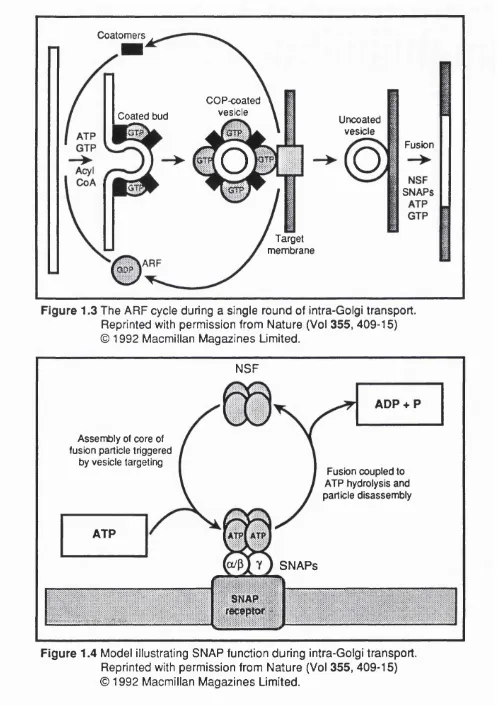
Using this assay Rothman and colleagues have gained considerable insight into the steps required during a single round of intercistemal transport. These steps may be broadly defined as coated vesicle budding at the donor compartment, vesicle scission, vesicle targeting, vesicle uncoating, and vesicle fusion at the acceptor compartment. A number of selective inhibitors of these successive steps were of crucial importance in the characterisation of the process. The use of GTP7S for example caused the accumulation of coated vesicles (Melançon, et al.,
1987). Use of this inhibitor therefore allowed the purification of Golgi coated vesicles (GCV) (Malhotra, eta!., 1989) and allowed the biochemical analysis of the proteins of the coat (Serafini, eta!., 1991). These proteins could be separated as a family of so-called "coat” proteins or "COPs", designated a,B,y,8 and a small
GTP-binding protein previously described as the ADP-ribosylation protein or "ARP". These proteins were found to be present in stoichiometric amounts on the outside of transport vesicles and that they were peripherally associated; these observations were all consistent with their role in the formation of vesicle coats.
Subsequent cloning and sequencing of the 8-COP gene (Duden, eta!., 1991)
revealed homology to a structural component of clathrin-coated vesicles of the endocytic pathway designated 8-adaptin, responsible for linking the clathrin lattice
to the underlying membrane. Although GCV lack clathrin, the discovery of this homology between structural components of these two sorts of coated vesicles at least raises the possibility that there may be other levels of structural similarity between the coats of vesicles isolated from different sections of the secretory pathway. However, there remains a basic difference in the fact that CCV are selective carriers while GCV are bulk carriers, seemingly without the capacity to concentrate proteins within their contents.
Immuno-electron microscopy (immuno-EM) reveals the fact that the coat proteins are absent from the surface of Golgi membranes prior to transport In vitro an6 that the bulk of 8-COP is present in the cytosol in a complex with other coat proteins of
650-700K (Duden, eta!., 1991 ; Waters, ef a/., 1991); this complex has been termed the "coatomer^. ARP is also freely soluble in the cytosol as a single protein independent of the coatomer. In the light of the myristylation of the amino terminus of the ARP protein (Kahn, et a!., 1987), the fact that only ARP in the GTP-bound but not the GDP-bound state will spontaneously insert into lipid bi layers (Kahn et
a/., 1991) and the necessity of ARP for 8-COP binding to Golgi membranes
attachment causes the binding of the other coat proteins. After scission of the coated bud and targeting to the acceptor membrane, the GTP is hydrolysed,
leading to uncoating of the vesicle and recycling of ARF to the inactive cytoplasmic pool. Such a model would explain the sensitivity of uncoating to GTPyS since without GTP hydrolysis there could be no ARF release, and
therefore no uncoating. Another possible feature of such a mechanism is that the specificity of targeting to the correct acceptor membrane might be generated by particular ARF proteins since there is known to be a family of these proteins.
1.2.9 Proteins required for fusion of transport vesicles
The first protein required for vesicle fusion at the acceptor membrane was
identified as a result of its sensitivity to N-ethylmaleimide (NEM). NEM causes an accumulation of uncoated vesicles and inhibition of transport-coupled
glycosylation. By incubating NEM-pretreated membranes with cytosolic extracts, it is possible to restore transport. The factor responsible for transport restoration has been purified to homogeneity and is termed the NEM-sensitive fusion protein (NSF). NSF is present in cytosol as a 76K homotetramer (Block, eta!., 1988) which is stabilised by ATP and is required for membrane fusion but not for budding (Malhotra, eta!., 1988). Sequence analysis shows that NSF has three domains (Tag ay a et a i, 1993). The two domains nearest to the G-terminus consist of two direct repeats which each contain an ATP-binding site. Mutation of these sites inhibits NSF function indicating that ATP hydrolysis is essential for fusion.
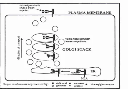
NSF is unable to bind to Golgi membranes on its own; a series of other factors mediate fusion. Of specific interest, three soluble NSF attachment proteins (SNAPs) have been purified: a-SNAP (35K), 8-SNAP(36K) and y-SNAP(39K)
(Weidman, et a i, 1989; Clary, et a i, 1990). The first two of these are very similar structurally while the third is very different, suggesting that either a-SNAP or 13- SNAP may act in concert with y-SNAP. SNAPs cannot bind NSF in solution but
require the activity of an integral membrane receptor in the acceptor Golgi membrane to mediate NSF membrane binding. NSF, a-SNAP, y-SNAP and the
receptor assemble together in a 20S particle which is likely to be the core of the fusogenic particle at the acceptor membrane (Wilson, et a i, 1992). The precise mechanism of fusion remains obscure for the moment, though Rothman and colleagues have postulated a working model for the interaction of NSF, SNAPs and the SNAP receptor (see Figure 1.4). Recent work from the Rothman
Coatomers
COP-coated vesicle Coaled bud
Ü
o
Uncoated vesicle
Fusion
o
SNAPs
Target membrane
Figure 1.3 The ARF cycle during a single round of intra-Golgi transport. Reprinted with permission from Nature (Vol 355, 409-15) © 1992 Macmillan Magazines Limited.
NSF
Assembly of core of fusion particle triggered
by vesicle targeting
ATP
ADP + P
Fusion coupled to ATP hydrolysis and particle disassembly
SNAPs
SNAP fe c e p w »
Figure 1.4 Model illustrating SNAP function during intra-Golgi transport. Reprinted with permission from Nature (Vol 355, 409-15) © 1992 Macmillan Magazines Limited.
may be the key to unlocking the question of targeting specificity required in
vesicular transport. The compartmental restriction of such vesicle SNAP receptors (VSNAREs") to appropriate donor membranes and target SNAP receptors f t- SNAREs") to corresponding acceptor membranes would provide an ideal mechanism for specifying targeting information within both the exocytic and
endocytic pathways (Warren, 1993a). Other cytosolic factors shown to be required for vesicle fusion after uncoating include acyl-CoA (Pfanner, etal., 1989) and a novel peripheral membrane protein termed pi 15 (Waters, etal., 1992).
1.2.10 Molecular dissection of ER to Golgi transport
The study of ER to Golgi transport in vitro required a different strategy to that of the Golgi system described above since homogenisation causes extensive
fragmention of the ER. Much milder conditions were therefore required and these were achieved by generating semi-intact cell populations in which perforations were made in the plasma membrane allowing the passage of small soluble molecules into the cytoplasm of cells but maintaining the complex intracellular architecture, particularly that of the secretory pathway.
Reconstitution of ER to Golgi transport in such a perforated system has shown a requirement for both ATP and cytosol (Beckers, et ai., 1987). More specifically, this transport has been also shown to be dependent on NSF (Beckers, et ai.,
1989; Wilson, et ai., 1989) and ARF (Balch, et ai., 1992), suggesting the universality of the emerging picture of transport mechanisms through the secretory pathway.
Further weight to confirm this conclusion has come from the extensive investigation of factors responsible for transport in the budding yeast,
Saccharomyces cerevisiae. This research has revealed the existence of over 40 essential components of the secretory pathway in yeast. Just as the use of selective inhibitors led to the accumulation of transport intermediates in the case of in vitro biochemical work, so mutagenesis of genes encoding essential
components of the secretory pathway led to a similar proliferation of such intermediates. These genes have been designated "sec" (short for secretory) genes and by isolating temperature-sensitive mutants defective in these genes, secretory transport can be shown to occur only at the permissive temperature.
Mutation of the sect 7, 18 or 22 genes for example blocks transport from the ER to the Golgi in yeast (Kaiser and Schekman, 1990), and causes the accumulation of SOnm vesicles which mediate this step of transport. The seel 8p has been shown
for intra-Golgi transport in yeast and also for Golgi to cell surface transport (Graham and Emr, 1991 ). The sec17p has been confirmed as the yeast homologue of a-SNAP (Clary, etal., 1990), consistent with the known genetic interaction of sec77 and s a d 8 (Kaiser and Schekman, 1990). The role of sec22p remains unknown.
Mutations in another subset of yeast genes also disrupts ER to Golgi transport but without the concomitant accumulation of SOnm transport vesicles. Epistatic
analysis demonstrates that these genes (sec12, s a d 3, s a d 6 , sac21 and sac23)
are responsible for budding rather than fusion reactions (Kaiser and Schekman, 1990). Sec1 2p is an integral membrane protein that appears to be required for the
recruitment of a small GTP-binding protein called sari p from the cytosol (Nakano,
at a!., 1988; d'Enfert, at a!., 1991). Transport from the ER to the Golgi in yeast has also been reconstituted in vitro and has in many cases confirmed the results of genetic analysis (Baker, at ai., 1988; Ruohola, at ai., 1988 ). GTP-yS inhibition experiments in such in vitro systems confirms a role for GTP hydrolysis in vesicle budding from the ER (Rexach and Schekman, 1991). The close homology
between sari p and ARF fuels the speculation that sec12p may be responsible for nucleating the budding site on ER membranes by acting as a receptor for sari p in the GTP-bound state, reminiscent of the mechanism proposed for recruitment of ARF from the cytosol to Golgi membranes to allow budding to occur. Once again, confirmation of these mechanisms would strengthen the notion that molecular mechanisms of vesicle-mediated traffic are conserved between organisms and also between different compartments of the secretory pathway.
Recently, sec2 1 p has been shown to be a component of the yeast homologue of
the mammalian coatomer (Hosobuchi, at ai., 1992) and is present in the cytosol as part of a 700-800 kD complex which also contains a family of other proteins of molecular weights 150,110, 73, 35 and 25kD. Sec21 p (105kD) and the 110kD
subunit are the yeast homologues of y-COP and p-COP respectively. The other
proteins do not correspond to other characterised SEC gene products. Since
sac21 function is essential for ER to Golgi transport, this proves that coatomer proteins are essential factors in this step of the secretory pathway. Although seel 3p and sec23p are also both present in yeast cytosol In large oligomeric complexes (Hicke and Schekman, 1989; Pryer at ai., 1993), their identity as yeast coatomer proteins has not yet been shown.
1.2.11 Glycosylation and the kinetics of transport
secreted proteins to pass from one compartment to the next. It is now of interest to explore in more detail the extent of our understanding of the processing of the secretory proteins themselves; in other words, to review what is known of the reactions which occur within these secretory compartments, as opposed to what happens in transit between the compartments.
These reactions of interest include the assembly of secretory molecules in the ER, the factors affecting the rate of exit from the ER, and the post-translational
modification of transported proteins as they pass through the ER and Golgi apparatus. Although there will be a number of general points made in the course of this discussion concerning these various reactions and processing steps, I propose to focus the main body of the following discussion on the assembly and processing of the Major Histocompatibility Complex (MHC) Class I proteins as examples of transported proteins since these molecules provided the main source of marker for monitoring biosynthetic protein transport in the work undertaken in this project.
7.2.12 Analysis of the glycosylation pathway
One of the most powerful tools for the study of the processing of secretory proteins along the secretory pathway has been the use of small lipid-enveloped viruses, such as Vesicular Stomatitus Virus (VSV), mentioned earlier in the context of the development of a cell-free system to monitor transport through the Golgi apparatus. These viruses have very restricted genomes and rely totally on the host cell biosynthetic machinery for the translation, processing and transport of their membrane proteins. The VSV genome, for example, encodes only five proteins: an integral membrane glycoprotein (G), a peripheral membrane protein (M), and three nucleocapsid-associated proteins (N,NS and L). As the virus shuts off the synthesis of the host cell proteins, the translational machinery is devoted entirely to synthesis of these proteins. Furthermore, the secretory pathway is deluged with these same proteins following their synthesis in the RER, facilitating the study of biosynthetic transport and post-translational modification of
transported proteins as they make their way to the cell surface. Evidence points to the similarity in behaviour of these viral glycoproteins to that of endogenous
Localisation of the processing events which occur within each of the compartments of the secretory pathway has also relied on the subcellular fractionation of these compartments to demonstrate reproducible partitioning of enzymes responsible for post-translational modification of secretory proteins. Such fractionation and recovery of enzymatic function relies on density gradient centrifugation in which dense ER membranes may be separated from lighter Golgi membranes. Refinement of the fractionation and centrifugation procedures allows finer differentiation between subcompartments and more detailed allocation of enzyme-mediated processing reactions to each of these subcompartments. Distribution of modifying enzymes across sucrose gradients has been found to be entirely consistent with the known sequence of modifications across the Golgi stack (Goldberg and Kornfeld, 1983), confirming the close relationship between structure and function along the secretory pathway. A number of different lines of evidence suggest the presence of a cholesterol gradient along the secretory pathway which is the most likely explanation of the density gradient distribution of subcompartments from the ER to the trans Golgi.
One of the most rigorous methods of locating the processing enzymes of the secretory pathway is the use of immunocytochemical labelling of ultrathin frozen sections, allowing resolution of enzyme residency at the electron microscope level. Such techniques rely either on the direct tagging of primary antibodies raised against enzymes of the secretory pathway, or the tagging of secondary antibodies which may bind first to the specific primary antibodies. Using a variety of these techniques, enzymes such as a-mannosidase II, N-
acetylglucosaminyltransferase I and galactosyltransferase have been localised to specific Golgi subcompartments in a number of different cell types.
Another method of mapping specifically the oligosaccharide processing steps is to label compartments with lectins that bind to specific carbohydrate moieties.
Although lectin-binding studies have the disadvantage of only localising oligosaccharide products rather than the enzymes mediating these structural changes, they do confirm the conclusions of other types of study, demonstrating the polar distribution of resident enzymes within the Golgi apparatus, consistent with the sequential processing of proteins as they traverse the Golgi stack in a cis
to trans direction.
1,2.13 Assembly and transport of the Major Histocompatibility Complex Class I proteins
multicellular organisms, the MHC glycoproteins are of particular significance because of their critical role in the immune system, and in turn, of the cellular mechanisms of defence against invading organisms.
In order to understand the assembly and transport of the MHC glycoproteins, it is necessary first to briefly describe their structure and function.
1.2.13.1 The structure and function of the MHC Class I proteins
The defining hallmark of the Immune system is the specific recognition of antigens. Specific receptors expressed on the surface of T lymphocytes are responsible for the recognition of short, linear peptides of around 8 - 1 0 amino
acids presented in the "jaws" of an MHC glycoprotein on the surface of other non lymphoid cells. Whereas the MHC Class II glycoproteins present peptides derived from exogenously synthesised proteins, the peptides presented by Class I
molecules are derived from endogenously synthesised proteins.
The Class I molecule is a bound heterotrimeric complex, comprising a 45kD heavy chain, a 1 2kD light chain and the peptide antigen itself. All three of these
components are required for the complex to assume its properly folded conformation in the ER.
The heavy chain is a type I integral membrane protein and is encoded within the MHC region. The extracellular portion of the molecule contains three domains, designated a1, oc2 and a3, of 90, 92 and 92 amino acid residues respectively, and
each encoded by separate exons (Malissen, et al., 1982). The membrane-
spanning region is about 25 residues long and there is a cytoplasmic tail of about 30 amino acids. The light chain, called p2-microglobulin (82m), is a non-MHC
encoded protein and is invariant between all Class I complexes. The fact that the a3 domain sequence is relatively conserved and that most of the sequence variations between heavy chains are confined to the a l and o2 domains,
suggested that the peptide-binding function would reside in these latter two domains, which would therefore also mediate the interaction with the T cell receptor.
The elucidation of the three dimensional structure of HLA-A2 at the 2.6Â level by
X-ray crystallography has been of prime importance in unravelling the
mechanisms of antigen presentation by Class I molecules (reviewed by Bjorkman and Parham, 1990). The structure is best described as two sets of homologous domains. The a3 domain along with B2m constitute one set proximal to the
membrane, while the a l and o2 domains constitute the other, distal to the
membrane. The membrane-proximal domains are folded into p-sandwich
structures similar to immunoglobulin constant regions, connected by an internal disulphide bond. The membrane-distal domains together form a platform
consisting of an eight-stranded p-pleated sheet, with two long a-helices which traverse the top of the sheet. The deep groove between these two a-helices forms
the peptide-binding site (Bjorkman, etal., 1987).
1.2.13.2 MHC Class I assembly
The co-translational translocation of the heavy chain into the ER follows the classically defined signal sequence-dependent pathway common to most
secretory proteins. Assembly of the trimeric complex requires the initial folding of the heavy chain and 82m into assembly-competent structures; association is rapid and follows 5-10 min after synthesis (Carlin and Merlie, 1986; Krangel, etal.,
1979; Owen, etal., 1980). Association of the heavy chain with both 82m and peptide is essential for the formation and subsequent transport of a stable Class I complex to the cell surface (Ploegh, etal., 1979; Owen, etal., 1980; Sege, etal.,
1981 ; Williams, et al., 1989). The necessity of correct folding and oligomeric assembly prior to exit from the ER has been shown for many other secretory glycoproteins, including VSV G protein and hemagglutinin (HA) which must form trimers in the ER before the protein becomes transport-competent (Copeland, et
a/.,1986,1988; Gething, etal., 1986; Doms, etal., 1987).
there would be rapid, quantifiable binding of viral peptides leading to efficient cell surface presentation of these peptides and a correspondingly swift activation of cytotoxic T cells (Parham, 1990).
The in vitro reconstitution of Class I biosynthesis has been a major advance in the study of the early stages of assembly of the trimeric complex, and in the
identification of the intermediates along the pathway (Kvist and Hamann, 1990; Schumacher, etal., 1990; Townsend, etal., 1990). These systems should also allow the determination of the particular residues responsible for subunit
interaction and subsequent folding. In vivo studies have already shown that neither the cytoplasmic nor the transmembrane domain of the heavy chain is required for association with B2m (Zuniga, etal., 1983; Krangel, etal., 1984). The precise sequence of subunit association is not currently known. Evidence points to substantial conformational changes in the structure of the heavy chain upon 82m binding (Krangel, etal., 1984; Yokoyama, etal., 1985; Allen, etal.,
1986), consistent with the finding that 82m contacts all three domains of the HLA- A2 heavy chain. In vivo (Ljunggren, etal., 1990) and in vitro (Townsend, etal.,
1990) studies point to the existence of heterodimers comprising the heavy chain and 82m in a conformation which has an empty binding groove. Such
heterodimers, in the so-called "open" state rapidly bind peptides forming stable "closed" heterotrimeric complexes in which the binding groove is virtually
inaccessible (Chen and Parham, 1989). The transition from open to closed state is likely to involve another conformational change triggered by peptide-binding
(Schumacher, etal., 1990). Investigation of Class I assembly in vitro using detergent lysates of whole cells shows that addition of either 82m or peptide promotes the formation of class I molecules which may be recognised by conformational specific antibodies (Townsend, etal., 1990). Townsend and co workers therefore favour a more general scheme of assembly involving equilibria between free subunits, heterodimers of heavy chain with 82m or peptide and the fully assembled heterotrimer consisting of all three components. In this scenario, peptide binding would not be required for initial association of 82m with heavy chain, but would stabilise the interaction. Equally, initial binding of peptide into the a1-o2 groove of the heavy chain would cause a conformational change which
LUMEN OF THE ENDOPLASMIC RETICULUM
Peptide transporter
Proteolytic machinery
Loaded Class I molecule
CYTOPLASM
Figure 1.5 MHC Class I assembly in the endoplasmic reticulum.
mature oligosaccharide structure present on protein
m edial
P L A S M A M E M B R A N E
O
vesicles mediating transport between compartmentsG O L G I S T A C K
E R
H L A
S u g a r residues a re re p res en ted b y : " • N-acetylglucosamine
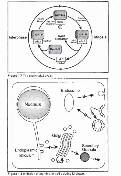
Figure 1.6 N-linked oligosaccharide processing of HLA during secretion.
1.2.14 Protein folding and chaperonlns
It has recently become clear that cells possess a class of proteins which function as molecular chaperones to temporarily stabilise partially folded proteins and to prevent illicit interactions with other proteins. These "chaperonins" have been implicated at a variety of stages of cellular metabolism such as protein synthesis, translocation and folding, cycles of macromolecular assembly and disassembly, protection against environmental stress and targeting proteins for degradation.
One such chaperonin molecule has been identified in the process of Class I assembly in mice (Degen and Williams, 1991). The essential requirement for proper assembly of the Class I trimeric complex for transport and cell surface function necessitates adequate quality control within the ER lumen to ensure that only functional complexes enter the secretory pathway. This Class I chaperonin (p88) undergoes rapid and quantitative association with newly synthesised heavy chains in the ER. Dissociation is not triggered by 82m binding to the heavy chain, but appears to occur while the complex is still in the ER. The rate of dissociation matches the rate of transport of the trimeric complex from the ER to the Golgi in each of the cell lines studied suggesting that p88 dissociation may be the rate- limiting step in the transfer of Class I in this step of the secretory pathway, consistent with a role in detaining "empty" heavy chains. As well as serving to stabilise and detain partially assembled Class I molecules, the p88 molecule may also have a role to play in directing peptide binding by physically interacting with the peptide transporters, though this theory remains speculative for the present.
1.2.15 Kinetics o f protein transport out o f the ER
Having considered the assembly of heavy chains, 82m and peptide into transport- competent heterotrimeric complexes, it is of interest to consider the rate of
transport from the ER to the Golgi apparatus (reviewed by Hurtley and Helenius, 1989; Pelham, 1989). The rate of secretion of different complexes shows allele- dependent variation (Williams, etal., 1985; Neefjes and Ploegh, 1988). Evidence points to the transport of at least some Class I molecules in the bulk flow of membrane traffic through the secretory pathway once they are fully assembled; the fastest measured rates of transport to the Golgi apparatus for certain Class I molecules correspond to the fastest secretion rates for other proteins, with a half- time for export to the mec/Za/Golgi of about 15 to 25 min (Fries, etal., 1984; Williams, etal., 1985).
accumulation of more slowly transported molecules to be the ER (Williams, etal.,
1985). Consistent with this finding, studies of the interaction of p88 with Class I complexes show that dissociation from this chaperonin may be the rate-limiting step for exit from the ER (Degen and Williams, 1991), which in turn, reflects the rate of proper assembly of the complex into a fully transport-competent
conformation. Studies of human allelic differences in secretion rates suggest a relationship between the binding affinity of Class I subunits and the rate of transport to the Golgi apparatus (Neefjes and Ploegh, 1988); HLA-A and B have greater affinity for 82m than HLA-C (Stam, etal., 1986) and are transported more rapidly in assembled trimeric complexes than HLA-C, despite the fact that HLA-C is synthesised in comparable amounts to HLA-A and B.
Mutagenesis of the H2K allele in mice and the non-classical HLA-E and HLA-F alleles in humans have shown that while 82m association is necessary for cell surface transport, it is not sufficient (Williams, etal., 1988; Shimizu, etal., 1988). It is clear that the peptides loaded onto Class I molecules also play a significant role in determining the overall conformation of the complex, and therefore the rate at which assembled complexes are transported to the cell surface (Townsend, etal.,
1989). As well as being an important observation with respect to the secretion rates of Class I molecules, the peptide-mediated inhibition of cell surface presentation has obvious implications for the possible strategies adopted by intracellular pathogens to evade immune surveillance.
Experiments with the glucosidase inhibitor 1-deoxynojirimycin shows that the lack of adequate oligosaccharide trimming prevents the export of certain glycoproteins from the ER (Gross, etal., 1983; Lemansky, etal., 1984; Lodish and Kong, 1984). These results suggest that carbohydrates might be involved in determining the native transport-competent conformation of the protein, highlighting the structural significance of oligosaccharide sidechains.
The variation in the rate of exit from the ER for many different secretory proteins has led to the suggestion that exit from the ER and more specifically, entry into transport vesicles destined for the Golgi apparatus is a receptor-mediated process (Lodish, etal., 1983; Fries, etal., 1984), involving complex protein interactions. However, since such delays may well be accounted for by oligomeric assembly and conformational changes within the ER which must precede the inclusion of secretory molecules into bulk flow carrier vesicles and since there has been no direct evidence to date for such receptors, the simpler explanation of
1.2.16 Glycosylation of secretory proteins
The polypeptide backbone of newly synthesised secretory proteins is often modified in a series of glycosylation reactions as the proteins traverse the secretory pathway, sequential modifications occurring as the proteins pass from one compartment to the next in a vectorial fashion via vesicular-mediated traffic. Since glycosylation of the heavy chain of the Class I complex was the basis for monitoring transport along the secretory pathway in the work I have undertaken here, I propose to describe the classical oligosaccharide processing sequence and how such oligosaccharides may be used to determine which compartments have been visited along the secretory pathway.
1.2.16.1 The types of glycosylation
The function of oligosaccharide modification of proteins is not very well understood. In the case of plasma membrane proteins, these carbohydrate moieties may facilitate cell-cell interactions via the extracellular matrix, or may confer structural stability on the fully mature protein, perhaps by increasing resistance to proteolysis. In the case of secreted proteins, they may increase the solubility of proteins by increasing the net charge on the protein. Certain sugar residues are involved in the sorting of some lysosomal proteins. The use of tunicamycin, an antibiotic which inhibits the first stage of protein glycosylation, allows the secretion of some glycoproteins but not others; this indicates that the carbohydrate side chains of some but not all glycoproteins have an important function to play in allowing passage along the exocytic pathway.
The two main types of glycosylation are N or (asparagine)-linked and O (threonine or serine)-linked. The latter modifications are less common and have been less well characterised as a result. Moreover, the heavy chain of Class I does not have any 0-linked sugars, and so I do not propose to discuss these modifications further.
Structural analysis of N-linked sugars using NMR has revealed that they fall into one of three classes, namely complex, hybrid or high mannnose. These three categories share a common pentasaccharide core. High mannose
N-linked oligosaccharides are usually added to the structural motif Asn-X-Ser/Thr, where X may be any amino acid other than proline or aspartate (Marshall, 1972, 1974). The capacity of this tripeptide structure to assume an appropriate
conformation in the context of short range interactions with nearby amino acids appears to have a major effect on the rate and extent of glycosylation. Such
interactions would have the effect of altering the accessibility of the tripeptide motif to the oligosaccharyl transferases. The addition of the most proximal residues is a co-translational event, which therefore occurs while the protein is still in the
process of folding. Once the protein assumes its fully native conformation , potential N-linked glycosylation sites may well be obscured, limiting the time during which such modifications may be initiated (Pless and Lennarz, 1977).
1.2.16.2 The sequence of oligosaccharide processing
The MHC Class I heavy chain has a single N-linked glycosylation site at Asn86 within the a l domain. This glycosylation site is conserved between the 25 HLA-A, 35 HLA-B and 18 HLA-C sequences that have so far been determined. The sequence of oligosaccharide modifications of the heavy chain is summarised in Figure 1.6 (reviewed by Komfeld and Kornfeld, 1985).
The trimming of the outer glucose residues and at least one of the outer mannose residues occurs in the ER, and have been shown to be co-translational events for the VSV G protein (Atkinson and Lee, 1984). Once within the cis Golgi, the high mannose sidechains are trimmed by the Golgi-specific mannosidase I to give a Man5GlcNAc2 structure. N-acetylglucosaminyltransferase I (NAGT I) catalyses the
addition of N-acetylglucosamine (GlcNAc) to one branch of the bi-antennary sugar chain in the med/a/Golgi, and the two outer mannose residues of the other branch are then trimmed by mannosidase II. A second med/a/N-
acetyIglucosaminyItransferase (NAGT II) adds another N-acetylglucosamine residue to the second branch and fucosyltransferase may mediate the transfer of a fucose residue to the innermost GlcNAc residue. Galactosyltransferase (GT) and a2,6-sialy Itransfe rase (ST) catalyse the addition of terminal galactose and
sialic acid residues in the trans cisternae and TGN.
1.2.17 Using oiigosaccharide processing to monitor giycoprotein transport