THE-ALLOSTERIC REGULATION OF
ADENOSINE RECEPTORS
Fiona R. Cohen
A thesis submitted in fulfilment of the requirements of the University of London for the degree of Doctor of Philosophy
1995
ProQuest Number: 10017241
All rights reserved
INFORMATION TO ALL USERS
The quality of this reproduction is dependent upon the quality of the copy submitted.
In the unlikely event that the author did not send a complete manuscript and there are missing pages, these will be noted. Also, if material had to be removed,
a note will indicate the deletion.
uest.
ProQuest 10017241
Published by ProQuest LLC(2016). Copyright of the Dissertation is held by the Author.
All rights reserved.
This work is protected against unauthorized copying under Title 17, United States Code. Microform Edition © ProQuest LLC.
ProQuest LLC
789 East Eisenhower Parkway P.O. Box 1346
ABSTRACT
The adenosine Ai receptor is one of the large superfamily of G protein coupled receptors. These receptors are generally considered to have 2 binding sites, one which binds the hormone or neurotransmitter, and one which binds the G protein. However it has recently been reported that an additional binding site exists on the Ai receptor. The binding of certain benzoylthiophenes (e.g. 2-amino-4, 5- dimethylthien-3-yl [3-(trifluoro methyl) phenyl] - methanone, PD81) to this site, allosterically modulate the binding and activity of agonists (and antagonists), thereby providing a novel means of modulating the actions of endogenous adenosine.
Initially, the binding and functional properties of the cloned human adenosine Ai receptor, expressed in Chinese hamster ovary cells, were characterised. A novel saponin pretreatment simplified the assay system and enabled for the first time a detailed characterisation of the binding properties of adenosine itself for the Ai receptor. The core of the study was a quantitative investigation of the effects of allosteric agents on binding and function of the adenosine Ai receptor. The experimental evidence is quantitatively compatible with PD81 binding to a specific allosteric site on the Ai receptor. Functional assays demonstrate that the cloned human A2A receptor does not appear to have the same allosteric site.
CONTENTS
Abstract
Page
2
Contents
List of Figures
List of Tables
3
10
16
Abbreviations 18
CHAPTER 1: INTRODUCTION
1.1 G protein coupled receptors 20
1.1.1 The structure of G protein coupled receptors 1.1.2 G proteins
1.1.3 The G protein activation cycle
22
26 28
1.2 Adenosine and adenosine receptors 31
1.2.1 Adenosine
1.2.2 Classification of receptor subtypes 1.2.3 Cloning of the adenosine receptors 1.2.4 Receptor Effector Coupling
1.2.4.1 Adenylate cyclase 1.2.4.2 K" channels 1.2.4.3 Other Effectors
31 32 34 37 38 39 42
Page
1.3.1 The mechanism of action and advantages of allosteric 43 acting drugs
1.3.2 Allosteric regulation of G protein coupled receptors 45 1.3.3 Allosteric regulation of adenosine receptors 46
1.4 Receptor domains 47
1.4.1 Receptor domains involved in G protein interactions 47
1.4.2 The ligand binding domain 49
1.4.2.1 Ligand binding domains of adenosine 53 receptors
1.4.3 The allosteric ligand binding domain 56
1.5 Aims of Research Project 58
CHAPTER 2: MA TERIALS AND METHODS
2.1 Studies on CHO cells stably transfected with Ai, Aza or A3 receptors59
2.1.1 Materials 59
2.1.2 CHO Cell Culture 59
2.1.3 Membrane Preparation 59
2.1.4 Radioligand Binding 60
2.1.5 Binding studies with [^^SjGTPyS 61
2.1.6 cAMP assays 62
2.1.6.1 cAMP assays on CHO.A1 and CHO.A2A cells 62 2.1.6.2 cAMP assays on CHO.A3 cells 62
2.1.7 Data analysis 63
Page
2.2.1 Materials 63
2.2.2 Strategy of splicing by overlap extension 64 2.2.3 Subcloning of the Ai and the A2A receptors 67
2.2.4 Primer design for PCR reactions 72
2.2.4.1 Design of general primers 72 2.2.4 2 Design of specific primers 73
2.2.5 Polymerase chain reaction 74
2.2.5.1 First generation chimeras 74
2.2.5.2 Second generation chimeras 75 2.2.6 Subcloning the chimeric constructs 75 2.2.7 Preparation of electrocompetent XL1 Blue cells 76 2.2.8 Transformation of electrocompetent cells 76
2.2.9 Plasmid DNA preparation 77
2.2.10 Sequencing 78
2.2.10.1 Sequencing primer design 78 2.2.10.2 Sequencing reactions, preparation and 80
loading the sequencing gel
2.2.10.3 Analysis of sequencing data 81
2.3 Electrophysiological Studies 84
2.3.1 Preparation of RNA 84
2.3.2 Preparation of oocytes 87
2.3.3 Injection of RNA 8 8
2.3.4 Electrophysiological recordings 89
2.4 Binding Studies on COS cells transiently transfected with Ai, A2A or 90 A1/A2A chimeric constructs.
2.4.1 Materials 90
2.4.2 COS cell culture 90
Page
2.4.4 Membrane Preparation 93
2.4.5 Radioligand binding 93
CHAPTER 3: CHARACTERISATION OF THE HUMAN A, ADENOSINE RECEPTOR
3.1 Introduction 94
3.2 Removal of some of the complexities associated with adenosine 95 receptor binding assays.
3.2.1 Adenosine is present in binding assays using CHO-Ai 96 membranes
3.2.2 Effects of saponin pretreatment on the binding of 96 [^H]DPCPX in the presence of and absence of ADA and GTP 3.2.3 Effects of saponin pretreatment on the binding of CHA to Ai 98
receptors
3.2.4 Effect of the presence of saponin in the assay on the 101 binding of CHA
3.2.5 Effects of saponin treatment on receptor function 102
3.3 Characterisation of the binding properties of human Ai receptors 106
3.3.1 Kinetics of radioligand binding 106
3.3.2 Saturation analysis 112
3.3.3 CHA binding properties in competition experiments under 116 various conditions
Page
3.5 Discussion 122
3.5.1 Endogenous adenosine in adenosine receptor binding assays 122 3.5.2 A possible permeability barrier between GTP and G proteins 123 3.5.3 Binding properties of the human Ai adenosine receptor 124
3.5.3.1 Kinetic studies 125
3.5.3 2 Effects of GTP on antagonist binding 127 3.5.3 3 Comparison of affinity constant values obtained 130
in this thesis with those obtained previously.
3.5.4 The binding properties of adenosine 131
CHAPTER 4: ALLOSTERIC ENHANCEMENT OF ADENOSINE Ai RECEPTOR BINDING AND FUNCTION BY PD81
4.1 Introduction 134
4.2 Binding Properties of CHO.Ai membranes in the presence 135 of PD81.
4.3 Dissociation Kinetics of flH ]PIA in the presence of PD81. 143
4.4 Functional Properties of CHO.Ai membranes in the presence 145 of PD81.
4.4.1 Modulation by adenosine agonists of [^^S]GTPyS binding to 145 G proteins.
4.4.2 Modulation by adenosine of [^^S]GTPyS binding to 149 G proteins.
Page
4.5 Discussion 154
CHAPTER 5: CONSTRUCTION AND ANALYSIS OF A ,/A2A
CHIMERIC RECEPTORS
5.1 Introduction 162
5.2 Construction of the chimeric receptors 164
5.3 Analysis of chimeric receptors using electrophysiology 168
5.3.1 The oocyte expression system 168
5.3.2 Expression of the adenosine Ai receptor and functional 168 coupling to GIRK1 in oocytes
5.3.3 Expression of the adenosine A2A receptor and functional 177 coupling to GIRK1 in oocytes
5.3.4 The effect of PD81 on the Ai adenosine receptor 179 5.3.5 Expression and function of the chimeric receptors 180
5.4 Analysis of constructs using radioligand binding 190
5.4.1 Initial screening of chimeric receptors 190 5.4.2 Binding affinities of agonists and antagonists 194 5.4.2.1 Determination of antagonist affinities 195 5.4.2.2 Determination of agonist affinities 199 5.4.3 Effect of PD81 on chimeric receptors 201 5.4.3.1 Effect of altering the buffer composition 206
on the binding of PD81
Page
5.5.1 The technique of chimeric receptors 211 5.5.2 Using electrophysiological techniques as a functional assay 212 5.5.3 Expression, binding and function of the chimeric receptors 213 5.5.4 The effect of PD81 on the wildtype and chimeric receptors 219 5.5.5 Modelling the position of the allosteric binding site 224
5.5.6 Conclusions 230
CHAPTER 6: SUMMARY, CRITICISM AND FUTURE DIRECTIONS
6.1 Summary 231
6.1.1 Characterisation of the human Ai adenosine receptor 231 6.1.2 The allosteric action of PD81 at the Ai adenosine receptor 232 6.1.3 The allosteric ligand binding site on the Ai adenosine 232
receptor
6.2 Critical Assessment and future directions 233
References 238
Appendix 1: Equations used in data analysis 264 Appendix 2; Structure of compounds used in the study 268
LIST OF FIGURES
CHAPTER 1
Page
1.1: General mechanism of action of G protein coupled receptors. 21
1.2: A helical wheel model of the seven transmembrane domains 24 of muscarinic receptors.
1.3: Receptor- G protein interactions 30
CHAPTER 2
2.1 : Mechanism of gene splicing by overlap extension; first 65 generation
2.2: Mechanism of gene splicing by overlap extension; second 6 6 generation
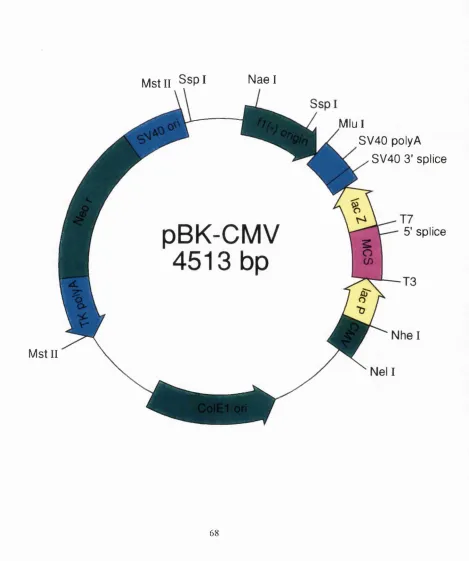
2.3: Map of the pBK-CMV plasmid 6 8
2.4: The strategy for subcloning the human Ai and A2A adenosine 70 receptor sequence into pBK-CMV from PGEM7 and pcDNAneo respectively.
2.5: Analysis of sequencing data from the ABI 373A DNA sequencer 82
Page
2.7: Photograph of the Cell Cube and confluent cells after 91 electroporation
CHAPTER 3
3.1 : Competition by CHA of f H]DPCPX binding to human Ai 99 adenosine receptors pretreated with varying saponin concentrations.
3.2: The effect of saponin pretreatment on CHA-stimulated binding 104 of f^SJGTPyS to G proteins.
3.3: The dissociation kinetics of fH]DPCPX and [^H]CHA binding to 107 Ai adenosine receptors.
3.4: Association of [^H]DPCPX to Ai adenosine receptors in the 110 presence or absence of GTP (1 mM).
3.5: Association of f H]CHA to Ai adenosine receptors. 111
3.6: Saturation Isotherms for agonist and antagonist binding to the 114 human Ai adenosine receptor.
3.7: Competition by CHA of [^H]DPCPX binding to C H O - A i 117
membranes pretreated with 1 0Oug/ml saponin in the presence and absence of GTP and ADA.
3.8: Competition of [^H]DPCPX binding to adenosine Ai receptors on 121 saponin pretreated CHO membranes by CHA and adenosine in
the presence and absence of GTP and ADA.
Page
CHAPTER 4
4.1 : The effect of varying concentrations of PD81 on the competition 136 by CHA of [^H]DPCPX binding to human Ai adenosine receptors.
4.2: The effect of varying concentrations of PD81 on the competition 141 by PI A, NECA and adenosine of f H]DPCPX binding to human
Ai adenosine receptors.
4.3: The effect of varying concentrations of PD81 on the dissociation 144 kinetics of [^H]PIA binding to Ai adenosine receptors.
4.4: The effect of PD81 on agonist-stimulated binding of f^S]GTPyS 146 to G proteins in AiAR membranes.
4.5: The effect of varying concentrations of PD81 on PIA-stimulated 149 binding of f^SJGTPyS to G proteins.
4.6: The effect of PD81 on adenosine-stimulated binding of 150 [^^S]GTPyS to G proteins in Ai adenosine receptor membranes.
4.7: The effect of PD81 on cAMP levels induced by the non- 153 selective adenosine receptor agonist NECA in CHO.Ai,
CHO.A2A and CHO.A3 cells.
CHAPTER 5
5.1 : The residues at the points of interchange between the Ai and 165 the A2A adenosine receptor sequences in the chimeric receptors.
Page
5.3: The construction of a current-voitage (l-V) relationship by using a 170 voltage-step protocol.
5.4: Current recordings as a result of increasing concentrations of 171 adenosine followed on a Maclab 14 recorder.
5.5: Dose response of adenosine acting at the Ai adenosine receptor 173 expressed in Xenopus oocytes.
5.6: Determination of whether endogenous adenosine is present in, or 175 released from Xenopus oocytes.
5.7: The possible coupling of Ai adenosine receptors to endogenous \C 176 channels in Xenopus oocytes.
5.8: Dose response of adenosine acting at the A2A adenosine receptor 178 expressed in Xenopus oocytes.
5.9: The effect of PD81 in the Xenopus oocytes injected with cRNA 181 from both the Ai adenosine receptor and GIRK1, and injected
with cRNA of GIRK1 only.
5.10: The inward rectification observed of the l-V responses of CHI 185 and CH4 both stimulated by lOOnM adenosine.
5.11: A qualitative comparison of the conductance size at 1 pM 186 adenosine at C H I, CH4 and CH3.
5.12: Dose response of adenosine acting at CH1, CH4, CH2a and 188 CH3a all expressed in Xenopus oocytes
Page
5.13: Screening of [^H]DPCPX binding to the Ai, A2A and A1/A2A 191 receptors.
5.14: Agonist binding to the Ai, A2A and A1/A2A receptors. 193
5.15: Binding of f H]NECA to CH3, CH2a and CH3a chimeras. 194
5.16: Comparison of the affinity of DPCPX at CH4 and the Ai and A2A 198 adenosine receptors.
5.17: Comparison of the affinity of CHA at CHI, CH4, CH4a and the 200 Ai adenosine receptors.
5.18: The effect of PD81 on the competition by CHA of [^H]DPCPX 202 binding to the Ai adenosine receptor.
5.19: The effect of PD81 on the competition by CHA of [^H]DPCPX 204 binding to the chimeric receptors, CHI, CH4 and CH4a.
5.20: Comparison of the affinity of PD81 at C H I, CH4, CH4a and the 205 Ai adenosine receptor.
5.21 : The effect of buffer composition on the PD81 effect on [^H]NECA 207 binding.
5.22: The effect PD81 on [^H]NECA binding to the Ai and A2A 209 adenosine receptors in a Mg^Vree buffer.
Page
5.24: Proposed model of the binding of PD81 and NECA to the 228 human Ai adenosine receptor
LIST OF TABLES
CHAPTER 3
Page
3.1 : Effect of saponin pretreatment on the binding of [^H]DPCPX 97 (0.2nM) to CHO-A1 membranes, measured in the presence or
absence of ADA and GTP.
3.2: Effect of saponin pretreatment on the competition by CHA of 100 [^H]DPCPX binding to human Ai adenosine receptors in the
presence and absence of GTP and ADA.
3.3: Competition by CHA of [^H]DPCPX binding to human Ai 101 adenosine receptors in the presence of varying concentrations
of saponin in the assay.
3.4: The effect of saponin pretreatment on CHA-stimulated binding 103 of [^^SJGTPyS to CHO - Ai membranes.
3.5: CHA-stimulated binding of [^^S]GTPyS to CHO - Ai membranes 105 in the presence of varying concentrations of saponin.
3.6: Affinity constants and Bmax values for ligand binding to saponin 113 pretreated human Ai Adenosine receptors.
CHAPTER 4
Page
4.2: The effect of PD81 on agonist-stimulated binding of [^®S]GTPyS 148 to G proteins in CHO.A1 membranes.
4.3: The effect of PD81 on PIA-stimulated binding of f^S]GTPyS to 148 G proteins in CHO.A1 membranes.
4.4: The effect of PD81 on adenosine-stimulated binding of 151 [^®S]GTPyS to G proteins in CHO.Ai membranes.
4.5: The effect of PD81 on cAMP levels induced by the non-selective 152 adenosine receptor agonist NECA in CHO.A1 and CHO.A3 cells.
CHAPTER 5
5.1 : Mean conductance values and EC50 values obtained for the 183 wildtype and chimeric receptors.
5.2: Comparison of the Hill Coefficients and EC50 values of the Ai, A2A 184 and CH4 receptors.
5.3: Parameter estimates of agonist and antagonist binding affinities 196 at the Ai, A2A and A1/A2A chimeric receptors.
5.4: The effect of PD81 on the competition between [^HJDPCPX and 203 either CHA or NECA at the Ai , C H I, CH4 and CH4a receptors.
5.5: The effect of PD81 on [^H]DPCPX binding to the Ai and three 206 chimeric adenosine receptors.
5.6: Effect of PD81 on [^H]NECA binding to the chimeric receptors in 210 a Mg^‘"-free buffer.
ABBREVIATIONS
[’“ l]HPIA
A-R
A-R-G
Ai / A2A AR
ADA bp
CGS 21680
C H I - C H 4 CH2a - CH4a CHA
CHO
CHO. Ai / A2A / A3
C0SM6 cells DMSO DNA DPCPX E Coli
EDTA G protein GIRK1 GDP Gpp(NH)P GTP GTPyS GTPase HEPES HighK or 90K
(-)-N®-3-([^^®l]-iodo-4-hydroxyphenyllsopropyladenosine agonist-receptor binary complex
agonist-receptor-G protein ternary complex Ai / A2A chimeric adenosine receptor
adenosine deaminase base pairs
2-[4-(2-carboxyethyl)-phenethylamino]-5’-N-ethylcarboxamidoadenosine
First generation chimeric A1/A2A adenosine receptor Second generation chimeric A1/A2A adenosine receptor N®-cyclohexyladenosine
Chinese hamster ovary
Chinese hamster ovary cells transfected with the cloned human A i, A2A or A3 adenosine receptors
African green monkey kidney cells dimethylsulfoxide
deoxyribonucleic acid
8-Cyclopentyl-1, 3-dipropylxanthine
Eschericia coli
ethylenediaminetetraacetic acid guanine nucleotide binding protein
Inwardly rectifying, G protein coupled potassium channel guanosine 5’-diphosphate
5’-guanylyl-imidodiphosphate guanosine 5’-triphosphate
guanosine-5’-0-(3-thio)triphosphate guanosine 5’-triphosphatase
MOPS 3-[N-morpholino]propanesulphonic acid NECA 5’-N-ethylcarboxamidoadenosine
CD optical density
0R2 83mM NaCI, 2.5mM KCI, 1mM NagHPO^, 5mM HEPES, pH 7.5
PBS phosphate buffered saline PCR polymerase chain reaction
PD71,605 2-amino-4, 5, 6, 7-tetrahydrobenzothien-3-yl [3-(chloro phenyl)] - methanone
PD81 2amino4, 5dimethylthien3yl [3(trifluoromethyl) phenyl] -methanone
PEI polyethylenimine
PIA R - N®-phenylisopropyladenosine R-G receptor-G protein binary complex
RNA ribonucleic acid
SOE Splicing by overlap extension
TM Transmembrane domain
Vent VentR DNA polymerase
XAC 8-{4-[({[(2-aminoethyl)amino]carbonyl}methyl)oxyl]phenyl}-1,3-dipropylxanthine
CHAPTER 1: INTRODUCTION
1.1 G protein coupled receptors
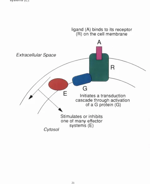
A wide variety of hormone and neurotransmitter receptors interact with heterotrimeric guanine nucleotide binding regulatory proteins (G proteins). These G protein coupled receptors form a large family of integral membrane proteins whose amino acid sequences have seven hydrophobic domains containing distinctive sequence patterns. Receptors of this class respond to a structurally diverse range of agonists, including small molecules such as biogenic amines or adenosine, peptides such as angiotensin or the neurokinins, and larger proteins such as lutotropin or thyrotropin. The binding of a ligand to its specific receptor triggers a signal transduction cascade via activation of a G protein which acts as an intermediate to stimulate or inhibit one of many effector systems (Figure 1.1). Effector enzymes and ion channels that are coupled to G protein-mediated pathways include adenylyl cyclase, phospholipases A and C, calcium and potassium channels, and phosphodiesterases. Thus the signal transduction pathway initiated by G protein coupled receptors represents a major mechanism for transmembrane signalling in cellular systems.
It is likely that there are between 400-1000 different G protein coupled receptors, of which %250 members have been cloned. This superfamily of
receptors appears to constitute one of the largest gene families of the mammalian genome. The cloning of G protein coupled receptors has facilitated the determination of receptor structure and the nature of the ligand binding site.
Figure 1.1: General mechanism of action of G protein coupled receptors.
The ligand (A) binds to its receptor (R) on the cell membrane. This interaction initiates a transduction cascade through activation of a G protein (G) inside the cell. The activated G protein is able to stimulate or inhibit one of many effector systems (E).
ligand (A) binds to its receptor (R) on the cell m em brane
Extracellular Space
Initiates a transduction cascade through activation
of a G protein (G)
Cytosol
Stim ulates or inhibits one of m any effector
system s (E)
1.1.1 The structure o f G protein coupled receptors
All members of the G protein coupled receptor family are thought to have the same basic structure in the transmembrane part of the protein because of their sequence similarities and their common function of binding and activation of G proteins. It has been shown that in two members of this receptor family, rhodopsin and the p2-adrenergic receptor, the N-terminus is on the extracellular side of the membrane and the C-terminus is on the intracellular side (Applebury and Hargrave; 1986, Wang etal. 1989).
Hydropathicity analysis of the primary structures of G protein coupled receptor proteins suggests that they contain seven transmembrane alpha-helices. Predictions of the structures of G protein coupled receptors has largely derived from the known structure of bacteriorhodopsin, a purple transmembrane protein from Halobacterium halobium. Although bacteriorhodopsin is a light-driven proton pump and does not couple to G proteins, electron microscopy has shown that it consists of a bundle of seven a-helices which span the membrane (Henderson and Unwin; 1975, Henderson etal. 1990).
Rhodopsin, a member of the family of G protein coupled receptors, shares with bacteriorhodopsin its response to light through the isomérisation of a covalently-bound retinal chromophore. A low resolution projection map of bovine rhodopsin, determined by electron crystallography of two-dimensional crystals, gives further evidence for the existence of this seven transmembrane a-helical structure (Schertler at a/. 1993). Four helices were shown to be nearly perpendicular to the membrane and three were tilted. When the projection density of a rhodopsin molecule is compared to that of bacteriorhodopsin, the projection structure of rhodopsin is found to be less elongated, slightly broader and the helices are tilted differently from those of bacteriophodopsin.
Baldwin (1993) has aligned the putative transmembrane domains of 200 sequences of the G protein coupled receptor family. The probable arrangement
position appropriate to the extent of its lipid-facing surface area. Helix III appears to be the most hidden within the helical bundle, whereas helices I, IV and V are more exposed to the membrane lipid environment. Some receptors have short inter-helical loops, so in order for all the receptor family to have the same arrangement of helices, each helix must be organised in the helical bundle according to its sequence order. Figure 1.2 shows a helical wheel model of the probable arrangement of the transmembrane a-helices based on the projection map (Schertler et al. 1993) and the sequence alignment studies (Baldwin et ai. 1993). The suggested arrangement gives a closely packed structure at the intracellular surface where the G protein interaction is thought to occur and a more open structure in the extracellular half of the receptor, producing a ligand binding pocket. However, the proposed three-dimensional arrangement is still fairly speculative (Hibert et ai. 1993) and three-dimensional crystallographic data is essential for a more accurate determination of the structure of G protein coupled receptors.
Most of the G protein coupled receptors cloned to date have at least one consensus sequence for N-linked glycosylation (Asn-X-Ser/Thr where X can be any amino acid except for proline and aspartate) in the extracellular domain. Prevention of receptor glycosylation, either by inhibition of glycosylation enzymes in the cell or by site-directed mutagenesis of the potential glycosylation sites on the receptor, results in a decrease in the level of expression of either the p2-adrenergic receptor or the lutropin receptor on the cell surface (Rands et ai. 1990, Liu et al. 1993) but not the m2 muscarinic acetylcholine receptor (van Koppen and Nathanson; 1990). However the lack of glycosylation does not appear to alter ligand binding or functional activity of those receptor molecules expressed in the plasma membrane.
G protein coupled receptors contain a number of conserved cysteine residues, some of which are thought to be important in maintaining receptor structure. There are two highly conserved cysteine residues in the first and second extracellular loops of the receptors that have been demonstrated to form an intramolecular disulphide bridge in both rhodopsin and muscarinic acetylcholine receptors (Karnik et al. 1988, Curtis ef a/. 1989).
Figure 1.2: A helical wheel model of the seven transmembrane domains of muscarinic receptors.
Eighteen sequences of m1-m5 receptors from various species including human, chick and Drosophila are considered. Each helix is shown as a helical wheel (angle 100°) with the short lines representing the orientation of the side- chains. Only the central 18 of the 20-25 residues thought to be present as a- helices are shown. The helices are viewed from the extracellular domain and are positioned approximately in the relative positions suggested by the projection map of rhodopsin (Schertler et al. 1993). Residues identical in all the sequences are shown by their single letter code and those found in most G protein coupled receptors are enclosed in a square. In addition the aspartate residue in transmembrane domain III (TMIII) conserved in all monoaminergic receptors, is enclosed in a hexagon. Residues that are hydrophobic (including serine) and not conserved between muscarinic subtypes and species are depicted by closed circles. Both the conserved and hydrophilic residues tend to cluster at helix-helix boundaries and towards the interior of the structure, whereas the non-conserved and hydrophobic residues face outwards. (From
Mutagenesis of either of these two cysteine residues with alanine or valine, results in a destabilising of the tertiary structure of both rhodopsin (Davidson et al. 1994) and the (3-adrenergic receptor (Dixon et al. 1987b, Dohlman et al.
1990). This suggests a role for these residues in maintaining the active conformation of the receptor which is involved with the binding and activation of the G protein.
1.1.2 G proteins
G proteins fall into two major families, the ‘small G proteins’, usually found as single polypeptides of about 200 amino acids (reviewed in Hall; 1990) and the heterotrimeric G proteins (reviewed in Gilman; 1987). The family of heterotrimeric G proteins are composed of a, p, and y subunits in order of
decreasing mass. The a subunit binds guanine nucleotides and has a GTPase function whilst the p and y subunits are always co-associated. At present, the family is known to contain at least sixteen different genes that encode the a - subunit, five that enocde the p-subunit and seven that encode y-subunits (Simon et a i 1991, Cali et al. 1992, Watson et al. 1994).
The a-subunits have conserved primary structure (50-90% identity) and share a common molecular mechanism but they have diverse signalling functions. The interaction of the a-subunit with receptors, guanine nucleotides, GPy-
subunits and effectors has been located to different structural regions of the a- subunit (reviewed by Bourne et al. 1991, Conklin and Bourne; 1993).
The a-subunit cycles between a resting (GDP-bound) conformation primed for interaction with agonist-stimulated receptors and an active (GTP-bound) conformation capable of activating or inhibiting downstream effectors (see
below). Crystal structures, refined to a resolution of 2.2Â, of the GDP- (Noel et
al. 1993) and 1.8Â of the GTP- (Lambright et al. 1994) bound forms of the a-
subunit of transducin, Gt« (the G protein that couples to rhodopsin) have shown that the nucleotide binding site is deep within the a-subunit between the
GTPase domain homologous to small GTPases and an a-helical domain unique to heterotrimeric G proteins. The structural differences induced by nucleotide exchange are localised to three regions on one face of the protein. The formation of hydrogen bonds between the y-phosphate of GTP and a glycine and threonine residue in the a-subunit of the G protein is thought to initiate the nucleotide exchange, which is then further propagated by a series of ionic interactions (Lambright et al. 1994). All of the critical residues for the conformational switch in Gt« are conserved in all G protein a subunits so it is anticipated that the same mechanism will hold for all members of the heterotrimeric G protein family.
Experimental evidence suggests that the amino-terminal region of the a- subunit interacts with the Py-subunits, either directly or by maintaining a conformation necessary for heterotrimer formation (Denker at al. 1992, Neer at al. 1988). A short highly conserved sequence, thought to be close in space to the amino-terminus, is also thought to be an important regulatory contact between Ga and Py. This region is more highly conserved than any other sequence in Ga, in keeping with the functional conservation of a-Py interactions in all G protein-based signalling pathways. As yet no putative contact sites have been shown to encode the specificity of an a subunit for interacting with a particular species of Py but this kind of specificity is suspected (Kleuss atal. 1992).
Initial studies using rhodopsin suggested that the guanine nucleotide binding site and rhodopsin binding site are located in different domains of the G protein (Fung and Nash; 1983). At least three regions of Ga, in particular the carboxyl terminus, are now thought to contact the receptor. Evidence for this comes from experiments using peptides mimicking the C-terminus and a small adjacent region of the a subunit (Hamm at al. 1988), and chimeric constructs
replacing amino acids of the aq C-terminus with those of aa (Conklin at al.
separate regions of primary structure involved in receptor-effector interactions (Berlot and Bourne; 1992).
Four of the mammalian Gp subunits share « 80% homology but the most recently found p-subunit is more divergent (Watson et al. 1994). Despite this close homology, they cannot form dimers with each kind of y-subunit; Pi
interacts with either yi or 7 2, P2 interacts with 7 2 but not 71 and pp cannot associate with either 71 or 72 (Schmidt at al. 1992, Pronin and Gautam; 1992). Furthermore, p-subunits are selective in coupling to a-subunits. Muscarinic
receptors couple to a voltage sensitive Ca^^ channel through OoiPsy (but not Pi, p2 or P4) whereas somatostatin receptors couple to the same channel through Oo2pi7 (Kleuss at al. 1992). Based on the amino acid sequence of the p- subunit, the amino-terminal region is thought to form an amphipathic a-helix which could be involved with coiled-coil interactions with both the a- and 7- subunits of the G protein (Lupas at al. 1992). The remainder of the protein consists of seven repeating units, each about 43 amino acids long of unknown function. Recent evidence suggests that interactions between the receptor and the p-subunit, as well as the a-subunit are important for receptor-G protein coupling (Taylor atal. 1994).
The seven y-subunits known to date are much more heterogeneous than the p- subunits (Cali at al. 1992) and have been shown to be crucial for determining the function of the Gpy-subunits (Clapham and Neer; 1993). The 73 subunit has been shown to be necessary for the coupling of the somatostatin receptor to a voltage sensitive Ca^'" channel whereas the 74 subunit is needed for coupling the muscarinic receptor to the same channels (Kleuss at al. 1993).
1.1.3 The G protein activation cycle
In the G protein activation cycle, Ga exists in at least three different conformational states (Figure 1.3); the inactive GDP bound state (a.GDP) , a state in which the guanine nucleotide site is empty, and as the active GTP bound state (a.GTP). a.GDP has a high affinity for binding py, and binding of
py increases its affinity for GDP. Ligand stimulated receptors bind the heterotrimeric a.GDP.Py complex and trigger release of GDP creating aPy with no guanine nucleotide bound, in the absence of added guanine nucleotides, the stimulated receptor binds the empty G protein tightly and stabilses the complex. The ratio of GTP:GDP in the cell is high and GTP fills the empty nucleotide binding site of Ga. A change in the conformation of a.GTP results, which causes it to dissociate from both the stimulated receptor and the py subunits, a.GTP or Py may then activate a number of relevant effectors (see later). Hydrolysis of the bound GTP terminates the regulation of the receptor (in the case where a.GTP is the active component) and Ga returns to its inactive GDP-bound state. GTP hydrolysis results from an intrinsic GTPase property of the a-subunit (Fung et al. 1981 ).
Figure 1.3: Receptor- G protein interactions
A general model for the interactions between agonist (A), receptor (high agonist affinity state, Rh, or low agonist affinity state, Rl), and G protein (Ga(3y) as described in the text.
Pi
GDP
GTP
GTP
— GDP
GTP
De Lean et al. (1980) have proposed a model for the (3-adrenergic receptor in which a receptor-G protein complex (R-G) is stabilized by binding of agonist to form a ternary complex, agonist - receptor - G protein (A-R-G). Binding of guanine nucleotide to the a-subunit of the G protein destabilizes the ternary
complex. The active a-subunit - GTP complex generated is then free to interact with the effector. The ternary complex A-R-G exhibits a high affinity for the agonist, whereas the binary A-R complex exhibits low affinity for the agonist. Thus, binding of G protein to the A-R complex allosterically affects agonist binding to the receptor through interconversion of a low affinity state into a high affinity state (De Lean at al. 1980).
Several biochemical tools which act within the G protein activation cycle are used to study G-protein linked signal transduction pathways. Cholera toxin from Vibrio choleras transfers the ADP-ribose moiety from NAD to a conserved arginine residue within Gas and transducin a chains (Van Dop et al. 1984). This modification requires the presence of an additional protein, ADP- ribosylating factor, and suppresses GTPase activity, locking the G protein in the active GTP-bound conformation (Kahn and Gilman; 1984, Milligan; 1988). Pertussis toxin (islet activating protein from Bordetella pertussis) ADP- ribosylates G protein a-chains at a cysteine located four residues from the C-
terminal of Gdi-a, Gaoi, G«o2, Gati, and Gat2- This modification appears dependent upon an apy heterotrimeric state and results in G protein uncoupling from the receptor (reviewed in Milligan; 1988).
1.2 Adenosine and adenosine receptors
1.2.1 Adenosine
Pearson; 1990). In addition to the role of adenosine in cellular energy metabolism and the importance of adenosine nucleotides in the transmission of genetic information, adenosine functions as a neuromodulator. Under appropriate conditions adenosine is released from cells where it can interact with specific cell surface receptors to modulate cellular function.
Adenosine is produced intracellularly by two different metabolic pathways, both of which involve hydrolases. The first involves the hydrolysis of AMP to adenosine by 5’-nucleotidase and the second the catabolism of S- adenosylhomocysteine (Olsson and Pearson; 1990). When the cellular energy state is depressed, under conditions of stress such as hypoxia, the level of intracellular adenosine rises dramatically and it is released from the cell. The released adenosine binds to and activates adenosine receptors, which in turn alter the activity of a second messenger pathway within the cell via coupling to G proteins, in an attempt to bring the tissue or cell back into a normal equilibrium. Once released from the cell, adenosine can diffuse across the cell membrane back into the cell, it can be transported back by a specific nucleoside transporter (Kwong et al. 1988) or it can be deaminated by adenosine deaminase to the receptor-inactive inosine (Olsson and Pearson; 1990).
Almost all organ systems of an animal are regulated by the local release of adenosine which as a consequence has extensive physiological functions. These include effects on cardiac rate and contractility, release of neurotransmitters, smooth muscle tone, sedation, platelet function, lipolysis, renal function and white blood cell function (Ramkumar at al. 1988). Because of the widespread nature of these effects there has been considerable interest in the classification of adenosine receptors and their second messenger responses.
1.2.2 Classification o f receptor subtypes
Purinoceptors (adenosine and adenine nucleotide receptors) were initially classified as Pi and P2 receptors by Burnstock in 1978. Adenosine
receptors (or Pi receptors) were distinguished from ATP receptors by; 1) the rank order of potency of adenosine > nucleotides; 2) a selective antagonism by methylxanthines; 3) coupling to adenylyl cyclase and 4) an absence of effect on prostaglandin synthesis. Adenosine receptors were subsequently subdivided into Ai and A2 subtypes based on their ability to inhibit (Ai) or stimulate (A2) adenylate cyclase (Van Calker et al. 1979). Early studies described the order of potency at Ai adenosine receptors as R- phenylisopropyladenosine (R-PIA) > adenosine > 5’-N-ethyl-carboxamide adenosine (NECA), whilst that at the A2 receptors exhibited the rank order; NECA > adenosine > R-PIA (Londos at al. 1980). Adenosine itself was shown to have nanomolar potency for Ai receptors but micromolar potency for A2 receptors (Van Calker at al. 1979, Daly at al. 1981).
The A2 adenosine receptors were further subdivided into A2A and A2b subtypes on the basis of high and low potency, respectively, for adenosine and the agonists, NECA and 2[p-(carboxyethyl)phenethylamino]-5’-N- ethylcarboxamidoadenosine (CGS21680) (Bruns at al. 1986, Jarvis at al.
1989).
Ribeiro and Sebastiao (1986) postulated the existence of a third subclass of adenosine receptor, the A3 adenosine receptor, based on observations that in atrial tissue, NECA and PIA were equipotent and that these agonists had no effect on adenylate cyclase. This contrasts with the characteristics of the Ai, A2A or A2b receptors. Recent reviews cast major doubt on the existence of this A3 receptor (Collis and Hourani; 1993, Carruthers and Fozard; 1993). The variable potencies may be the result of agonist- or tissue-related factors, and adenosine receptors have since been shown to couple to a variety of other effectors beside adenylate cyclase (Stiles; 1992, and see ‘receptor effector coupling’ below).
R-PIA = NECA but did not bind the Ai selective antagonist 1,3-dipropyl-8- cyclopentylxanthine (DPCPX). It has since been cloned in a variety of species, is considered distinct from the established Ai and A2 receptors and has been named the A3 receptor (Carruthers and Fozard; 1993). A3 receptors have been identified on a tumour cell line derived from rat mast cells and the receptor transcript has been shown to be present in this cell line. Combining this information with the pharmacological profile of mediator release from rat mast cells, Ramkumar et al. (1993) have postulated a physiological function of A3 receptors in mast cell degranulation.
In addition to the adenosine receptors, a purine-site (P-site) for adenosine is located at the cytoplasmic face of the catalytic subunit of adenylate cyclase which mediates inhibitory effects of high concentrations of adenosine (Johnson
at al. 1991, Linden; 1994a). Another group of adenosine binding proteins, has recently been identified by fH]NECA binding in several mammalian tissues. These have been called adenotin (Lorenzen at al. 1992) and Ax (Schwabe at al. 1993). The physiological roles of these proteins are as yet unknown but because of their intracelllular location they are not considered to be adenosine receptors.
Thus the Pi adenosine receptor family can be subdivided, based on pharmacological criteria into Ai, A2A, A2B and A3 subfamilies. This classification has also been found by molecular cloning of the receptor subtypes.
1.2.3 Cloning of the adenosine receptors
To date thirteen adenosine receptors have been cloned: the Ai receptor from five species, the A2A receptor from three species, the A2B receptor from two species and the A3 receptor from three species (reviewed by Tucker and Linden; 1993, Linden at al. 1993a, Linden; 1994b). All of the cloned adenosine receptors belong to the superfamily of G protein coupled receptors.
Adenosine receptors were two of the first four G protein coupled orphan receptors identified by a PCR homology screening strategy (Libert et al. 1989). This strategy involves the construction of oligonucleotides on the basis of similarities among genes that encode G protein coupled receptors. Two orphan receptors , RDC7 and RDC8, were cloned using this PCR technique from a canine thyroid cDNA library (Libert atal. 1990a, 1990b).
The molecular species RDC7 was shown to correspond to the canine adenosine Ai receptor based on the binding of [^H]N®cyclohexyladenosine and inhibition of forskolin stimulated adenylyl cyclase in transfected cells (Libert at al. 1991a). In the same year Reppert at al. (1991) and Mahan at al. (1991) simultanously cloned the rat A^ adenosine receptor. The amino acid sequence is 91% identical to the corresponding canine receptor. Both Olah at al. (1992) and Tucker at al. (1992) simultaneously cloned the bovine Ai receptor. It is > 90% identical to the analogous rat and canine clones but possesses the species specific ligand binding characteristics expected from previous ligand binding studies. Three groups have subsequently cloned the human Ai adenosine receptor (Salvatore at al. 1992, Libert at al. 1992, Townsend- Nicholson and Shine; 1992). It shows 95% sequence identity to the rat Ai receptor and 94% identity to the canine and bovine Ai receptors. Finally, an adenosine Ai receptor from rabbit has been cloned (Bhattacharya at al. 1993).
Among cloned G protein coupled receptors, some have been found to be intronless, such as the p-adrenergic receptor (Kobilka at al. 1987), some have no intron in their coding region, such as muscarinic receptors (Bonner at al.
indicate the involvement of alternative splicing with exons 3 and 4 being mutually exclusive within a transcript. The Ai receptor has been mapped to the human chromosome 22 (Libert et al. 1991 b).
Examination of the distribution of rat Ai adenosine receptor mRNA by Northern blot analysis showed that it is highly expressed in brain, spinal cord, testis and white adipose tissue. In situ hybridisation studies revealed an extensive hybridisation pattern in the central nervous system, with high levels in cerebral cortex, hippocampus, cerebellum, thalamus, brainstem and spinal cord (Reppert atal. 1991).
Schiffmann at al. (1990) used in situ hybridisation to show that RDC8 (Libert at a/. 1989) has a tissue distribution similar to A2A binding sites in brain, that is, limited primarily to the striatum, nucleus accumbens, and olfactory tubercle. Subsequently, RDC8 was expressed and identified as an A2A receptor based on binding of [^H]NECA and [^H]CGS21680 and adenylyl cyclase activation of transfected cells (Maenhaut at al. 1990). In 1992, the homologous rat A2A receptor was simultaneously cloned by Chern at al. (1992) and Fink at al.
(1992) and found to be 82% identical to the canine receptor. The human A2A adenosine receptor was cloned by Furlong at al. (1992). Stehle at al. (1992) cloned a cDNA from rat brain (RFL9) that is 46% and 45% identical to rat A2A and Al receptors respectively. Northern blot analysis of the tissue distribution of RFL9 mRNA showed highest levels of expression in the large intestine, cecum and urinary bladder - a distinct pattern from those of either the Ai or the A2A adenosine receptor mRNAs. This receptor has now been identified as an A2b receptor (Rivkees and Reppert; 1992).
Meyerhof and co-workers (1991) identified a novel receptor clone designated tgpcrl from a rat testis cDNA library which had > 40% amino acid identity with canine Ai and A2A adenosine receptors, but no ligand for the receptor was identified. Zhou at al. (1993) detected a clone designated R226 in a rat striatal library which was found to encode an adenosine receptor. This was called the A3 receptor and was subsequently found to be the same clone as that found by Meyerhof at al. (1991) but there appears to be an error in one of these
sequences, resulting in a 33bp frame shift within the coding region. Clones encoding a sheep A3 receptor (Linden et al. 1993b) and a human A3 receptor (Salvatore at al. 1993) have also been isolated. In contrast to rat A3 mRNA, found primarily in testis, the sheep transcript is most abundant in lung and spleen, whereas the human transcript is most abundant in liver and lung. This receptor does not correspond to the pharmacologically proposed A3 receptor of Ribeiro and Sebastiao (1986) despite its A3 designation.
The cloned adenosine Ai receptors are small (Mr «36,500) compared with other G protein coupled receptors, for example, the adrenergic receptors (O’ Dowd at al. 1989). The predicted molecular masses of the cloned Ai receptor (%37kd) and the cloned A2a receptor («45kd) closely match that determined using photaffinity labeling and purification techniques (Klotz at al. 1985, Nakata; 1989, Barrington at al. 1989). The Ai adenosine receptor has several unusual structural properties compared with other G protein coupled receptors. These include; one or two consensus glycosylation sites on the second extracellular loop rather than site(s) on the amino terminus; a very short third intracellular loop (34 amino acids compared to a usual of 140-180 amino acids for other receptors known to inhibit adenylate cyclase); a relatively low molecular weight with 326 amino acids and a potential fatty acylation site on the carboxyl tail. The A2 adenosine receptor also has glycosylation site(s) in the second extracellular loop but in contrast to the Ai adenosine receptor, has a fairly normal sized third intracellular loop for stimulatory receptors. A potential site for fatty acid acylation (cys-) is found on the A2b receptor but is not present on the A2a receptor (Libert at al. 1989), which does however have an unusually long carboxyl terminus domain.
1.2.4. Receptor Effector Coupling
couple to a variety of effector systems including ion channels and phospholipases.
1.2.4.1. Adenylate cyclase
Ai adenosine receptor mediated inhibition of adenylate cyclase has been observed in many tissues including the brain (Van Calker et al. 1979), adipocytes (Londos at al. 1980), cardiac myocytes (Martens at al. 1987) and
DDTi MF-2 smooth muscle cells (Ramkumar at al. 1990). Cloned Ai adenosine receptors, expressed in COS cells have also been shown to inhibit adenylate cyclase indicating that the cloned species are functionally identical to the Ai adenosine receptors found in tissues (Reppert at al. 1991 ).
Agonist affinity chromatography of detergent-solubilised bovine brain membranes results in the co-purification of Ai receptors and at least two different species (Go and Gj) of tightly bound pertussis toxin-sensitive G
proteins (Munshi and Linden; 1989). Functional reconstitution of G proteins and Ai adenosine receptors was almost completely abolished in membranes treated with pertussis toxin (Munshi and Linden; 1990) suggesting that Ai adenosine receptors couple selectively to the pertussis toxin-sensitive subset of G proteins.
Inhibition of adenylate cyclase has also been seen with the cloned A3 receptor which has been shown to occur via a pertussis toxin-sensitve type of G protein (Zhou at al. 1992, Salvatore at al. 1993).
1.2.4.2. K* channels
After the discovery that acetylcholine activates inward rectifying potassium channels (K" channels ) via G proteins in cardiac atria, Karachi and co-workers (1986) examined the effect of adenosine and discovered that it was able to activate the same \C channels as acetylcholine. The acetylcholine and adenosine responses are mediated by muscarinic and Ai adenosine receptors, respectively. Adenosine has also been found to activate similar \C channels in other tissues such as the brain (Trussel and Jackson; 1985).
The \C channel activated by muscarinic agonists in the atrium was cloned from a rat atrium cDNA library by functional coupling to co-expressed serotonin 1A receptors in Xenopus oocytes. The channel, denoted KGA, is activated by serotonin 1A, muscarinic m2, and 5-opioid receptors via G proteins (Dascal et al 1993a, 1993b). The same channel was cloned simultaneously by a different group, based on sequence homology with R0MK1 and IRK1 ( two other cloned members of the family of inwardly rectifying \C channels) and named GIRK1 (Kubo et al. 1993b). R0MK1 and IRK1 are inwardly rectifying \C channels but are not thought to be G protein gated (Ho et al. 1993, Kubo et al. 1993a). The prominent features of the secondary structure of these inwardly rectifying \C
channels are a pore forming region (P) characteristic of all voltage-dependent channels flanked by two hydrophobic membrane-spanning domains. M l and M2 and cytoplasmic amino- and carboxyl- termini.
Activation of Ai adenosine receptors in rat or guinea pig ventricular myocytes has been shown to modulate the function of an ATR-sensitive potassium (fC[ATP]) channel [via the a-subunit of the Gj protein] (Kirsch et al. 1990, Ito et al. 1994). In either whole cells or excised patches, this channel, which typically opens when ATP levels are low, was activated by application of adenosine. K*[ATP] channels have also been shown to be involved in adenosine Az
which displays an ATP sensitive \C current (denoted I k a t p ) . This channel has
the same structural features as the other members of the inward rectifier family (Ashford et aL 1994).
When GIRK1 is expressed in xenopus oocytes, the protein encoded by GIRK1 displays many of the properties of the K* current activated by muscarinic agonists in the atria (denoted Ikach ), including the magnitude of the single channel conductance and Mg^"‘-dependent inward rectification (Kubo et al.
1993b). However, there are unexplained differences in its behaviour such as weak stimulation by G proteins and strong acivation via a co-expressed p- adrenergic receptor (Lim et al. 1995). These discrepancies may be due to an unidentified subunit of the channel which is missing in the oocyte expression system. Krapivinsky et al. (1995) have shown that a protein homologous to members of the inwardly rectifying \C channel family co-immunoprecipitates with atrial GIRK1. This protein (CIR) corresponds to the protein recently cloned by Ashford et al. (1994). CIR forms a unique \C channel when expressed in Xenopus oocytes, but does not seem to constitute I k a t p by itself.
When CIR is coexpressed with GIRK1 in oocytes, G protein gated inwardly rectifying \C current is significantly increased and shows properties similar to those of atrial Ikacit It is thought that the atrial Ikach may be a heteromultimer composed of GIRK1, CIR and maybe additional subunits (Krapivinsky et al.
1995, see also Duprat et al. 1995).
Several related G protein-coupled K"" channels have recently been cloned from mouse brain and expressed in Xenopus oocytes (Lesage et al. 1994). The expression of these channels in Xenopus oocytes has facilitated their studies but the recent expression of a functional GIRK1 channel in a stable mammalian cell line provides a far more convenient and homologous system for analyses of G protein-K"" channel interactions (Philipson et al. 1995). While GIRK1 polypepetides were expressed in two transfected cell lines, only one showed consistent GIRK1 current characteristics, suggesting, as above, that another component may be necessary for GIRK1 channel expression (Philipson et al. 1995).
Since it was first demonstrated that a purified pertussis-toxin sensitive G protein (apy) activates potassium channels in isolated inside-out patches of atrial cell membranes (Yatani et al. 1987), there has been an ongoing controversy over whether it is the a-subunit (G J or the Py-subunit (Gp^) of the G protein that activates GIRK1. There have been reports showing the involvement of G« (Birnbaumer; 1987, Yatani at al. 1988, Yatani at al. 1990), Gpy(Logethetis at al. 1987, Kurachi at al. 1989, Yamada at al. 1994), and even reports that both G« and Gpy are responsible (Logethetis at al. 1988).
After the initial report of an effector activated by Gpy (Logethetis at al. 1987), the list of Gpy targets has grown steadily until now as many effectors are known
to be regulated by this subunit as by Ga. Effectors can be regulated by G« only, by Gpy only, by Ga or Gpy (that is, both subunits can regulate the effector) or by Ga and Gpy together (i.e. regulation by one subunit depends on prior priming by the other), (reviewed in Clapham and Neer; 1993). With the availability of the cloned GIRK1 channel, Reuveny at al. (1994) again tried to resolve whether the Ga or the Gpy, or both, activate the channel. Co-expression of GIRK1 with Gpy but not Gapy in xenopus oocytes resulted in channel activity that persisted in the absence of cytoplasmic GTP. Application of Gp., but not Gai -GTPyS, activated GIRK1 channels. Thus Gpy appears to be sufficient for the activation of GIRK1 (Reuveny ef a/. 1994).
Since all activated G protein coupled receptors liberate G(3y subunits from the
heterotrimeric Gapy protein, the above result suggests that many different receptor types, acting on different heterotrimeric G proteins, would liberate GPy subunits and therefore activate GIRK1. In fact, Lim at al. (1995) have shown that the Pz-adrenergic receptor, a Gs-linked receptor, activates GIRK1 when co expressed in xenopus oocytes (Lim at al. 1995). This pathway is novel because GIRK1 is being activated via a Gs-linked receptor. Furthermore, Lim
If the assumption is made that the coupling of the P2-adrenergic receptor to GIRK1 is limited by the endogenous level of available Gs heterotrimers, this coupling would be enhanced when the level of Gs heterotrimers is raised. Their explanation further assumes that oocytes contain less endogenous Gas than other G„ subunits. In fact, it has been shown that there is less endogenous mRNA for Gas than for other G« subunits (Onate et al. 1992). These results reinforce the present concepts that Py subunits activate GIRK1, and also suggest that Py subunits may provide a link between GIRK1 and receptors not previously known to couple to inward rectifiers. However, many assumptions have to be made to complete the logic of this interaction and a more rigorous analysis is necessary to probe the mechanism of action. There are no known instances in normal physiology where a GIRK1-type current can be activated by Pz-adrenergic receptor stimulation. If the mechanism that has been proposed by Lim at al. (1995) is correct it is probably due to the relative concentrations and/or localisations of different receptors and G-protein subunits in the cells in which GIRKI-type expression has been studied.
1.2.4.3. Other Effectors
Ai adenosine receptors have been reported to inhibit Ca^" channel opening in neurons although no precise mechanism of action is known (Fredholm and Dunwiddie; 1988, Olsson and Pearson; 1990). It may occur indirectly as a result of membrane hyperpolarisation caused by increases in \C conductance, or directly by the action of an activated G protein on the Ca^* channel itself (Dolphin at al. 1986, Macdonald at al. 1986, Wu and Saggau; 1994). Ai adenosine receptors have also been shown to couple to a Cl' conductance both in the brain (Schubert and Mager; 1989) and in kidney cells (Kelley at al.
1990).
Several G protein coupled receptors are coupled to the stimulation of phosphoinositidase and to the subsequent production of the intracellular messengers, the inositol phosphates and diacylglycerol. Ai adenosine
receptors appear to regulate the activity of a phosphoinositidase, phospholipase C, but the exact mechanism is unclear. Adenosine primarily appears to modulate the responses of other neurotransmitters. For example, in guinea pig cerebral cortical cells, adenosine enhances histamine stimulated inositol phosphate accumulation (Alexander et al. 1990). However, in the mouse cortex, activation of Ai adenosine receptors inhibits inositol phosphate accumulation produced by histamine (Alexander at al. 1990). Depending on the species or tissue studied, activation of Ai adenosine receptors has been shown to directly stimulate (White at al. 1992, Gerwins and Fredholm; 1992) or inhibit (Delahunty at al. 1988, Linden and Delahunty; 1989) inositol phosphate accumulation (reviewed by Olsson and Pearson; 1990). In summary, the effects of adenosine on inositol phosphate metabolism are extremely varied, possibly due to the presence of different subunits of G proteins in the different tissues or species studied.
A variety of other effector systems have been reported to be coupled to Ai adenosine receptors, and these include, guanylate cyclase (Kurtz; 1987), phospholipase A2 (Akbar at al. 1994) and a low Km cAMP phosphodiesterase (Wong atal. 1985).
1.3 Allosterism
1.3.1 The mechanism of action and advantages of aliosteric acting drugs
SCHEME 1.1
R + L
Ks
« R.L
D
D
Ki
Kz
D.R + L
D.R.L.
K4
R is a receptor and D and L represent the aliosteric ligand and the competitive ligand respectively. K1-K4 represent the affinity constants for the binding steps. Aliosteric interactions occur if it is possible to form the ternary complex DRL. If Ki > K2 there is negative co-operativity between the binding of the two ligands L and D to the receptor, R. However, if Ki < K2 , the two ligands bind to the receptor in a positive co-operative manner. The degree of co-operativity depends on both of the ligands, D and L and is represented by K2/ Ki. Neutral co-operativity occurs when Ki = K2. In this case, the equilibrium binding of D is unaffected by L but the kinetics of binding of D and/or L may change.
Drugs which act allosterically to tune up or down ongoing neurotransmission have several potential advantages over drugs which act directly on the recognition sites for neurotransmitters. In terms of the above scheme, direct
acting agonists mimic the actions of L by binding to the same site and activating the receptor. Antagonists also bind at the same site, thus competing with both L and other agonists, but are unable to activate the receptor.
In vivo, neurotransmitter receptors respond to the stimulated release of the endogenous transmitter and are thus not continuously stimulated. A direct acting agonist, designed to mimic the endogenous transmitter, however, will produce a continuous stimulation, often resulting in desensitisation of the maximal response. Furthermore, widespread side effects may often be seen because the wide distribution of receptors allows the direct acting agonist to activate all those to which it gains access.
Aliosteric acting drugs only affect receptor function when an agonist/neurotransmitter (L) binds. Thus they synergise with the endogenous neurotransmitter but have minimal effects alone. This has two advantages; firstly, the temporal component of neurotransmission is retained. D enhances or diminishes the endogenous signal depending, respectively, on whether it acts in a positive or negative co-operative manner. Secondly, the action of aliosteric agents is limited to the location of significant neurotransmitter release, and so systemic side effects are largely avoided.
An example from the GABA receptor field demonstrates the potential usefulness of aliosteric acting drugs. Diazepam and other benzodiazepines act as aliosteric enhancers of the endogenous transmitter, GABA at GABAa receptors. They potentiate the response to GABA by stabilising a high affinity state of the GABA receptor (Study and Barker 1981; Barker et ai. 1986). Benzodiazepines have an acceptable side effect profile and are used clinically. However, direct-acting GABAa agonists have so far failed to reach the clinic.
1.3.2 Aliosteric regulation o f G protein coupled receptors
acetylcholine receptors (Clark and Mitchelson; 1976). Most research on the aliosteric regulation of muscarinic acetylcholine receptors has been carried out using gallamine, a drug which also acts as a neuromuscular blocker at nicotinic acetylcholine receptors. Stockton et al. (1983) showed that gallamine interacted with membrane-bound M2 receptors (one of the 5 subtypes of muscarinic receptors) and a variety of muscarinic ligands, L, in accordance with scheme 1.1. For all the ligands analysed, the interactions with gallamine were negative co-operative. The aliosteric site has since been found on all five receptor subtypes (Ellis etal. 1991).
Associated with the aliosteric interaction of gallamine with muscarinic receptors is a change in the kinetics of the radioligand used to monitor the interaction. Gallamine slowed down the dissocation rate and the association rate of a tritiated antagonist, [^H]NMS, in a dose-dependent manner (Stockton et ai.
1983) with a rank order of potency of m2> m4> m1> m3> m5 (Ellis et ai. 1991). A further observation is that the binding of agents such as gallamine to the aliosteric site is very sensitive to changes in ionic strength (Redder et al.
1991).
Not only have drugs causing negative aliosteric effects been found, but positive co-operativity has been shown in the case of alcuronium binding to M2 muscarinic receptors (Tucek et al. 1990). The ability of alcuronium, a neuromuscular blocking agent, to induce positive co-operativity appears to depend on the particular ligand used and the subtype of receptor involved (Tucek et al. 1990; Proska and Tucek; 1994).
1.3.3 Aliosteric regulation of adenosine receptors
2-amino-3-benzoylthiophenes have been shown to enhance the binding of f H]- labelled agonists to Ai adenosine receptors in rat brain membranes and slow down the dissociation kinetics of fH]-labelled agonist binding. They also enhance Ai receptor-mediated inhibition of forskolin-stimulated cAMP accumulation in a rat thyroid cell line (Bruns and Fergus; 1990). Bruns and


![Figure 3.1: Competition by CHA of [^H]DPCPX binding to human AiAdenosine receptors pretreated with varying saponin concentrations.](https://thumb-us.123doks.com/thumbv2/123dok_us/9045060.1440608/101.596.68.505.273.719/figure-competition-binding-aiadenosine-receptors-pretreated-varying-concentrations.webp)
![TABLE 3.2: Effect of saponin pretreatment on the competition by CHA of f H]DPCPX binding to human Ai adenosine receptors in the presence and absence of GTP and ADA.](https://thumb-us.123doks.com/thumbv2/123dok_us/9045060.1440608/102.595.70.384.357.729/effect-saponin-pretreatment-competition-binding-adenosine-receptors-presence.webp)
![TABLE 3.3: Competition by CHA of [^H]DPCPX binding to human Ai adenosine receptors in the presence of varying concentrations of saponin](https://thumb-us.123doks.com/thumbv2/123dok_us/9045060.1440608/103.596.90.449.535.760/competition-binding-adenosine-receptors-presence-varying-concentrations-saponin.webp)
![TABLE 3.4: The effect of saponin pretreatment on CHA-stimulatedbinding of f ®S]GTPyS to CHO - Ai membranes.](https://thumb-us.123doks.com/thumbv2/123dok_us/9045060.1440608/105.596.63.480.320.626/table-effect-saponin-pretreatment-cha-stimulatedbinding-gtpys-membranes.webp)
