99
EFFECTS OF CHRONIC PRENATAL RESTRAINT STRESS ON
ANXIETY IN POST WEANED MALE AND FEMALE WISTAR RATS
Saju Binu Cheriana, Bairy KLb, Muddanna S Raoc , Somayaji SNd, Ramnarayan Ke a
Department of Anatomy, Melaka Manipal Medical College, Manipal University, Manipal.576104,India
b
Professor and Head of Pharmacology, Kasturba Medical College, Manipal University, Manipal.576104, India*
c
Department of Anatomy, Faculty of Medicine, Kuwait University,P.O. Box 24923 Safat 13110, Kuwait
d
Professor and Head of Anatomy, Melaka Manipal Medical College, Manipal University, Manipal.576104,India
e
Dean and Professor of Pathology, Melaka Manipal Medical College, Manipal University, Manipal.576104,India
* Corresponding author
Summary
Stress in adulthood can have a profound effect on physiology and behavior, but the extent to which prolonged maternal stress affect brain function of offspring when they are adult remains primarily unknown. Controversies exist in literature regarding sexual dimorphism in the effects of prenatal stress on the postnatal cognitive behavioral development. To investigate the effect of prenatal stress on locomotor, exploratory and emotional development, pregnant rats of Wistar strain were subjected to restraint stress from E11 till delivery. Male and female pups born to these stressed rats were subjected to open field test on 21st day of postnatal life. Results were compared with rats of the same age and sex born to control mothers, which were not stressed. The results showed that prenatal maternal restraint stress affected both male and female offsprings during young age. These results suggests that prolonged maternal stress leads to long lasting malfunction of the hippocampus, which extends to and is manifested in adulthood. Prenatally stressed males exhibited higher anxiety levels when compared to the stressed females suggesting that prenatal stress effects are gender- specific.
Pharmacologyonline 2: 99-122 (2010) Cherian et al.
Introduction
Development is shaped by a highly complex process involving the interplay of complex
biological and environmental factors. Prenatal or intrauterine development plays critical
role in normal physical, mental and behavioral development of an individual. Maternal
nutrition (1), exposureto environmental toxicants (2,3), and stressful disturbances(4,5) of
thepregnant female are among the many variables that can affectin utero conditions and impair the maturational trajectory ofthe fetus. All sorts of early environmental influences
can leave indelible imprints and influence the development of an offspring. In most of the
cases, affects of such insults will be carried to the young age or even to the whole life
span of the individual (6). Though any system of the body is the target of flawed
development, nervous system becomes the main target of faulty development.
A substantial body of evidence indicates that prenatal stress is known to increase anxiety,
behavioral and cognitive functions, in postnatal life (7). Gestational stress is reported to
increase the anxiety like behavior in elevated plus maze or in open field (8) and
decrease the spatial learning and memory in T-maze (9), diminution of time spent in
target quadrant in the water maze, spontaneous alternation test in Y-maze (10) and
passive avoidance learning (11).Thus there are many instances in which neural function
and cognition are either facilitated by prenatal stress (12) or even not affected (13).
101 In male rats, prenatal stress is reported to decrease the learning ability in water maze
(14) and increase the tight rope test score (15), increase the emotionality in open field
(Abe H et al 2007),and depression like behavior in forced swim test(16). In female rats,
prenatal stress results in increased learning in water maze(14), decreased learning ability
and memory (17) and elevated anxiety like behavior (18). Both male and female
prenatally stressed rats showed decreased performance in spontaneous alternation and
delayed alternation in Y-maze (19), delayed memory deficit, spatial and non spatial
memory and short and long term memories. Thus controversies exist in the literature
regarding differences in behavior according to the gender of offspring and mechanisms
underlying these cognitive behavioral teratology. Hence an attempt was made here to find
out the effect of chronic prenatal stress paradigm on anxiety and the mechanisms
underlying these effects in both male and female rat pups.
Materials and Methods
In-house bred male and female rats of Wistar strain were used in the study. Animals were
bred in Central Animal House of Kasturba Medical College, Manipal. Breeding and
maintenance of animals were done according to the guidelines of Committee for the
purpose of Control and Supervision of Experiments on Animals (CPCSEA). Institutional
Animal Ethical Committee (I.A.E.C) approval (IAEC/KMC/06/2005-2006) was obtained
before the conduct of the study and care was taken to handle the rats in humane manner.
Adult female rats (3 months old) were housed in presence of a male rat in polypropylene
Pharmacologyonline 2: 99-122 (2010) Cherian et al.
constant light-dark cycle (12:12 h) with controlled temperature (22±3ºC) and
approximately 50±5% humidity in an air conditioned animal house. Animals were fed on
laboratory feed (Gold Mohur; Lipton India Ltd.) and water ad libitum.
Timed pregnancy in rats
To get the pregnant rats of known gestational day, all female rats were subjected to
vaginal smear test (20). The rats in the estrus cycle were placed with adult males
overnight for breeding. A vaginal smear was examined on the next day of breeding. The
presence of sperms in the smear confirmed the mating and that day is taken as day zero of
pregnancy for further counting the days. Each pregnant female was separated and kept in
an individual cage and fed with standard feed. Pregnant females were assigned randomly
into control and stressed mothers groups.
Prenatal stress protocol
Pregnant rats in the stressed group were stressed daily from embryonic day E11 till
delivery. They were exposed to a regimen of restraint stress by placing in a wire mesh
restrainer for 6 hours per day (21).The wire mesh restrainer has a wooden base and
stainless steel wire mesh restrainer hinged to the base. A padlock and latch will help to
secure the rat in the restrainer. The restrainer with dimensions11cm×6cm (B) ×6cm (H)
103 suffocation. Control mothers were left undisturbed in the home cage for the duration of
their pregnancies. All dams delivered at term (21-22 days of gestation). The offspring of
both groups were raised by their biological mothers until weaning (21 days after birth).
Experimental design
After weaning, two male pups, and two female pups were selected from each of the
control mother and designated as normal control (NC, n=12) group. Similarly, two male
pups and two female pups were selected from each of the stressed mother and designated
as stressed (ST, n=12) group. Rats in both NC, and ST group were subjected to open field
test at 21st postnatal day as described below:
Open field test
To assess the locomotor,exploratory activities and emotional reactivity, neonatal rats at
21 days were subjected to open field test as described by (23).The apparatus consists of a
rectangular box(100×100×40 cms),the floor area marked into 25 squares out of which 9
are central and 16 are peripheral. In this novel environment, the fraction of total
exploratory time spent in peripheral area (close to the wall) and in the central area was
measured for individual rats. The critical measure was the time the animal spent
exploring the inner area of the novel arena. The open field was cleaned between each
subject to prevent olfactory cues from affecting the behavior of subsequently tested rats.
Total time for exploration in each session was 5 minutes and each rat had 3 sessions with
Pharmacologyonline 2: 99-122 (2010) Cherian et al.
In addition in this novel open field, Rearing (elevated hind limb & pelvis with elevation
of fore limb) and grooming (use of head, tongue and fore limb for the process of cleaning
various part of the body) behaviour and fecal pellets was also quantified .
Statistical analysis:
Data were presented as Mean ± SE. Results obtained from the present study were
correlated and analyzed by one way Analysis of Variance (ANOVA) followed by
Bonferroni’s post hoc test. Values of P<0.05 were considered statistically significant.
Results
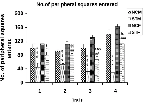
Number of peripheral squares entered during 5 minutes
Stressed rats [both male (STM) and female (STF)] entered significantly less number of
peripheral squares compared to respective control rats [control male (NCM), and control
female (NCF)] in all trial days. Stressed females entered significantly higher number of
peripheral squares compared to stressed male rats in all trials (Table R-1 and Fig. R- 1).
NCM vs STM: ***P<0.001; NCF vs STF: $ P<0.05, $$ P<0.01, $$$ P<0.001; STM vs
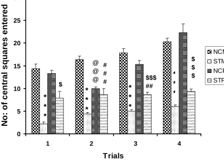
105 Number of central squares entered during 5 minutes
Stressed rats [both male (STM) and female (STF)] entered significantly less number of
central squares compared to respective control rats [control male(NCM),and control
female(NCF)] in all trial days. Stressed females entered significantly higher number of
central squares compared to stressed male rats in all trials (Table R-2 and Fig. R- 2).
NCM vs STM: ***P<0.001; NCF vs STF: $ P<0.05, $$ P<0.01; STM vs STF:## P
<0.01, NCM vs NCF :@@@ P< 0.001
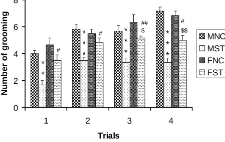
Number of grooming events during 5 minutes
Stressed rats [both male (STM) and female (STF)] showed less number of grooming
compared to respective control rats [control male(NCM),and control female(NCF)] in all
trial days. Stressed females showed significantly higher number of grooming events
compared to stressed male rats in all trials (Table R-3 and Fig. R- 3).NCM vs STM:,
**P<0.01***P<0.001; NCF vs STF: $ P<0.05, $$ P<0.01; STM vs STF: # P<0.05,## P
<0.01
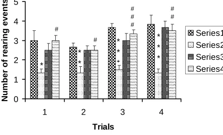
Number of rearing events during 5 minutes
Stressed male rats (STM) showed significantly less number of rearing compared to
respective control rats,control male(NCM) in all trial days. Stressed females showed
significantly more number of rearing compared to stressed male rats (Table R-4 and Fig.
R- 4). NCM vs STM: *P<0.05;**P<0.01, ***P<0.001 ; STM vs STF: # P<0.05,
Pharmacologyonline 2: 99-122 (2010) Cherian et al.
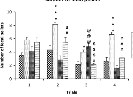
Defecation scores during 5 minutes
Stressed rats [both male (STM) and female (STF)] showed significantly more number of
fecal pellets compared to respective control rats[control male(NCM),and control
female(NCF)] in all trial days. Stressed females showed significantly lower number of
fecal pellets compared to stressed male rats (Table R-5 and Fig. R- 5). NCM vs STM:
***P<0.001, *P<0.05 ; NCF vs STF: $ P<0.05,$$$ P<0.001; STM vs STF: # P<0.05,
107
No.of peripheral squares entered
0
40
80
120
160
200
1
2
3
4
Trails
No.
of
per
ipher
al
squar
es
ent
e
re
d
NCM
STM
NCF
STF
*
*
*
*
*
*
*
*
*
$$ ###*
*
*
$ # $$ ## $$$ ##Figure. R- 1 : Number of peripheral squares entered by the control (n=6) and stressed (n=6) rats in
the open field test. Note that stressed rats [both male (STM) and female (STF)] entered significantly
less number of peripheral squares compared to respective control rats [control male (NCM), and
control female (NCF)] in all trial days. Stressed females entered significantly higher number of
peripheral squares compared to stressed male rats in all trials, though there was no significant
difference between control male and control female. NCM vs STM: ***P<0.001; NCF vs STF: $
P<0.05, $$ P<0.01, $$$ P<0.001; STM vs STF: # P<0.05,## P <0.01, ### P <0.001( One way ANOVA,
Pharmacologyonline 2: 99-122 (2010) Cherian et al.
Number of peripheral squares entered
Trial Day
Groups Day 1 Day 2 Day 3 Day 4
NCM 100.5±11.90 92.15±2.54 101.0±10.80 140.7±13.83
STM 44.50 ± 2.46
***
53.00±7.39***
32.33±7.79***
29.17±7.54***
NCF 112.7±2.20 112.0±3.20 130.3±7.01 161.7±16.00
STF 79.00±9.49$,# 79.33±5.48$$,## 68.00±8.06$$$,## 113.2±4.39$$,###
F value 14.72 24.13 24.51 25.85
Anova Significance
P<0.0001 P<0.0001 P<0.0001 P<0.0001
109
No: of central squares entered
0 5 10 15 20 25 30
1 2 3 4
Trials
N
o
: o
f cen
tr
al
sq
u
a
res en
te
re
d
NCM STM NCF STF $*
*
*
@ @ @ # # #*
*
*
$$$ ##*
*
*
$ $ $*
*
*
Figure R- 2: Number of central squares entered by the control (n=6) and stressed
(n=6) rats in the open field test. Note that stressed rats [both male (STM) and female
(STF)] entered significantly less number of central squares compared to respective
control rats [control male(NCM),and control female(NCF)] in all trial days. Stressed
females entered significantly higher number of central squares compared to stressed
male rats in all trials. NCM vs STM: ***P<0.001; NCF vs STF: $ P<0.05, $$ P<0.01;
STM vs STF:## P <0.01, NCM vs NCF :@@@ P< 0.001 ( One way ANOVA,
Pharmacologyonline 2: 99-122 (2010) Cherian et al.
Number of central squares entered
Trial Day
Groups Day 1 Day 2 Day 3 Day 4
NCM 14.33±1.03 16.33±0.84 17.83±0.94 20.33±0.71
STM 2.16 ± 0.47
***
4.33±0.42***
5±0.36***
6±0.36***
NCF 13.33±0.66 10±0.36@@@ 15.33±0.88 22.33±1.9
STF 7.83±1.53$ 8.66±1.33### 8.66±0.55$$$,### 9.33±0.55###
F value 15.88 35.19 66.08 56.33
Anova Significance
P<0.0001 P<0.0001 P<0.0001 P<0.0001
111
Number of grooming
0
2
4
6
8
1
2
3
4
Trials
N
u
mbe
r of
gr
ooming
MNC
MST
FNC
FST
*
*
#*
*
*
#*
*
*
$ ##*
*
*
$$ #Figure R- 3: Number of grooming events by the control (n=6) and stressed (n=6) rats in the
open field test. Note that stressed rats [both male (STM) and female (STF)] showed less
number of grooming compared to respective control rats[control male(NCM),and control
female(NCF)] in all trial days. Stressed females showed significantly higher number of
grooming events compared to stressed male rats in all trials, though there was no significant
difference between control male and control female. NCM vs STM:, **P<0.01***P<0.001;
NCF vs STF: $ P<0.05, $$ P<0.01; STM vs STF: # P<0.05,## P <0.01 ( One way ANOVA,
Pharmacologyonline 2: 99-122 (2010) Cherian et al.
Number of grooming events
Trial Day
Groups Day 1 Day 2 Day 3 Day 4
NCM 4±0.25 5.83±0.3 5.66±0.42 7.16±0.30
STM 1.66± 0.33
**
3.5±0.22***
3.33±0.33***
3.33±0.33**
NCF 4.66±0.49 5.5±0.22 6.33±0.33 6.83±0.4
STF 3.5±0.42# 4.83±0.3 # 5.16±0.3## 5±0.36$,##
F value 10.93 14.74 13.39 25.29
Anova Significance
P<0.001 P<0.001 P<0.001 P<0.001
113
Number of rearing events
0
1
2
3
4
5
1
2
3
4
Trials
N
u
mb
er
o
f r
ear
in
g event
s
Series1
Series2
Series3
Series4
*
#
*
*
#
*
*
*
#
#
#
*
*
*
#
#
Figure R-4: Number of rearing events by the control (n=6) and stressed (n=6) rats
in the open field test. Note that stressed male rats (STM) showed significantly less
number of rearing compared to respective control rats[control male(NCM) in all
trial days. Stressed females showed significantly more number of rearing
compared to stressed male rats. NCM vs STM: *P<0.05;**P<0.01, ***P<0.001 ;
STM vs STF: # P<0.05, P<0.05, ### P <0.001 (One way ANOVA, Bonferroni’s
Pharmacologyonline 2: 99-122 (2010) Cherian et al.
Number of rearing events
Trial Day
Groups Day 1 Day 2 Day 3 Day 4
NCM 3.0±0.51 4.33±0.66 2.16±0.40 2.33±0.55
STM 1.33± 0.21 8.16±0.30
***
4.0±0.36
*
6.66±0.33***
NCF 2.5±0.34 2.83±0.60 4.83±0.47@@@ 1.66±0.47
STF 3.0±0.25 5.5±0.67$,# 2.16±0.16$$$,#
3.16±0.40###
F value 5.00 15.05 13.11 38.73
Anova Significance
P<0.01 P<0.0001 P<0.0001 P<0.0001
115
Number of fecal pellets
0
2
4
6
8
10
1
2
3
4
Trials
N
u
m
b
e
r o
f fe
c
a
l p
e
lle
ts
MNC
MST
FNC
FST
*
*
*
$
#
@
@
@
*
$
$
$
#
*
*
*
#
#
#
Figure R-5: Defecation scoresby the control (n=6) and stressed (n=6) rats in the
open field test. Note that stressed rats [both male (STM) and female (STF)]
showed significantly more number of fecal pellets compared to respective
control rats[control male(NCM),and control female(NCF)] in all trial days.
Stressed females showed significantly lower number of fecal pellets compared to
stressed male rats . NCM vs STM: ***P<0.001, *P<0.05 ; NCF vs STF: $
P<0.05,$$$ P<0.001; STM vs STF: # P<0.05, ### P <0.001( One way ANOVA,
Pharmacologyonline 2: 99-122 (2010) Cherian et al.
Number of fecal pellets
Trial Day
Groups Day 1 Day 2 Day 3 Day 4
NCM 3.5±0.42 4.33±0.66 2.16±0.40 2.33±0.55
STM 5.8± 0.4 8.16±0.30
***
4.0±0.36
*
6.66±0.33***
NCF 4.16±0.70 2.83±0.60 4.83±0.47@@@ 1.66±0.47
STF 5.5±0.76 5.5±0.67$,# 2.16±0.16$$$,# 3.16±0.40###
F value 3.41 15.05 13.11 38.73
Anova Significance
P<0.05 P<0.0001 P<0.0001 P<0.0001
117
Discussion
The results of the present study revealed that prenatal stress affected the
locomotor, exploratory and emotional reactivity in both male and female young adult
rats. There is significantly decreased exploration in the peripheral and central squares by
the stressed male and female with reduced grooming and rearing effects and more
number of fecal pellets .But when the stressed males were compared with the stressed
females, it was observed that the male rats exhibited higher anxiety levels with less
exploration and more number of fecal pellets when compared to the stressed females.
The method of stress used in the present study is one of the well known methods
of stress(19,22). Different methods of stress procedures have been used such as forced
immersion in cold water(24), social stress by exposing the rats to cat (25,26), electric foot
shock(27,12). Stress by restrainer method used in the present study is convenient and
animals will not suffocate, but at the same time stress them. Stress by this method is
known to increase the adrenal gland weight and glucocorticoid hormone level(19,22).
Open field activity is one of the behavioral assays for emotionality. Emotional states are
also accompanied by various vegetative phenomena (acceleration of heart rate, dilatation
of pupils, etc.) An autonomic function, which can be conveniently evaluated together
with the activity measurement, is defecation (28). Those animals which ambulate less and
defecate more in the open field situation are considered more emotional than animals
with high ambulation and low defecation scores (23). The results of our study showed
that stressed rats [both male (STM) and female (STF)] were more emotional as they
Pharmacologyonline 2: 99-122 (2010) Cherian et al.
Gestational stress increases circulating maternal hormones that produces changes in
behavior (29). Adult rats exposed to excess endogenous glucocorticoids in utero display decreased grooming and rearing in an open field and increased immobility in a forced
swim test (30). It has been shown that HPA axis of prenatally stressed rats is
dysregulated which is manifested by increase plasmacorticosterone in response to stress
(31). Dysregulation of the HPA axis and abnormal behaviour in response to a stressful
situation is a prominent feature of patients with mental illness. Chronic elevation of
plasmacorticosterone can impair the negative feedback regulation of the HPA axis by
reducing the number of mineralocorticoid (MR) and glucocorticoid (GR) receptors in the
hippocampus and other brain regions (32). High levels of corticosterone also facilitate
conditioned fear- induced freezing behavior by increasing corticotropin –releasing
hormone (CRH) gene expression in the central nucleus of amygdala and in the bed
nucleus of stria terminalis (33). These brain regions were known to be involved in the
generation of fear – related behavior (34) It is therefore possible that exposure of the
developing fetal brain to higher than normal concentrations of glucocorticoid at a critical
period during development can sensitise it to fear- inducing stimuli at adulthood which
would be the possible mechanism underlying our findings.
Increased anxiety-related behaviour following prenatal stress has been reported in rats
and primates(30,6). Anxiety, shows sex-dependent changes following chronic prenatal
119 masculinized behavior of the STF (stressed female) rats when compared to their NCF
(normal control female counterparts). Prenatal stress influenced this measure of anxiety
in a sex- specific manner. This PS influence on anxiety is similar to previous reports(5).
PS appears to have masculinized the female performance and this finding would be
consistent with others who have observed PS- induced masculinization of the female
offspring (36,37). Changing levels of estradiol in the sexes over the lifespan appear to
contribute to the differences in response to stress (35). It is also becoming increasingly
clear that maternal stress during pregnancy can influence childhood behaviour (36).
Conclusion
Our results reinforce the hypothesis that many psychopathological affections have their
origin in early developmental influences. More generally, they show the heuristic value
of accurate animal models to better understand the mechanism by which early stress and
epigenetic risk factors promote anxiety disorder and depression in children and that these
effects are gender- specific, thus revealing the decisive importance of nine months of
pregnancy for the rest of the child’s life and that of the adult it will become.
Pharmacologyonline 2: 99-122 (2010) Cherian et al.
References
1. Firth EC, Rogers CW, Vickers M, Kenyon PR, Jenkinson CM, Blair HT, Johnson PL, Mackenzie DD, Peterson SW, Morris ST. The bone-muscle ratio of fetal lambs is affected more by maternal nutrition during pregnancy than by maternal size. Am J Physiol Regul Integr Comp Physiol. 2008 ;294(6):1890-4.
2. Gilbert ME, Kelly ME, Samsam TE and Goodman J. H. Chronic developmental lead exposure reduces neurogenesis in adult rat hippocampus but does not impair spatial learning. Toxicol Sci. 2005; 86(2):211-3.
3. Wormser U, Izrael M, Van der Zee, Brodsky B,Yanai J . A chick model for the mechanism of mustard gas neurobehavioral teratogenecity. Neurotoxicol Teratol. 2005; 27(1): 65-71.
4. Igosheva N, Klimova O, Anischchenko T, Glover V. Prenatal stress alters cardiovascular responses in adult rats .J. Physiol. 2003; 557(1):273.
5. Bowman RE, MacLusky RJ, Sarmiento Y, Frankfurt M, Gordon M, Luine V.N. Sexually dimorphic effects of prenatal stress on cognition, hormonal responses and central neurotransmitters. Endocrinol . 2004;145 (8): 3778-3787
6. Coe CL, Kramer M, Czeh B, Gould E, Reeves AJ, Kirschbaum C & Fuchs E. Prenatal stress diminishes neurogenesis in the dentate gyrus of juvenile rhesus monkeys. Biol Psychiatry. 2003 ; 54: 1025–1034.
7.
Hellemans KG, Sliwowska JH, Verma P, Weinberg J. Prenatal alcohol exposure: fetal programming and later life vulnerability to stress, depression and anxiety disorders. Neurosci Biobehav Rev. 2010 ;34(6):791-8078. Kohman RA, Tarr AJ, Day CE, McLinden KA & Boehm GW. Influence of prenatal stress on behavioral, endocrine, and cytokine responses to adulthood bacterial endotoxin exposure. Behav Brain Res.2008;193(2): 257.
9. Son GH, Geum D, Chung S, Kim EJ, Jo JH, Kim CM, Lee KH, Kim H, Choi S, Kim HT, Lee CJ & Kim K. Maternal stress produces learning deficits associated with impairment of NMDA receptor-mediated synaptic plasticity. J Neurosci,2006;26(12): 3309.
10. Darnaudéry M, Perez-Martin M, Bélizaire G, Maccari S & Garcia-Segura LM , Insulin-like growth factor 1 reduces age-related disorders induced by prenatal stress in female rats. Neurobiol Aging. 2006;27:(1):119.
11. Wu J, Song TB, Li YJ, He KS, Ge L & Wang LR, Prenatal restraint stress impairs learning and memory and hippocampal PKCbeta1 expression and translocation in offspring rats. Brain Res.2007; 1141:205.
121 14. Baker S, Chebli M, Rees S, Lemarec N, Godbout R & Bielajew C, Effects of gestational stress: 1 Evaluation of maternal and juvenile offspring behavior, Brain Res. 2008; 1213: 98.
15. Pallarés ME, Scacchi Bernasconi PA, Feleder C & Cutrera RA. Effects of prenatal stress on motor performance and anxiety behavior in Swiss mice. Physiol Behav.2007; 92(5): 951.
16. Abe H, Hidaka N, Kawagoe C, Odagiri K, Watanabe Y, Ikeda T, Ishizuka Y, Hashiguchi H, Takeda R, Nishimori T & Ishida Y. Prenatal psychological stress causes higher emotionality, depression-like behavior, and elevated activity in the hypothalamo-pituitary-adrenal axis. Neurosci Res.2007; 59(2):145.
17. Li H, Li X, Jia N, Cai Q, Bai Z, Chen R, Song T, Zhu Z & Liu J, NF-kappaB regulates prenatal stress-induced cognitive impairment in offspring rats, Behav Neurosci , 122:2 (2008) 331.
18. Mueller BR & Bale TL. Early prenatal stress impact on coping strategies and learning performance is sex dependent.Physiol Behav.2007;91(1): 55.
19. Gué M, Bravard A, Meunier J, Veyrier R, Gaillet S, Recasens M & Maurice T, Sex differences in learning deficits induced by prenatal stress in juvenile rats. Behav Brain Res.2004; 150 (1-2): 149.
20. Lesage J, Del- Favero F, Leonhardt M. Prenatal stress induces intrauterine growth restriction and programmes glucose intolerance and feeding behavior disturbances in the aged rat. J Endocrinol.2004; 181:291.
21. Igosheva N, Klimova O, Anischchenko T, Glover V. Prenatal stress alters cardiovascular responses in adult rats .J. Physiol. 2003; 557(1):273.
22. Sunanda, Rao M.S, Raju T.R. Effect of chronic restraint stress on dendritic spines and excrescences of hippocampal CA3 pyramidal neurons- a quantitative study. Brain res.1995;694: 312.
23. Bures J. Buresova O. Huston J P. Techniques and basic experiments for the study of brain and behavior .2nd ed.Amsterdam- Newyork :Elsevier,1983:95-97, 148-160.
24. Drago F, Di Leo F, & Giardina L. Prenatal stress induces body weight deficit and behavioural alterations in rats: the effect of diazepam. Eur Neuropsychopharmacol.1999; 9: 239.
25. Lordi B, Protais P, Mellier D & Caston J. Acute stress in pregnant rats: effects on growth rate, learning, and memory capabilities of the offspring. Physiol Behav.1997;62(5):1087.
26. Patin V, Lordi B, Vincent A & Caston J. Effects of prenatal stress on anxiety and social interactions in adult rats. Brain Res Dev Brain Res.2005;160; 2:265.
27. Estanislau C & Morato S . Prenatal stress produces more behavioral alterations than maternal separation in the elevated plus-maze and in the elevated T-maze. Behav Brain Res,2005;163(1):70.
Pharmacologyonline 2: 99-122 (2010) Cherian et al.
29. Weinstock M. Can the behaviour abnormalities induced by gestational stress in rats be prevented or reversed? Stress. 2002;5(3):167-76.
30. Welberg LA, Seckl JR ,Holmes MC. Inhibition of 11beta-hydroxysteroid dehydrogenase, the foeto-placental barrier to maternal glucocorticoids, permanently programs amygdala GR mRNA expression and anxiety-like behaviour in the offspring. Eur J Neurosci.2002; 12: 1047–1054.
31. Kapoor A, Matthews SG. Short periods of prenatal stress affect growth, behaviour and hypothalamo–pituitary–adrenal axis activity in male guinea pig offspring. The Journal of Physiology. 2005; 566: 967-977.
32. Jacobsen L and Sapolsky R. ‘’The role of hippocampus in the feedback regulation of the hypothalamic-pituitary-adrenocortical axis”. Endocrinol. Rev. 1991;12:118-134
33
.
Schulkin J, Gold PW and Mc Ewen BS. Induction of corticotrophin releasing hormone gene expression by glucocorticoids : implication for understanding the states of fear and anxiety and allostatic load. Psychoneuroendocrinology. 1998; 23: 219-24334. Le Doux, J.E.” Emotion: clues from the brain”, Annu Rev. Psychol. 1995; 46: 209-235
35. Luine VN, Beck KD, Bowman RE, Frankfurt M, Maclusky NJ. Chronic stress and neural function: accounting for sex and age. J Neuroendocrinol.2007;19(10):743-51
36. O'Connor TG, Heron J, Golding J & Glover V .Maternal antenatal anxiety and behavioural/emotional problems in children: a test of a programming hypothesis. J Child Psychol Psychiatry, 2003; 44; 1025–1036.
37. Sachser N, Kaiser S. Prenatal social stress masculinizes the females behavior in guinea pigs . Physiol Behav . 1996; 60:589-594