Nitric Oxide, Carbonyl Protein, Lipid
Peroxidation and Correlation Between
Antioxidant Vitamins in Different
Categories of Pulmonary and Extra
Pulmonary Tuberculosis
Shubhangi Mahesh Dalvi1, Vinayak Wamanrao Patil1,
Nagsen Nirgun RamRaje2, Jaising Marutrao PhaDtaRe3, Sarita Uday GujaRathi4
1 Department of Biochemistry Grant Government Medical College and Sir J.J.
Group of Hospitals Byculla Mumbai 400008, India
2 Pulmonary Medicine Department, Grant Government Medical College and
Sir J.J. Group of Hospitals Byculla Mumbai 400008, India
3 Pulmonary Medicine Department, Gokuldas Tejpal Government Hospital
Mumbai 400001, India
4 Employment State Insurance Scheme Hospital Mohan Nagar Chinchwad
Pune 411035, India
Submitted:21 May 2012 Accepted:18 Sep 2012
Abstract
Background: Lipid peroxidation, nitric oxide, carbonyl protein, causing production of reactive oxygen and reactive nitrogen intermediates that lead to oxidative, nitrosative stress. The stress is found to cause deterioration in the cellular function, mutagenesis, and DNA damage. The oxidative stress is correlated with the antioxidant vitamins status.
Methods: Newly diagnosed cultured positive sputum pulmonary category I, II, III (n = 100
each), extra pulmonary category I (n = 35) before and after directly observed short course treatment
of six months vitamins, by HPLC.
Results: Oxidative parameter levels were significantly increased, and activities of vitamins were found to be significantly decreased in subjects of all categories of pulmonary and extra pulmonary tuberculosis. Positive correlations between nitric oxide, carbonyl protein, and lipid peroxidation were seen among them. Negative correlations between nitric oxide, carbonyl protein, lipid peroxidation with vitamin E, C, A were seen in tuberculosis (two sided P < 0.01).
Conclusion: Increase oxidative stress and nitrosative stress, leading to protein carbonyl formation in tuberculosis. The increased protein carbonyl, hampers many important functions of proteins. The changes were reversed after six months of antitubercular treatment in patients with good recovery but increase stress was not completely reversed.
Keywords: carbonyl protein, lipid peroxidation, Nitric oxide, tuberculosis, vitamin E, C, A
Introduction
Tuberculosis pandemics increase morbidity, mortality, and the frequency of opportunistic infections. The infection is established in the alveolar macrophages of the distal alveoli (1).
Oxygen free radical species are generated in the normal course of a variety of essential biochemical reactions that progress at the sub-cellular organelle level. Peroxidative damage to the membranes manifests itself as loss in membrane fluidity, increased fragility of biomembranes, loss of membrane secretory functions, and
breakdown of the transmembrane ionic gradient. The hydroxyl radical is the most damaging oxygen free radical species, and can initiate the lipid peroxidation phenomenon. If not checked, the lipid peroxidation reaction is a self-perpetuating vicious cycle, as the lipid peroxyl radical, in an attempt to stabilise itself, attracts hydrogen atoms from the adjacent polyunsaturated fatty acids (2).
The lipid peroxidation metabolites can have an adverse effect on immune responses. Hence, high levels of polyunsaturated fatty acids in the diet are immuno-depressive, and also, the higher the level of the acids, the greater is the lipid peroxidation. Since prostaglandin and leukotriene synthesis is connected with free radical generation, the antioxidants can affect the concentration of these immuno-modulators, such as prostaglandin E2 (3).
Upon engulfment by macrophages,
Mycobacterium tuberculosis inhibits phagosome maturation and phagolysosome fusion. However, the pathogen remains enclosed in these phagocytic vacuoles, where it survives and multiplies despite the hostile surrounding environment of phagosomes. Production of reactive oxygen and reactive nitrogen intermediates is a major response of macrophages to inflammatory stimuli or infection (4). Reactive oxygen and reactive nitrogen intermediates include highly toxic molecules such as hydrogen peroxide, nitric oxide, and peroxynitrite (5); this results in deterioration of cellular function. Normally, these chemically reactive molecules are produced in the cell at basal levels as a result of metabolic processes, but they are contained and neutralised by different antioxidant systems existing in every cell. However, upon infection, reactive oxygen and reactive nitrogen intermediates are produced at high concentrations, and they disrupt cellular homeostasis by damaging numerous components of the cell, including lipids, nucleic acids, proteins, and metal cofactors leading to mutagenesis, necrosis, and apoptosis (4,5).
The beneficial effect of vitamin E in the immunological response is due precisely to the protection against immuno-suppression produced by the pro-oxidant reactions in the cell membrane. It should also be pointed out that when macrophages are subjected to oxidative stress, their vitamin E content is reduced considerably (6). The fat-soluble vitamin retinol is crucial for immune defence, enhancing white blood cell function and resistance to infection and carcinogens (7). Vitamin C can enhance the body’s resistance to an assortment of diseases, including infectious disorders, and many types of cancer (8). The present work was undertaken to study the correlation among the extent of oxidative stress, antioxidant vitamin status, lipid peroxidation, nitric oxide, and carbonyl protein in tuberculosis patients in different categories, with and without directly observed short-course treatment, in people of lower socio-economic status.
Materials and Methods
The study was undertaken in subjects between 16 and 60 years of age, of lower socio-economic status, from different categories of pulmonary and extra-pulmonary tuberculosis, diagnosed by newly cultured positive sputum pulmonary category I (n = 100; 50 male and 50 female). Extra-pulmonary patient category (n = 35; 19 males and 16 females), before and after treatment of six months, untreated category II (n = 100), untreated category III (n = 100), and normal control subjects (n = 100), were selected from the pulmonary tuberculosis and chest diseases department, outpatient department, and inpatient department of Sir J.J. Group of Hospitals, Gokuldas Tejpal Government Hospital, and the Municipal Corporation Group of Tuberculosis Hospitals in Shewri Mumbai Maharashtra, India. Approval of the medical ethical committees of the institutes was obtained for the study (No – IEC/Pharm/379/07, dated 30/8/2007). Informed consent was taken from the subjects. Statistical analysis using software Mini-tab 16 for student’s t test, ‘correlation’ was significantly different from zero (two-sided
P < 0.01).
Patients with chronic diseases, hepatitis, diabetes, renal impairment, cardiovascular co-morbidities, neurological psychiatric disorders, human immuno-deficiency virus infection, various malignancies, as well as heavy smokers, alcoholics, and tobacco-chewers were excluded from the study. Lipid peroxidation, nitric oxide, carbonyl protein, and vitamin A, E, and C parameters were estimated using UV-spectrophotometry (Jasco-V670), HPLC (Jasco – 2075+/ PU2089+).
chromogen was centrifuged and the intensity of the colour developed in the supernatant was measured spectrophotometrically at 535 nm. Malondialdehyde levels were expressed in nmol/mL:
Malondialdehyde
(nmol/mL) =
Effective volume of serum used × Abs. at 535 nm
0.156
Nitric oxides (10) by Najwa K Cortas, nitrates in serum were assayed by a modification of the cadmium-reduction method; the nitrite produced was determined by diasotisation of sulfanilamide and coupling to naphthylethylene diamine. Samples were deproteinised with Somogyi reagent, and the nitrate was reduced by Cu-coated Cd in glycine, buffer at pH 9.7 with pseudo-first-order reaction kinetics. Time interval for the assay was 75 min to 90 min. The colour formed by nitrite diazotization was measured at 540 nm, carbonyl protein (11); carbonyl groups in protein reacted with 2, 4 – dinitrophenylhydrazine to form 2, 4 – dinitrophenylhydrazone, which was estimated spectrophotometrically at 360–390 nm.
Protein carbonyl conc. in nm/mg =
Carbonyl conc. in nmol/mL (Protein conc. in mg/mL)
Vitamin A and E (12) ethyl alcohol was added to plasma (1:1; v/v), heptane, and internal
standard 50 µL of 0.2 mM (10 nmol) and 25 µL of 0.6 mM (15 nmol), centrifuged using the top layer for HPLC. Column – C18, 5 μ (micron), 4.6 mM × 250 mm (Hypersilgold), 1.0mL/min, 280 nm, mobile phase-acetonitrile, methanol, and ethyl acetate in a ratio of 88:10:2 respectively, (degassed). The result was then prepared with ethanol for vitamin A, E at 20 mg% and 100 mg% respectively of standard solution (Figure 1a).
Sample
concentration =
Peak area sample × standard concentration × Factor Peak area internal standard (internal standard) in the sample
Factor calculation
F =
Peak area internal standard (internal concentration) of the standard-mixture Peak area of the standard-mixture (STD)
Vitamin C (12) ethyl alcohol was added to plasma (1:1; v/v), and internal standard 50 mg% with dilution 1:25, the tubes were vortex incubated for 10 min at 2–8 °C and then centrifuged at × 10.000 g for 10 min. 20 µL of the supernatant was injected into column C18, 5 μ (micron), 4.6mM × 250 mM, 0.5 mL/min, 245 nm, buffer –2.3 gm% of ammonium dihydrogen phosphate, pH7.0 with ortho-phosphoric acid, mobile phase-buffer and acetonitrile in a ratio of 9:1 (Figure 1b).
Sample
Concentration =
Peak area sample × Concentration of standard
Peak area standard
Results
In the present study, there is a significant increase in malondialdehyde, nitric oxide, and protein carbonyl. The highest levels were found
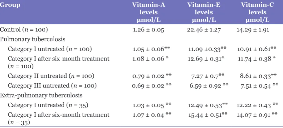
to be in subjects in category III of pulmonary tuberculosis (Table 1). Significant reductions in vitamin E, C, and A (Table 2) status show increased protective effects (13–17). There is a positive correlation between malondialdehyde, nitric oxide, and protein carbonyl among them (Table 3) (Figure 2, 3, and 4). There is a negative correlation between malondialdehyde (18–25), nitric oxide, and protein carbonyl markers with vitamins E, C, and A (Table 4, 5, and 6) (Figure 5).
Table 1: Malondialdhyde, nitric oxide, and carbonyl protein in different types of tuberculosis
Group Malondialdhyde
levels nmol/mL
Nitric oxide
µmol/L carbonylProtein
nmol/mg
Control (n = 100) 2.46 ± 0.13 35.16 ± 1.04 4.37 ± 0.15
Pulmonary tuberculosis
Category I untreated (n = 100) 5.46 ± 0.33** 58.67 ± 3.63** 6.65 ± 0.22** Category I after six-month treatment
(n = 100) 3.43 ± 0.35* 45.73 ± 1.29* 5.43 ± 0.25*
Category II untreated (n = 100) 6.52 ± 0.27** 59.80 ± 3.4** 6.87 ± 0.19** Category III untreated (n = 100) 8.61 ± 0.61** 70.48 ± 2.63** 7.08 ± 0.21** Extra-pulmonary tuberculosis
Category I untreated (n = 35) 5.46 ± 0.33** 58.67 ± 3.63** 6.65 ± 0.22** Category I after six-month treatment
(n = 35) 3.43 ± 0.35** 45.73 ± 1.29** 5.43 ± 0.25**
** P ≤ 0.001 – Highly significant.
* P ≤ 0.05 – Significant; values shown are Mean ± SD.
Table 2: Vitamins in different types of tuberculosis
Group Vitamin-A
levels µmol/L
Vitamin-E levels µmol/L
Vitamin-C levels µmol/L
Control (n = 100) 1.26 ± 0.05 22.46 ± 1.27 14.29 ± 1.91
Pulmonary tuberculosis
Category I untreated (n = 100) 1.05 ± 0.06** 11.09 ±0.33** 10.91 ± 0.61** Category I after six-month treatment
(n = 100) 1.08 ± 0.06 * 12.69 ± 0.31* 11.74 ± 0.38 *
Category II untreated (n = 100) 0.79 ± 0.02 ** 7.27 ± 0.7** 8.61 ± 0.33** Category III untreated (n = 100) 0.69 ± 0.02 ** 6.59 ± 0.92 ** 7.51 ± 0.54 ** Extra-pulmonary tuberculosis
Category I untreated (n = 35) 1.03 ± 0.05 ** 12.49 ± 0.53** 12.22 ± 0.43 ** Category I after six-month treatment
(n = 35) 1.07 ± 0.04 ** 15.44 ± 0.51** 14.07 ± 0.91 **
** P ≤ 0.001 – Highly significant.
Table 3: Correlation among malondialdehyde, protein carbonyl, and nitric oxide
Group r-values
malondialdehyde/ protein carbonyl
r-values malondialdehyde/
nitric oxide
r-values Nitric oxide/protein
carbonyl
Control (n = 100) 0.971 0.951 0.947
Pulmonary tuberculosis
Category I untreated (n = 100) 0.949** 0.852** 0.894**
Category I after six-month
treatment (n = 100) 0.962* 0.983* 0.974*
Category II untreated (n = 100) 0.926** 0.961** 0.964**
Category III untreated (n = 100) 0.954** 0.893** 0.957** Extra-pulmonary tuberculosis
Category I untreated (n = 35) 0.930** 0.927** 0.982**
Category I after six-month
treatment (n = 35) 0.961* 0.958* 0.964*
* P ≤ 0.05 – Significant. ** P ≤ 0.01 – Highly significant.
Figure 3: (a) Scatter diagram of nitric oxide and vitamin E in category I pulmonary tuberculosis and extra pulmonary untreated. (b) Scatter diagram of nitric oxide and vitamin C in category I pulmonary tuberculosis and extra pulmonary untreated. (c) Scatter diagram of nitric oxide and vitamin A in category I pulmonary tuberculosis and extra pulmonary untreated.
Figure 4: (a) Scatter diagram of Carbonyl protein and vitamin E in category I pulmonary tuberculosis untreated. (b) Scatter diagram of Carbonyl protein and vitamin C in category I pulmonary tuberculosis untreated. (c) Scatter diagram of Carbonyl protein and vitamin A in category I pulmonary tuberculosis untreated.
Table 4: Correlation between malondialdehyde and vitamin A, E, and C
Group r-values
Malondialdhyde/ Vitamin A
r-values Malondialdhyde/
Vitamin E
r-values Malondialdhyde/
Vitamin C
Control (n = 100) –0.923 –0.842 –0.902
Pulmonary tuberculosis
Category I untreated (n = 100) –0.899** –0.940** –0.902** Category I after six-month
treatment (n = 100) –0.927* –0.958* –0.968*
Category II untreated (n = 100) –0.949** –0.832** –0.934** Category III untreated
(n = 100) –0.922** –0.641** –0.941**
Extra-pulmonary tuberculosis
Category I untreated (n = 35) –0.918** –0.958** –0.824** Category I after six-month
treatment (n = 35) –0.942* –0.884* –0.884*
* P ≤ 0.05 – Significant. ** P ≤ 0.01 – Highly significant.
Discussion
Tuberculosis remains one of the top killers among infectious diseases. It is the most feared disease in the world, and spreads from person to person via aerosol distribution (26). In modern medicine, more emphasis is being made on biochemical changes such as oxidative, hyperoxidant, and nitrosative stress due to more liberation of free radicals, leading to increased concentrations of malondialdehyde, nitric oxide products, and protein carbonyl formation (tight relationship). Antioxidant vitamins E, C, and A inhibit and neutralise stress and balance the equilibrium (14,27–33).
Dietary antioxidant vitamin status is poor in tuberculosis patients of lower socio-economic status, and a need was felt to study the correlation of antioxidant and oxidative stress parameters with and without directly-observed short-course treatment.
Figure 5: (a) Scatter diagram of lipid peroxidation (MDA) with carbonyl protein in category I pulmonary tuberculosis untreated. (b) Scatter diagram of lipid peroxidation (MDA) with nitric oxide in category I pulmonary tuberculosis untreated. (c) Scatter diagram of nitric oxide with carbonyl protein in category I pulmonary tuberculosis untreated.
Table 5: Correlation between nitric oxide and vitamin A, E, and C
Group r-values
Nitric oxide/ Vitamin A
r-values Nitric oxide/
Vitamin E
r-values Nitric oxide/
Vitamin C
Control (n = 100) –0.886 –0.881 –0.906
Pulmonary tuberculosis
Category I untreated (n = 100) –0.958** –0.952** –0.941**
Category I after six-month treatment (n = 100) –0.928* –0.966* –0.972* Category II untreated (n = 100) –0.909** –0.811** –0.968** Category III untreated (n = 100) –0.988** –0.587** –0.907** Extra-pulmonary tuberculosis
Category I untreated (n = 35) –0.986** –0.923** –0.882**
Category I after six-month treatment (n = 35) –0.916* –0.862* –0.862* * P ≤ 0.05 – Significant.
** P ≤ 0.01 – Highly significant.
Table 6: Correlation between carbonyl protein and vitamin A, nitric oxide and vitamin E, and nitric oxide and vitamin C
Group r-values
Carbonyl protein/ Vitamin A
r-values Carbonyl
protein/ Vitamin E
r-values Carbonyl
protein/ Vitamin C
Control (n = 100) –0.938 –0.859 –0.929
Pulmonary tuberculosis
Category I untreated (n = 100) –0.951** –0.951** –0.970** Category I after six-month treatment (n = 100) –0.962* –0.981* –0.978* Category II untreated (n = 100) –0.954** –0.841** –0.948** Category III untreated (n = 100) –0.948** –0.516** –0.833** Extra-pulmonary tuberculosis
Category I untreated (n = 35) –0.974** –0.937** –0.911**
Category I after six-month treatment
(n = 35) –0.949* –0.903* –0.903*
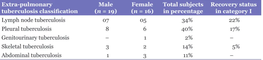
Table 7: Comparative statement of recovery in extra-pulmonary category I tuberculosis
Extra-pulmonary
tuberculosis classification (nMale = 19) (Femalen = 16) Total subjects in percentage Recovery status in category I
Lymph node tuberculosis 07 05 34% 22%
Pleural tuberculosis 8 6 40% 17%
Genitourinary tuberculosis – 1 2% –
Skeletal tuberculosis 3 2 14% 5%
Abdominal tuberculosis 1 3 11% –
Mycobacterium tuberculosis in a natural way, by causing lipid peroxidation in the polyunsaturated fatty acids of the phospholipids, present in the cell membrane of the pathogenic microorganism.
During directly observed short-course treatment in category I (pulmonary and extra-pulmonary), severity of the disease was decreased; hence there was a decrease in oxidative stress parameters (lipid peroxidation, nitric oxide, and carbonyl protein). During the treatment, there was a progressive decrease in the bacillary load, because of which the oxidative stress seemed to be reduced.
Nitric oxide reversibly inhibits enzymes containing transition metals or free radical intermediates in their catalytic cycle. Catalase, cytochrome P450 and cytochrome oxidase, ribonucleotide reductase, are important enzymes for the synthesis of DNA. Inhibition of DNA synthesis by nitric oxide inhibits viral replication. Oxygen and its derivatives hydrogen peroxide, singlet oxygen, hydroxyl radical, nitrite, and peroxynitrite radicals have a cytotoxic and genotoxic effect, and could be the primary sources of oxidative damage to a number of tissues and organs in tuberculosis. The toxic effect of the reactive species is neutralised by antioxidant defence.
Nitric oxide causes oxidation of thiols, and has damaging effects on proteins, converting them into protein carbonyls; this hampers many important functions of proteins, and also leads to enhancement of the process of ageing (11). Oxidatively-modified proteins are not repaired and must be removed by proteolytic degradation. A decrease in the efficiency of proteolysis will cause an increase in the cellular content of oxidatively-modified proteins, which has been shown to increase in the disease process (14). Introduction of carbonyl groups in proteins means the formation of carbonyls (aldehydes, ketones) on the side chains of amino acids, e.g., l Lys, Arg, Pro, and Thr, which makes them susceptible to
degradation by proteolytic enzymes, leading to deficiency of the proteins (15,20).
The decreased concentration of serum vitamin A may result from hypozincemia, as zinc is required for vitamin A metabolism (19). The present study has demonstrated statistically significant decreases (P < 0.001) in the levels of vitamin E, C, and A in tuberculosis. Vitamin E assists in restricting the self-perpetuation of the vicious lipid peroxidation phenomenon (25). Vitamin C is the first-line antioxidant defence against free oxygen radicals, functioning as an ‘electron sink’ as it donates its electron to the free radical species, thereby converting it to less harmful forms and hence, preventing the chain reaction of lipid peroxidation (12,19).
Extra-pulmonary tuberculosis subjects with lymph node involvement (aspirated whitish material), who followed a proper category regime with a nutritious diet showed better recovery as compared to those with pleural effusion, skeletal, and abdominal involvement. Subjects with abdominal tuberculosis showed poor recovery and were entered in category II (Table 7). The recovery of subjects with category I tuberculosis was totally dependent on proper treatment and nutritional status irrespective of age and sex (14,34–36).
Conclusion
There is increasing oxidative stress, nitrosative stress, and lipid peroxidation, leading to protein carbonyl formation from category I to category III, in tuberculosis in untreated subjects. With directly observed short-course treatment in category I, good recovery from increased stress was found.
Equilibrium between free radical generation and quenching mechanisms is the characteristic of health; if this equilibrium is disturbed, free radicals increase. After six months of directly observed short-course treatment in tuberculosis patients; if equilibrium is not maintained, this leads to a shift of disease classification to category II, and III. An in-depth gene expression study at the molecular level in terms of lipid peroxidation, nitric oxide, protein carbonyl, and vitamin status with directly observed short-course treatment is needed (37).
Acknowledgement
No financial support provided for this research work.
Conflict of interest
None.
Funds
Not received any funds.
Authors' Contribution
Conception and design, drafting of the article and collection and assembly of data: SMD
Analysis and interpretation of the data: SMD, VWP
Final approval of the article: VWP
Provision of study materials or patient: NNR, JMP Statistical expertise: SUG
Correspondence
Dr Dalvi Shubhangi Mahesh
BSc, MSc Medical Biochemistry (University of Pune) Department of Biochemistry
Grant Government Medical College Byculla Mumbai, 400008, India
Tel: +919-89218 2028 Fax: +022-2373 5599
Email: [email protected]
References
1. Flynn JL, Chan J. Immunology of tuberculosis.
Methods Enzymol. 2001;19:93–129.
2. Meister A. Methods in systems biology. Methods Enzymol. 1995;251:3–7.
3. Meade CJ, Mertin J. The mechanism of immunoinhibition by arachidonic and linoleic acid: effects on the lymphoid and reticulo-endothelial systems. Int Arch Allergy Appl Immunol. 1976;51(1):2–24.
4. Nathan C, Shiloh MU. Reactive oxygen and nitrogen intermediates in the relationship between mammalian hosts and microbial pathogens. Proceedings of the Proc Natl Acad Sci US. 2000;97(16):8841–8848.
5. Gwinn MR, Vallyathan V. Respiratory burst: role in signal transduction in alveolar macrophages. J Toxicol Environ Health. 2006;9(1):27–39.
6. Traber MG, Atkinson J. Vitamin E, antioxidant and nothing more. Free Radical Biology and Medicine. 2007;43(1):4–15.
7. Zeba AN, Sorgho H, Rouamba N, Zongo I, Rouamba J, Guiguemde RT, et al. Major reduction of malaria morbidity with combined vitamin A and zinc supplementation in young children in Burkina Faso: a randomised double blind trial. Nutr J 2008;7(7):7.
8. Combs GF. The Vitamins: Fundamental aspects in nutrition and health. 3rd ed. Burlington (US): Elsevier Academic Press; 2008.
9. Buege JA, Aust SD. Microsomal lipid peroxidation.
Methods Enzymol. 1978;52:302–310.
10. Cortas NK, Wakid NB. Determination of inorganic nitrate in serum and urine by kinetic cadmium reduction method. Clin Chem. 1990;36(8): 1440–1443.
11. Levine RL, Garland D. Determination of carbonyl content in oxidatively modified proteins. Methods Enzymol. 1990;186:464–478.
12. Islam SN, Hossain KJ, Ahsan M. Serum vitamin E, C and A status of the drug addicts undergoing detoxification: influence of drug habit, sexual practice and lifestyle factors. Eur J Clin Nutr. 2001;55(11):1022–1027.
13. Turanlahti ME, Lassus P, Andersson S. Nitric oxide and hyperoxia in oxidative lung injury. Acta Paediatr. 2000;89(8):966–970.
14. Kulkarni A, Madrasi NA. Relationship of nitric oxide and protein carbonyl in tuberculosis. Indian J Tuberc. 2008;55(3):138–144.
15. Mohod K, Dhok A, Kumar S. Status of oxidants and antioxidants in pulmonary tuberculosis with varying bacillary load. J Exp Sci. 2011;2(6):35–37.
16. Khan MA, Tania M, Zhang D, Chen H. Antioxidant enzymes and cancer. Chin J Cancer Res. 2010;22(2):87–92.
17. Reddy YN, Murthy SV, Krishna DR, Prabhakar MC. Role of free radicals and antioxidants in tuberculosis patients. Indian J tuberc. 2004;51(4):213–218.
18. Kulkarni R, Deshpande A, Saxena K, Sinha ARS, Verma M. Role of Tumor necrosis factor alpha, Malondialdehyde & serum Iron in Anemic Tuberculosis Patients. Biomed Res. 2011;22(1): 69–72.
20. Lamsal M, Narayan G. Evaluation of lipid peroxidation product, nitrite and antioxidant levels in newly diagnosed and two months follow-up patients with pulmonary tuberculosis. Southeast Asian J Trop Med Public Health. 2007;38(4):695–703.
21. Singhahi R, Arora D, Singh R. Oxidative Stress And Ascorbic Acid Levels in Cavitary Pulmonary Tuberculosis. J Clin Diagn Res. 2010;4(6): 3437–3441.
22. De Souza TP, Oliveira JR, Pereira B. Physical exercise and oxidative stress. Effect of intense physical exercise on the urinary chemiluminescence and plasmatic malondialdehyde. Rev Bras Med Exporte. 2005;11(1):91–96.
23. DelRio D, Stewart AJ, Pellegrini N. A review of recent studies on malondialdehyde as toxic molecule and biological marker of oxidative stress. Nutr Metab Cardiovasc Dis. 2005;15(4):316–328.
24. Deveci F, Iihan N. Plasma malondialdehyde and serum trace element concentration in patients with active pulmonary tuberculosis. Biol Trace Elem Res.
2003;95(1):29–38.
25. Traber M. The biological activity of vitamin E.
Oregon: The Linus Pauling Institute; 2011.
26. Kaufmann SH. A short history of Robert Koch’s fight against tuberculosis: those who do not remember the past are condemned to repeat it. Tuberculosis. 2003;83(1-3):86–90.
27. Drancourt M, Raoult D. Palaeomicrobiology: current issues and perspectives. Nat Rev Microbiol.
2005;3(1):23–35.
28. Starosta V, Griese M. Protein oxidation by chronic pulmonary disease in children. Pediatr pulmonology. 2006;41(1):67–73.
29. Rai RR, Phadke MS. Plasma oxidant antioxidant status in different respiratory disorder. Indian J Clin Biochem. 2006;21(2):161–164.
30. Madebo T, Lindtjorn B, Pal A. Circulating antioxidants and lipid peroxidation products in untreated tuberculosis patients in Ethiopia. Am J Clin Nutr.
2003;78(1):117-122.
31. Akiibinu MO, Arinola OG. Non–Enzymatic antioxidants and nutritional profiles in newly diagnosed pulmonary tuberculosis patients in Nigeria.
Afr J Biomed Res. 2007;10(2):223–228.
32. Ramesh SK, Amareshwara M. Study of protein oxidation and antioxidants status in pulmonary tuberculosis patients. Int J Pharma and Bio Scis.
2011;2(3):104–109.
33. Avery SV. Molecular targets of oxidative stress.
Biochem J. 2011;434(2):201–210.
34. Srinivasan S, Pragasam V, Jenita X, Kalaiselvi P, Muthu V, Varalakshmi P. Enhanced oxidative stress in urogenital tuberculosis patients—A predisposing factor for renal stone formation amelioration by vitamin E supplementation. Clin Chimica Act.
2004;109 350(1):57–63.
35. Sudha K, Rao AV, Rao SN. Oxidative stress and antioxidants in tubercular meningitis. Indian J Clin Biochem. 2002;17(1):34–41.
36. Yeager ME, Reddy MB. Activation of the unfolded protein response is associated with pulmonary hypertension. Pulmonary Circ. 2012;2(2):229–240.
37. Connolly LE, Edelstein PH, Ramakrishnan L. Why is long-term therapy required to cure tuberculosis?