R E V I E W
Open Access
Glial contributions to neurodegeneration in
tauopathies
Cheryl E. G. Leyns and David M. Holtzman
*Abstract
Tauopathies are a broad set of neurodegenerative dementias characterized by aggregation of the tau protein into filamentous inclusions that can be found in neurons and glial cells. Activated microglia, astrocytes and elevated levels of proinflammatory molecules are also pathological hallmarks that are found in brain regions affected by tau pathology. There has been abundant research in recent years to understand the role of gliosis and neuroinflammation in neurodegenerative diseases, particularly in Alzheimer’s disease (AD) which is the most common form of dementia. AD is a tauopathy characterized by both extracellular amyloid-β plaques in addition to intracellular neurofibrillary tangles and neuropil threads containing aggregated tau protein. Accumulating evidence suggests that neuroinflammation offers a possible mechanistic link between these pathologies. Additionally, there appears to be a role for neuroinflammation in aggravating tau pathology and neurodegeneration in tauopathies featuring tau deposits as the predominant pathological signature. In this review, we survey the literature regarding inflammatory mechanisms that may impact neurodegeneration in AD and related tauopathies. We consider a physical role for microglia in the spread of tau pathology as well as the non-cell autonomous effects of secreted proinflammatory cytokines, specifically interleukin 1 beta, interleukin 6, tumor necrosis factor alpha and complement proteins. These molecules appear to have direct effects on tau pathophysiology and overall neuronal health. They also indirectly impact neuronal homeostasis by altering glial function. We conclude by proposing a complex role for gliosis and neuroinflammation in accelerating the progression of AD and other tauopathies.
Keywords:Tau, Tauopathy, Alzheimer’s disease, Neurodegeneration, Neuroinflammation, Gliosis, Microglia, Astrocyte
Background
Abnormal accumulation of the tau protein into intracellu-lar, fibrillar aggregates is observed across a broad spectrum of neurodegenerative disorders that are collectively referred to as tauopathies. Over twenty-five syndromes are currently classified as a tauopathy, which highlights the heterogeneity of these diseases and their vast impact in the dementia field. Primary tauopathies feature tau deposits as the predominant pathological signature and include pro-gressive supranuclear palsy (PSP), corticobasal degener-ation (CBD), frontotemporal dementia and parkinsonism linked to chromosome 17 (FTDP-17), Pick’s disease (PiD), chronic traumatic encephalopathy (CTE) and argyrophilic grain disease (AGD) [1, 2]. Alzheimer’s disease (AD), the most prevalent cause of dementia, is categorized as a
secondary tauopathy due to the additional presence of
amyloid-β (Aβ) plaques and their hypothesized role in
initiating AD pathogenesis [3]. In addition to toxic pro-tein aggregates, activated astrocytes and microglia as well as elevated proinflammatory markers are other common pathological hallmarks of tauopathies [4, 5].
The notion that neuroinflammatory processes contribute to disease was once provocative, yet recent studies have uncovered multiple mechanisms by which aberrant gliosis causes detrimental neuroinflammation that can influence tau pathology and accelerate neurodegeneration. One hypothesized mechanism postulates that early activation of microglia stimulates the release of cytokines and chemo-kines that in turn co-activate astrocytes [6]. Potentially, chronic glial activation alters tau biology, encouraging tangle formation, and diminishes neuronal fitness [4–6]. Furthermore, glial cells may also be physically contributing to the spread of tau pathology [7]. This review summarizes the literature pertaining to the effects of neuroinflammation
* Correspondence:holtzman@neuro.wustl.edu
Department of Neurology, Washington University, Hope Center for Neurological Disorders, Knight Alzheimer’s Disease Research Center, 660 S. Euclid Ave, St. Louis, MO 63110, USA
and gliosis on tau pathology and vice versa. Both in vitro and in vivo mechanistic studies are discussed alongside evi-dence from neuropathology and neuroimaging reports in AD and non-AD tauopathy patients. We further explore potential mechanisms by which inflammatory processes may alter the neurodegenerative process.
Tau pathobiology
The MAPT gene on chromosome 17q21.31 encodes the tau protein which can be alternatively spliced into six distinct isoforms [8, 9]. These isoforms differ by inclu-sion of either three or four conserved repetitive domains (termed R) as well as the addition of one or two amino-terminal inserts [8]. Aggregates composed of both 3R and 4R tau are seen in disease states, though curiously several tauopathies including PSP, CBD, FTDP-17, and AGD exclusively feature 4R tau deposits. 4R tau does display a higher propensity for aggregation [10] which has led some to postulate that it is more pathogenic [11–13]. A recent study reported significantly higher levels of aggregated hyperphosphorylated tau (p-tau) and less-soluble tau species after using anti-sense oligonucle-otides to increase the ratio of 4R:3R tau in mice express-ing human tau under the endogenous promoter [12]. While this data supports 4R tau pathogenicity, other factors still contribute to 3R tau aggregation which is seen exclusively in PiD and in the mixed 3R and 4R tau inclusions in AD and CTE [1, 2].
In a healthy brain, tau is predominantly localized in mature neuronal axons and primarily functions to pro-mote microtubule assembly and stability as well as vesicle and organelle transport along microtubules [14–16]. Phos-phorylation of serine and threonine residues flanking the microtubule binding domain of tau regulate its interac-tions with tubulin and influence its conformational state [2, 17]. Therefore, inappropriate phosphorylation of tau at these regions can lower its affinity for tubulin and inhibit its ability to promote microtubule assembly [15]. Free tau species are vulnerable to hyperphosphorylation, which can leave the intrinsically disordered protein more prone to formingβ–sheet conformations that promote aggregation into filamentous neurofibrillary tangles (NFTs) that fill neuronal soma and dense neuropil threads (NTs) that line neuronal processes. In addition to phosphorylation, tau can undergo a variety of other post-translational modifica-tions such as acetylation [18–20], glycosylation [21, 22], methylation [23, 24], nitration [25, 26], O-glycosylation [27–29], polyisomerisation [30, 31], SUMOylation [32, 33], truncation [34–37], and ubiquitination [38–40]. These modifications alter tau structure, function and cellular localization which influence its pathophysiology [2, 14].
Seminal studies by Braak and Braak first described a spatial and temporal pattern in the appearance of tangles in AD patient brains that follow neuronal networks and
correlate with cognitive decline. In the AD pattern, NFTs first appear in the transentorhinal region and pro-gress along anatomical pathways to the hippocampus and eventually the neocortex [41, 42]. An analogous pat-tern has been recapitulated in two independent mouse models, rTgECtau mice, where mutant tau was exclu-sively expressed in the entorhinal cortex and neurons containing aggregates but lacking tau mRNA were found downstream in the dentate gyrus and hippocampus [43, 44]. Similar temporal progression of tau pathology is observed in AGD, though the brain regions involved dif-fer [45]. Likewise, the spatial distribution of tangles is distinct in other tauopathies [1, 2] indicating additional mechanisms involving the vulnerability of certain neur-onal populations contribute to disease.
Although tau is predominantly produced by neurons in the brain, it is expressed at low levels in oligodendro-cytes and astrooligodendro-cytes and tau pathology is prevalent in these cells across tauopathies [74]. Tau accumulates to form fine, branching coiled bodies and argyrophilic threads that line myelinating processes in oligodendro-cytes [75, 76]. Astrocytic tau pathology occurs in several tauopathies but can appear differently. Diffuse granular p-tau clustered around a nucleus of dense tangles illus-trates tufted astrocytes specific to PSP. Alternatively, circular tau puncta localized to distal processes compose astrocytic plaques in CBD while ramified bushy astro-cytes are typical to AGD. Thorn-shaped astroastro-cytes fea-ture perinuclear tau deposits and are relatively more common as they are observed in PSP, AGD, PiD, AD and in the brains of the cognitively normal elderly [74, 77, 78]. The diversity of astrocytic tau pathology and the implications of each subtype are still largely unknown as is the percentage of tau in glial inclusions that are derived from astrocytes and oligodendrocytes versus neu-rons. Interestingly, glial fibrillary acidic protein (GFAP), an astrocyte-specific marker commonly up-regulated in acti-vated states, is redistributed differently per each astrocytic phenotype [79]. In addition, reactive gliosis correlates more closely with thorn-shaped astrocytes as opposed to tufted astrocytes [77, 78] suggesting the first may be a common pathological response while the latter independ-ent of the reactive gliotic process [74]. Furthermore, tau lesions impact glial functions leading to an array of dele-terious consequences both within the glia themselves as well as non-cell autonomous effects on neuronal health. Tau inclusions have also been reported in microglia [80–82] despite a lack of tau expression, providing fur-ther evidence that pathological tau may also be trans-ferring between cells in the brain.
Risk factors for tauopathies implicate a role for gliosis and neuroinflammation
Reactive gliosis and neuroinflammation were historically considered secondary events in tauopathies and other neurodegenerative diseases. Since the start of the twenty-first century however, accumulating evidence has sug-gested that aberrant activation of microglia and astrocytes
drives chronic neuroinflammation which negatively
impacts disease progression. Genetic studies have further implicated roles for the innate immune system in neuro-degenerative diseases, particularly AD.
Whole exome sequencing studies have identified nu-merous gene variants that influence risk for developing AD with varying degrees. Notably, variants of TREM2, an immunoglobulin-like cell-surface receptor primarily expressed on microglia in the brain, were recently found to confer a 2 to 4-fold increased risk for AD [83]. Exactly how TREM2 variants confer AD risk is still under
investigation, but current studies indicate it may be due to a loss-of-function in lipid sensing, microglia
prolifera-tion and or microglial response to Aβ plaques [84].
However, TREM2’s effect on AD risk is still second to the greatest risk factor for late-onset sporadic AD, apoli-poprotein E (ApoE). In the brain, ApoE is predominantly secreted by glial cells and functions as a major trans-porter of lipoproteins between cells in the brain. Of the three ApoE alleles, ε2, ε3, and ε4, the ApoEε4 allele is associated with a 4–12-fold increased risk based on allele dosage [85, 86]. ApoEε4 is largely thought to
influ-ence AD pathogenesis by decreasing Aβ turnover and
clearance as well as by directly influencing Aβ aggrega-tion [87]. Addiaggrega-tionally, ApoEε4 has been found to have reduced ability to suppress inflammatory stimuli and higher densities of NFTs have been reported in ApoEε4 carriers [88]. Interestingly, the ApoEε4 genotype has also been found to be over-represented in FTD [89, 90] including correlating with increased brain atrophy in patients [91]. Therefore, one possibility is that ApoEε4may increase neuroinflammation which may enhance tau pathology and/or neurodegeneration independent of its influences on Aβ. The exact contributions of ApoE and TREM2 on tau pathogenesis remains unclear and should be more thoroughly assessed in future research. Other gene variants associated with influencing AD risk that impact microglia function and complement signaling in-clude CD33, CR1, ABCA7, SHIP1, BIN1, CD2AP, CLU, HLA-DRB5/DRB1, INPP5D, SORL1, EPHA1, PLD3, PICALM, and MS4A [2, 92, 93]. While in some cases pro-tein products of these genes have been found to influence
Aβ accumulation and structure such as CD33 and CLU
[94, 95], additional studies are needed to understand their implications in primary tauopathies.
activate gliosis and impact tau pathophysiology [100–102]. Altogether, genetic and environmental risk factors for AD and other tauopathies implicate that glial cells and chronic inflammation may have a more active role in the degen-erative process than previously thought. In AD, Aβplaque deposition may initially provoke gliosis [4, 103] while repetitive mild TBIs have been shown to prime microglia and lead to exacerbated inflammatory responses that are speculated to contribute to the development of CTE [99]. The hypothesis that chronic neuroinflammation plays a causal role in neurodegeneration is rapidly changing the way the field approaches disease research.
Microgliosis in tauopathy
Microglia are the resident immune cells in the brain and have a nuanced role in neuroprotection and mainten-ance of homeostasis. Yet under pathological conditions microglia become activated and transform into a rami-fied, branched state. These cells have the capacity to migrate, proliferate and efficiently phagocytose patho-gens and cellular debris, including protein aggregates [104]. Furthermore, activated microglia may release a host of proinflammatory cytokines including interleukin (IL) 1-beta (IL-1β), tumor necrosis factor alpha (TNF-α), IL-6, IL-18 and interferon gamma (IFN-γ) as well as pro-duce nitric oxide, reactive oxygen species and many others associated with a neurodegenerative phenotype [105]. Microglia can also take on a state that is believed to promote tissue remodeling and repair through release of anti-inflammatory cytokines like IL-4, transforming growth factor beta (TGF-β), YM1, arginase 1, and IL-10. Both phenotypes have been reported in disease states making their roles in neurodegeneration unclear [105]. For instance, one study found significantly higher levels of
IL-1β and TGF- β in the frontal cortex of AD patient
brains corresponding with the presence of ramified, acti-vated microglia and increased levels in TNF-α and IL-6. Conversely in PSP, only IL-1βwas significantly increased in the substantia nigra and subthalamic nucleus [92, 106]. This highlights several inherent differences between AD and other tauopathies. First, the spatial distribution of neuroinflammation is dependent on the deposition of pro-tein aggregates specific to each disease [107]. Second, increases in TGF- βand other cytokines associated with remodeling and repair are often reported in response to plaque deposition and are hypothesized to be protective against Aβ pathology [108, 109]. This indicates neuroin-flammation may arise differently in AD due to Aβ depos-ition as opposed to pure tauopathies like PSP and CBD where only tau deposits are seen. In contrast, expression of IL-1β, TNF-αand IL-6 all feed into a cascade that leads to increases in tau hyperphosphorylation, reduction in synapse markers, and neuronal loss [110]. Limited data is available regarding cytokine transcript levels in human
tissue of other rarer tauopathies, however neuropatholo-gists note morphologically activated glial cells routinely accompany tau deposition [74]. It is possible that both inflammatory and repair associated microglia coexist in disease states as they try to combat the accumulation of misfolded proteins while also attempting to counteract neuroinflammation. Meanwhile, experimental evidence does demonstrate that dysregulation of proinflammatory molecules is detrimental for tau pathology.
Recently, studies have begun using PET to further examine neuroinflammation in the neurodegenerative process. The most popular tracers investigated to date bind to translocator protein (TPSO) which is expressed on activated microglia, astrocytes, and other infiltrating immune cells in the brain. TPSO signal has been shown to increase with microglia activation in tauopathies in-cluding AD, PSP, PiD, and FTDP-17 [111–113] as well as several other neurodegenerative diseases and injury models such as other frontotemporal dementias (FTDs), Parkinson’s disease, stroke and TBI [114]. Interestingly, despite many reports characterizing microglial
activa-tion in response to Aβ plaque deposition, Maeda et
al. observed stronger correlation of TPSO tracer 11C– AC-5216 binding in response to NFTs and NTs in AD and non-AD tauopathy brains. These findings were further investigated in PS19 human-tau trans-genic mice, which carry a P301S mutation in the human MAPT gene that is causative for FTDP-17 [115]. The authors observed that radiotracer accumulation in the hippocampus and entorhinal cortex strongly corre-lated to tau deposition. Importantly, TPSO signaling pre-ceded thioflavin-S-positive tangles and MRI-measureable regional atrophy in the mice implying TPSO PET may be more adept at detecting neuroinflammation induced by tau aggregation [111]. Other PET tracers are also being further developed for imaging of neuroinflammation. Future studies are needed to understand how signal changes spatially and temporally relate to pathology pro-gression in human AD and other primary tauopathies. However, these data do support that tau-induced gliosis is an intrinsic process in tauopathies and that further research should aim to elucidate potential causal relation-ships between microglia activation, tauopathy progression, and neurodegeneration.
Microglia may contribute to tau spreading
lacking tau expression outside of the entorhinal cortex did not have tau deposits. Yet by 24 months, tau aggregates were noted in non-synaptically connected neurons as well as glial cells that surrounded degenerating axon terminals [43]. Similarly, Braak staging has also described tau path-ology in unconnected brain regions in AD patients past Braak stage III [41] and glial tau pathology is widely observed across tauopathies [74]. Glial cells are potentially affected by tau pathology as neuronal axons and dendrites degenerate and release toxic, aggregated tau species. Additionally, recent work suggests that microglia play a plausible role in the synaptic and non-synaptic spread of tau pathology.
Microglia readily take up both soluble and insoluble forms of tau [7, 56, 82, 116]. Once engulfed, tau is either degraded [116] or re-released in exocytosing microvesi-cles called exosomes [7, 55]. Interestingly, increased levels of exosome-associated tau have been found by some groups in the CSF and blood of individuals with AD and FTDs [55, 117]. This led one group to hypothesize that microglia actively contribute to tau propagation by phagocytosing and exocytosing tau pro-tein [7]. They observed a significant reduction in patho-logically phosphorylated tau staining by AT8 following pharmacological depletion of microglia in two different tauopathy mouse models. Furthermore, microglial abla-tion rescued neuronal excitability deficits and resulted in significantly lower levels of proinflammatory cytokines. The authors went on to show that microglia rapidly phagocytose tau and secrete it in exosomes. Inhibition of exosome synthesis reduced tau secretion from microglia in vivo and impeded the development of tau pathology. They concluded that microglia play a significant role in non-synaptic tau propagation and neurotoxicity. While intriguing, this study only examined p-tau species, not fibrillar aggregates, and the number of animals used in each experiment was small. In addition, reducing micro-gliosis and thereby the levels of proinflammatory cyto-kines may have also altered the progression of tau pathology independent of exosome synthesis. Further work is needed to truly understand the contribution of microglia-derived exosomes in the spread of tauopathy.
Astrogliosis in tauopathy
Astrocytes are the most abundant cell type in the brain and are instrumental in supporting neuronal health and function. However, astrogliosis can also be a major con-tributor to chronic neuroinflammation that diminishes neuronal integrity [118]. Many of the proinflammatory cytokines secreted by microglia can also be synthesized and secreted by astrocytes. Furthermore, signals secreted by microglia, such as IL-1β, TNF-α, IL-6, and C1q have been shown to co-activate astrocytes leading to neuronal dysfunction and death [107]. For instance, reduction of
astrocyte-derived cytokine S100βwas reported in a study that inhibited IL-1R signaling in mice [119]. The authors
demonstrated that IL-1β stimulated S100β secretion
that activated GSK-3β in neurons to reduce neuronal
β-catenin signaling which has been implicated in tau phosphorylation [120]. Yet, blocking IL-1R restored β-catenin levels by inhibiting GSK-3β. This demon-strates that IL-1β has indirect effects on neurons via affecting cross-talk with astrocytes. Interestingly, IL-1β has also been shown to impact inflammatory responses of astrocytes by binding to and stabilizing IL-6 and COX-2 mRNA, which was shown to be dependent on PKC kinase [121]. Recent studies have further demon-strated how inflammatory signaling can regulate toxic gain-of-function and loss-of-function in astrocytes. These studies illustrated activated microglia induce
what was termed an “A1” astrocytic subtype by
secre-tion of IL-1α, TNF, and C1q. A1 astrocytes lost their ability to promote neuronal survival, growth, synapto-genesis, and phagocytosis and were also highly toxic to neurons, though the exact mechanism of toxicity is still unknown [122, 123]. Importantly, this group has dem-onstrated that A1 astrocytes are upregulated in AD and other neurodegenerative disorders, though additional tauopathies were not tested. This study highlighted the array of consequences that microglial-derived cytokines can have on astrocyte gene expression and function which can ultimately impact neuronal integrity.
neurofilament staining revealed ballooned neurons and axonal degeneration in areas with abundant tau pathology [126]. Likewise, expressing tau in either glia
or neurons in a Drosophila model was neurotoxic and
co-expression in both cell types synergistically enhanced cell death [74, 127]. While these are admittedly artificial models of astrocytic tauopathy, they demonstrate that tau accumulation in astrocytes is sufficient to cause neuronal degeneration.
Secreted proinflammatory factors
Interleukin 1 beta (IL-1β)
Arguably the most prominent cytokine consistently upregulated in AD and related tauopathies is IL-1β. This proinflammatory marker is expressed by multiple cell types in the brain, yet pro-IL-1βtranscripts are thought to first be synthesized by microglia in response to insult or injury [128, 129]. Pro-IL-1β is cleaved into its bio-active form by interaction with caspase 1 proteases that are activated by inflammasomes. Once released, IL-1β binds to its cognate receptor, the type 1 IL-1β receptor (IL-1R), which is expressed on many cell types through-out the brain including neurons, though generally IL-1β exerts its primary actions on microglia, astrocytes, and endothelial cells. Binding of IL-1βto IL-1R elicits trans-duction signaling that activates nuclear factor kappa B (NF-kB) and mitogen-activated protein kinase (MAPK) pathways to promote production of itself as well as induce expression of other proinflammatory cytokines like TNFαand IL-6 [129].
In accordance with upregulation of IL-1β transcripts, caspase 1 levels are elevated in cortical and hippocampal AD brain lysate compared to age-matched controls [109]. Fibrillar Aβ has also been shown to activate cas-pase 1 via the NALP3 inflammasome, leading to the release of IL-1β [130]. One study further investigated genetic deletion of NALP3 or caspase 1 in APP/PS1 mutant mice (expressing mutant form of the amyloid precursor protein and presenilin 1 genes) and found that gene deficiency increased Aβ phagocytosis by microglia which led to a reduction in plaque deposition and pro-tective effects on learning and memory [109]. Likewise, another group reported overexpression of IL-1βenhanced plaque-associated microglia and attenuated Aβpathology in the 3xTg mouse model [131]. These mice express mu-tant forms of APP, PS1 and tau and therefore develop both plaque and tangle pathology. These data indicate that IL-1β signaling may be protective in the context of Aβ pathology in AD. In contrast, p-tau was increased in
the IL-1β-3xTg mice despite reductions in Aβ plaques
[131]. Although there may be confounds due to the
concurrent Aβ pathology in the mouse model, this
result suggests a detrimental relationship between neu-roinflammation and tauopathy.
Both in vitro and in vivo studies have demonstrated
that IL-1β signaling mediates tau phosphorylation by
multiple kinases and results in synapse loss and neuronal dysfunction. In culture, microglia activated by
lipopoly-saccharide (LPS) produced high levels of IL-1β and
TNF-α that resulted in greater p38-MAPK signaling.
This led to increases in p-tau as well as reduced synap-tophysin levels in neuron-microglia co-cultures. Analo-gous effects were observed when cultures were treated with recombinant IL-1β. Importantly, treatment with an IL-1βreceptor agonists or an anti-IL-1βantibody atten-uated the effects of activated microglia on neuronal tau
and synaptophysin, while anti-TNF-α antibodies were
ineffective [132]. This demonstrates that activated microglia secrete IL-1β that is critical for enhancing inflammation and prompting neuronal damage via kin-ase transduction pathways in vitro. Similar increkin-ases in p38-MAPK and glycogen synthase kinase 3 (GSK-3β) signaling were also observed in the IL-1β-3xTg mice that displayed higher levels of p-tau [131]. In addition, age-related microglial activation has been reported in Tg4510 human-tau transgenic mice corresponding with the appearance of insoluble tau aggregates. LPS treat-ment markedly exacerbated glial activation and p-tau in these mice and microglia were observed to cluster in p-tau burdened areas such as the hippocampus, though cell to cell association was rarely observed with tau-positive neurons [133]. Likewise, another group found that LPS-induced microglial activation further
exacer-bated IL-1β levels and tau hyperphosphorylation in
3xTg mice by activation of cyclin-dependent kinase 5 (cdk5) and formation of a p25 fragment. Administration of the cdk5 inhibitor, roscovitine, markedly blocked tau phosphorylation [134]. In a follow-up study, an IL-1R
blocking antibody reduced IL-1β and TNF-α
concentra-tions in 3xTg mice as well as p-tau levels. Suppression of these effects corresponded with significantly reduced
p38-MAPK, GSK-3β and cdk5/p25 activity which are
significantly hypomethylated in blood samples of patients with FTD and PSP compared to cognitively normal con-trols and that methylation at these sites correlated with increases in IL-1β[135]. Altogether, these studies reveal a critical role for IL-1βin the regulation of neuroinflamma-tion and tau pathogenesis. Future studies utilizing pure tauopathy models and tissue samples from primary tauo-pathies will help elucidate the specific effects of IL-1βon tau-mediated neurodegeneration.
The interaction between IL-1βdriven
neuroinflamma-tion and neuronal tau hyperphosphorylaneuroinflamma-tion may be par-tially regulated by the microglial-specific fractalkine receptor (CX3CR1). Genetic deletion of CX3CR1 in mice expressing human tau under its endogenous pro-moter (htau mice) led to substantially increased micro-glial activation indicated by CD68 staining as well as elevated levels of p-tau and insoluble tau aggregates [136]. Additionally, p38-MAPK levels were significantly increased in CX3CR1 deficient htau mice, though no
changes were detected in GSK-3β or p25. To examine
the possibility that CX3CR1 deficient microglia were affecting tau phosphorylation in neurons, the authors
placed conditioned media from CX3CR1−/− microglia
on wildtype neurons and observed comparable increases in p38-MAPK and p-tau. However, pretreating the neu-rons with an IL-1R antagonist attenuated the effects of
CX3CR1−/− conditioned media, thereby suggesting
microglial-derived IL-1β promotes tau phosphorylation via p38-MAPK in neurons [136]. More recently, another group also reported increased microgliosis and IL-1β levels in young CX3CR1 deficient htau mice which appeared to precede tau pathology, accelerate p38-MAPK activation and p-tau accumulation, and result in reduced hippocampal weight in aged mice [137]. These observa-tions corresponded with a reduction in synaptosome associated protein 25, critical for synaptic vesicle fusion, as well as learning and memory deficits. Finally,
adoptive transfer of microglia from CX3CR1−/− htau
mice into non-transgenic recipients resulted in increased AT8 p-tau staining and p-38-MAPK signaling which was blocked by co-injection with an IL-1R agonist [137]. These results add further credence to the hypothesis that neuroinflammation may accelerate tau pathology by influencing its phosphorylation state, cause neur-onal dysfunction, and ultimately lead to neurodegen-eration. However, it should be noted that IL-1β is not sufficient to cause neurotoxicity or neurodegeneration in the absence of tau [136, 138]. Additionally, further experiments are needed to truly elucidate if neuroin-flammation is sufficient to induce tau seeding or accelerate the spread of tau pathology. Nevertheless, IL-1β is clearly a pivotal cytokine capable of driving chronic gliosis, influencing tauopathy progression and impacting tau-induced neurodegeneration.
Tumor necrosis factor alpha (TNF-α)
TNF-αis another proinflammatory marker that has been
implicated in neurodegenerative diseases. It is a key immunocytokine known to orchestrate communication between immune cells and control their many functions throughout the body. In the brain, TNF-α is critical for development, physiology, synaptic plasticity, sleep and circadian cycling, and normal behavior [139, 140]. It is expressed at low basal levels, but can be rapidly
upregu-lated in response to injury. TNF-α has two primary
receptors, TNFR1 which is constitutively expressed throughout the brain and TNFR2 which is inducible and primarily localized to glial and endothelial cells [141]. Binding of TNFR1 induces apoptotic signaling while TNFR2 activation results in nuclear entry of NF-kB and promotes transcription of pro-survival genes [140, 142].
In disease, TNF-α has been shown to lead to neuronal
apoptosis by activation of caspases 1 and 3, overstimula-tion of glutamate receptors and inhibioverstimula-tion of early long term potentiation dependent on p38-MAPK activation. Additionally, induction of the NF-kB pathway by TNF-α stimulates the release of the proinflammatory enzyme cyclooxygenase 2 (COX-2) [140], as well as activation of c-Jun N-terminal kinase (JNK) which has been shown to phosphorylate tau [143]. These pathways and kinases have also been implicated in affecting tau pathophysi-ology and neuronal dysfunction.
The majority of studies to date have investigated the role of TNF-α in relation to Aβpathology in AD, yet a few reports also detail effects on tau. Investigation into TNF-αsignaling in AD first began when it was found to co-localize with plaques in post-mortem analysis of AD
brains [144]. TNF-α levels were also found to be
ele-vated in the CSF of AD patients and correlate with dis-ease progression [145, 146]. Therefore, one group began
by investigating the interaction between Aβand TNF-α
signaling. Aβ is capable of binding to TNFR1 which
ultimately leads to activation of NF-kB and neuronal apoptosis [147]. Furthermore, overexpression of TNF-α in 3xTg mice led to enhancement of the local inflamma-tory environment, increased intracellular Aβ levels and tau hyperphosphorylation. These ultimately led to neur-onal death marked by a loss of NeuN-positive neurons in the injected region [148]. Conversely, another group found that global knockout of TNFR1 and TNFR2
receptors in 3xTg mice worsened Aβand tau pathology
[149]. This indicates that TNF-α signaling may be
important in early disease states or that there were pos-sibly developmental deficits due to the loss of TNFR1 and 2 that led to long term consequences in protein aggregation. Unfortunately, no studies have truly tested the effects of TNF-αsignaling in pure tauopathy models
despite TNF-α being implicated in activating pathways
p38-MAPK and JNK kinases. One study did combine fluorescence lifetime imaging microscopy with Förster resonance energy transfer techniques (FRET) to study tau aggregation in response to TNF-α stimulus in vitro.
They reported that microglial-derived TNF-α was
cap-able of inducing tau aggregation in neurites [150]. Future studies are needed to fully understand the role of TNF-α in tauopathy-driven neurodegeneration and whether it is a viable drug target to slow disease progression.
Interleukin 6 (IL-6)
IL-6 is a crucial cytokine for micro- and astrogliosis in the brain conveying paradoxical proinflammatory and neurotrophic effects. It has been shown to support pro-liferation of both astrocytes and microglia [151, 152] and enhance microglial phagocytosis [153, 154]. Like TNF-α, IL-6 has been found in Aβplaques and is elevated in the CSF and plasma of AD patients [105]. Interestingly though, increased IL-6 levels have been shown to correl-ate more closely with NFT burden in AD patients rather than neuritic plaques [155] as well as age-related cognitive decline in humans [156]. In cell culture, Aβ stimulates IL-6 release which leads to microglial differentiation, thought to further enable them to degrade Aβ [154]. In fact, IL-6 overexpression in APP transgenic mice reduced plaque deposition [157]. Conversely, treatment of hippo-campal neurons with IL-6 led to tau phosphorylation via cdk5 and p35 [158]. IL-6 can also activate JAK-STAT pathways, NMDA receptors, and p38-MAPK kinases which all have been shown to contribute to p-tau forma-tion [154, 159]. Therefore, IL-6 is another example of how cytokine signaling may prove protective in the context of
Aβ pathology yet detrimental for tau. Additional work
exploring the influences of IL-6 on the development and spread of tau pathology will help clarify this cytokine’s role in tau-stimulated pathogenesis and degeneration.
Complement proteins
The complement system is composed of many proteins that react with one another to opsonize pathogens and signal immune cells in order to combat infectious agents. Activation of the complement cascade is initiated by one of over 30 soluble factors that all can lead to assembly of C3 convertase, which results in C3a and C3b products. These peptides can either signal immune cells to phagocytose opsonized antigens as well as induce cell death [160]. Additionally, complement sig-naling may lead to a host of other cellular functions including release of proinflammatory cytokines such as IL-1β, TNF-α, IL-6 and IL-18 [161]. Interestingly, C1q has also been shown to interact with protein aggregates
including Aβ and tau. A study dating back to 1996
described the localization of C1q with Aβplaques as well as C1q-positive structures along NFTs in human AD brain
tissue [162]. The authors speculated that C1q was binding to extracellular NFTs, though at the time tau was thought to strictly be an intracellular protein. Given current know-ledge in the field regarding tau release and propagation, it would be interesting to investigate if C1q may label tau once it is released into the ISF prior to being taken up by another cell. The authors also observed C1q staining along apical dendrites of otherwise apparently healthy neurons. This finding is intriguing in the context of later work that has uncovered a role for complement signaling to mediate synaptic pruning by microglia. While this occurs normally in the developing brain [163, 164], Hong et al. found that C1q was also upregulated prior to plaque formation in multiple mouse models of Aβpathology and co-localized with synaptic markers. Furthermore, oligomeric Aβ induced C1q deposition while C3 was necessary for oligomeric-Aβ-dependent engulfment of synapses by microglia. Therefore, the authors proposed a model where
C1q and Aβ operate to activate the complement cascade
and drive synapse elimination by microglia in AD [165]. Interestingly, another recent publication reported C1q deposition was dependent on ApoE isoforms, with aged human ApoEε4 knock-in mice accumulating significantly more C1q in the hippocampus than ApoEε2 mice. This may have interesting implications in the context of AD considering ApoEε4 is the greatest risk factor for late-onset AD dementia. Additionally, ApoEε2 enhanced syn-apse elimination by astrocytes while ApoEε4 prevented it [166]. Astrocytes are the major source of ApoE in the brain but the implications for impaired astrocyte-mediated synapse phagocytosis require further experiments. How-ever, these studies do suggest that both microglia and astrocytes have important, active roles in disease processes. It will also be interesting to see if future studies reveal a role for complement signaling in mediating synapse loss in primary tauopathies or in aggravating tau path-ology and neuronal loss as it has been shown for other proinflammatory molecules.
Additional cytokines and factors
There are vast arrays of additional cytokines that are dysregulated in AD and related tauopathies. Interleukins such as IL-18, IL-34, IL-4, IL-10 IL-13, and others have been reported as either up or downregulated in patient brain tissue, CSF or blood [92, 103, 167]. Specifically, increased IL-18 signaling has been shown to activate JNK and p38-MAPK pro-apoptotic pathways [105]. Another study also found that IL-18 may impact hyper-phosphorylation of tau via cdk5/p35 and GSK-3βkinases [168]. Meanwhile, anti-inflammatory molecules like IL-4 and IL-10 may antagonize the proinflammatory effects
of IL-1β and IL-6 [103]. Other factors such as TGFβ,
aspects of inflammation, tauopathy and
neurodegenera-tion. For instance, IFNγ signaling has been shown to
lead to tau phosphorylation and acceleration of neuritic
tangle pathology while TGFβ has been shown to be a
key regulator of various microglial factors including CX3CR1 and numerous interleukins [110, 169]. Continued research in these and other aforementioned molecules will illuminate the role of neuroinflammation in tauopathy and neurodegeneration.
Neuroinflammation in tauopathies: Cause or effect?
Gliosis and neuroinflammation are prevalent in tauopa-thy patient brains [4, 5, 106] and recapitulated across many animal models [127, 170–174]. Furthermore, microgliosis, astrogliosis and inflammatory markers like
IL-β, TNF-α and IL-6 have been shown increase in
response to tau pathology [171]. However, it is still a matter of debate whether aberrant neuroinflammation causes tau pathophysiology or if glial cells respond first to tau toxicity. Yoshiyama et al. has provided the most com-pelling evidence to date that microgliosis can precede tau tangle formation and is capable of driving neurodegenera-tion. In their initial paper describing the PS19 tau trans-genic mouse, the authors were surprised by the striking increase in CD11b immunoreactivity in 3-month-old ani-mals, prior to the accumulation of tau deposits [115]. Additional radiograms utilizing [3H]DAA1106 clearly demonstrated an age-dependent microglial-activation in the hippocampus, amygdala and entorhinal cortex. More-over, CA3 neurons in the hippocampus of 4-month-old
mice were immunoreactive for IL-β and COX-2. To
further test the hypothesis that microgliosis was capable of driving tauopathy, the immunosuppressant FK506 was given to the mice beginning at 2 months of age. Not only did treatment significantly reduce tau pathology and brain atrophy, it dramatically increased the life-span of the mice. While these data offer a mechanistic link between aber-rant microglial activity and tauopathy progression, more recent studies have revealed earlier forms of tau aggrega-tion in PS19 tau transgenic mice using a cellular FRET-based biosensor assay that utilizes recombinant repeat domain tau (RD-tau) fused to either yellow or cyan fluor-escent protein. In the presence of tau seeds, the RD-tau aggregates and FRET signal can be measured by flow cytometry [66]. This assay has led to new insights regard-ing tau toxicity and disease progression. For instance, it has revealed tau seeding activity in PS19 mice as early as 1.5–2 months of age [66]. Therefore, it is possible that tau seeds invoke early microglial activation, which in turn accelerate tau pathology and neurodegeneration. It also raises interesting questions regarding microglial activation and tau seeding, especially considering the dramatic effects of FK506 treatment. Do activated microglia physically con-tribute to tau seeding or spread? Do proinflammatory
molecules activate pathways that encourage tauopathy development? Is gliosis required for neurodegeneration in tauopathies or does it exacerbate it? Future studies should investigate the link between microgliosis, neuroinflam-mation and tau seeding as well as consider the pos-sible effects of tau strains which have been shown to have different degrees of seeding activity and provoke unique microglial phenotypes [175].
Implications for therapies
Despite the significant clinical and economic burden tauopathies place on society, there are currently no treatments capable of curing or even slowing disease progression. The pursuit for tau-based therapies has rap-idly expanded over the past ten years and today drug discovery efforts are fervently ongoing. Drug develop-ment is currently investigating tau immunotherapies, small molecule inhibitors, and microtubule stabilizers [176–179]. There have been many preclinical studies published in these areas and some agents have just started progressing through clinical trials.
While drug discovery is an active area of research in the dementia field, it is also important to have a full understanding of the mechanisms underlying disease. Initial immunization studies for Aβ were halted due to severe neuroinflammatory adverse events, some of which
resulted in death. Additionally, many of the Aβ
to neuronal toxicity. A recent paper also showed that single chain fragment variables (scFvs) derived from an anti-tau antibody decreased p-tau accumulation in the brain of PS19 tau transgenic mice indicating that micro-glial activation via the Fc domain of an antibody is not required for the protective effect of such a treatment [185]. Additional work is needed to stringently test if Fc-effectorless tau antibodies can prevent formation of aggregated tau and neurodegeneration in vivo as well as characterize the effects of inflammation stimulated by tau immunotherapies.
There is also renewed interest in targeting inflammatory pathways since the discovery of TREM2-mediated risk for AD. In the past, clinical trials with various NSAIDs and glucocortoroids failed to rescue cognitive deficits in AD patients or prevent disease progression despite promising data from preclinical animal studies [186]. However, it is
possible that more targeted therapies or starting earlier in the disease process will have positive effects. Current data indicates that inflammation is initially stimulated by Aβin AD and that chronic gliosis influences tau pathogenesis. If this is true, it is possible that targeted therapies that inter-rupt neuroinflammation may be utilized after Aβ accumu-lation begins to delay or prevent tauopathy in AD. In the context of primary tauopathies, targeting specific proin-flammatory molecules or pathways may alter the progres-sion of disease and symptoms.
Conclusions
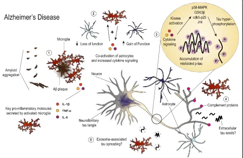
Accumulating evidence clearly illustrates a role for glio-sis and neuroinflammation in tau pathogeneglio-sis and neurodegeneration. However, initiation of inflammatory pathways may occur differently depending on the dis-ease. Fig. 1 summarizes the proposed roles of gliosis
Fig. 1Illustration summarizing the hypothesized roles of gliosis and neuroinflammation in AD. Aggregation of Aβlikely stimulates microglia early
and neuroinflammation in AD that haven been
dis-cussed in this review. In AD, Aβ aggregation likely
stimulates early gliosis and release of inflammatory
mediators such as IL-1βand C1q. These molecules may
act via autocrine and or paracrine signaling to increase levels of other proinflammatory cytokines such as
TNF-α or IL-6 from microglia, astrocytes, neurons, and
potentially other cell types in the brain. Early comple-ment signaling has also been shown to stimulate microglial-mediated phagocytosis of synapses. Together these mechanisms may lead to early neuronal dysfunc-tion and synapse loss. Microgliosis can also co-activate astrocytes, provoking both loss and gain of functions impacting neuronal health. Additionally, neuroinflamma-tion offers one way by which tau biology may be altered in AD through increasing phosphorylation that may promote protein misfolding, though other mechanisms likely also co-exist. Tau pathology then progresses through the spread of toxic tau species, neuronal vulnerability, or by
combination of both mechanisms. Altogether, Aβand tau pathology combine with gliosis to drive neurodegenera-tion and cell death in AD.
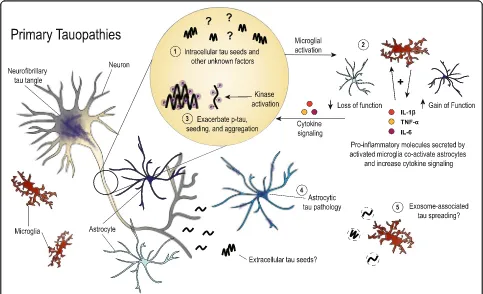
Primary tauopathies share many common features with AD, but early mechanisms of neuroinflammation in disease may differ due to the lack of amyloid pathology in pure tauopathies. Fig. 2 illustrates current thinking regarding the role of glial cells in these diseases, though there is a need for more literature directly pertaining to tau-mediated neurodegenerative mechanisms outside of the AD field. Microgliosis may be sparked by early tau aggregates, possibly tau seeds, in primary tauopathies. This may then initiate a positive feedback loop, simi-lar to that for AD, which amplifies microglial activa-tion, co-activates astrocytes, and aggravates pathways influencing tau hyperphosphorylation and aggregation. Tau accumulation in glial cells leads to further dys-function that impacts neuronal viability in a non-cell-autonomous fashion, However, it remains unclear why
Fig. 2Depiction of the roles that have been described for glial cells in primary tauopathies. In the absence of amyloid pathology, early microgliosis
there are phenotypic differences in the brain regions affected by tauopathy and the types of tau aggregates that have been described in neuropathology studies. Together, tau pathology and neuroinflammation synergistically drive neurodegeneration and clinical symptoms in tauopathies such as PSP, CBD, FTDP-17, PiD, AGD and CTE.
Clearly tau pathogenesis significantly contributes to neurodegenerative diseases. However, there are many outstanding questions that require further research and clarification. For instance, do the genetic risk variants recently found to associate with AD play a role in pri-mary tauopathies? Certain features are shared between AD and other diseases that feature tau aggregation, but there are also some clear distinctions. Therefore, it is also necessary to understand how neuroinflammatory mechanisms, such as cytokine and complement signal-ing, function in AD and in the absence of amyloid path-ology. Furthermore, the influence of these signaling pathways should be explored further, beyond tau phos-phorylation, to truly understand if neuroinflammation can contribute to the formation of insoluble, fibrillar tau aggregates. In extension, it is unclear what, if any, role gliosis plays in neurodegeneration. Is gliosis required for brain degenerative phenotypes or are neuroinflammatory molecules released from activated glial cells the main contributors? It may be that tau aggregates are the spark needed for cellular dysfunction in the brain, and neuro-inflammation the accelerant for disease progression. Additionally, the phenotypic differences in astrocytic tau pathology are intriguing and the functional conse-quences should be further explored. Finally, recent evi-dence has indicated that microglia may be contributing to the spread of tau aggregates. The extent to which microglia may physically contribute to disease progression or be influencing tau seeding or spread via neuroinflam-mation needs further investigation. Addressing these questions will ultimately help explain the relationship be-tween microglial activation, tauopathy progression, and neurodegeneration and hopefully facilitate the creation of drugs that will be effective in treating tauopathy disorders.
Abbreviations
AD:Alzheimer’s disease; AGD: Argyrophilic grain disease; ApoE: Apolipoprotein E; Aβ: Amyloid beta; CBD: Corticobasal degeneration; cdk5: Cyclin-dependent kinase 5; CSF: Cerebral spinal fluid; CTE: Chronic traumatic encephalopathy; CX3CR1: Microglial-specific fractalkine receptor; Fc: Fragment crystallizable; FRET: Förster resonance energy transfer; FTDP-17: Frontotemporal dementia with parkinsonism linked to chromosome 17; GFAP: Glial fibrillary acidic protein; GSK-3β: Glycogen synthase kinase 3; IFNγ: Interferon gamma; IL: Interleukin; IL-1β: Interleukin 1 beta; ISF: Interstitial fluid; JNK: c-Jun N-terminal kinase; MAPK: Mitogen activated protein kinase; NF-κB: NF-kappaB;
NFT: Neurofibrillary tangle; NT: Neuropil thread; p-tau: Phosphorylated tau; PSP: Progressive supranuclear palsy; TGF-β: Transformation growth factor beta; TNF-α: Tumor necrosis factor alpha; TPSO: Translocator protein
Acknowledgements
The authors would like to thank Dr. Gilbert Gallardo for assisting in editing the manuscript and Katie Campbell for helping with figure design.
Funding
Funding for this work was provided by NIH grants NS090934 (DMH), AG04867801 (DMH), F31 AG053976 (CEGL) and grants from the Tau Consortium and the JPB Foundation.
Availability of data and materials Not applicable.
Authors’contributions
CEGL proposed the topic of the paper, reviewed the literature, drafted and revised the manuscript. CEGL constructed the figures. DMH critically revised the manuscript. All authors read and approved the final manuscript.
Ethics approval and consent to participate Not applicable.
Consent for publication Not applicable.
Competing interests
CEGL and DMH are listed as inventors on a patent licensed by Washington University to C2N Diagnostics on the therapeutic use of anti-tau antibodies. DMH co-founded and is on the scientific advisory board of C2N Diagnostics, LLC. C2N Diagnostics, LLC has licensed certain anti-tau antibodies to AbbVie for therapeutic development. DMH is on the scientific advisory board of Pro-clara and consults for Genentech, Eli Lilly, Denali, AbbVie, and GlaxoSmithK-line. The authors have no additional financial interests.
Publisher’s Note
Springer Nature remains neutral with regard to jurisdictional claims in published maps and institutional affiliations.
Received: 10 April 2017 Accepted: 20 June 2017
References
1. Spillantini MG, Goedert M. Tau pathology and neurodegeneration. Lancet Neurol. 2013;12:609–22.
2. Arendt T, Stieler JT, Holzer M. Tau and tauopathies. Brain Res Bull. 2016; 126:238–92.
3. Musiek ES, Holtzman DM. Three dimensions of the amyloid hypothesis: time, space and“wingmen.”. Nat Neurosci. 2015;18:800–6.
4. Wyss-Coray T, Mucke L. Inflammation in neurodegenerative disease–a double-edged sword. Neuron. 2002;35:419–32.
5. Ransohoff RM. How neuroinflammation contributes to neurodegeneration. Science. 2016;353:777–83.
6. Lull ME, Block ML. Microglial activation and chronic neurodegeneration. Neurotherapeutics. 2010;7:354–65.
7. Asai H, Ikezu S, Tsunoda S, Medalla M, Luebke J, Haydar T, et al. Depletion of microglia and inhibition of exosome synthesis halt tau propagation. Nat Neurosci. 2015;18:1584–93.
8. Goedert M, Spillantini MG, Jakes R, Rutherford D, Crowther RA. Multiple isoforms of human microtubule-associated protein tau: sequences and localization in neurofibrillary tangles of Alzheimer’s disease. Neuron. 1989;3:519–26. 9. Neve RL, Harris P, Kosik KS, Kurnit DM, Donlon TA. Identification of cDNA
clones for the human microtubule-associated protein tau and chromosomal localization of the genes for tau and microtubule-associated protein 2. Brain Res. 1986;387:271–80.
10. Combs B, Voss K, Gamblin TC. Pseudohyperphosphorylation has differential effects on polymerization and function of tau isoforms. Biochemistry Mosc. 2011;50:9446–56.
11. Adams SJ, DeTure MA, McBride M, Dickson DW, Petrucelli L. Three repeat isoforms of tau inhibit assembly of four repeat tau filaments. PLoS One. 2010;5:e10810.
12. Schoch KM, DeVos SL, Miller RL, Chun SJ, Norrbom M, Wozniak DF, et al. Increased 4R-tau induces pathological changes in a human-tau mouse model. Neuron. 2016;90:941–7.
14. Morris M, Maeda S, Vossel K, Mucke L. The many faces of tau. Neuron. 2011; 70:410–26.
15. Witman GB, Cleveland DW, Weingarten MD, Kirschner MW. Tubulin requires tau for growth onto microtubule initiating sites. Proc Natl Acad Sci U S A. 1976;73:4070–4.
16. Tau Regulates the Attachment/Detachment but Not the Speed of Motors in Microtubule-Dependent Transport of Single Vesicles and Organelles [Internet]. PubMed J. [cited 2017 Feb 22]. Available from: https://ncbi.nlm. nih.gov/labs/articles/10381391/.
17. Spires-Jones TL, Stoothoff WH, de Calignon A, Jones PB, Hyman BT. Tau pathophysiology in neurodegeneration: a tangled issue. Trends Neurosci. 2009;32:150–9.
18. Min S-W, Cho S-H, Zhou Y, Schroeder S, Haroutunian V, Seeley WW, et al. Acetylation of tau inhibits its degradation and contributes to tauopathy. Neuron. 2010;67:953–66.
19. Irwin DJ, Cohen TJ, Grossman M, Arnold SE, Xie SX, Lee VM-Y, et al. Acetylated tau, a novel pathological signature in Alzheimer’s disease and other tauopathies. Brain J Neurol. 2012;135:807–18.
20. Cohen TJ, Guo JL, Hurtado DE, Kwong LK, Mills IP, Trojanowski JQ, et al. The acetylation of tau inhibits its function and promotes pathological tau aggregation. Nat Commun. 2011;2:252.
21. ŠimićG, BabićLeko M, Wray S, Harrington C, Delalle I, Jovanov-MiloševićN, et al. Tau protein hyperphosphorylation and aggregation in Alzheimer’s disease and other Tauopathies, and possible Neuroprotective strategies. Biomol Ther. 2016;6:6.
22. SimićG, Diana A, Hof PR. Phosphorylation pattern of tau associated with distinct changes of the growth cone cytoskeleton. Prog Mol Subcell Biol. 2003;32:33–48.
23. Thomas SN, Funk KE, Wan Y, Liao Z, Davies P, Kuret J, et al. Dual modification of Alzheimer’s disease PHF-tau protein by lysine methylation and ubiquitylation: a mass spectrometry approach. Acta Neuropathol Berl. 2012;123:105–17.
24. Funk KE, Thomas SN, Schafer KN, Cooper GL, Liao Z, Clark DJ, et al. Lysine methylation is an endogenous post-translational modification of tau protein in human brain and a modulator of aggregation propensity. Biochem J. 2014;462:77–88.
25. Horiguchi T, Uryu K, Giasson BI, Ischiropoulos H, LightFoot R, Bellmann C, et al. Nitration of tau protein is linked to neurodegeneration in tauopathies. Am J Pathol. 2003;163:1021–31.
26. Reynolds MR, Reyes JF, Fu Y, Bigio EH, Guillozet-Bongaarts AL, Berry RW, et al. Tau nitration occurs at tyrosine 29 in the fibrillar lesions of Alzheimer’s disease and other tauopathies. J Neurosci. 2006;26:10636–45.
27. Arnold CS, Johnson GV, Cole RN, Dong DL, Lee M, Hart GW. The microtubule-associated protein tau is extensively modified with O-linked N-acetylglucosamine. J Biol Chem. 1996;271:28741–4.
28. Liu F, Iqbal K, Grundke-Iqbal I, Hart GW, Gong C-X. O-GlcNAcylation regulates phosphorylation of tau: a mechanism involved in Alzheimer’s disease. Proc Natl Acad Sci U S A. 2004;101:10804–9.
29. Li X, Lu F, Wang J-Z, Gong C-X. Concurrent alterations of O-GlcNAcylation and phosphorylation of tau in mouse brains during fasting. Eur J Neurosci. 2006;23:2078–86.
30. Lu PJ, Wulf G, Zhou XZ, Davies P, Lu KP. The prolyl isomerase Pin1 restores the function of Alzheimer-associated phosphorylated tau protein. Nature. 1999;399:784–8.
31. Nakamura K, Greenwood A, Binder L, Bigio EH, Denial S, Nicholson L, et al. Proline isomer-specific antibodies reveal the early pathogenic tau conformation in Alzheimer’s disease. Cell. 2012;149:232–44.
32. Dorval V, Fraser PE. Small ubiquitin-like modifier (SUMO) modification of natively unfolded proteins tau and alpha-synuclein. J Biol Chem. 2006; 281:9919–24.
33. Dorval V, Fraser PE. SUMO on the road to neurodegeneration. Biochim Biophys Acta. 1773;2007:694–706.
34. Wischik CM, Novak M, Thøgersen HC, Edwards PC, Runswick MJ, Jakes R, et al. Isolation of a fragment of tau derived from the core of the paired helical filament of Alzheimer disease. Proc Natl Acad Sci U S A. 1988;85:4506–10.
35. Gamblin TC, Chen F, Zambrano A, Abraha A, Lagalwar S, Guillozet AL, et al. Caspase cleavage of tau: linking amyloid and neurofibrillary tangles in Alzheimer’s disease. Proc Natl Acad Sci U S A. 2003;100:10032–7. 36. Johnson GVW. Tau phosphorylation and proteolysis: insights and
perspectives. J. Alzheimers Dis. 2006;9:243–50.
37. Zhao X, Kotilinek LA, Smith B, Hlynialuk C, Zahs K, Ramsden M, et al. Caspase-2 cleavage of tau reversibly impairs memory. Nat Med. 2016;22:1268–76. 38. Mori H, Kondo J, Ihara Y. Ubiquitin is a component of paired helical
filaments in Alzheimer’s disease. Science. 1987;235:1641–4.
39. Cripps D, Thomas SN, Jeng Y, Yang F, Davies P, Yang AJ. Alzheimer disease-specific conformation of hyperphosphorylated paired helical filament-tau is polyubiquitinated through Lys-48, Lys-11, and Lys-6 ubiquitin conjugation. J Biol Chem. 2006;281:10825–38.
40. Myeku N, Clelland CL, Emrani S, Kukushkin NV, Yu WH, Goldberg AL, et al. Tau-driven 26S proteasome impairment and cognitive dysfunction can be prevented early in disease by activating cAMP-PKA signaling. Nat Med. 2016;22:46–53. 41. Braak H, Braak E. Neuropathological stageing of Alzheimer-related changes.
Acta Neuropathol. Berl. 1991;82:239–59.
42. Braak H, Thal DR, Ghebremedhin E, Del Tredici K. Stages of the pathologic process in Alzheimer disease: age categories from 1 to 100 years. J Neuropathol Exp Neurol. 2011;70:960–9.
43. de Calignon A, Polydoro M, Suárez-Calvet M, William C, Adamowicz DH, Kopeikina KJ, et al. Propagation of tau pathology in a model of early Alzheimer’s disease. Neuron. 2012;73:685–97.
44. Liu L, Drouet V, Wu JW, Witter MP, Small SA, Clelland C, et al. Trans-synaptic spread of tau pathology in vivo. PLoS One. 2012;7:e31302.
45. Saito Y, Ruberu NN, Sawabe M, Arai T, Tanaka N, Kakuta Y, et al. Staging of argyrophilic grains: an age-associated tauopathy. J Neuropathol Exp Neurol. 2004;63:911–8.
46. Walsh DM, Selkoe DJ. A critical appraisal of the pathogenic protein spread hypothesis of neurodegeneration. Nat Rev Neurosci. 2016;17:251–60. 47. Brettschneider J, Del Tredici K, Lee VM-Y, Trojanowski JQ. Spreading of
pathology in neurodegenerative diseases: a focus on human studies. Nat Rev Neurosci. 2015;16:109–20.
48. Guo JL, Lee VMY. Cell-to-cell transmission of pathogenic proteins in neurodegenerative diseases. Nat Med. 2014;20:130–8.
49. Polymenidou M, Cleveland DW. Prion-like spread of protein aggregates in neurodegeneration. J Exp Med. 2012;209:889–93.
50. Jucker M, Walker LC. Self-propagation of pathogenic protein aggregates in neurodegenerative diseases. Nature. 2013;501:45–51.
51. Kim W, Lee S, Hall GF. Secretion of human tau fragments resembling CSF-tau in Alzheimer’s disease is modulated by the presence of the exon 2 insert. FEBS Lett. 2010;584:3085–8.
52. Kim W, Lee S, Jung C, Ahmed A, Lee G, Hall GF. Interneuronal transfer of human tau between lamprey central neurons in situ. J. Alzheimers Dis. 2010;19:647–64.
53. Karch CM, Jeng AT, Goate AM. Extracellular tau levels are influenced by variability in tau that is associated with tauopathies. J Biol Chem. 2012;287:42751–62. 54. Pooler AM, Phillips EC, Lau DHW, Noble W, Hanger DP. Physiological
release of endogenous tau is stimulated by neuronal activity. EMBO Rep. 2013;14:389–94.
55. Saman S, Kim W, Raya M, Visnick Y, Miro S, Saman S, et al. Exosome-associated tau is secreted in tauopathy models and is selectively phosphorylated in cerebrospinal fluid in early Alzheimer disease. J Biol Chem. 2012;287:3842–9.
56. Wu JW, Herman M, Liu L, Simoes S, Acker CM, Figueroa H, et al. Small Misfolded tau species are internalized via bulk Endocytosis and Anterogradely and Retrogradely transported in neurons. J Biol Chem. 2013;288:1856–70.
57. Frost B, Jacks RL, Diamond MI. Propagation of tau misfolding from the outside to the inside of a cell. J Biol Chem. 2009;284:12845–52.
58. Yamada K, Cirrito JR, Stewart FR, Jiang H, Finn MB, Holmes BB, et al. In vivo microdialysis reveals age-dependent decrease of brain interstitial fluid tau levels in P301S human tau transgenic mice. J Neurosci. 2011;31:13110–7. 59. Yamada K, Holth JK, Liao F, Stewart FR, Mahan TE, Jiang H, et al. Neuronal
activity regulates extracellular tau in vivo. J Exp Med. 2014;211:387–93. 60. Yamada K, Patel TK, Hochgräfe K, Mahan TE, Jiang H, Stewart FR, et al. Analysis of in vivo turnover of tau in a mouse model of tauopathy. Mol Neurodegener. 2015;10:55.
61. Magnoni S, Esparza TJ, Conte V, Carbonara M, Carrabba G, Holtzman DM, et al. Tau elevations in the brain extracellular space correlate with reduced amyloid-βlevels and predict adverse clinical outcomes after severe traumatic brain injury. Brain. 2012;135:1268–80.
63. Holmes BB, DeVos SL, Kfoury N, Li M, Jacks R, Yanamandra K, et al. Heparan sulfate proteoglycans mediate internalization and propagation of specific proteopathic seeds. Proc Natl Acad Sci U S A. 2013;110:E3138–47. 64. Guo JL, Lee VM-Y. Seeding of normal tau by pathological tau conformers
drives pathogenesis of Alzheimer-like tangles. J Biol Chem. 2011;286:15317–31. 65. Michel CH. Kumar S, Pinotsi D, Tunnacliffe a, St. George-Hyslop P,
Mandelkow E, et al. extracellular Monomeric tau protein is sufficient to initiate the spread of tau protein pathology. J Biol Chem. 2014;289:956–67. 66. Holmes BB, Furman JL, Mahan TE, Yamasaki TR, Mirbaha H, Eades WC, et al. Proteopathic tau seeding predicts tauopathy in vivo. Proc Natl Acad Sci U S A. 2014;111:E4376–85.
67. Takeda S, Wegmann S, Cho H, DeVos SL, Commins C, Roe AD, et al. Neuronal uptake and propagation of a rare phosphorylated high-molecular-weight tau derived from Alzheimer’s disease brain. Nat. Commun. [Internet]. 2015 [cited 2017 Feb 23];6. Available from: https://www.ncbi.nlm.nih.gov/ pubmed/26458742.
68. Takeda S, Commins C, DeVos SL, Nobuhara CK, Wegmann S, Roe AD, et al. Seed-competent high-molecular-weight tau species accumulates in the cerebrospinal fluid of Alzheimer’s disease mouse model and human patients. Ann Neurol. 2016;80:355–67.
69. Clavaguera F, Bolmont T, Crowther RA, Abramowski D, Frank S, Probst A, et al. Transmission and spreading of tauopathy in transgenic mouse brain. Nat Cell Biol. 2009;11:909–13.
70. Clavaguera F, Akatsu H, Fraser G, Crowther RA, Frank S, Hench J, et al. Brain homogenates from human tauopathies induce tau inclusions in mouse brain. Proc Natl Acad Sci U S A. 2013;110:9535–40.
71. Iba M, Guo JL, McBride JD, Zhang B, Trojanowski JQ, Lee VM-Y. Synthetic tau fibrils mediate transmission of Neurofibrillary tangles in a transgenic mouse model of Alzheimer’s-like Tauopathy. J Neurosci. 2013;33:1024–37. 72. Lasagna-Reeves CA, Castillo-Carranza DL, Sengupta U, Guerrero-Munoz MJ,
Kiritoshi T, Neugebauer V, et al. Alzheimer brain-derived tau oligomers propagate pathology from endogenous tau. Sci. Rep. [Internet]. 2012 [cited 2017 Feb 23];2. Available from: https://www.ncbi.nlm.nih.gov/pubmed/ 23050084.
73. Guo JL, Narasimhan S, Changolkar L, He Z, Stieber A, Zhang B, et al. Unique pathological tau conformers from Alzheimer’s brains transmit tau pathology in nontransgenic mice. J Exp Med. 2016;213:2635–54.
74. Kahlson MA, Colodner KJ. Glial tau pathology in Tauopathies: functional consequences. J Exp Neurosci. 2015;9:43–50.
75. Komori T. Tau-positive glial inclusions in progressive supranuclear palsy, corticobasal degeneration and Pick’s disease. Brain Pathol Zurich Switz. 1999;9:663–79.
76. Arima K. Ultrastructural characteristics of tau filaments in tauopathies: immuno-electron microscopic demonstration of tau filaments in tauopathies. Neuropathol Off J Jpn Soc Neuropathol. 2006;26:475–83. 77. Ikeda K, Akiyama H, Kondo H, Haga C, Tanno E, Tokuda T, et al.
Thorn-shaped astrocytes: possibly secondarily induced tau-positive glial fibrillary tangles. Acta Neuropathol. Berl. 1995;90:620–5.
78. Togo T, Dickson DW. Tau accumulation in astrocytes in progressive supranuclear palsy is a degenerative rather than a reactive process. Acta Neuropathol. Berl. 2002;104:398–402.
79. Ferrer I, López-González I, Carmona M, Arregui L, Dalfó E, Torrejón-Escribano B, et al. Glial and neuronal tau pathology in tauopathies: characterization of disease-specific phenotypes and tau pathology progression. J Neuropathol Exp Neurol. 2014;73:81–97.
80. Odawara T, Iseki E, Kosaka K, Akiyama H, Ikeda K, Yamamoto T. Investigation of tau-2 positive microglia-like cells in the subcortical nuclei of human neurodegenerative disorders. Neurosci Lett. 1995;192:145–8.
81. Ghoshal N, García-Sierra F, Fu Y, Beckett LA, Mufson EJ, Kuret J, et al. Tau-66: evidence for a novel tau conformation in Alzheimer’s disease. J Neurochem. 2001;77:1372–85.
82. Bolós M, Llorens-Martín M, Jurado-Arjona J, Hernández F, Rábano A, Avila J. Direct evidence of internalization of tau by microglia in vitro and in vivo. J. Alzheimers Dis. 2016;50:77–87.
83. Guerreiro R, Wojtas A, Bras J, Carrasquillo M, Rogaeva E, Majounie E, et al. TREM2 variants in Alzheimer’s disease. N Engl J Med. 2013;368:117–27. 84. Ulrich JD, Holtzman DM. TREM2 function in Alzheimer’s disease and
Neurodegeneration. ACS Chem Neurosci. 2016;7:420–7.
85. Corder EH, Saunders AM, Risch NJ, Strittmatter WJ, Schmechel DE, Gaskell PC, et al. Protective effect of apolipoprotein E type 2 allele for late onset Alzheimer disease. Nat Genet. 1994;7:180–4.
86. Corder EH, Saunders AM, Strittmatter WJ, Schmechel DE, Gaskell PC, Small GW, et al. Gene dose of apolipoprotein E type 4 allele and the risk of Alzheimer’s disease in late onset families. Science. 1993;261:921–3. 87. Castellano JM, Kim J, Stewart FR, Jiang H, DeMattos RB, Patterson BW, et al.
Human apoE isoforms differentially regulate brain amyloid-βpeptide clearance. Sci Transl Med. 2011;3:89ra57.
88. Ohm TG, Kirca M, Bohl J, Scharnagl H, Gross W, März W. Apolipoprotein E polymorphism influences not only cerebral senile plaque load but also Alzheimer-type neurofibrillary tangle formation. Neuroscience. 1995;66:583–7. 89. Ji Y, Liu M, Huo YR, Liu S, Shi Z, Liu S, et al. Apolipoprotein Eε4 frequency is
increased among Chinese patients with Frontotemporal dementia and Alzheimer’s disease. Dement Geriatr Cogn Disord. 2013;36:163–70. 90. Fabre SF, Forsell C, Viitanen M, Sjögren M, Wallin A, Blennow K, et al.
Clinic-based cases with frontotemporal dementia show increased cerebrospinal fluid tau and high apolipoprotein E epsilon4 frequency, but no tau gene mutations. Exp Neurol. 2001;168:413–8.
91. Agosta F, Vossel KA, Miller BL, Migliaccio R, Bonasera SJ, Filippi M, et al. Apolipoprotein Eε4 is associated with disease-specific effects on brain atrophy in Alzheimer’s disease and frontotemporal dementia. Proc Natl Acad Sci U S A. 2009;106:2018–22.
92. López González I, Garcia-Esparcia P, Llorens F, Ferrer I. Genetic and Transcriptomic Profiles of Inflammation in Neurodegenerative Diseases: Alzheimer, Parkinson, Creutzfeldt-Jakob and Tauopathies. Int J Mol Sci. [Internet]. 2016 [cited 2017 Feb 27];17. Available from: https://www.ncbi.nlm. nih.gov/pmc/articles/PMC4783939/.
93. Malik M, Parikh I, Vasquez JB, Smith C, Tai L, Bu G, et al. Genetics ignite focus on microglial inflammation in Alzheimer’s disease. Mol
Neurodegener. 2015;10:52.
94. Griciuc A, Serrano-Pozo A, Parrado AR, Lesinski AN, Asselin CN, Mullin K, et al. Alzheimer’s disease risk gene CD33 inhibits microglial uptake of amyloid beta. Neuron. 2013;78:631–43.
95. DeMattos RB, O’dell MA, Parsadanian M, Taylor JW, Harmony JAK, Bales KR, et al. Clusterin promotes amyloid plaque formation and is critical for neuritic toxicity in a mouse model of Alzheimer’s disease. Proc Natl Acad Sci U S A. 2002;99:10843–8.
96. McKee AC, Cairns NJ, Dickson DW, Folkerth RD, Keene CD, Litvan I, et al. The first NINDS/NIBIB consensus meeting to define neuropathological criteria for the diagnosis of chronic traumatic encephalopathy. Acta Neuropathol Berl. 2016;131:75–86.
97. Giunta B, Obregon D, Velisetty R, Sanberg PR, Borlongan CV, Tan J. The immunology of traumatic brain injury: a prime target for Alzheimer’s disease prevention. J Neuroinflammation. 2012;9:185.
98. Lozano D, Gonzales-Portillo GS, Acosta S, de la Pena I, Tajiri N, Kaneko Y, et al. Neuroinflammatory responses to traumatic brain injury: etiology, clinical consequences, and therapeutic opportunities. Neuropsychiatr Dis Treat. 2015;11:97–106.
99. Cherry JD, Tripodis Y, Alvarez VE, Huber B, Kiernan PT, Daneshvar DH, et al. Microglial neuroinflammation contributes to tau accumulation in chronic traumatic encephalopathy. Acta Neuropathol. Commun. [Internet]. 2016 [cited 2017 Feb 27];4. Available from: https://www.ncbi.nlm.nih.gov/pmc/ articles/PMC5084333/.
100. Cannon JR, Greenamyre JT. The role of environmental exposures in neurodegeneration and neurodegenerative diseases. Toxicol Sci Off J Soc Toxicol. 2011;124:225–50.
101. Cho Y-E, Lee M-H, Song B-J. Neuronal cell death and degeneration through increased Nitroxidative stress and tau Phosphorylation in HIV-1 transgenic rats. PLoS One. 2017;12:e0169945.
102. Reid AH, McCall S, Henry JM, Taubenberger JK. Experimenting on the past: the enigma of von Economo’s encephalitis lethargica. J Neuropathol Exp Neurol. 2001;60:663–70.
103. Weisman D, Hakimian E, Ho GJ. Interleukins, inflammation, and mechanisms of Alzheimer’s disease. Vitam Horm. 2006;74:505–30.
104. Kettenmann H, Hanisch U-K, Noda M, Verkhratsky A. Physiology of microglia. Physiol Rev. 2011;91:461–553.
105. Wang W-Y, Tan M-S, Yu J-T, Tan L. Role of pro-inflammatory cytokines released from microglia in Alzheimer’s disease. Ann Transl Med. [Internet]. 2015 [cited 2017 Mar 6];3. Available from: https://www.ncbi.nlm.nih.gov/ pmc/articles/PMC4486922/.