R E S E A R C H A R T I C L E
Open Access
Influence of different application protocols
of universal adhesive system on the clinical
behavior of Class I and II restorations of
composite resin
–
a randomized and
double-blind controlled clinical trial
Andreia Assis Carvalho
*, Murillo Martins Leite, Jessica Karla Maia Zago,
Carla Aparecida Bernardes Costa Meneses Nunes, Terezinha de Jesus Esteves Barata, Gersinei Carlos de Freitas,
Érica Miranda de Torres and Lawrence Gonzaga Lopes
Abstract
Background:Multimode adhesives incorporate the versatility of adapting to various clinical situations by its capacity to be used in different protocols. This study aimed to evaluate the clinical behavior of composite resin direct restorations (Class I and II) performed with different universal dentin adhesive application protocols comparing adapted FDI and adapted USPHS criteria.
Methods:The current study is a randomized, double-blind, split-mouth, and convenience sample controlled clinical trial. The participants (age≥18 years) had restorative need of Class I and/or II due to the presence of carious lesions and/or unsatisfactory restorations in at least three dental elements. Each participant received three application protocols for Scotchbond Universal adhesive (3M ESPE), one in each tooth to be restored: ER = etch-and-rinse + adhesive (n = 50); SEE = selective enamel etch + adhesive (n = 50) and SE = self-etch adhesive (n = 50). All teeth were restored in a similar way using Filtek™Supreme composite resin (3M ESPE). Restorations were evaluated using the adapted FDI and adapted USPHS criteria, at baseline after 7 to 21 (12.02 ± 5.68) days (T1; n = 50 per group) and after 12 to 20 (15.8 ± 2.7) months (T2; n = 46 per group) by two previously calibrated evaluators (Kappa > 0.80). The statistical tests were performed between groups (Friedman), intragroup (Wilcoxon), and between the criteria considering acceptable and not acceptable restorations (McNemar),α= 0.05.
Results:A statistically significant difference was observed only for the property“superficial staining”, between groups at T2 (p= 0.01) for ER (n = 13 with score 2 or more) and SEE (n = 3 with score 2 or more) and intragroup for ER (T1, n = 1 with score 2 or more; T2, n = 13 with score 2 or more,p= 0.001) and SE (T1, n = 0 with score 2 or more; T2, n = 8 with score 2 or morep= 0.007). For the other comparisons between groups, intragroup, and between the adapted FDI and adapted USPHS criteria, there were no statistically significant differences (p≥0.05).
(Continued on next page)
© The Author(s). 2019Open AccessThis article is distributed under the terms of the Creative Commons Attribution 4.0 International License (http://creativecommons.org/licenses/by/4.0/), which permits unrestricted use, distribution, and reproduction in any medium, provided you give appropriate credit to the original author(s) and the source, provide a link to the Creative Commons license, and indicate if changes were made. The Creative Commons Public Domain Dedication waiver (http://creativecommons.org/publicdomain/zero/1.0/) applies to the data made available in this article, unless otherwise stated.
* Correspondence:[email protected]
(Continued from previous page)
Conclusions:It can be concluded that the different application protocols of the universal adhesive resulted in clinically“acceptable”restorations after 15.8 ± 2.7 months of follow-up. Adapted FDI and adapted USPHS criteria provided similar results to each other.
Trial registration.
Number in Brazilian Registry of Clinical Trials (ReBEC): RBR-9p3hdp. Registered 24 May 2015.
Keywords:Dental materials. Clinical trial. Dental bonding. Adhesives
Background
According to the form of demineralization of the dental substrate and the treatment given to the smear layer, the traditionally available adhesive systems can be divided into: etch-and-rinse (ER), self-etch (SE), and universal/ multimode [1,2]. ER systems profess the removal of the smear layer by etching of the dental substrate in a separ-ate step from the application of the adhesive [1, 2]. SE adhesive systems are capable of modifying and incorpor-ating the smear layer, simultaneously to dentin demineralization [3–5]. The universal adhesive can be used in different application modes: ER, SE, or selectice enamel etch (SEE) [6, 7]. The universal adhesive repre-sents one type of one-step SE adhesives, and the durabil-ity and stabildurabil-ity of bonded interfaces created by them continue to be questionable [8].
For a better comprehension of this process, it shall be understood that SE monomers are often less acidic than phosphoric acid, so that some minerals remain attached to the collagen fibers, allowing chemical bonds between the dental substrate and functional groups of the adhesive monomers [9]. Chemical bonding occurs with the inter-action of functional acid monomers (carboxylic groups, phosphonic or phosphate) with hydroxyapatite crystals in dentin [10]. 10-methacryloyloxydecyl dihydrogen phos-phate (10-MDP) is an example of a functional monomer that can interact with calcium ions in hydroxyapatite crys-tals in a process that is conducted until the formation of calcium salts of MDP (MDP-Ca salts) [10].
One of the advantages of using the universal adhesive in the clinical routine would be allowing the dental sur-geon to choose the type of application protocol accord-ing to the clinical situation, optimizaccord-ing the final result of the procedure [11]. However, while the development of universal adhesive systems has been an innovation in ad-hesive dentistry, it is questionable whether or not they are appropriate for all adhesive procedures [12]. Cur-rently, there are only a few clinical trials that evaluate the bonding performance of universal adhesives [8]. There is still a lack of literature about these materials, with few clinical studies in Class V restorations [13–16].
It should be observed that the strength of the dentin bond is also variable according to the location of the cavity in the tooth (cervical or occlusal), and this is due to the variability of the dentinal tissue itself [17].
According to Purk et al. (2007) [18], the photopolymer-izable composite resin may bond differently to the dentin, depending on the amount of cavity walls and there may be more cracks through the adhesive in the gingival wall than in the axial wall. Thus, clinical studies with universal adhesives performed in Class I and II cav-ities are necessary.
In this context, the clinical evaluation of restorations involves the use of criteria developed for some factors considered relevant in the clinical performance of dental restorative materials [19]. The evaluations can be judged according to different factors, among which are the USPHS (United States Public Health Service) criteria [19] and the FDI (Fédération Dentaire Internationale/ World Dental Federation), being that the latter is divided into aesthetic, functional, and biological parameters of the restorations [20]. According to the FDI criteria, the restorations can be classified into five scores: 1 = clinic-ally very good; 2 = clinicclinic-ally good; 3 = clinicclinic-ally suffi-cient/satisfactory; 4 = clinically unsatisfactory; and 5 = clinically bad [20]. According to the USPHS criteria, the restorations can be classified into three scores: Alpha, Bravo and Charlie [19]. Studies comparing the clinical behavior of different bonding strategies using modified FDI and USPHS criteria have concluded that FDI criteria are more sensitive than the modified USPHS criteria for small variations in clinical outcomes of noncarious cer-vical lesions [13,14,16].
Therefore, this work aimed to evaluate the clinical be-havior of composite resin restorations performed with a universal adhesive system used in different application protocols comparing the adapted FDI and adapted USPHS criteria. Null hypotheses (is) were tested: (1) the adhesive application protocol (ER, SEE, or SE) does not influence the clinical behavior of Class I and II compos-ite resin restorations over time, and (2) there is no dif-ference between the adapted FDI and adapted USPHS evaluation criteria of the clinical behavior.
Methods
Study design
guidelines. Clinical procedures were performed at the School of Dentistry of the Federal University of Goiás, Brazil, from June 2015 to April 2018. The research project was approved by the Research Ethics Committee (CAAE: 36829814.0.0000.5083), registered in the Brazilian Registry of Clinical Trials (ReBEC - http://www.ensaiosclinicos. gov.br; RBR-9p3hdp) and the participants signed a written informed consent form.
Sample size
The sample size calculation was based on the reten-tion rates, postoperative sensitivity, adaptareten-tion, and marginal coloration of the Scotchbond Universal ad-hesive (3M ESPE, St Paul, MN, USA) for Class V res-torations after 18 months [14], using α of 0.05 and a power of 80%. Thereby, the number of 50 teeth per group was determined to detect a difference of 20% in the tested groups.
Sample selection
Inclusion criteria were age equal to or older than 18 years; clinical and radiographic need for Class I and/or II restorations in posterior permanent teeth due to the presence of carious lesions and/or unsatisfactory preex-isting restoration; presence of at least three teeth (one for each adhesive protocol) to be restored per partici-pant, which antagonistic teeth were present; and at least 20 teeth in functional occlusion.
Exclusion criteria were periodontitis; users of braces or removable orthodontic appliances; removable prosthesis wearers; signs of parafunctional habits; pregnant or lac-tating women; and fluorosis in the enamel.
Radiographic examination included the use of inter-proximal and periapical digital radiographs as auxiliary means of diagnostic for participants who presented in-clusion criteria during the clinical examination. Oral hy-giene guidelines were given to all study participants prior to restorative procedures.
Randomization and blinding
The randomization process was performed using manu-ally generated tables by a member of the research group not involved in performing the clinical procedures. De-tails of the allocated group, including the adhesive protocol to be used and the teeth sequence to be re-stored, were written on sheets placed in sequentially numbered, opaque, sealed envelopes, which were opened at the time of the restorative procedure. The operator knew which type of intervention would be performed, however, the participants did not know which treatment they received for a given tooth, and the evaluators were also unaware of the adhesive protocols used (double-blind study).
The option for split-mouth study design [21] was per-formed so that the same participant could receive the three adhesive protocols to be evaluated.
Restorative procedures
The clinical procedures were performed by two oper-ators who are experts in restorative dentistry. One of them was responsible for implementing the operative procedures and the other was responsible for the re-storative procedures. The basic composition of the main materials used in this study is described in Table 1.
With absolute isolation, the operative approach of carious lesion or unsatisfactory restoration was ini-tially performed with spherical diamond tips (KG Sorensen, Cotia, SP, Brazil), of compatible number to carious lesion or unsatisfactory restoration, in high rotation under refrigeration (Kavo, Rio de Janeiro, RJ, Brazil), and then, the carious dentin were removed with carbide bur (Maillefer, Dentsply, Rio de Janeiro RJ, Brazil) in low-speed (Contra-angle, Kavo, Rio de Janeiro, RJ, Brazil). After cavity prepar-ation, cavity cleaning was done with 2% chlorhexi-dine solution (Reymer from Brazil, Aparecida de Goiânia, GO, Brazil) with sterile cotton ball for 1 min.
Cavities considered very deep, the protection of the dentinopulpar complex was done with hydroxide of calcium cement (Hydro C, Dentsply, Rio de Janeiro RJ, Brazil) associated with resin-modified glass iono-mer cement (Ionoseal, Voco, Cuxhaven, Germany), lining the bottom wall only. Deep cavities, the resin-modified glass ionomer cement was used. For medium or shallow depth cavities the adhesive proto-cols were applied directly. The Scotchbond Universal adhesive system (3M ESPE, St Paul, MN, USA) was applied to the tooth surface according to the drawn protocol, as detailed in Table 2.
Clinical evaluation
The first evaluation (T1) was performed 7 to 21 (12.02 ± 5.68) days after the restoration and, the second evalu-ation (T2) was performed after 12 to 20 months (15.8 ± 2.7) of monitoring. The evaluations were performed by two properly calibrated evaluators who were not in-volved with the restorative procedures. Excellent interex-aminer agreement was obtained for all the criteria under analysis (Kappa> 0.80). Restorations were evaluated using the adapted USPHS (Table 3) [19] and adapted FDI (Table4) criteria [20]. To evaluate the postoperative sensitivity of the teeth under analysis, air jets were ap-plied for 10s with the triple syringe 2 cm from the occlu-sal surface [13, 14, 20], and the participant was asked if there was sensitivity caused by the air jet or if there was postoperative pain at some other time prior to the evalu-ation. Both examiners evaluated the restorations inde-pendently, and in the absence of agreement during the evaluations, consensus was sought before dispensing the participant.
The scores were dichotomized as “acceptable” for restorations that did not require intervention
(clinically very good, clinically good, clinically suffi-cient/satisfactory, Alpha and Bravo) and “not accept-able” (clinically unsatisfactory, clinically bad, and Charlie) for those that needed repair or replacement [14]. To compare the adapted FDI and adapted USPHS criteria, considering the “acceptable” and
“not acceptable” restorations, the properties of each criterion that could be compared were separated: adapted FDI (marginal staining, material fracture and retention, marginal adaptation, postoperative hyper-sensitivity and dental vitality, and recurrence of car-ies, erosion, or abfraction) and adapted USPHS (marginal discoloration, retention, fracture, marginal adaptation, postoperative sensitivity, and recurrence of caries).
Data analysis
Statistical analysis followed the CONSORT protocol, considering the intention to treat, including all ran-domized participants and including those who did not participate in the evaluation appointments. De-scriptive analysis was performed for the evaluated criteria. The Friedman test was used for the com-parison between groups (ER, SEE, and SE) at T1 and T2. The Wilcoxon test was used for the intragroup comparison (i.e., each group between the times T1 and T2). The McNemar test was used for the com-parison between the adapted FDI and adapted USPHS criteria considering acceptable and not ac-ceptable restorations. All statistical tests were per-formed with SPSS software version 24 (IBM SPSS Statistics, Chicago IL, USA) at a significance level of 5% (α= 0.05).
Results
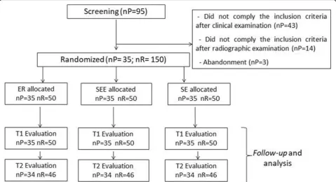
Initially, 95 participants were evaluated. After screen-ing, a final sample consisted of 35 subjects, account-ing for total of 150 restorations (Fig. 1). Data corresponding to participants and to restored teeth are show in Table 5. The majority of participants Table 1Composition of the main materials used in this study informed by the manufacturers
Material Manufacturer Lot Composition*
Condac 37
(Joinville, SC, Brazil)
FGM 090715 37% Phosphoric acid, thickeners.
Filtek™Supreme
(St Paul, MN, USA)
3M ESPE N595157 (color B2E) 176149
(color B2B)
Bis-GMA, UDMA, TEGDMA, Bis-EMA, non-agglomerated silica, non-agglomerated and agglomerated zirconia, and aggregated particles of zirconia/silica.
Scotchbond Universal Adhesive
(St Paul, MN, USA)
3M ESPE 565520 HEMA, MDP (10-methacryloyloxydecamethylene phosphoric acid), phosphate monomer, dimethacrylate, hydroxyethyl methacrylate, copolymer of polyalkanoic acid methacrylate, ethanol, water, initiators and silane.
*According to information from the manufacturers
Table 2Clinical protocol of adhesive application in the three groups tested
Technique Clinical sequence
Etch-and-rinse (ER) - Total acid etching for 30s in enamel and 15s in dentin;
- Abundant washing for 10s with water jet and removal of humidity excess with absorbent paper; - Application of the adhesive for 20s;
- Air jet for 5s;
- Photopolymerization for 20s.
Selective Enamel Etch (SEE)
- Selective acid etching in enamel for 30s; - Abundant washing for 10s with water jet and
removal of humidity excess with absorbent paper; - Application of the adhesive for 20s;
- Air jet for 5s;
- Photopolymerization for 20s.
Self-Etch (SE) - Application of the adhesive for 20s; - Air jet for 5s;
were female (60%), 20–29 years old (77%), and the restored teeth were mainly molars (81%) with Class I type cavities (64%).
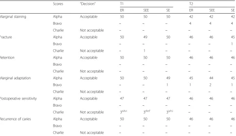
Restorations were evaluated using the adapted FDI and adapted USPHS criteria 7 to 21 (12.02 ± 5.68) days after the restoration (T1; n = 50 per group) and after 12 to 20 (15.8 ± 2.7) months (T2; n = 46 per group) by two previously calibrated evaluators (Kappa > 0.80). The data obtained during the two-time evaluations (T1 and T2) are summarized in Table 6 (adapted USPHS criteria) and Table7(adapted FDI criteria).
For the“surface staining” property, a significant differ-ence was observed at T2 between groups ER (n = 13 with score 2 or more) and SEE (n = 3 with score 2 or more; p= 0.01) and intragroup for ER (T1, n = 1 with score 2 or more; T2, n = 13 with score 2 or more;p= 0.001) and SE (T1, n = 0 with score 2 or more; T2, n = 8 with score 2 or more; p= 0.007). For the other comparisons be-tween groups and intragroup, there were no significant differences (p≥0.05).
In each group, postoperative sensitivity (initial assess-ment) was reported for three restored teeth, with
“clinically good” (adapted FDI criteria) and “Charlie” (adapted USPHS criteria) scores. It is noteworthy that one participant reported postoperative sensitivity after the restorations with the three adhesive protocols tested. In the T2 assessment, no participant reported pain sensi-tivity in the period.
There was no significant difference when the adapted FDI and adapted USPHS criteria were compared (p≥ 0.05), with“acceptable” restorations in adapted FDI (ER n = 44, SEE n = 46, and SE n = 46) and in adapted USPHS (ER n = 46, SEE n = 46, and SE n = 46) after 15.8 ± 2.7 months of follow-up.
Discussion
The present study makes an important contribution to the literature on universal adhesive systems since it was performed in Class I and II cavities of permanent teeth, which differs from the studies of Mena-Serrano et al. (2013) [13], Perdigão et al. (2014) [14], Lawson et al. (2015) [15], and Lopes et al. (2016) [16] performed on Class V restorations. Considering that the most import-ant parameter for evaluating the performance of an Table 3The adapted USPHS criteria with their categories and grading
Categories Grading “Decision”
Marginal discoloration 1. Alpha (clinically ideal)
2. Bravo (showing minor deviations from the ideal, nevertheless acceptable–except for retention and secondary caries)
3. Charlie (should be replaced to avoid future damage or requiring immediate replacement)
1. Acceptable (1, 2) 2. Not acceptable (3) Fracture
Retention
Marginal integrity
Postoperative sensitivity
Recurrence of caries
Table 4The adapted FDI criteria with their categories and grading
Categories Sub-categories Grading “Decision”
A. Aesthetic properties 1. Surface lustre 2. Staining a. surface b. margin
3. Color match and translucency 4. Esthetic anatomical form
1. Clinically excellent/very good 2. Clinically good
3. Clinically sufficient/satisfactory (minor shortcomings, no
unacceptable effects but not adjustable without damage
to the tooth)
4. Clinically unsatisfactory/(but reparable) 5. Clinically poor (replacement necessary)
1. Acceptable (1, 2, 3) 2. Not acceptable (4, 5)
B. Functional properties 5. Fracture of material and retention 6. Marginal adaptation
7. Occlusal contour and wear a) qualitatively b) quantitatively 8. Approximal anatomical form a. contact point b. contour
9. Radiographic examination (when applicable) 10. Patient’s view
C. Biological properties 11. Postoperative (hyper-)sensitivity and tooth vitality
12. Recurrence of caries (CAR), erosion, abfraction 13. Tooth integrity (enamel cracks, tooth fractures) 14. Periodontal response (always compared to a
adhesive system is the retention [16], these studies justi-fied the evaluation of Class V noncarious cervical lesions [13–16]. Nevertheless, the location factors of the cavities and type of masticatory forces could also influence the maintenance of the adhesive interface [17, 22], which highlights the importance of the evaluation performed here in Class I and II cavities.
Additionally, another factor that could interfere with the adhesive quality would be cavity cleaning after completion of the cavity preparation. In this study, 2% chlorhexidine solution was used before acid etching in the ER and SEE groups and prior to the application of the adhesive in the SE group, in order to disinfect the dentin surface, remove microorganisms, and minimize the possibility of recurrence of dental cavity [23]. However, chlorhexidine can also be
used after etching, to minimize degradation by the metallo-proteinases of collagen matrices that are incompletely infil-trated by the adhesive system [24]. There is no consensus in the literature concerning the efficacy of chlorhexidine use when applying SE adhesives or even when or how the chlorhexidine solution should be applied [25–27]. A recent clinical study with 36-month follow-up showed that cavity pre-treatment with chlorhexidine for inhibition of hybrid layer degradation does not add any beneficial effect to the clinical performance of restorations [28]. However, other la-boratory studies have reported that for cavity cleaning prior to etching, chlorhexidine showed no significant change in microtensile bond strength of adhesive, even when the SE application mode was used [25,29,30]. Therefore, in the present work the option was to perform its prior Fig. 1Characterization of the participants of the study (nP- number of Participants; nR- number of Restorations; ER- Etch-and-rise; SEE- Selective Enamel Etch; SE- Self-Etch; T1- first evaluation; T2- second evaluation)
Table 5Distribution of participants and restored teeth
Groups Gender Age (years) Tooth Arcade distribution Type of preparation Cavity Depth
M F 20–29 30–50 PM M Jaw Mand I II S M D VD
ER 14 21 27 8 10 40 33 17 34 16 15 4 16 15
SEE 8 42 29 21 30 20 19 1 18 12
SE 11 39 26 24 32 18 26 6 11 7
A Freq 14 21 27 8 29 121 88 62 96 54 60 11 45 34
R Freq 40% 60% 77% 23% 19% 81% 58% 42% 64% 36% 40% 7% 30% 23%
Abbreviations:ER- Etch-and-rinse;SEE- Selective Enamel Etch;SE- Self-Etch;A Freq-Absolute frequency;R Freq-relative frequency;M- male;F- female;PM- premolar;
application to the use of the adhesive system independently of its protocol.
In this context, although in the present study the ap-plication of the adhesive in a single layer was used, as in-dicated by the manufacturer, the possibility of its use in two layers is reported [31], in order to to enhance the bond strength. A laboratory work performed by Fujiwara et al. (2018) [31], the application of an universal adhe-sives in two layers improved the adhesive quality when compared to the application in a single layer. According to the authors of the study, this result may be related to the possibility of the double application protocol produ-cing a more uniform adhesive layer and compensating for possible defects of single application [31]. This may be an important point of discussion for other long-term longitudinal follow-up clinical trials.
Based on the results obtained in this study, the first null hypothesis that “the adhesive application protocol (ER, SEE or SE) does not influence the clinical behav-ior of Class I and II composite restorations over time” had to be accepted, since the clinical behavior of composite resin restorations was similar, resulting in clinically acceptable restorations after 15.8 ± 2.7 months of follow-up, independently of the different application protocols of the universal adhesive (ER,
SEE, and SE). Similar results to those reported by Mena-Serrano et al. (2013) [13] and Perdigão et al. (2014) [14] were obtained. These studies demon-strated that the clinical behavior of the Scotchbond Universal adhesive used in the form of SEE and SE, in Class V restorations, did not depend on the adhe-sive protocol tested in clinical follow-up of 6 and 18 months, respectively. These data may be related to the short period of clinical evaluation of these studies (6 and 18 months), which was similar to the present study (15.8 ± 2.7 months). In a study by Loguercio et al. (2015) [32], in 36 months of evaluation, there were signs of degradation when the universal adhesive was applied in the SE mode. Therefore, the restora-tions need to be re-evaluated after a long follow-up period (4–5 years) with the aim of confirming the re-sults [33]. For Cuevas-Suarez et al., 2019 [8], the pub-lished data from the clinical trials suggested that the clinical performance of the universal adhesive did not depend on the bonding strategy in up to 36 months of follow-up.
Although statistically significant differences were ob-served for the surface staining property between T1 and T2 for the ER and SE groups and between ER and SEE techniques at T2, the scores remained within the Table 6Number of restorations evaluated, for each experimental group, classified according to the adapted USPHS criteria at T1 and T2 times
Scores “Decision” T1 T2
ER SEE SE ER SEE SE
Marginal staining Alpha Acceptable 50 50 50 42 42 42
Bravo ̶ ̶ ̶ 4 4 4
Charlie Not acceptable ̶ ̶ ̶ ̶ ̶ ̶
Fracture Alpha Acceptable 50 49 50 46 46 45
Bravo ̶ ̶ ̶ ̶ ̶ 1
Charlie Not acceptable ̶ 1 ̶ ̶ ̶ ̶
Retention Alpha Acceptable 50 50 50 46 46 46
Bravo ̶ ̶ ̶ ̶ ̶ ̶
Charlie Not acceptable ̶ ̶ ̶ ̶ ̶ ̶
Marginal adaptation Alpha Acceptable 50 50 49 45 44 45
Bravo ̶ ̶ 1 1 2 1
Charlie Not acceptable ̶ ̶ ̶ ̶ ̶ ̶
Postoperative sensitivity Alpha Acceptable 47 47 47 46 46 46
Bravo ̶ ̶ ̶ ̶ ̶ ̶
Charlie Not acceptable 3a,b,c 3d,e,f 3g,h,i ̶ ̶ ̶
Recurrence of caries Alpha Acceptable 50 50 50 46 46 46
Bravo ̶ ̶ ̶ ̶ ̶ ̶
Charlie Not acceptable ̶ ̶ ̶ ̶ ̶ ̶
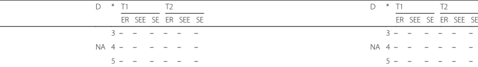
Table 7Number of restorations evaluated, for each experimental group, classified according to the adapted FDI criteria at T1 and T2 times
D * T1 T2 D * T1 T2
ER SEE SE ER SEE SE ER SEE SE ER SEE SE
Surface lustre A 1 50 50 49 45 46 44 Approximal anatomical form contact point (nR Class II = 54)
A 1 14 19 16 11 14 12
2 ̶ ̶ 1 1 ̶ 2 2 ̶ ̶ ̶ ̶ 1 –
3 ̶ ̶ ̶ ̶ ̶ ̶ 3 2 1 2 3 2 4
NA 4 ̶ ̶ ̶ ̶ ̶ ̶ NA 4 ̶ ̶ ̶ ̶ ̶ ̶
5 ̶ ̶ ̶ ̶ ̶ ̶ 5 ̶ ̶ ̶ ̶ ̶ ̶
Staining surface A 1 49 50 50 33 43 38 Approximal anatomical form contour (nR Class II = 54)
A 1 15 20 16 13 16 14
2 1 ̶ ̶ 10 2 7 2 1 ̶ 2 1 1 2
3 ̶ ̶ ̶ 2 1 1 3 ̶ ̶ ̶ ̶ ̶ ̶
NA 4 ̶ ̶ ̶ 1a ̶ ̶ NA 4 ̶ ̶ ̶ ̶ ̶ ̶
5 ̶ ̶ ̶ ̶ ̶ ̶ 5 ̶ ̶ ̶ ̶ ̶ ̶
Staining margin A 1 50 50 49 42 42 42 Patient’s view A 1 50 50 49 46 46 46
2 ̶ ̶ 1 3 4 3 2 ̶ ̶ 1 ̶ ̶ ̶
3 ̶ ̶ ̶ 1 ̶ 1 3 ̶ ̶ ̶ ̶ ̶ ̶
NA 4 ̶ ̶ ̶ ̶ ̶ ̶ NA 4 ̶ ̶ ̶ ̶ ̶ ̶
5 ̶ ̶ ̶ ̶ ̶ ̶ 5 ̶ ̶ ̶ ̶ ̶ ̶
Color match and translucency
A 1 47 46 46 42 41 41 Postoperative (hyper-)sensitivity and tooth vitality
A 1 47 47 47 45 46 46
2 2 4 4 2 4 4 2 3 3 3 1 ̶ ̶
3 1 ̶ ̶ 1 1 1 3 ̶ ̶ ̶ ̶ ̶ ̶
NA 4 ̶ ̶ ̶ 1a ̶ ̶ NA 4 ̶ ̶ ̶ ̶ ̶ ̶
5 ̶ ̶ ̶ ̶ ̶ ̶ 5 ̶ ̶ ̶ ̶ ̶ ̶
Esthetic anatomical form A 1 49 47 47 45 42 43 Recurrence of caries (CAR), erosion, abfraction A 1 50 50 50 46 46 46
2 1 3 3 1 4 3 2 ̶ ̶ ̶ ̶ ̶ ̶
3 ̶ ̶ ̶ ̶ ̶ ̶ 3 ̶ ̶ ̶ ̶ ̶ ̶
NA 4 ̶ ̶ ̶ ̶ ̶ ̶ NA 4 ̶ ̶ ̶ ̶ ̶ ̶
5 ̶ ̶ ̶ ̶ ̶ ̶ 5 ̶ ̶ ̶ ̶ ̶ ̶
Fracture of material and retention
A 1 50 49 50 46 46 45 Tooth integrity (enamel cracks, tooth fractures) A 1 50 50 50 46 46 46
2 ̶ 1 ̶ ̶ ̶ 1 2 ̶ ̶ ̶ ̶ ̶ ̶
3 ̶ ̶ ̶ ̶ ̶ ̶ 3 ̶ ̶ ̶ ̶ ̶ ̶
NA 4 ̶ ̶ ̶ ̶ ̶ ̶ NA 4 ̶ ̶ ̶ ̶ ̶ ̶
5 ̶ ̶ ̶ ̶ ̶ ̶ 5 ̶ ̶ ̶ ̶ ̶ ̶
Marginal adaptation A 1 50 50 49 45 44 45 Periodontal response (nR Class II = 54)
A 1 16 20 18 14 17 16
2 ̶ ̶ 1 1 1 1 2 ̶ ̶ ̶ ̶ ̶ ̶
3 ̶ ̶ ̶ ̶ 1 ̶ 3 ̶ ̶ ̶ ̶ ̶ ̶
NA 4 ̶ ̶ ̶ ̶ ̶ ̶ NA 4 ̶ ̶ ̶ ̶ ̶ ̶
5 ̶ ̶ ̶ ̶ ̶ ̶ 5 ̶ ̶ ̶ ̶ ̶ ̶
Occlusal contour and wear (qlt)
A 1 48 50 49 44 45 45 Adjacent mucosa A 1 50 50 50 46 46 46
2 2 ̶ 1 1 1 1 2 ̶ ̶ ̶ ̶ ̶ ̶
3 ̶ ̶ ̶ ̶ ̶ ̶ 3 ̶ ̶ ̶ ̶ ̶ ̶
NA 4 ̶ ̶ ̶ 1b ̶ ̶ NA 4 ̶ ̶ ̶ ̶ ̶ ̶
5 ̶ ̶ ̶ ̶ ̶ ̶ 5 ̶ ̶ ̶ ̶ ̶ ̶
Occlusal contour and wear (qtt)
A 1 49 50 50 45 46 46 Oral and general health A 1 50 50 50 46 46 46
clinically acceptable range. Possibly the observed surface staining was related to oral hygiene and eating habits of the participants, which was similarly observed by other authors [34,35].
On the other hand, Lawson et al. (2015) [15], in an evaluation after 24 months, tested the Scotchbond Univer-sal in the ER and SE technique and the Scotchbond Multi-Purpose (3 M ESPE) (ER) in Class V restoration and con-cluded that both tested adhesives deteriorated in relation to the adaptation and marginal discoloration. The authors also affirmed that Scotchbond Universal in the SE tech-nique and Scotchbond Multi-Purpose presented similar clinical behavior, however, they stated that Scotchbond Universal with ER had the best results [15].
With the increase of the clinical evaluation period to 60 months, Baracco et al. (2016) [36] observed more fre-quent and severe marginal staining in Class I and Class II restorations when they tested the SE adhesive protocol (Adper Scotchbond SE and P90, 3 M ESPE) compared to the ER protocol (Adper Scotchbond XT, 3 M ESPE), as well as the presence of marginal adaptation deterioration in all groups according to the adhesive systems tested. Van Dijken and Pallesen (2017) [37], after 6 years of evaluation of Class II restorations, found failures related to fracture and recurrence of dental caries when com-paring a three-step ER application mode adhesive system (cmf-els, Saremco AG) and an SE without HEMA (Adh-eSe OneF, Ivoclar Vivadent). The failures corresponded to 11.4% of restorations with prior acid etching and to 20% in restorations with SE adhesive [37]. The authors inferred that the fracture of the restorations was related to the extension of the restorations [37]. In the present study, a case of fracture of the restoration was observed in the initial evaluation, which also suggests that the cause is due, probably, to the extension of the restor-ation. In in vitro study of Jacker-Guhr et al. (2019) [38], cohesive fractures were observed for ER, mainly in the enamel, probably due to higher bond strength using uni-versal adhesives after additional phosphoric acid etching. On the other hand, the present clinical trial differs from the studies of Mena-Serrano et al. (2003) [13], Per-digão et al. (2014) [14] and Loguercio et al. (2018) [33] in which the deterioration of the marginal adaptation of
the restorations with the use of MDP universal adhesive was observed after 6 and 18 months of clinical evalu-ation, both for the adhesive protocol with ER and SE ap-plication modes. This difference may be related to the incidence of masticatory forces and complexity of the restorations, once the restorations were performed in Class I and II cavities, which is different from the cited works in which the restorations were performed in Class V cavities.
In a follow-up period of 18 months, Perdigão et al. (2014) [14] reported a 2.5% loss of retention in Class V restorations, and the universal adhesive protocol did not influence the loss of retention. In the 24-month follow-up of Class V restorations, retention rates of 87.6, 94.9, and 100% were reported for Scotchbond Multi-Purpose (ER), Scotchbond Universal in the SE application mode, and Scotchbond Universal with ER, respectively [15]. They concluded that Scotchbond Universal, in the SE and ER application mode, was similar to or better than Scotchbond Multi-Purpose [15]. In the present study, in-dependently of the adhesive protocol applied, 100% re-tention of all the performed restorations was observed. This result should probably also be related to the loca-tion and type of the restoraloca-tions (Class I and II). Al-though there was still no difference between the adhesive protocols, a systematic review showed that the enamel bond strength of universal adhesive is improved with prior phosphoric acid etching (SEE) [39]. SEE followed by the application of a mild universal adhesive currently appears to be the best choice to effectively achieve a durable bond to tooth tissues [8]. However, this effect was not evident for dentin with the use of mild adhesive with the ER [39].
In relation to postoperative sensitivity, in the present study there were no statistically significant differences between the groups, nor was there a rela-tionship between postoperative sensitivity and cavity depth (47% shallow and medium/53% deep and very deep). The teeth that presented sensitivity at the baseline were classified with a “Charlie” score, since for the USPHS criterion there is no “Bravo” score for this criterion [19]. Thus, the restorations are classified as “Alpha” for no reports of postoperative sensitivity Table 7Number of restorations evaluated, for each experimental group, classified according to the adapted FDI criteria at T1 and T2 times(Continued)
D * T1 T2 D * T1 T2
ER SEE SE ER SEE SE ER SEE SE ER SEE SE
3 ̶ ̶ ̶ ̶ ̶ ̶ 3 ̶ ̶ ̶ ̶ ̶ ̶
NA 4 ̶ ̶ ̶ ̶ ̶ ̶ NA 4 ̶ ̶ ̶ ̶ ̶ ̶
5 ̶ ̶ ̶ ̶ ̶ ̶ 5 ̶ ̶ ̶ ̶ ̶ ̶
or “Charlie” for when there is a complaint. These pa-tients were followed up, observing a progressive re-gression of sensitivity not requiring intervention. In the T2 evaluation, no provoked sensitivity was ob-served in all restored teeth. These results are in agreement with other works in the literature using the same adhesive system in the different etching pro-tocols [13–15] and with other adhesive systems also in the mode with ER and SE [37, 40].
To compare the criteria, the properties evaluated were dichotomized as“acceptable” and“not acceptable”, simi-lar to other studies [13, 14]. As there were no statisti-cally significant differences for the comparisons between the adapted FDI and adapted USPHS criteria, the second null hypothesis that “there is no difference between the adapted FDI and adapted USPHS evaluation criteria of the clinical behavior” was also accepted. The USPHS and FDI clinical evaluation criteria are described in pre-vious studies, such as the FDI being more sensitive than the USPHS in the “marginal adaptation” parameter [13, 14]. However, the option to maintain both criteria is to allow the comparison of results with studies that used only one of the criteria [16].
The clinical follow-up period of the present study may be considered a limitation, however, the divulga-tion of results is important for comparison with simi-lar follow-up studies [14, 33]. Longitudinal clinical follow-ups for longer periods are necessary to evalu-ate the influence of different application protocols of the universal adhesive system (Scotchbond Universal) on the clinical behavior of Class I and II composite resin restorations. The datasets generated during the current study are available in the Federal University of Goiás Repository [41].
Conclusions
It was concluded that the application protocols of the Scotchbond Universal adhesive system did not influence the clinical behavior of Class I and II composite resin restorations during the follow-up period (15.8 ± 2.7 months); and, the adapted FDI and adapted USPHS clin-ical evaluation criteria were similar to each other.
Abbreviations
10-MDP:10-methacryloyloxydecyl dihydrogen phosphate;
CONSORT: Consolidated Standards of Reporting Trials; ER: Etch-and-rinse; FDI:Fédération Dentaire Internationale/World Dental Federation; MDP-Ca salt: metacriloiloxidecil dihidrogênio fosfato-cálcio; ReBEC: Brazilian Registry of Clinical Trials; SE: Self-Etch; SEE: Selective Enamel Etch; T1: first evaluation; T2: second evaluation; USPHS: United States Public Health Service
Acknowledgements
The authors thank to 3M ESPE for providing the adhesive system and composite resins.
Authors’contributions
AAC, MML, JKMZ and TJEB performed the screening procedures and restorative procedures, being that AAC was a major contributor in writing
the manuscript. CABCMN performed the radiographic examinations and reports for initial diagnosis. EMT performed the statistical analysis of the data obtained. GCF and LGL were the evaluators during the clinical evaluations, in addition to that LGL was the guideline of the work. All authors read and approved the final manuscript.
Funding
The study was financed by the research fomentation agency FAPEG (Foundation for Research Support of the State of Goiás) through a scholarship grant. The funding agencies were not involved in the design of the study; the collection, analysis, and interpretation of data; or in writing the manuscript.
The scholarship was for a single postgraduate student, in the monthly amount of R$ 2000.00 (two thousand reais). It started in June 2015 and ended in April 2018.
Availability of data and materials.
The datasets generated during the current study are available in the Federal University of Goiás Repository [Federal University of Goiás Repository. Goiânia, Goiás. 2018.http://repositorio.bc.ufg.br/tede/handle/tede/8506. The dataset used is also available from the corresponding author on reasonable request.
Ethics approval and consent to participate.
This study was approved by the Research Ethics Committee (CAAE: 36829814.0.0000.5083), registered in the Brazilian Registry of Clinical Trials (ReBEC -http://www.ensaiosclinicos.gov.br; RBR-9p3hdp). Written informed consent to participate was obtained from all study participants.
Consent for publication
Not applicable.
Competing interests.
The authors declare that they have no competing interests.
Received: 28 February 2019 Accepted: 20 September 2019
References
1. Salvio LA, Di Hipólito V, Martins AL, De Goes MF. Hybridization quality and bond strength of adhesive systems according to interaction with dentin. Eur J Dent. 2013;7(3):315–26.https://doi.org/10.4103/1305-7456.115416. 2. Van Meerbeek B, Perdigão J, Lambrechts P, Vanherle G. The clinical
performance of adhesives. J Dent. 1998;26(1):1–20.
3. Perdigão J, Dutra-Corrêa M, Saraceni CH, Ciaramicoli MT, Kiyan VH, Queiroz CS. Randomized clinical trial of four adhesion strategies: 18-month results. Oper Dent. 2012;37:3–11.https://doi.org/10.2341/11-222-C.
4. Tay FR, Pashley DH. Aggressiveness of contemporary self-etching systems. I: depth of penetration beyond dentin smear layers. Dent Mat. 2001;17:296–308.
5. Vermelho PM, Reis AF, Ambrosano GMB, Giannini M. Adhesion of multimode adhesives to enamel and dentin after one year of water storage. Clin Oral Invest. 2016.https://doi.org/10.1007/s00784-016-1966-1. 6. Wagner A, Wendler M, Petschelt A, Belli R, Lohbauer U. Bonding
performance of universal adhesives in different etching modes. J Dent. 2014;42:800–7.https://doi.org/10.1016/j.jdent.2014.04.012.
7. Marchesi G, Frassetto A, Mazzoni A, Apolonio F, Diolosa M, Cadenaro M, Di Lenarda R, Pashley DH, Tay F, Breschi L. Adhesive performance of a multi-mode adhesive system: 1-year in vitro study. J Dent. 2014;42:603–12.https:// doi.org/10.1016/j.jdent.2013.12.008.
8. Cuevas-Suárez CE, da Rosa WLO, Lund RG, Silva AF, Piva E. Bonding performance of universal adhesives: an update systematic review. J Dent. 2019;21(7):7–26.https://doi.org/10.3290/j.jad.a41975.
9. Geerts S, Bolette A, Seidel L, Guéders A. An in vitro evaluation of leakage of two etch and rinse and two self-etch adhesives after thermocycling. Int J Dent. 2012:1–7.https://doi.org/10.1155/2012/852841.
11. Grégoire G, Sharrock P, Prigent Y. Performance of a universal adhesive on etched and non-etched surfaces: do the results match the expectations? Mat Scien Eng. 2016;C66:199–205.https://doi.org/10.1016/j.msec.2016.04.022. 12. Jang J-H, Lee MG, Woo SU, Lee CO, Yi J-K, Kim D-S. Comparative study of
the dentin bond strength of a new universal adhesive. Dent Mat J. 2016; 35(4):606–12.https://doi.org/10.4012/dmj.2015-422.
13. Mena-Serrano A, Kose C, Paula EA, Tay LY, Reis A, Loguercio AD, Perdigão J. A new universal simplified adhesive: 6-month clinical evaluation. J Esthet Restor Dent. 2013;25:55–69.https://doi.org/10.1111/jerd.12005.
14. Perdigão J, Kose C, Mena-Serrano AP, De Paula EA, Tay LY, Reis A, Loguercio AD. A new universal simplified adhesive: 18-month clinical evaluation. Oper Dent. 2014;39(2):113–27.https://doi.org/10.2341/13-045-C.
15. Lawson NC, Robles A, Fu C-C, Lin CP, Sawlani K, Burgess JO. Two-year clinical trial of a universal adhesive in total-etch and self-etch mode in non-carious cervical lesions. J Dent. 2015;43(10):1229–34.https://doi.org/10.1016/ j.jdent.2015.07.009.
16. Lopes LS, Calazans FS, Hidalgo R, Buitrago LL, Gutierrez F, Reis A, Loguercio AD, Barceleiro MO. Six-month follow-up of cervical composite restorations placed with a new universal adhesive system: a randomized clinical trial. Oper Dent. 2016;41(5):465–80.https://doi.org/10.2341/15-309-C. 17. Masarwa N, Mohamed A, Abou-Rabii I, Zaghlan RA, Steier L. Longevity of
self-etch dentin bonding adhesives compared to etch-and-rinse dentin bonding adhesives: a systematic review. J Evid Base Dent Pract. 2016:96– 106.https://doi.org/10.1016/j.jebdp.2016.03.003.
18. Purk JH, Dusevich V, Glaros A, Eick JD. Adhesive analysis of voids in class II composite resin restorations at the axial and gingival cavity walls restored under in vivo versus in vitro conditions. Dent Mater. 2007;23(7):871–7. https://doi.org/10.1016/j.dental.2006.07.001.
19. Cvar JF, Ryge G. Reprint of criteria for the clinical evaluation of dental restorative materials. Clin Oral Invest. 2005;9:215–32.https://doi.org/10.1007/ s00784-005-0018-z.
20. Hickel R, Peschke A, Tyas M, Mjör I, Bayne S, Peters M, Hiller K-A, Randall R, Vanherle G, Heintze SD. FDI world dental federation: clinical criteria for the evaluation of direct and indirect restorations-update and clinical examples. Clin Oral Invest. 2010;14:349–66.https://doi.org/10.1007/s00784-010-0432-8. 21. Antczak-Bouckoms AA, Tulloch JF, Berkey CS. Split-mouth and cross-over
designs in dental research. J Clin Periodontol. 1990;17(7 Pt 1):446–53. 22. Schwendicke F, Göstemeyer G, Blunck U, Paris S, Hsu L-Y, Tu Y-K. Directly
placed restorative materials: review and network meta-analysis. J Dent Res. 2016;95(6):613–22.https://doi.org/10.1177/0022034516631285.
23. Suma NK, Shashibhushan KK, Reddy VVS. Effect of dentin disinfection with 2% chlorhexidine gluconate and 0.3% iodine on dentin bond strength: an in vitro study. Int J Clin Pediatr Dent. 2017;10(3):223–8.https://doi.org/10. 5005/jp-journals-10005-1440.
24. Farhadpour H, Sharafeddin F, Akbarian S, Azarian B. Combination effect of hemostatic and disinfecting agents on micro-leakage of restorations bonded with different bonding systems. J Dent Biomater. 2016;3(3):292–8. 25. Alves dos Santos R, Lima EA, Montes MAJR, Braz R. Pre-treating dentin with
chlorhexidine and CPP-ACP: self-etching and universal adhesive systems. Acta Biomater Odontol Scand. 2016;2(1):79–85.https://doi.org/10.1080/ 23337931.2016.1203265.
26. Montagner AF, Sarkis-Onofre R, Pereira-Cenci T, Cenci MS. MMP inhibitors on dentin stability: a systematic review and meta-analysis. J Dent Res. 2014; 93(8):733–43.https://doi.org/10.1177/0022034514538046.
27. Zheng P, Zaruba M, Attin T, Wiegand A. Effect of different matrix metalloproteinase inhibitors on microtensile bond strength of an etch-and-rinse and a self-etching adhesive to dentin. Oper Dent. 2015;40(1):80–6. https://doi.org/10.2341/13-162-L.
28. Favetti M, Schroeder T, Montagner AF, Correa MB, Pereira-Cenci T, Cenci MS. Effectiveness of pre-treatment with chlorhexidine in restoration retention: a 36-month follow-up randomized clinical trial. J Dent. 2017;60:44–9.https:// doi.org/10.1016/j.jdent.2017.02.014.
29. Flury S, Peutzfeldt A, Schmidlin PR, Lussi A. Exposed dentin: influence of cleaning procedures and simulated pulpal pressure on bond strength of a universal adhesive system. PLoS One. 2017;12(1):e0169680.https://doi.org/ 10.1371/journal.pone.0169680.
30. Mokhtari F, Anvar E, Mirshahpanah M, Hemati H, Kazemi AD. The probable effect of irrigation solution and time on bond strength to coronal dentin: an in vitro evaluation. Iranian Endod J. 2017;12(4):439–42.https://doi.org/10. 22037/iej.v12i4.10106.
31. Fujiwara S, Takamizawa T, Barkmeier WW, Tsujimoto A, Imai A, Watanabe H, et al. Effect of double-layer application on bond quality of adhesive systems. J Mech Behav Biomed Mater. 2018;77:501–9.https://doi.org/10. 1016/j.jmbbm.2017.10.008.
32. Loguercio AD, de Paula EA, Hass V, Luque-Martinez I, Reis A, Perdigão J. A new universal simplified adhesive: 36-month randomized double-blind clinical trial. J Dent. 2015;43(9):1083–92.https://doi.org/10.1016/j. jdent.2015.07.005.
33. Loguercio AD, Luque-Martinez IV, Fuentes S, Reis A, Muñoz MA. Effect of dentin roughness on the adhesive performance in non-carious cervical lesions: a double-blind randomized clinical trial. J Dent. 2018;69:60–9. https://doi.org/10.1016/j.jdent.2017.09.011.
34. Lopes LG, Cefaly DFG, Franco EB, Mondelli RFL, Lauris JRP, Navarro MFL. Clinical evaluation of two“packable”posterior composite resins: two-year results. Clin Oral Invest. 2003;7:123–8.https://doi.org/10.1007/s00784-003-0218-3. 35. Marques IP, Oliveira FBS, Souza JGS, Ferreira RC, Magalhães CS, França FMG,
Popoff DAV. Influence of surface treatment on the performance of silorane-based composite resin in class I restorations: a randomized clinical trial. Clin Oral Invest. 2018.https://doi.org/10.1007/s00784-018-2390-5.
36. Baracco B, Fuentes MV, Ceballos L. Five-year clinical performance of a silorane- vs a methacrylate-based composite combined with two different adhesive approaches. Clin Oral Invest. 2016;20:991–1001.https://doi.org/10. 1007/s00784-015-1591-4.
37. Van Dijken JWV, Pallesen U. Durability of a low shrinkage TEGDMA/HEMA-free resin composite system in class II restorations. A 6-year follow up. Dent Mat. 2017;33:944–53.https://doi.org/10.1016/j.dental.2017.04.021.
38. Jacker-Guhr S, Sander J, Luehrs AK. How "universal" is adhesion? Shear bond strength of multi-mode adhesives to enamel and dentin. J Adhes Dent. 2019;21(1):87–95.https://doi.org/10.3290/j.jad.a41974.
39. da Rosa WLO, Piva E, Silva AF. Bond strenght of universal adhesives: a systematic review and meta-analysis. J Dent. 2015;43:765–76.https://doi.org/ 10.1016/j.jdent.2015.04.003.
40. Swift EJ Jr, Rittern AV, Heymann HO, Sturdevant JR, Wilder AD Jr. 36-month clinical evaluation of two adhesives and microhybrid resin composites in class I restorations. Am J Dent. 2008;21(3):148–52.
41. Federal University of Goiás Repository. Goiânia, Goiás. 2018.http:// repositorio.bc.ufg.br/tede/handle/tede/8506. .
Publisher’s Note