Open Access
Research
Translocation of particles and inflammatory responses after
exposure to fine particles and nanoparticles in an epithelial airway
model
Barbara Rothen-Rutishauser*, Christian Mühlfeld, Fabian Blank,
Claudia Musso and Peter Gehr
Address: Institute for Anatomy, Division of Histology, University of Bern, Bern, Switzerland
Email: Barbara Rothen-Rutishauser* - rothen@ana.unibe.ch; Christian Mühlfeld - muehlfeld@ana.unibe.ch; Fabian Blank - blank@ana.unibe.ch; Claudia Musso - claudia.musso@bluewin.ch; Peter Gehr - gehr@ana.unibe.ch * Corresponding author
Abstract
Background: Experimental studies provide evidence that inhaled nanoparticles may translocate over the airspace epithelium and cause increased cellular inflammation. Little is known, however, about the dependence of particle size or material on translocation characteristics, inflammatory response and intracellular localization.
Results: Using a triple cell co-culture model of the human airway wall composed of epithelial cells, macrophages and dendritic cells we quantified the entering of fine (1 µm) and nano-sized (0.078
µm) polystyrene particles by laser scanning microscopy. The number distribution of particles within the cell types was significantly different between fine and nano-sized particles suggesting different translocation characteristics. Analysis of the intracellular localization of gold (0.025 µm) and titanium dioxide (0.02–0.03 µm) nanoparticles by energy filtering transmission electron microscopy showed differences in intracellular localization depending on particle composition. Titanium dioxide nanoparticles were detected as single particles without membranes as well as in membrane-bound agglomerations. Gold nanoparticles were found inside the cells as free particles only. The potential of the different particle types (different sizes and different materials) to induce a cellular response was determined by measurements of the tumour necrosis factor-α in the supernatants. We measured a 2–3 fold increase of tumour necrosis factor-α in the supernatants after applying 1 µm polystyrene particles, gold nanoparticles, but not with polystyrene and titanium dioxide nanoparticles.
Conclusion: Quantitative laser scanning microscopy provided evidence that the translocation and entering characteristics of particles are size-dependent. Energy filtering transmission electron microscopy showed that the intracellular localization of nanoparticles depends on the particle material. Both particle size and material affect the cellular responses to particle exposure as measured by the generation of tumour necrosis factor-α.
Published: 25 September 2007
Particle and Fibre Toxicology 2007, 4:9 doi:10.1186/1743-8977-4-9
Received: 27 April 2007 Accepted: 25 September 2007
This article is available from: http://www.particleandfibretoxicology.com/content/4/1/9 © 2007 Rothen-Rutishauser et al; licensee BioMed Central Ltd.
Background
Besides the generation of ultrafine particles from combus-tion processes (UFP), an increasing number of manufac-tured nanoparticles (NP), defined as structures with a diameter of 1–100 nm, are released into air, water and soil [1,2]. Manufactured NP have many novel applications, thus furthering the progress of nanotechnology [3]. One aim of nanotechnology is to deliver therapeutic and diag-nostic agents and is referred to as nanomedicine [4]. NP are already present in many products, such as suncream, other cosmetics or leisure wear [5], and human exposure to NP is strongly increasing.
Upon inhalation, airborne UFP or NP come into contact with a series of structural and functional barriers that pro-tect the respiratory system against harmful and innocuous particulate material [6]. This is important as the internal surface area of the lungs is vast (alveoli and airways approximately 140 m2) [7] facilitating efficient access to the lung tissue. However, despite the existence of these barriers, respiratory diseases related to inhalation of air-borne UFP are frequent and increasing [8-10]. The physi-ological barriers of the respiratory system may not be effective to protect the body from particles < 0.1 µm in size. Deposition as well as the subsequent fate of inhaled UFP and NP is different from that of larger particles. It has been shown that titanium dioxide(TiO2) particles with a diameter of less than 0.1 µm are able to cross cellular membranes in a rat lung exposure model that did not involve commonly known phagocytotic mechanisms [11], and that a small fraction of TiO2 NP are rapidly transported from the airway lumen to the connective tis-sue and subsequently released into the systemic circula-tion [12]. As these particles were also found inside pulmonary capillary erythrocytes it is not surprising that in other studies UFP could be localized in many other organs of the body, including the liver, the heart and the nervous system within a few hours after deposition in the respiratory system [13-15]. Once inside the organism ambient particulate matter may cause adverse health effects due to increased pulmonary and cardiovascular morbidity as shown by a number of epidemiological stud-ies [8,10,16,17]. In this context, a specific toxicological effect has been attributed to UFP recently [18]. It has been described that inhaled combustion-derived UFP provoke oxidative stress causing inflammation as well as oxidative adducts in the epithelium that may contribute to carcino-genesis [19]. A growing body of literature supports the concept that manufactured NP share the toxic potential of UFP and it is generally accepted that the toxicity of NP depends on a variety of their properties [20,21], such as size [22], bulk material, surface charge [23].
NP have the capacity to enter different cell types and evade endocytotic pathways [11,24,25]. In vitro experiments
revealed penetration of NP into mitochondria of macro-phages and epithelial cells, associated with oxidative stress and mitochondrial damage [26]. In addition, pene-tration of NP into the nucleus has been shown in a number of studies [11,27-29]. Once inside the cells, NP may cause several biological responses including the enhanced expression of pro-inflammatory cytokines [30], the generation of reactive oxygen species [31] and DNA strand breaks [32]. Further associations between oxidative stress and inflammation responses are described in the lit-erature [19,33,34] and inflammation responses are asso-ciated again with adverse health effects [35,36].
In order to determine the importance of particle size and material on the translocation behaviour of NP and their potential to induce cellular responses we have used a tri-ple cell co-culture model of the airway wall composed of monocyte derived macrophages (MDM), epithelial cells and monocyte derived dendritic cells (MDDC) [37]. First, the intracellular localization of fluorescently labeled pol-ystyrene fine particles (1 µm) and NP (0.078 µm) within the different cell types was analyzed by laser scanning microscopy (LSM) combined with digital image restora-tion. The quantitative distribution of the different parti-cles among the different cell types was compared using a contingency table analysis. Second, the intracellular local-ization of NP made of different materials (gold, and TiO2) was studied using energy filtering transmission electron microscopy (EFTEM). TiO2 NP were among the earliest industrially produced and applied NP in everyday appli-cations [5]. Gold NP are in use for optical, electronic, magnetic, catalytic, and biomedical applications [38]. Finally, the potential of the particles to induce a pro-inflammatory response in dependence on size and mate-rial was investigated by measurement of the tumor necro-sis factor alpha (TNF-α).
Results
Translocation of polystyrene fine particles and nanoparticles
particles in MDM as well as in MDDC, and again only few particles in epithelial cells (Fig. 1).
Special attention was paid to the integrity of the epithelial layer. Transepithelial electrical resistance (TEER) measure-ments were performed before and at various times during incubation with the particles. Addition of particles did not influence the integrity of the tight junctions when com-pared to control cultures and TEER values within a range from 140 to 190 Ωcm2 in the control cultures and in cul-tures treated with particles were measured.
Quantitative analysis of intracellular polystyrene fine particles and nanoparticles
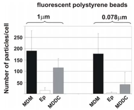
Since we had seen a qualitative difference in the number of intracellular particles within the different cell types, the number of fluorescently labeled polystyrene particles of different sizes (1 µm, and 0.078 µm) was counted using a specific software, called "DiaCount", which allows to pre-cisely and reliably count thousands of objects in 3D image stacks created by LSM. As for 1 µm particles we found 190 ± 90 (SD) particles in MDM, 116 ± 41 particles in MDDC, and 15 ± 12 particles in epithelial cells (Fig. 2). For 0.078
µm particles we found: 177 ± 90 particles in MDM, 41 ± 55 particles in MDDC, and 4 ± 4 particles in epithelial cells (Fig. 2).
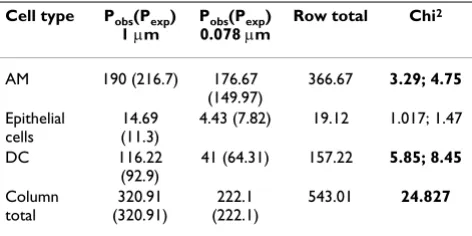
Contingency table analysis for polystyrene fine particles and nanoparticles (Table 1)
With 3-1 compartments (cell types) times 2-1 groups (dif-ferently sized particles) = 2 degrees of freedom, and a total
chi squared value of 24.83 the null-hypothesis that the differently sized particles are similarly distributed among the different cells has to be rejected (p < 0.01). The partial chi-squared value contributed 10% or more to the total chi-squared for both particle sizes in macrophages and MDDC. For 1 µm particles there were fewer particles than expected in macrophages and more particles than expected in MDDC. In contrast, for 0.078 µm particles, there were more particles than expected in MDM and fewer particles than expected in MDDC.
Intracellular localisation of differently composed nanoparticles
In order to analyse whether the material of the NP deter-mines its intracellular localization, TiO2 NP(mean diame-ter of 0.032 µm) and colloidal gold NP (diameter of 0.025
µm) were visualized and analysed in cells using EFTEM [25]
Bigger membrane-bound aggregates (>0.2 µm) of TiO2 NP were identified by analytical TEM in all cell types, i.e. MDM, epithelial cells and MDDC (Fig. 3A). In addition we found single particles and smaller (<0.2 µm) aggre-gates that were not membrane bound (Fig. 3A). Interest-ingly, silver enhanced gold particles were not observed in vesicles they were only detected as single particles or as small aggregates (<0.1 µm) free in the cytoplasm (Fig. 3B).
Quantification of particles inside individual cells
Figure 2
Quantification of particles inside individual cells. Intracellular particle numbers were analysed in cultures exposed to 1 µm, and 0.078 µm polystyrene particles with the software Diacount. Epithelial cells (Ep). Number of intracellular parti-cles was counted in individual cells. Data are expressed as the mean of 3–4 experiments (10–14 cells scanned per experiment by LSM).
Intracellular particle localisation in tripe cell co-cultures visu-alised by LSM
Figure 1
Some gold particles were even found in the cell nucleus (Fig. 3B arrow).
Influence of particle size and composition on the inflammatory potential
As shown above, the morphological examination of the particle-cell interactions showed that all particle types
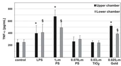
used in this study are able to enter the different cells of the co-culture system. Therefore, we determined the pro-inflammatory cytokine TNF-α in the culture supernatants after incubations with particles for 24 h (Fig. 4). In the control cultures minor TNF-α concentrations were meas-ured. When LPS (lipopolysaccaride, a positive control) and 1 µm particles were added to the cell cultures, the TNF-α signal increased significantly. No increase in
TNF-α concentration was seen when 0.078 µm polystyrene par-ticlesor TiO2 NP were added to the cultures. We found a significant increase of TNF-α in the supernatants after applying gold NP.
Discussion
The understanding of the possible functional and patho-logical disorders induced in the respiratory tract by NP requires the investigation of the direct effects of these par-ticles on state and activity of lung cells. Therefore, we have used our established in vitro model of the airway wall [37] to compare the translocation behaviors of fine particles and NP, to study the intracellular localization of NP and to investigate the potential of the particles in dependence on size and material to induce a pro-inflammatory response.
First, fine fluorescently labeled polystyrene particles (1
µm) and NP (0.078 µm) have been added to the cells and visualized by LSM combined with digital image restora-tion [25]. Both particle sizes were found in all three cell types, however, the number of particles was always con-siderably smaller in the epithelial cells than in both other cell types. Quantitative analysis revealed that the number
TNF-α release in triple cell co-cultures upon particle incuba-tion
Figure 4
TNF-α release in triple cell co-cultures upon particle incuba-tion. TNF-α levels in the supernatants (upper chamber, lower chamber) were measured by ELISA. TNF-α release in cells exposed to LPS, 1 µm, and 0.078 µm polystyrene parti-cles, TiO2, and gold NP. Values are means ± SD of 3 experi-ments. * indicates a statistical difference to the levels in the supernatants in the control of the upper chamber, § indicates
a statistical difference to the levels in the supernatants in the control of the lower chamber.
EELS images of cells containing TiO2 and silver enhanced gold
particles
Figure 3
EELS images of cells containing TiO2 and silver enhanced gold
particles. TiO2 particles (A) were found inside all cell types,
of 1 µm and 0.078 µm particles inside MDM was twice the number found in MDDC. This finding confirms what was reported by Kiama et al. [39] who showed that MDM are twice as phagocytic as immature MDDC in vitro. Only a small particle number was found inside epithelial cells for both particle sizes. The quantitative analysis for the NP distribution in the different cell types was performed as described by Mühlfeld et al. [40]. One main conclusion that can be drawn from this analysis is that the distribu-tion of particles within the different cell types is not equal among the different particle sizes. The difference is char-acterized by a significant lower number of NP to be local-ized in the MDDC in comparison to the larger particles. Conversely, the number of particles localized in MDM was higher than expected for 0.078 µm NP and lower than expected for the larger particles. Recently, we have shown that MDDC and MDM collaborate as sentinels against fine particles by building a transepithelial interdigitating network of cell processes [41], so the current data under-line that fine particles might actively be transported from MDM to MDDC, whereas the nano-sized material has dif-ferent translocation characteristics. It is tempting to spec-ulate that the unique entering mechanisms of NP may prevent the physiological interplay between macrophages and dendritic cells to a certain degree. Concluding from our results and from earlier published findings [11,25] we confirmed the phagocytic uptake for 1 µm particles whereas particles < 0.1 µm may have the property of enter-ing cells by unknown mechanisms (what we called adhe-sive interaction).
All polystyrene particle types we used in this study were observed in MDDC in the triple cell co-culture system, although the MDDC, residing underneath the epithelial monolayer, were thought not to be directly exposed to particles. Since the TEER did not decrease during the experiment we assume that the tight junctions were not opened after addition of particles to the medium. How-ever, our studies showed that MDDC formed fine cyto-plasmic processes towards the luminal side or even migrated as complete cells to the apical side of the epithe-lial barrier wall to take up particles of a diameter of 1 µm deposited on the epithelial apical surface [41]. However, it was the tightness of the epithelium which influenced the migration index of the MDDC, but not the mecha-nism of particle uptake [41]. Whether the cells are inter-acting in a similar manner after exposure of the cultures to NP is the aim of further investigations.
In order to draw any conclusions whether NP induce cel-lular responses it is indispensable to know if particles are attached to the cell membrane or have entered the cells. Sophisticated microscopic methods have been applied for the visualization of manufactured NP as described in Rothen-Rutishauser et al. [25]: LSM in combination with image restoration for fluorescently labeled polystyrene NP, and EFTEM for gold NP as well as for TiO2 NP. As shown in LSM and TEM micrographs, polystyrene, gold and TiO2 NP can enter cells, even the non-phagocytic A549 epithelial cells. This confirms previous reports that NP can enter many cell types [11,42], even non-cytic cells like red blood cells which have neither phago-cytic receptors at the surface nor are they equipped with a phagocytic apparatus [25]. For some NP the cell mem-brane does not seem to exert a barrier function; it seems as if they just pass through the membrane. We have currently no idea how these particles penetrate through the cell membrane. The entering mechanisms are currently under debate. Some assume these particles to enter cells by any endocytic processes other suggest mechanisms different from endocytosis like diffusion, membrane fluidity, pass-ing through channels or further by adhesive interactions as for instance electrostatic forces, Van der Waals- or steric interactions [11,43,44]. Clearly, the entering mechanisms of NP need to be further investigated.
TiO2 was found inside all three cell types of the triple cell co-culture model, i.e. MDM, epithelial cells, and MDDC, as membrane-bound larger aggregates as well as free as smaller aggregates or individual particles in the cyto-plasm. Other studies have shown vesicles containing mostly large aggregates of TiO2 in A549 cells [45] or mem-brane-bound ceria NP agglomerates in human lung fibroblasts [46]. However, in all these studies only con-ventional TEM was used to detect the particles, and it might well be that single NP or small aggregates of few
Table 1: Contingency table analysis of particle numbers in different cell types among the particle sizes
Cell type Pobs(Pexp)
1 µm
Pobs(Pexp)
0.078 µm
Row total Chi2
AM 190 (216.7) 176.67
(149.97)
366.67 3.29; 4.75
Epithelial cells
14.69 (11.3)
4.43 (7.82) 19.12 1.017; 1.47
DC 116.22
(92.9)
41 (64.31) 157.22 5.85; 8.45
Column total
320.91 (320.91)
222.1 (222.1)
543.01 24.827
In the first column, the different cell types, i.e. MDM, epithelial cells, or MDDC, are denoted. In each of the cell types, the mean number of particles was counted (Pobs). According to the equation given in
the methods section the number of expected particles was calculated (Pexp). To see whether the distribution of counted particles within the three cell types was different for the different particle sizes, partial chi2 values and total chi2 were calculated. For 2
particles could not be identified. In our study gold NP were detected only free in the cytoplasm of all cell types. These findings are in contrast to the study from Takenaka et al. [47] who detected gold NP aggregates inside small vesicles in macrophages of the rat lung. It is not clear why we could not detect vesicle containing gold particles, how-ever, it might be that in the study from Takenaka et al. [47] single NP or small aggregates were not detectable by con-ventional TEM in the cytoplasm. We also found gold par-ticles inside the cell nuclei. Penetration of NP into the nucleus has been shown in a number of studies [11,27-29]. Tsoli and colleagues[28] have shown that 1.4 nm Au55 particle clusters interact with DNA in a way which may be the reason for the strong toxicity of these tiny par-ticles towards human cancer, since it is generally known that DNA double-strand breaks may cause cancer [48]. Of great interest is the stereological analysis of TiO2 and gold NP, which is planned in future studies.
A crucial reaction of cells to particles deposited in the lung is the release of cytokines from activated epithelial cells, MDM and MDDC. The essential role of the pro-inflamma-tory chemokine TNF-α in relation to lung injuries caused by ambient UFP or manufactured NP was described by Donaldson and colleagues [19]. In our studies TNF-α release after exposure to 1 µm polystyrene particles was significantly higher than in cultures exposed to 0.078 µm particles. Moreover, varying responses have been found with different types of NP, like polystyrene, gold, and TiO2 particles. Interestingly, only the exposure to gold NP was found to induce a pro-inflammatory response.
In conclusion, all particle types were detected inside the three cells of the triple cell co-culture model independent of their composing material and size. However, the distri-bution of fine and nano-sized polystyrene particles among the cells of the culture system was different sug-gesting size to be an important factor determining the translocation characteristics of particles. Additionally, the intracellular localization differed between TiO2 and gold NP suggesting different modes of intracellular trafficking and, possibly, entering mechanisms depending on the particle material. In accordance, both size and material of the particles affected the toxicological potential of the par-ticles, i.e. the TNF-α generation. Whether the different inflammatory potential between differently composed NP is related to their entering mechanism and intracellular target structures needs further research.
Methods
Triple cell co- cultures
Cultures were prepared as previously described by Rothen-Rutishauser et al. [37]. Briefly, A549 cells (passage 10–40) were grown on cell culture inserts (surface area of 4.2 cm2, pores of 3.0 µm in diameter, high pore density
PET membranes for 6-er well plates; (BD Biosciences, Basel, Switzerland). MDM and MDDC were derived from human blood monocytes as already described [37]. Briefly, peripheral blood monocytes were isolated from buffy coats (blood donation service, Berne, Switzerland) and cultured in the same medium as used for the epithe-lial cells except for the supplementation of 5% human serum (blood donation service Bern, Switzerland) instead of 10% foetal calf serum. For the generation of MDDC the monocytes were cultured for 7–10 d in medium supple-mented with 34 ng/mL IL-4 (Sigma, Fluka Chemie GmbH, Buchs, Switzerland) and with 50 ng/mL GM-CSF (R&D Systems, Oxon, UK), whereas the MDM were obtained without any additional supplements for 7–10d. Epithelial cells were cultured for 7 days before MDM were added on top of the epithelial monolayer and MDDC underneath the insert membrane. The triple cell co-cul-tures were kept overnight in medium supplemented with 1% L-Glutamine, 1% penicillin/streptomycin, and 5% heat inactivated (pooled) human serum at 37°C in 5% CO2 humidified atmosphere.
Particle incubation
Commercially available particles were used: Fluoresbrite™ plain yellow green microspheres with diameters of 1 µm (Polysciences, Chemie Brunschwig AG, Basel, Switzer-land); Fluorescent particles, yellow green with a mean diameter of 0.078 µm (KiskerGbR, Chemie Brunschwig AG, Basel, Switzerland); Bovine Serum Albumin (BSA) gold tracers with a diameter of 0.025 µm (Aurion, Anawa Trading SA, Wangen, Switzerland); Titanium (IV) oxide (TiO2), anatase 99.9% (metal basis)) with a mean diame-ter of 0.02–0.03 µm (Alfa Aesar, Johnson Matthey GmbH, Karlsruhe, Germany).
All particle dilutions were sonicated for 2 min prior to incubation with cells in order to avoid aggregation. Poly-styrene particles were diluted in RPMI 1640 medium and adjusted to a concentration of 1010 particles/mL. As rec-ommended from the company gold particles were first dialyzed in PBS for 24 hours to remove the sodium acid and were diluted in RPMI 1640 to a final dilution of 1010 particles/mL. A stock solution of TiO2 particles in milli-pore water (2.5 mg/mL) was diluted in RPMI 1640 to a final concentration of 5 µg/mL. One mL of this suspen-sion was then added to cell cultures. Incubations were done for 24 h before analysis of the cells. Each experiment was repeated between 3 to 5 times.
TEER measurements
the surface area of the inserts (4.2 cm2). Electrical resist-ance was measured in triple cultures to follow the epithe-lial tightness as described in Rothen-Rutishauser et al. [49].
Cell labeling and fixation
Cells were washed in phosphate buffered saline (PBS, 10 mM, pH 7.4: 130 mM NaCl, Na2HPO4, KH2PO4) and fixed for 15 min. at room temperature in 3% paraformal-dehyde in PBS. Fixed cells were treated with 0.1 M glycine in PBS for 5 min. and permeabilized in 0.2% Triton X-100 in PBS for 15 min. The cells were incubated with the first and second antibodies for 60 min each at room tempera-ture. Preparations were mounted in PBS:glycerol (2:1) containing 170 mg/mL Mowiol 4–88 (Calbiochem, VWR International AG).
Antibodies were diluted in PBS as follows: mouse anti-human CD14 1:20 (Clone UCHM-1, C 7673, Sigma), mouse anti-human CD86 1:20 (Clone HB15e, 36931A, PharMingen, BD Biosciences,), goat anti-mouse cyanine 5 1:50 (AP124S, Chemicon, VWR International AG, Life Sci-ences, Lucerne, Switzerland), and phalloidin rhodamine 1:100 (R-415, Molecular Probes, Invitrogen AG, Basel, Switzerland).
Tumor necrosis factor alpha detection
Following particle incubation, supernatants from triple cell co-cultures in the upper and lower chamber were col-lected separately and stored at -70°C. After centrifugation, TNF-α was quantified by a commercially available DuoSet ELISA Development kit (R&D Systems, Catalogue Number: DY 210, Oxon, UK) according to the manufac-turer's recommendations. The assay was repeated twice, each in duplicates. Between three to five experiments were carried out for each combination of particles and cell types. The incubation time of the cells with the particle suspension was 24 h.
The mouse anti-human TNF-α capture antibody was
coated overnight in 96-well immunoassay plates (NUNC, MaxiSorp) at a concentration of 4 µg/mL in PBS at room temperature. Differing to the producer's protocol the plate was blocked with PBS supplemented with 1% BSA, 5% sucrose and 0.05% sodium acid for 1 h at room tempera-ture (as opposed to the reagent dilution provided with the test kit). After washing with buffer, supernatants from samples and the standards (recombinant human TNF-α, concentrations from 0.02 to 10 ng/mL of TNF-α) were pipetted into the wells and incubated at room tempera-ture for 2 h. After washing, biotinylated goat anti-human TNF-α detection antibodies were added to the wells and incubated for 2 h at room temperature. After washing, horseradish peroxidase conjugated streptavidin was added to the plates, which were then incubated for 20 min
at room temperature in the dark. Finally, the substrate solution (Tetramethylbenzidine/H2O2 (R&D Systems, Art.Nr. DY999)) was added. After 20 min in darkness, the colour development was stopped by 2NH2SO4 and the plate was put on the shaker for 10 min (differing from the protocol). Then the absorbance was read at 450 nm using an ELISA reader (SpectraMax 340 PC or Benchmark Plus Microplate Spectrophotometer (BioRad, Hempel Hemp-stead, UK)). The concentration of TNF-α was determined by comparing the absorbance of the samples with stand-ard recombinant human TNF-α and calculated with the Office Excel program from Microsoft.
Laser scanning microscopy and image restoration
A Zeiss LSM 510 Meta with an inverted Zeiss microscope (Axiovert 200 M, Lasers: HeNe 633 nm, HeNe 543 nm, and Ar 488 nm) was used. Image processing and visuali-zation was performed using IMARIS, a 3D multi-channel image processing software for confocal microscopic images (Bitplane AG, Zurich, Switzerland). For the locali-zation and visualilocali-zation of particles at high resolution a deconvolution algorithm was applied [25] using the Huy-gens 2 software (Scientific Volume Imaging B. V., Hilver-sum, Netherlands) in order to increase axial and lateral resolutions and to decrease noise.
Particle quantification
After the image acquisition the total particle number in the scans was counted with the particle tracking software Diacount (Semasopht, Lausanne, Switzerland; http:// www.semasopht.com). For each experimental sample cells were randomly scanned with LSM. Experiments were performed in triplicates or quadruplicates, and 10–15 cells were scanned for each data point. The particles were counted within individually defined cell types, which were labelled for specific cell surface markers (CD14 for MDM, and CD86 for MDDC, F-Actin for the epithelial cells).
Energy filtering transmission electron microscopy
reagent (AURION R-GENT SE-LM; Aurion, Anawa trading SA).
Statistics
The results of the TEER and the ELISA measurements are expressed as mean values with the standard deviation of the mean (SD). The statistical analysis was performed using SigmaStat for Windows (Version 3.10, Systat Soft-ware, Inc., Richmond, California, USA) statistical soft-ware. Two groups were compared using Student's t-Test. p < 0.05 was considered to be significant.
The distribution of the different particles among the dif-ferent cell types was compared using a contingency table analysis as described by Mühlfeld et al. [40]. The observed numbers of polystyrene particles were compared by use of a contingency table analysis to depict changes in particle distribution among the different cell types according to the size group of the particles. From the number of observed particles, the number of expected particles was calculated according to the equation: column total x row total/grand total = expected number of particles. Partial chi-squared values for each cell type and particle size are given by: (observed particles – expected particles)2/ expected particles = partial chi squared value. From the partial chi squared values the total chi squared value was calculated by summing up the partial values. The total chi squared value indicated whether the distributions of par-ticles among the different cell types differed depending on particle size. The partial chi squared values helped to iden-tify those compartments that contributed substantially to the different distributions. A convenient cut-off value for a substantial contribution is given by 10% or more of the total chi-squared.
List of abbreviations
BSA Bovine serum albuminEFTEM Energy filtering transmission electron microscopy
LSM Laser scanning microscopy
MDDC Monocyte derived dendritic cells
MDM Monocyte derived macrophages
NP Nanoparticles
PBS Phosphate buffered saline
TiO2 Titanium dioxide
TEM Transmission electron microscopy
TNF-α Tumour necrosis factor alpha
UFP Ultrafine particles
Competing interests
The author(s) declare that they have no competing inter-ests.
Authors' contributions
BRR carried out the design of the study, has done the acquisition of the data, the analysis and interpretation of data, and drafted the manuscript. ChM has performed the contingency table analysis; he has been involved in the analysis and interpretation of data, as well as drafting the manuscript. FB has been involved in the analysis and interpretation of data, and in revising the manuscript crit-ically for important intellectual content. CM has per-formed the ELISA measurements and has done the data analysis. PG was the project leader, he has intellectually accompanied the experimental work; he has been involved in revising the manuscript critically for impor-tant intellectual content and has given final approval of the version to be published.
Acknowledgements
We are grateful to Barbara Tschirren, Andrea Stokes, Claudia Haller, Beat Haenni and Sandra Frank for their excellent technical assistance.
This work was supported by the Swiss National Science Foundation, the Swiss Agency for the Environment, the Silva Casa and the Johanna Dürmüller-Bol Foundation.
References
1. Mazzola L: Commercializing nanotechnology. Nat Biotechnol
2003, 21:1137-1143.
2. Paull R, Wolfe J, Hebert P, Sinkula M: Investing in nanotechnol-ogy. Nat Biotechnol 2003, 21:1144-1147.
3. Gwinn MR, Vallyathan V: Nanoparticles: Health Effects-Pros and Cons. Environ Health Perspect 2006, 114:1818-1825.
4. Moghimi SM, Hunter AC, Murray JC: Nanomedicine: current sta-tus and future prospects. FASEB J 2005, 19:311-330.
5. Maynard A, Michelson E: The Nanotechnology Consumer Prod-ucts Inventory. 2005 [http://www.nanotechproject.org/ index.php?id=44].
6. Nicod LP: Lung defenses: an overview. Eur Respir Rev 2005,
95:45-50.
7. Gehr P, Bachofen M, Weibel ER: The normal human lung: ultrastructure and morphometric estimation of diffusion capacity. Respir Physiol 1978, 32:121-140.
8. Peters A, Wichmann HE, Tuch T, Heinrich J, Heyder J: Respiratory effects are associated with the number of ultrafine particles. Am J Respir Crit Care Med 1997, 155:1376-1383.
9. Wichmann HE, Spix C, Tuch T, Wolke G, Peters A, Heinrich J, Krey-ling WG, Heyder J: Daily mortality and fine and ultrafine parti-cles in erfurt, germany part I: role of particle number and particle mass. Res Rep Health Eff Inst 2000, 98:5-86.
10. Schulz H, Harder V, Ibald-Mulli A, Khandoga A, Koenig W, Krombach F, Radykewicz R, Stampfl A, Thorand B, Peters A: Cardiovascular effects of fine and ultrafine particles. J Aerosol Med 2005,
18:1-22.
11. Geiser M, Rothen-Rutishauser B, Kapp N, Schurch S, Kreyling W, Schulz H, Semmler M, Im Hof V, Heyder J, Gehr P: Ultrafine parti-cles cross cellular membranes by nonphagocytic mecha-nisms in lungs and in cultured cells. Environ Health Perspect 2005,
113:1555-1560.
Publish with BioMed Central and every scientist can read your work free of charge "BioMed Central will be the most significant development for disseminating the results of biomedical researc h in our lifetime."
Sir Paul Nurse, Cancer Research UK
Your research papers will be:
available free of charge to the entire biomedical community
peer reviewed and published immediately upon acceptance
cited in PubMed and archived on PubMed Central
yours — you keep the copyright
Submit your manuscript here:
http://www.biomedcentral.com/info/publishing_adv.asp
BioMedcentral clearance through microvasculature. Particle Fibre Toxicol 2007
in press.
13. Brown JS, Zeman KL, Bennett WD: Ultrafine particle deposition and clearance in the healthy and obstructed lung. Am J Respir Crit Care Med 2002, 166:1240-1247.
14. Kreyling WG, Semmler M, Erbe F, Mayer P, Takenaka S, Schulz H, Oberdorster G, Ziesenis A: Translocation of ultrafine insoluble iridium particles from lung epithelium to extrapulmonary organs is size dependent but very low. J Toxicol Environ Health A
2002, 65:1513-1530.
15. Oberdoerster G, Sharp Z, Atudorei V, Elder A, Gelein R, Kreyling W, Cox C: Translocation of inhaled ultrafine particles to the brain. Inhal Toxicol 2004, 16:437-445.
16. Pope CA III, Dockery DW, Schwartz J: Review of epidemiological evidence of health effects of particulate air pollution. Inhal Toxicol 1995, 7:1-18.
17. Lighty JS, Veranth JM, Sarofim AF: Combustion aerosols: factors governing their size and composition and implications to human health. J Air Waste Manag Assoc 2000, 50:1565-1618. 18. Borm PJ, Kreyling W: Toxicological hazards of inhaled
nanopar-ticles – potential implications for drug delivery. J Nanosci Nan-otechnol 2004, 4:521-531.
19. Donaldson K, Tran L, Jimenez LA, Duffin R, Newby DE, Mills N, Mac-Nee W, Stone V: Combustion-derived nanoparticles: a review of their toxicology following inhalation exposure. Part Fibre Toxicol 2005, 2:10.
20. Nel A, Xia T, Madler L, Li N: Toxic potential of materials at the nanolevel. Science 2006, 311:622-627.
21. Oberdoerster G, Oberdoerster E, Oberdoerster J: Nanotoxicol-ogy: an emerging discipline evolving from studies of ultrafine particles. Environ Health Perspect 2005, 113:823-839.
22. Kreyling WG, Semmler-Behnke M, Moller W: Ultrafine particle-lung interactions: does size matter? J Aerosol Me 2006, 19:74-83. 23. Foged C, Brodin B, Frokjaer S, Sundblad A: Particle size and sur-face charge affect particle uptake by human dendritic cells in an in vitro model. Int J Pharm 2005, 298:315-322.
24. Kapp N, Kreyling W, Schulz H, Im Hof V, Gehr P, Semmler M, Geiser M: Electron energy loss spectroscopy for analysis of inhaled ultrafine particles in rat lungs. Microsc Res Tech 2004,
63:298-305.
25. Rothen-Rutishauser BM, Schürch S, Haenni B, Kapp N, Gehr P: Inter-action of fine and nanoparticles with red blood cells visual-ized with advanced microscopic techniques. Environ Sci Technol
2006, 40:4353-4359.
26. Li N, Sioutas C, Cho A, Schmitz D, Misra C, Sempf J, Wang M, Ober-ley T, Froines J, Nel A: Ultrafine particulate pollutants induce oxidative stress and mitochondrial damage. Environ Health Per-spect 2003, 111:455-460.
27. Gurr JR, Wang AS, Chen CH, Jan KY: Ultrafine titanium dioxide particles in the absence of phopactivation can induce oxida-tive DNA damage to human bronchial epithelial cells. Toxicol
2005, 15:66-73.
28. Tsoli M, Kuhn H, Brandau W, Esche H, Schmid G: Cellular uptake and toxicity of Au55 clusters. Small 2005, 8–9:841-844. 29. Liu Y, Meyer-Zalka W, Franzka S, Schmid G, Leis M, Kuhn H:
Gold-Cluster Degradation by the Transition of B-DNA into A-DNA and the Formation of Nanowires. Angew Chem Int Ed
2003, 42:2853-2857.
30. Muller J, Huaux F, Moreau N, Misson P, Heilier J-F, Delos M, Arras M, Fonseca A, Nagy JB, Lison D: Respiratory toxicity of multi-wall carbon nanotubes. Toxicol Appl Pharmacol 2005, 207:221-231. 31. Gonzalez-Flecha B: Oxidant mechanisms in response to
ambi-ent air particles. Mol Aspects Med 2004, 25:169-182.
32. Vinzents PS, Møller P, Sørensen M, Knudsen LE, Hertel O, Jensen FP, Schibye B, Loft S: Personal exposure to ultrafine particles and oxidative DNA damage. Environ Health Perspect 2005,
113:1485-1490.
33. Rahman I, MacNee W: Role of transcription factors in inflam-matory lung diseases. Thorax 1998, 53:601-612.
34. Brown DM, Wilson MR, MacNee W, Stone V, Donaldson K: Size-dependent proinflammatory effects of ultrafine polystyrene particles: a role for surface area and oxidative stress in the enhanced activity of ultrafines. Toxicol Appl Pharmacol 2001,
175:191-199.
35. Pope CA III, Burnett RT, Thurston GD, Thun MJ, Calle EE, Krewski D, Godleski JJ: Cardiovascular mortality and long-term
expo-sure to particulate air pollution: epidemiological evidence of general pathophysiological pathways of disease. Circulation
2004, 109:71-77.
36. Brown DM, Donaldson K, Borm PJ, Schins RP, Dehnhardt M, Gilmour P, Jimenez LA, Stone V: Calcium and ROS-mediated activation of transcription factors and TNF-alpha cytokine gene expression in macrophages exposed to ultrafine particles. Am J Physiol Lung Cell Mol Physiol 2004, 286:L344-L353.
37. Rothen-Rutishauser BM, Kiama SG, Gehr P: A three-dimensional cellular model of the human respiratory tract to study the interaction with particles. Am J Respir Cell Mol Biol 2005,
32:281-289.
38. Daniel MC, Astruc D: Gold Nanoparticles: Assembly, Supramolecular Chemistry, Quantum-Size-Related Proper-ties, and Applications toward Biology, Catalysis, and Nan-otechnology. Chem Rev 2004, 104:293-346.
39. Kiama SG, Cochand L, Karlsson L, Nicod LP, Gehr P: Evaluation of phagocytic activity in human monocyte-derived dendritic cells. J Aerosol Med 2001, 14:289-299.
40. Mühlfeld Ch, Mayhew TM, Gehr P, Rothen-Rutishauser B: A novel quantitative method for analysing the distribution of nano-particles between different tissue and intracellular compart-ments. J AerosolMed in press.
41. Blank F, Rothen-Rutishauser B, Gehr P: Dendritic cells and mac-rophages form a transepithelial network against foreign par-ticulate antigens. Am J Respir Cell Mol Biol 2007, 36:669-77. 42. Wottrich R, Diabate S, Krug HF: Biological effects of ultrafine
model particles in human macrophages and epithelial cells in mono- and co-culture. Int J Hyg Environ Health 2004, 207:353-361. 43. Rimai DS, Quesnel DJ, Busnaia AA: The adhesion of dry particles in the nanometer to micrometer–size range. Colloids and Sur-faces A. Physicochemical and Engineering aspects 2000, 165:3-10. 44. Rothen-Rutishauser B, Schurch S, Gehr P: Interaction of particles
with membranes. In The toxicology of particles Edited by: Donaldson K, Borm P. CRC Press LCC; 2007:139-160.
45. Stearns RC, Paulauskis JD, Godleski JJ: Endocytosis of ultrafine particles by A549 cells. Am J Respir Cell Mol Biol 2001, 24:108-115. 46. Limbach LK, Li Y, Grass RN, Brunner TJ, Hintermann MA, Muller M, Gunther D, Stark WJ: Oxide nanoparticle uptake in human lung fibroblasts: effects of particle size, agglomeration, and diffu-sion at low concentrations. Environ Sci Technol 2005,
39:9370-9376.
47. Takenaka S, Karg E, Kreyling WG, Lentner B, Moller W, Behnke-Semmler M, Jennen L, Walch A, Michalke B, Schramel P, Heyder J, Schulz H: Distribution pattern of inhaled ultrafine gold parti-cles in the rat lung. Inhal Toxicol 2006, 18:733-740.
48. Agarwal S, Tafel AA, Kanaar R: DNA double-strand break repair and chromosome translocations. DNA Repair (Amst) 2006,
5:1075-1081.