USING IN VITRO CULTURED LUNG CELL EXPOSURE
SYSTEMS TO COMPARE THE TOXICITY OF FRESH AND
AGED DIESEL EXHAUST UTILIZING AN
OUTDOOR SMOG CHAMBER
Kim Maureen Lichtveld
A dissertation submitted to the faculty of the University of North Carolina at Chapel Hill in partial fulfillment of the requirements for the degree of Doctor of Philosophy in the Gillings School of Global Public Health, the Department of Environmental Sciences and Engineering.
Chapel Hill 2012
Approved by:
Harvey E. Jeffries
Kenneth G. Sexton
David Leith
Ilona Jaspers
Michael Madden
ii
© 2012
iii
Abstract
KIM MAUREEN LICHTVELD: Using In Vitro Cultured Lung Cell Exposure Systems to Compare the Toxicity of Fresh and Aged Diesel Exhaust Utilizing an Outdoor Smog Chamber
(Under the direction of Harvey E. Jeffries and Kenneth G. Sexton)
Previous observations indicated that as urban-like atmospheres were oxidized, the
modifications to their chemical composition also affected their toxicological potential.
Based on these studies, I hypothesized that atmospheric oxidative processes will alter
the composition of urban-like atmospheres containing diesel exhaust, resulting in a
modification of biological responses from exposure.
The Electrostatic Aerosol in Vitro Exposure System (EAVES) efficiently deposits
particles with no significant biological response from any internal processes and only
produces a biological response when toxic PM pass through the sampler with the
depo-sition voltage field on. Direct comparison of direct exposure EAVES with the commonly
used traditional particle exposure method using a resuspension technique clearly
demonstrated that the biological response in the latter was changed by extensive
sam-ple handling during the resuspension method, Furthermore, the EAVES is produces a
biological effect from a much smaller sample.
This new methodology was coupled with a gas phase only exposure system to the
outdoor smog chamber to further test the overall hypothesis. Two test atmospheres
conditions and these systems were subsequently modified by either thermal (dark) or
photochemical oxidation. Changes were evident in biological effect and chemical
com-position, but varied by source and oxidative environment. The gas phase exposures
bio-logical responses were increased only from the Mercedes thermally aged condition and
in the fresh conditions for the Volkswagen. To understand the biological response from
the PM exposures I examined three methods of comparison to the biological response.
This indicated that aged atmospheres are significant in biological response and that the
primary and secondary carbonyls present during the exposure are important.
From these studies, I have demonstrated my hypothesis that atmospheric
pro-cesses do alter the chemical composition and biological effect of diesel exhaust oxidized
in an urban-like environment, with the qualifier that the degree of the effect is
depend-ent on the amount of toxic compondepend-ents deposited on the cells. Further, I can conclude
that the source and aging environment of the test atmosphere play important roles in
v
Dedication
To my family, for all their support through these past years. My mother who has been a guiding light and a source of encouragement. My father who has waited so patiently for me to finally finish school and get my papers.
My sister who always makes me laugh and keeps me grounded.
My brother whose inquisitive mind keeps me guessing bringing me laughter and thought.
To my fiancé for whom without I could not have made it. You are my rock and I know that there is no obstacle big or small that we cannot get through together, come out the
other end, and through it all keep calm and most of all never stop laughing. To IJE your sarcasm, wit, humor, and artistry pull me into your world of wonder and for
that I thank you for my mini mind vacations.
To the Wandering E’s I am so thankful to have you in my life. Your warm welcome and support is utterly amazing.
To my friends who have gone on this journey with me, thank you for all your support and encouragement! Thank you for the adventures, laughter, and gatherings that
ena-bled me to escape the throws of dissertating.
vi
Acknowledgements
I would like to thank Wenli Zhang, Missy Brighton, and Jonathan Ciencewicki from
CEMALB, and Maryanne Boundy from the Department of Environmental Sciences and
Engineering for all of their technical advice; and Beth Fowler from the Department of
Environmental Sciences and Engineering for administrative assistance; Richard Kamens
from Department of Environmental Sciences and Engineering and Southern States
Volkswagen for the use of their vehicles.
Chapter 2: This work was supported in part by the US Environmental Protection
Agency Cooperative Agreements R829762 and CR829522, American Chemistry Council
Long Range Research Initiative, Project ID CIE-0102-02, and Exxon Mobil Foundation
Basic research support gift. Although the research described in this article has been
funded wholly or in part by the United States Environmental Protection Agency through
cooperative agreement CR829522 with the Center for Environmental Medicine, Asthma,
and Lung Biology, it has not been subjected to the Agency's required peer and policy
review, and therefore does not necessarily reflect the views of the Agency and no
offi-cial endorsement should be inferred. Mention of trade names or commeroffi-cial products
vii
Chapter 3 and 4: This work was supported in part by the U.S. Environmental
Pro-tection Agency Cooperative Agreements R829762 and CR829522; American Chemistry
Council Long Range Research Initiative, project ID CIE-0102-02; National Institute of
En-vironmental Health Sciences T32 ES007018; the Gillings School of Global Public Health’s
Gillings Innovation Laboratory Program for Research and Innovations Solutions; and
Exxon Mobil Foundation Basic Research support gift. Although the research described in
this article has been funded in part by the U.S. Environmental Protection Agency
through cooperative agreement CR829522 with the Center for Environmental Medicine,
Asthma, and Lung Biology (CEMALB), it has not been subjected to the agency’s required
peer and policy review, and therefore does not necessarily reflect the views of the
agency and no official endorsement should be inferred. Mention of trade names or
viii
Table of Contents
List of Tables ... xi
List of Figures ... xii
Chapter 1 : Background and Significance ... 2
Rationale ... 2
Diesel Exhaust Composition ... 2
Particulate Matter in the Lung ... 4
Cell Lines as a Model for the Lung ... 5
In Vitro Particulate Exposure Methods ... 7
Other ESP Exposure Devices and Ambiguities ... 11
Cascaded Health Effects Models ... 12
Hypothesis... 17
Approach ... 18
Objectives... 19
Chapter 2 : Design and Testing of Electrostatic Aerosol In Vitro Exposure System (EAVES): An Alternative Exposure System for Particles ... 21
ix
Materials and Methods ... 24
Results ... 29
Discussion... 33
Figures and Tables ... 37
Chapter 3 : In Vitro Exposures in Diesel Exhaust Atmospheres: Resuspension of PM from Filters Verses Direct Deposition of PM from Air ... 43
Introduction ... 43
Materials and Methods ... 46
Results ... 53
Discussion... 55
Supporting Information ... 60
Figures and Tables ... 61
Chapter 4 : Atmospheric Chemistry Modifies Diesel Exhaust's Biological Effects ... 71
Introduction ... 71
Materials and Methods ... 76
Results ... 82
Discussion... 88
Conclusions ... 100
Figures and Tables ... 102
x
Hypothesis... 114
Summary of findings ... 116
Conclusions ... 120
Limitations... 124
Implications ... 127
Future work ... 130
Appendix Example Calculations ... 133
xi
List of Tables
Table 2-1. EAVES tests results for particle deposition using two different size fluorescent-labeled polystyrene latex spheres aerosolized in air.
Sample duration was one hour. ... 41
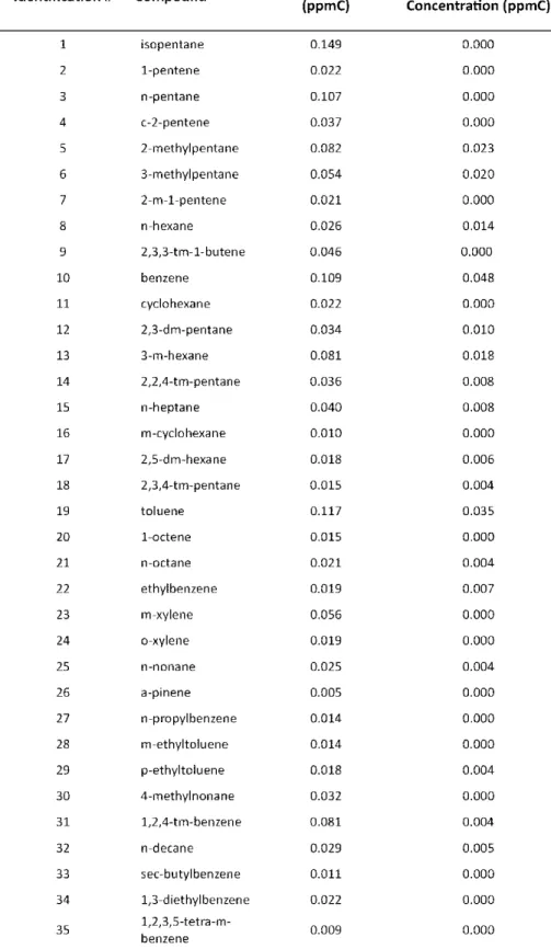
Table 3-1. Peak identifications of the hydrocarbon peaks in Figure 2 ... 63
Table 3-2. Identifications of the carbonyl peaks in Figure 3 ... 65
Table 4-1. List of carbonyl compounds detected in the Mercedes fresh (M-F) and photochemically oxidized (M-P) exposure test atmospheres.
Measurements reported in ppmC. ... 105
Table 4-2. List of carbonyl compounds detected in the Volkswagen fresh (V-F) and thermally oxidized (V-D) exposure test atmospheres.
Measurements reported in ppmC. ... 107
Table 4-3. List of carbonyl compounds detected in the Volkswagen fresh (V-F) and photochemically oxidized (V-P) exposure test atmospheres.
xii
List of Figures
Figure 1-1. Conceptual model ... 14
Figure 2-1: Top view image of the Electrostatic Aerosol in Vitro Exposure System
(EAVES) holding the four Millicells ... 37
Figure 2-2: Side view schematic of the EAVES with key components labeled. (1) inlet, (2) diffusers, (3) corona wire, (4) corona power plate, (5) in vitro cell holding well, (6) repellant plate, (7) particle collection plate, (8) outlet, (9) particle pathway. Air flow through the system is from
left to right. ... 37
Figure 2-3: Schematic of outdoor environmental irradiation chamber, through-the-roof sample lines, in-laboratory incubator holding the in vitro
exposure systems. ... 38
Figure 2-4: Effects of EAVES deposition field on cytotoxicity and inflammatory mediator production. A549 cells were exposed to clean air in the EAVES for one hour and tested for LDH and IL-8 release 9 hours post-exposure. Grey bars = EAVES turned on; black bars = EAVES turned off. LDH and IL-8 levels are expressed as fold increase over
non-exposed incubator control measurements. ... 38
Figure 2-5: Reconstructed relative ion current of m/z 181 ion chromatograms of carbonyls detected by PFBHA-derivatization of diesel exhaust gases
before (top) and after (bottom) passing through the EAVES. ... 39
Figure 2-6: Histograms of particle number per size interval of particles entering and exiting EAVES. Black histogram = PSL spheres exiting the EAVES with the power off; Grey histogram = PSL spheres exiting the EAVES
with the charging and deposition field on... 39
Figure 2-7: IL-8 release from A549 epithelial cells exposed to charged
polystyrene latex spheres (PSL). Clear bar = incubator control; Grey bar = cells exposed to PSL spheres operating with the power applied to the charging plate. A549 cells were exposed in the EAVES for one hour and examined for IL-8 release 9 hours post-exposure. Data
represent means + S.E.M. ... 40
xiii
increases over non-exposed incubator control measurements. * statistically different from non-exposed incubator control; #
statistically different from EAVES power off; p < 0.05. ... 40
Figure 3-1. Schematic diagram of the outdoor smog chamber, the laboratory sampling systems, and the biological exposure system (EAVES). Filters were collected directly under the chamber floor and
processed post-collection for the exposures to resuspended PM. The diesel exhaust sample injection lines into the chamber are also
depicted. ... 61
Figure 3-2. Gas chromatogram of VOC species in outdoor chamber. Top: after injection of exhaust from 1980 Mercedes (50 seconds) and 2.0 ppmC injection of SynUrb54 mixture, but before sunrise. Bottom: in dark after daylong sunlit reaction. The peak numbers correspond to the species listed in Table 1. Major compounds present in the
chromatogram labeled injected were: 1 – iso-pentane, 10 – benzene, 19 – toluene, 23 – m-xylene, and 31 – 1,2,4-tri-methyl-benzene. After photochemical aging the species either completely reacted away or
had significantly decreased. ... 62
Figure 3-3. Selective-ion chromatogram for the injections from the 1980 Mercedes and 2.0 ppmC of SynUrb54 in the chamber. of carbonyls from mister samples. Samples were collected after daylong
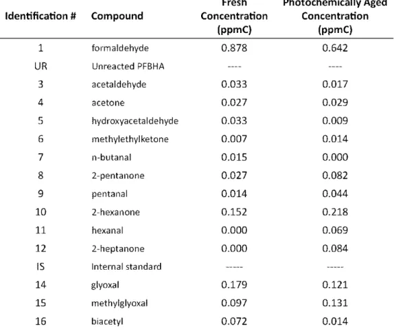
irradiation. The peak numbers correspond to the species identified in Table S2. UR – Unreacted PFBHA solution, IS – Internal Standard, and some of the major peaks are 1 – formaldehyde, 3 – acetaldehyde, 6 – methyl ethyl ketone, 8– 2-pentanone, 10 – 2-hexanone, 14 – glyoxal,
and 15 – methylglyoxal. ... 64
Figure 3-4. Selective-ion chromatogram of carbonyls from a water blank, a F12K media blank, and a filter sample taken after day-long irradiation of the injections from the 1980 Mercedes and 2.0 ppmC of SynUrb54 in the chamber. The peak numbers over the peaks correspond to the identities in Table 2. UR – Unreacted PFBHA reagent, IS – Internal
Standard. ... 66
Figure 3-5. COX-2 and IL-8 m-RNA expression induced by exposure to directly deposited PM from the aged exposure in the 1980 Mercedes with SynUrb54 experiment and induced by resuspension exposures. The symbols * indicates statistically different from non-exposed
incubator control; # indicates statistically significant difference compared to resuspension exposures. The error bars represent the
xiv
Figure 3-6. COX-2 and IL-8 m-RNA expression induced by exposure to directly deposited PM from the aged exposure in the 2006 Volkswagen with SynUrb54 experiment and induced by resuspension exposures. The symbols * indicates statistically different from non-exposed
incubator control; # indicates statistically significant difference compared to resuspension exposures. The error bars represent the
mean ± standard error from the mean. ... 68
Figure 3-7. COX-2 and IL-8 m-RNA expression induced PM from the AGED Exposure in the 1980 Mercedes with SynUrb54 experiment from resuspension exposures at concentrations of 2.65 µg/cm2 and 42.48 µg/cm2. The symbols * indicates statistically different from non-exposed incubator control; # indicates statistically significant difference compared to resuspension exposures. The error bars
represent the mean ± standard error from the mean. ... 69
Figure 4-1. Each bar represents a full chamber experiment conducted on the date(s) and times indicated. M represents the use of the older Mercedes and V represents the use of the newer Volkswagen. Duration of diesel exhaust injections are given at the beginning of each bar. The green windows represent the exposure to the ‘fresh’ conditions (F); the dark gray bar represents aging in the dark; the yellow bar represents aging in sunlight; the purple windows represents the exposure to the ‘dark aged’ conditions; the blue
windows represents exposure to the “sun aged” conditions. ... 102
Figure 4-2.Each bar height represents average conditions during the five exposure periods labeled in Fig. 1. The left side plots are for the Mercedes emissions along with 2 ppmC of SynUrb54 VOC mixture and the right are the same for the Volkswagen with 2 ppmC of
SynUrb54 injected. ... 103
Figure 4-3. Carbonyls Detected in Photochemically Aged SynUrb54 Mixture and Exhaust of 1980 Mercedes. Selective-ion chromatogram for the injections from the 1980 Mercedes and 2.0 ppmC of SynUrb54 in the chamber. Mercedes fresh chamber contents is labeled (M-F), and Mercedes photochemically aged chamber contents is labeled (M-P). Samples were collected after daylong irradiation. The peak numbers correspond to the species identified in Table 1. UR – Unreacted PFBHA solution, IS – Internal Standard, and some of the major peaks are 1 – formaldehyde, 3 – acetaldehyde, 6 – methyl ethyl ketone, 8–
2-pentanone, 10 – 2-hexanone, 14 – glyoxal, and 15 – methylglyoxal. ... 104
xv
chamber. Samples were collected after thermal aging in the dark. The peak numbers correspond to the species identified in Table 2. UR – Unreacted PFBHA solution, IS – Internal Standard, and some of the major peaks are 1 – formaldehyde, 2 – acetaldehyde, 3 – acetone, 4– acrolein, 20 – glyoxal, and 21 – methylglyoxal Table2. List of carbonyl compounds detected in the Volkswagen fresh (V-F) and thermally oxidized (V-D) exposure test atmospheres. Measurements reported
in ppmC. ... 106
Figure 4-5. Carbonyls Detected in Photochemically Aged SynUrb54 Mixture and Exhaust of 2006 Volkswagen. Selective-ion chromatogram for the injections from the 2006 Volkswagen and 2.0 ppmC of SynUrb54 in the chamber. Samples were collected after daylong irradiation. The peak numbers correspond to the species identified in Table 3. UR – Unreacted PFBHA solution, IS – Internal Standard, and some of the major peaks are 1 – formaldehyde, 18 – 2-cyclohexene-1one, 20 –
glyoxal, and 21 – methylglyoxal Table 3. ... 108
Figure 4-6. All bars are IL-8 m-RNA expression induced by exposure and controls. The exposure conditions are labeled the same as in Figure 1. In both sets, the top bar plots are responses for gas-phase only exposures and the bottom bar plots are responses for the particle-only exposures. Exposure duration for gas-only exposures were 3-h. Exposure for particle phase was ca. 2.0 µg for the Mercedes and 1µg for the Volkswagen. The symbol * indicates statistically different from non-exposed control; # indicates statistically significant difference between fresh and oxidized exposures. The error bars
represent the mean ± standard error from the mean. ... 110
Figure 4-7. Relative PM exposure relative to the IL-8 inflammatory response using the EAVES device for the Mercedes and Volkswagen and all exposure conditions (fresh, thermally aged, and photochemically aged). The red diamonds represent the relative PM exposure to increase in response for the Mercedes exposures highlighted in the blue box, and the orange diamonds represent the Volkswagen exposures highlighted in the green box. Incubator controls are all at zero. A trend line was added to show association with an R2 value of
0.204. ... 111
Figure 4-8. Inflammatory response relative to PM exposure that was grouped by chamber condition (fresh, thermally aged, and photochemically aged) using the EAVES device. All Volkswagen exposures are
highlighted in green and Mercedes in blue. Incubator controls are all at zero. Trend lines were added to each grouping: Photochemical and thermal aging with a first order fit of R2 of 0.8905 and 0.8680, and
xvi
Figure 4-9. Inflammatory response relative to the carbonyl-weighted PM exposure concentration and subsequently grouped by chamber conditions (fresh, thermally aged, and photochemically aged). Trend lines were added to each grouping: Photochemical aging with an R2 of 0.9954, thermal aging with an R2 of 0.9763 and fresh exposures
Chapter 1
: Background and Significance
Rationale
Epidemiological studies have shown a positive relationship between
measure-ments of ambient particulate matter (PM) and adverse health effects in susceptible
hu-man populations (patients with chronic obstructive pulmonary disease, chronic
bronchi-tis, asthma, and cardiovascular disease) (Dockery 1994; Bates 1995; Pope 1995; Sarnat
2001; Ayres 2008; HEI Panel on the Health Effects of Traffic-Related Air Pollution 2010).
Epidemiology has used the increases in hospital admissions and daily mortality reported
during short term episodes of high PM in the ambient air to find a positive relationship
between PM and human health effects. Even though there seems to be a positive
corre-lation there is still no established biological mechanisms to explain the toxicity of PM to
humans and the evidence of carcinogenic potential is limited because of the difficulty in
eliminating the influence of confounding factors and estimating exposure (Oberdorster
1990; Salvi 1999; Bunger 2000). Despite confounding factors like cigarette smoke,
Sil-verman et al. reported a correlation between lung cancer in miners and their exposures
to diesel exhaust (Silverman 2012). This study went on to be an influential part of the
decision by the International Agency for Research on Cancer (a part of the World Health
Organization) to assign a Group 1 designation to diesel exhaust emissions (International
Agency for Research on Cancer 2012). A Group 1 classification means that diesel
2
While epidemiological studies have suggested that a number of sources or source
characteristics are correlated with observed health effects, subsequent
laboratory-based toxicological studies often fail to confirm these as causes at levels that might
oc-cur in the ambient environment (Morgan 1997, Salvi 1999, Laks 2008, Dybdahl 2004,
Carlsten 2008, Sunil 2009). Sarnet et al. suggests that the ambient measurements may
be a weak indicator for the actual amount a person may be exposed to during the
course of the day (Sarnat 2001).
In 2001, Mauderly requested that the scientific community take a “more
integrat-ed, or holistic, view of the air quality-health relationship” and that “epidemiology is
largely limited to testing associations between health outcomes and the few pollutants
that are measured routinely” (Mauderly 2001). This study took the approach of being
more holistic and integrated to obtain understanding the inflammatory response of
die-sel exhaust in an urban-like environment.
Diesel Exhaust Composition
Diesel exhaust (DE) is a highly complex mixture of gases and particles,
continuous-ly changing in composition as it ages in the atmosphere. PM from diesel exhaust
con-tains an elemental carbon core surrounded by “organic compounds (such as polycyclic
aromatic hydrocarbon (PAHs)), sulfate, nitrate, metals and other trace elements
(man-ganese, sulfur, and iron)” (EPA 2002). Elemental carbon is inert to atmospheric
degra-dation, whereas organic compounds can degrade by reactions with sunlight, ozone, and
other atmospheric processes (McDow 1995, Fan 1996a, Fan 1996b, Lee 2004). The
3
water vapor, nitrogen oxides, sulfur compounds, benzene, 1-3, butadiene, nitrosamines,
PAHs, nitro-PAHs, and other volatile organic compounds (VOCs) (EPA 2002). Among the
gaseous components emitted from diesel engines, VOCs containing a carbonyl
(alde-hydes and ketones) functional group are of particular interest because some carbonyls
are considered probable carcinogens that are also capable of producing non-cancer
health effects (International Agency for Research on Cancer 1989).
The characteristics and composition of engine exhaust emissions from gasoline-
and diesel-fueled motor vehicles differ significantly. Many factors---including engine
type, fuel, engine load and after-treatment--- influence the composition of the primary
atmospheric emissions. These composition changes will affect both photochemical
re-activity and toxicity of the aged atmospheric mixture ultimately inhaled by humans. For
example, the primary emissions from gasoline fueled engines contain fewer particulates
than the equivalent volume of diesel-fueled engine emissions. Gasoline exhaust is more
photochemically reactive, however, because it contains a large fraction of gaseous
monocyclic aromatic compounds, which after reaction, contribute significantly to
sec-ondary particle growth (Odum 1997a, Odum 1997b, Kleindinest 2002). Diesel emissions
also react photochemically, especially in the presence of other urban VOCs, and such
reactions are known to modify the diesel PM surface and composition significantly
(McDow 1995, Fan 1996a, Fan 1996b). While both gasoline and diesel emissions react
in the atmosphere, their chemical mechanisms can be very different, making it difficult
for modelers to predict the composition and toxic characteristics of the respective aged
4
Particulate Matter in the Lung
In the lung, particles have five major mechanisms of deposition: inertial impaction,
gravitational settling, Brownian diffusion, electrostatic attraction, and interception.
While these mechanisms all play a role in particle deposition in the lung, particle size
mass has the greatest impact as well as the ultimate location of deposition (EPA 2002).
Deposition in the respiratory tract may occur in various regions of the lung from
the extrathoracic, the tracheobronchial, and the alveolar regions (Lippmann 1977).
When ~ 2.5 µm particles are inhaled, the particles’ velocity are increased, and abrupt
directional changes caused by airway branching makes inertial impaction in the mucus
layer lining the extrathoracic and tracheobronchial regions of the lung likely. Particles
trapped in extrathoracic and tracheobronchial regions are carried to the back of the
throat on a pulsing ciliary membrane covered in mucus (Shaw 2005). Upon arrival,
par-ticles are swallowed and then cleared via the gastrointestinal tract. Removal of the PM
in the pulmonary region does not mean that there could not be a whole body burden of
exposure to PM as it passes through the body. As particle diameters decrease to less
than 1 µm, the particles are more likely to behave like gases, and therefore are most
likely to deposit by diffusion. This allows particles to travel deep into the alveolar region
of the lung, where the particles impair gas exchange (Shaw 2005).
In the lung, particle exposure will cause injury and the recovery process begins
with the recruitment of cells associated with inflammation which is regulated by lipid
and protein mediators such as cytokines. Cytokines are relatively small proteins that
5
walls and activate signal transduction mechanisms involved in intracellular
communica-tion. These cytokines are responsible for regulating cell differentiation, proliferation,
and cell secretory activities. Inflammatory markers, such as, Interleukin-6, Interleukin-8,
cyclooxygenase-II enzyme, and Tumor Necrosis Factor-α (IL-6, IL-8, Cox-2, and TNF-α),
are early mediators of the body’s response to injury and are expressed rapidly after
ex-posure to toxic agents (Salem 2006; Cao 2007; Zoran D. Ristovski 2011). These can be
expressed over hours and measured either in the supernatants or through mRNA
analy-sis. Injury of pulmonary epithelial cells results in loss of cell membrane integrity, which
in turn results in the release of intercellular lactate dehydrogenase (LDH). Thus,
in-creased release of LDH is considered to be a sign of cellular death and can be measured
directly in the lung lining fluid.
Cell Lines as a Model for the Lung
Two advantages of using in vitro models are that these models allow investigators: 1) to study the response of individual components of the respiratory system to inhaled
toxins, 2) to examine mechanisms by which these toxicants cause inflammation, and 3)
to address questions of lung toxicity in human lung cells at levels that might be unethical
for human subject involvement. Although in vitro models do not have the ability to ac-count for all of the interactions in the cells’ natural environment, they respond to a
stressful environment as they would in the body (by releasing inflammatory markers).
Therefore, cellular release of these cytokines represents a reasonable measurement to
6
despite the absence of these immune response-related cells from in vitro cultures (Lieber 1976; ATCC 2011).
Immortalized cell lines are useful because they can be maintained under cultured
conditions for an extended period of time. In addition, they are readily available
allow-ing for optimization of replication of research without uncertainty introduced by
inter-personal variability in response or interspecies extrapolation. A549 cells are a model of
alveolar epithelial cells with type II cell-like characteristics, and have been extensively
used to assess the toxicity of air pollutants (Doyle 2004; Sexton 2004; Bitterle 2006;
Doyle 2007). Type II cells are involved in pulmonary defense mechanisms by secreting
protein mediators that contribute to pulmonary inflammation (Lumb 2005). In short,
Type II cells are responsible for sending out help signals. In this project, A549 cells were
grown on membranous support using complete media. Upon confluence, the growth
medium was replaced by serum-free media several hours before exposure to slow
cellu-lar proliferation. The cell culture inserts (Transwells or Millicells) can be used to
estab-lish an air-liquid interface, which facilitates direct exposure to pollutants without
inter-ference from media covering the cells.
The liquid under the membrane of these inserts allows cells to be adequately
hy-drated and supplied with nutrients while being exposed to pollutants on the cells’ apical
side. Cellular signaling can be measured using the supernatants or the ribonucleic acid
(RNA) collected from the cells. Laboratory assays such as the LDH essay and Enzyme
re-7
leased by the cells. Real-time Reverse Transcription-Polymerase Chain Reaction (RT-PCR)
uses the RNA collected from the cells to measure intercellular production of mRNA
re-sponsible for mediator production.
In Vitro Particulate Exposure Methods
There are many methods for particle collection: gravitational settling, thermal
dif-fusion, thermophoresis, electrostatic precipitation, inertial impaction, and filter
collec-tion. The characteristics of each method affect their suitability for in vitro exposures. Each particulate exposure method was to be evaluated to assess the following: their
ability to efficiently collect and deposit particulate matter for specific particles of
inter-est (ie. 200-500nm diameter); any potential for damage to cell cultures during exposure;
and how each collection method might alter the particles’ primary state. A review of the
suitability of candidate exposure methods with respect to these three criteria is
pre-sented below. See appendix for all calculations.
To gain an understanding of the various PM depositions onto human epithelial
lung cells, assumptions need to be made to compare each methodology. The size range
of DE used in this study ranges between (200-500 nm) and for the purpose of method
comparisons, each method comparison will use the particle diameter of 200 nm with a
PM concentration in the chamber of 1.2 mg/m3. In addition, 4 µg per insert with a
membrane area of 1 cm2 was used as the area and amount of PM to be deposited onto
the cell culture inserts. In these calculations the assumption for the density of the PM is
8
Gravitational settling is considered acceptable for collecting super-micron
parti-cles (above 1.0 μm). Particles in the size range of DE (200-500 nm) have a slow terminal
settling velocity (taking into account slip correction). This method is not appropriate for
in vitro exposures since the time required for 4 µg of 200 nm DE particles to deposit would be 1.49x10⁷ sec or 172.5 days. The air space above the cell culture insets would
need to be 33.33 m to deposit enough material down on the each 1 cm2 insert. This is
obviously unacceptable for several reasons, including changes in the particles’ chemistry
that would occur over that time. While the particles’ settling velocity range (2.26x10 -6
m/sec and 9.91x10 -6 m/sec for 0.2 μm and 0.5μm particles respectively) (Hinds 1999),
would not damage the integrity of the cells, the time required for deposition makes its
use unrealistic.
Thermal diffusion is possible when considering particles less than 100 nm in
diam-eter, but the distance to be traveled must be short (0.1 cm), and the time that the
parti-cles have available to travel the distance must be very long (i.e. hours). In DE, only a
fraction of the particles are small enough to move far by thermal diffusion, and the
dis-tance to the surface of the cells is not small enough for collection, i.e. from the air to the
inserts on the collection plate. For example, for particles to diffuse 1 cm from the
repel-ler plate to the cell culture inserts, it would take approximately 59.35 hours. Although
the deposition onto the cells would be gentle, thermal diffusion is not a viable method
for particle deposition onto cell culture inserts for the size range of interest, and
9
Thermophoresis occurs when the movement of a particle is altered by a
tempera-ture gradient introduced to a gas and particle mixtempera-ture (particles will move in the
direc-tion of decreasing temperature). A typical thermophoresis precipitator houses a heating
element with deposition plates on each side (0.12 mm from the heating element) (Hinds
1999). This method could be useful for collecting particles in the size range of DE and
the deposition velocity is low enough that the cells would not be damaged (1.964x10⁻⁶ m/s)(Calculator 2009). However, the heating element may change the particle
composi-tion prior to deposicomposi-tion. In addicomposi-tion, the colleccomposi-tion plate is (typically) too close to the
heating element to permit the inclusion of cell culture inserts.
Inertial impaction works well for particles larger than 1 µm. When given enough
lateral velocity the particles can deposit onto a wall or collection plate. Usually,
parti-cles pass through a nozzle which aims a stream of air (at high velocity) at an impaction
plate. A certain size fraction of particles is deposited on each plate, with deposition of
fractions with decreasing diameter on subsequent plates. This method allows cells to be
simultaneously exposed to various-sized particles, allowing the researcher to determine
the hazards of various sized particles dynamically. There are three significant problems
with this method. First, the air flow over the cells (above 1 L/min) can cause the cell
cul-tures to rapidly desiccate compared to their environment. Secondly, if the inserts were
placed on the collection plate of the impactor, damage to the cells’ integrity is likely to
occur (Sillanpää 2008; Cooney 2011). For instance, the velocity of a 0.2 μm particle u
s-ing the area of the nozzle at 1.05×10⁻⁷ (like the Cultex system) was calculated, and found
10
to the velocity the Cultex system has a 0.7 % collection efficiency for particles in the 200
nm range (Paur 2011). Finally, using inertial impaction is likely to cause loss of VOCs
pri-or to exposure because after collection of PM out of the air stream pripri-or to exposure the
PM is no longer in equilibrium with the ambient air (Sillanpää 2008). For all of these
reasons, this method is not a viable way to deposit diesel particles directly onto cell
cul-ture inserts.
Impingers pass air containing PM through a liquid in which the particles are
col-lected (Li 2002, Madden 2003, Jaspers 2005). Compounds of interest and surface
fea-tures may be altered or lost by the particles’ transfer into the liquid media. Additionally,
collecting particles in the liquid medium can collect portions of the gas and PM, but not
all particles would be collected – further reducing the utility of impinger collection for in
vitro exposures (Madden 2008). While the instillation of the collection solution should
not cause structural damage to the cells, particles would be likely to agglomerate,
changing the size of the individual particles originally sampled (Stewart 1995; Sillanpää
2008; Cooney 2011). In addition, air liquid interface exposures are not possible with this
type of exposure method. These issues together discount this method.
Electrostatic precipitation (ESP) occurs when charged particles are subjected to an
electric field, causing the particles to drift across the flow and ultimately deposit on a
collection plate (Whitby 1974; Mainelis 1999). Traditionally, ESP has been used as a
method to control airborne dust in residential and industrial settings (Boelter and
collec-11
tion surface is orders of magnitude lower than that of an impactor sampling at the same
flow rate, resulting in a gentler deposition than found with impaction methods (Mainelis
1999). The calculation of settling velocity was determined by using the assumptions
above with the additional parameters listed in the appendix and was calculated to be
0.86 cm/s. Some potential issues that might change the particle exposure using ESP
could be: a charge on the particle, ozone produced by the corona wire, and the
possibil-ity of a size gradient on the collection plate. However, ESP has the abilpossibil-ity to provide a
gentle and effective method for direct particle in vitro exposures.
Other ESP Exposure Devices and Ambiguities
Other researchers have used ESP-based devices as PM exposure methods. In one
study, Volckens et al., (2009) used a modified version of the Electrostatic in Vitro Expo-sure System (EAVES) expoExpo-sure device (described herein) to expose concentrated
ambi-ent aerosols (PM 10) to primary human bronchial epithelial cells. Volkens found that,
compared to the more conventional resuspension method, the EAVES exposures
result-ed in an enhancresult-ed sensitivity to the toxicity of the ambient environment (the modifiresult-ed
EAVES needed less PM to cause a significant response than did the resuspension
tech-nique). Although there were significant responses from the primary cells exposed in the
modified EAVES, the variability in these direct exposure responses was large. This
varia-bility could be the result of a number of causes, including the study’s use of primary cells
and that the EAVES device used was of a prototypic design. Even with that variability,
however, the authors reported that effects were evident at concentrations orders of
12
Sillanpaa et.al. (2008) also used ambient exposures to test their ESP device with
the addition of a particle concentrator to enrich ambient particles by making them
larg-er. During this study they did not report any exposures to ambient PM, only to clean air
to determine if the ESP was causing an effect (Sillanpää 2008).
Another ESP device described in the literature used a bi-polar Kr-85 source to
charge the PM prior to deposition (Savi 2008). This system used a delivery tube to bring
the charged particles directly over the cells and, as the PM flowed over the surface of
the cells, an electric field below the cells attracted PM to the cellular surface. They used
a cell line to reduce ambiguity from the exposures and focus on the technique, but did
not include descriptions of exposures to test atmospheres. The goal of their paper was
to show that this method was viable for deposition of PM, and that non-toxic particles
deposited at an air-liquid interface would not cause a response from cells in their
de-vice.
Cascaded Health Effects Models
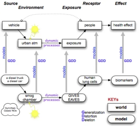
Figure 1-1 provides a schematic of a conceptual model for relating the real world
to a model system designed to study human health effects of exposure to gases and PM.
This part of the model illustrates that effects begin with the source of the pollutant,
fol-lowed by environmental reactions. The original source and environmentally altered
source are then available for exposure to people resulting in possible health outcomes.
The bottom illustrates the series of models used to implement the concept of exposure
from source to response. The models represent the available pieces in our laboratory to
13
realistic conditions, can result in clearer causal relationships from the source to the
ob-served health effects that are seen in epidemiological studies. In my discussion below,
each component and a realistic model that can simulate parts of the real world
condi-tions are described. With the use of these models, I hope to suggest future studies that
can assist with the connection from the source, reactions with the ambient
environ-ment, and subsequent biological response from human exposures.
Source Models
Because of the strong epidemiological findings connecting ambient PM 2.5 and
detrimental health effects, much attention has focused on diesel exhaust (Bates 1995;
Sarnat 2001). Ambient diesel PM has a mean aerodynamic diameter that ranges from 10
to 300 nm. These particles are readily inhaled and about 20 % are deposited into the
alveolar region of the lung (Bunger 2000; Kim 2006). Current studies of diesel exhaust
have used single stationary vehicles and carried out biological exposures to what is
es-sentially freshly emitted exhaust (Li 2002; Baulig 2003; Knebel 2001; Salvi 1999; Laks
2008; Sawant 2008; Carlsten 2008; Liu 2008; Sunil 2009). In this study, I have chosen to
use two diesel vehicles as primary pollution sources with different characteristics: an
older vehicle representing the cars and trucks running on diesel fuel prior to the
catalyt-ic converter, and a newer vehcatalyt-icle representing the cars with emission controls. By using
two different diesel vehicles I obtained samples from real-world sources that are on the
road today, which can be more representative than other DE research that uses altered
14
role of heavy-duty trucks on the road or the heavy machinery powered by diesel, as I am
initially focused on passenger cars as sources for test atmospheres.
Figure 1-1. Conceptual model
Atmosphere Models
In this study, outdoor smog chambers were used to simulate, using natural
sun-light, a set of urban-like test atmospheres that allow diesel exhaust to be
photochemi-cally altered as it likely is in the real world, but under controlled and repeatable
condi-tions (Jeffries 1976; Kamens 1981; Jeffries 1985; McDow 1995; Lee 2004; Doyle 2007;
Ebersviller 2012a; Ebersviller 2012b). To provide a gaseous chemical input typical of US
cities, I used a well-studied mixture of representative urban volatile organic compounds
15
(NO and NO2). Various amounts of diesel exhaust were added to this mixture to
pro-duce reactive urban-like atmospheres. Nevertheless, all important atmospheric
chemi-cal and chemichemi-cal phase change processes did occur in the chamber [24, 27-29, 29-46],
Hu 2007). This approach was critical to relevant outcomes in my study in that it was
able to simulate the complex dynamic processes occurring in the sunlight that results in
a large change in the diesel exhaust gases and particles as a function of time. Doing
ex-posures in the dark just after injection in these model systems also permit the
compari-son of primary (fresh) emissions in contrast to the secondary (oxidized) emissions later
in the simulation.
Exposure System Model and Biological Receptor Model
In this study, I applied the methodology and findings gained from development
and implementation of the Electrostatic Aerosol in Vitro Exposure System (EAVES), and the Gas in Vitro Exposure System (GIVES). These in vitro exposure systems are methods to elicit a response from human epithelial lung cells (A549 cells) exposed the chamber
contents. In the EAVES system, cells are exposed to particles alone, without interrupting
their equilibrium with the gases around them (de Bruijne 2009). Conversely, in the
GIVES system, no significant amount of PM comes in contact with the epithelial lung
cells, resulting in a virtually gases only exposure at the cellular surface.
These two exposure systems reside in a 37 °C incubator with sampling lines
cou-pled to the outdoor chamber. These systemsallow epithelial lung cells to be exposed to
diesel particles and gases separately, facilitating comparison of the inflammatory
16
were grown on membranous supports resulting in a monolayer of cells that were
ex-posed across an air-liquid interface analogous to the process that occurs in the lung
(Leiber 1976, Doyle 2006, Bakand 2007). In these exposure systems, cultured human
epithelial lung cells were used as a targeted biological system that was capable of
re-sponding to ambient gases and particles in ways that mimic the response in the human
body (Doyle 2006, Jaspers 1997). These cells represent the lower alveolar region of the
lung that would be exposed to ambient air in a human (Lumb 2005, Jasper 1997, Bakand
2007). In vitro models have been well studied as a method to examine the mechanisms induced by exposure. These models respond as they would in the body, but may not
give the true toxicological potential since they are not able to include the series of
cas-cade systematic effects in the body. Although this system lacks this ability, inter-cellular
signaling is still active despite the absence of organ-level multi-cellular complexity. The
release of these inter-cellular signals served as the health effect model.
Health Effect Model
The response at the cellular level serves as a reliable predictor of the acute
ad-verse health effects seen in the respiratory tract (Bakland 2007). Various intercellular
chemical signals occur in exposed human epithelial lung cells as a consequence of
inter-actions with gases and particles. As these signals are expressed both within the cell and
in the media supporting the cells, they can be used as markers of exposure. These same
markers of inflammation and cellular death have been monitored in other diesel
17
2001, Baulig 2003, Salvi 1999, Laks 2008, Sawant 2008, Carlsten 2008, Liu 2008, Sunil
2009).
Beneficial Attributes and Limitations of Cascaded Model
The overall benefit of this cascaded modeling system is the use of various sources
in a controlled, repeatable, and complex urban-like atmosphere. In addition, it has the
ability to separate the inflammatory response caused by gases and PM, using two
dif-ferent in vitro exposure systems. The inclusion of photochemical aging in the outdoor chamber allowed for dynamic chemical processes to occur as they would in the real
world. This, coupled to the two in vitro exposure systems, allowed for the separation of inflammatory response induced by the gases and PM. This model has given us better
insight to explain the differences in response seen between epidemiological studies and
current diesel research.
My cascade model approach allowed the research to be understandable,
quantifi-able, and controllquantifi-able, and thus likely to contribute to new insights about processes of
importance that might be obscured in the real (fully complex) world. The findings of this
study are not intended to be conclusive and definitive as is required by policy makers,
but to be indicative of the direction that future research needs to go.
Hypothesis
Atmospheric processes will alter the composition of urban-like atmospheres
con-taining diesel exhaust, which will result in a modification of biological responses from
exposure. Further, an older vehicle lacking emission controls will elicit a higher biological
18
Approach
Current in vitro exposure methods may not be suitable for observations of particle uptake of species of interest. Widely used methods of particle collection involve steps
that could result in the loss of volatile species from samples (Volckens 2002). Due to
these shortcomings, a new exposure technology was developed to provide a viable
method for in vitro PM exposures. To improve upon existing exposure methods, the new method maintains the equilibrium between gases and particles until the pollutants
arrive at the cellular interface. Exposures performed in such a way minimize artifacts
that affect volatile species. This new technology provides a biological response monitor
to more-accurately estimate the health effects of exposure to airborne mixtures of
gas-es and PM.
Once the new method was evaluated and optimized for PM exposure onto human
epithelial lung cells, it was further investigated to determine if the results were similar
to a widely used PM method. Specifically, one of the most widely used methods for in vitro exposures to PM is to collect PM on filters, resuspend the collected PM in a liquid medium, and subsequently add the mixture to the cell culture (Bayram 1998; Boland
1999; Abe 2000; Knebel 2002; Mazzarella 2007; Seagrave 2007). Ultimately this study
evaluated the differences in handling of PM from source to exposure noting the number
of steps prior to exposure. In addition, the composition of the PM delivered to the cells
was evaluated, as well as the observed response from the cells at a similar exposure
concentration. These comparisons permitted an evaluation to determine the relative
19
When the new direct exposure method had been tested it was used along with
the gas phase exposure system to explore the effects of atmospheric aging of diesel
ex-haust and the resulting toxicological effects on human epithelial lung cells (Doyle 2004;
Sexton 2004; Doyle 2007; Ebersviller 2012a; Ebersviller 2012b). The goal of this portion
of the study was to detect any differences in the toxicological responses of the human
epithelial cells through a series of experiments comparing different conditions: 1) older
and newer diesel vehicle emissions, 2) fresh and aged emissions (dark and
photochemi-cally aged), and 3) observe the differences in inflammation in the gas and particle
phas-es. This highly complex study not only used the two exposure tools but incorporated
chemical analyses to attempt to explain observed biological responses.
Objectives
My overall objective was to adopt a more integrated, holistic approach to
demon-strating the features and attributes that might be causally related to human health
ef-fects. This objective was realized through a series of cascaded physical and biological
modeling systems (described above).
The specific objectives listed below allow for the evaluation of the hypothesis.
1. Evaluate and validate a novel in vitro exposuremethod using electrostatic precipitation (ESP) to expose human lung cells to particulate matter (PM).
2. Compare a conventional PM in vitro exposure method to the in vitro ESP de-vice from Objective 1 using diesel exhaust to determine the sensitivity of the
20
3. Compare the toxic effects induced by exposure to fresh and aged emissions
(in the sunlight and in the dark) of old and modern diesel vehicles using
sepa-rate in vitro exposure methods developed for gases and PM.
4. Determine the specific toxicological effects independently induced by
partic-ulate and gas-phase components and their respective contribution to the
biological response observed in Objective 3.
By achieving these objectives, this research project addressed a number of
im-portant questions regarding the toxicity of diesel emissions: 1) Would the new in vitro PM method successfully remove sampling artifacts, thereby increasing sensitivity? 2) To
what extent did aging, either in the presence or absence of photochemistry, change the
toxicity of vehicle emissions? 3) What were the roles of particle and gas phase
Chapter 2
: Design and Testing of Electrostatic
Aer-osol
In Vitro
Exposure System (EAVES): An
Alterna-tive Exposure System for Particles
1Introduction
It has been shown that particulate matter (PM) is responsible for a significant
frac-tion of air pollufrac-tion-induced health effects, yet there remain many quesfrac-tions concerning
mechanisms of injury and what sources and components of this complex pollution are
most responsible (Pope 1995; U.S. Environmental Protection Agency 1999). Laboratory
animal and human in vivo studies have shown that inhalation of diesel exhaust (DE) in-creases markers of inflammation, including inflammatory cytokine production (Salvi
2000; Singh 2004). Studies using in vitro models to expose epithelial cells to DE have also shown increases in the production of inflammatory cytokines, such as Interleukin-8
(IL-8) and tumor necrosis factor-alpha (TNF-α), following exposure (Kim 2005; Seagrave
2007). Although in vitro models lack the ability to account for all intercellular interac-tions in the cells’ natural environment, studies using in vitro exposure models enable investigators to examine the effects of inhaled toxins on specific cell types, and are
im-portant to determine potential cellular mechanisms mediating these responses. Over
22
pollutants, such as ozone, similarly to the way these cells would be exposed in vivo (Jaspers 1997; Doyle 2004; Seagrave 2004; Sexton 2004; Doyle 2007).
Several methods to conduct in vitro exposures to PM have appeared in the litera-ture (Bayram 1998; Boland 1999; Abe 2000; Aufderheide 2000; Li 2002; Madden 2003;
Jaspers 2005; Seagrave 2005; Mazzarella 2007). All of these methods have known
dis-advantages and may, therefore, not accurately represent PM-induced health effects in vivo. Specifically, one of the most widely used methods for in vitro exposures to PM is to collect PM on filters, re-suspend the collected PM in a liquid medium, and subsequently
add the mixture to the cell culture (Bayram 1998; Boland 1999; Abe 2000; Knebel 2002;
Mazzarella 2007). Filters collect particulate matter efficiently, and particles are easily
re-suspended in a liquid for subsequent contact with cells. Major shortcomings of filter
col-lection, however, include the loss of VOCs (volatile organic compounds) from the PM,
agglomeration of small particles during collection, and the possible alteration of the
par-ticles during the recovery process and while in the liquid medium. Impactors collect
large diameter PM on plates relatively efficiently (Tsien 1997), but VOCs can again be
lost during collection, and, as with filters, the collected PM needs to be transferred to a
liquid medium before use with cells. In addition, impactors can only be used to sample
particles of relatively large diameter due low collection efficiency for small particles
(Hinds 1999; McDonald 2004). Alternatively, impingers have been used to sample air
containing PM through a liquid in which the particles are collected (Li 2002; Madden
2003; Jaspers 2005). Again, compounds and surface features of interest may be altered
23
been collected in the liquid medium in this manner, it is difficult to accurately determine
the concentration of PM in solution – further reducing the utility of impinger collection
for in vitro exposures. Recently, an in vitro system using impaction to deposit PM direct-ly onto cells was developed and tested (Cultex® Laboratories, Hannover, Germany)
(Aufderheide 2000; Seagrave 2005). While this exposure system presents a
much-improved method for in vitro PM exposures, there remain a number of disadvantages---including that impaction methods, while being efficient deposition methods for large
particles, have a much lower utility for small particles (Knebel 2002).
Electrostatic precipitation (ESP) is a widely used method of PM collection and
monitoring. Traditionally, ESP has been used as a method for aerosol collection in the
control of airborne dust in residential and industrial settings (Boelter 1997). Particles
are electrically charged and then subjected to a strong electric field that causes the
par-ticles to drift across the flow, and ultimately to deposit on a grounded collection plate
(Whitby 1974; Knutson 1975). When PM is collected with ESP, the velocity
perpendicu-lar to the collection surface is orders of magnitude lower than that of an impactor
sam-pling at the same flow rate. Mainelis, et al. (2002) modified a commercial ESP sampler
to examine its utility as a bioaerosol sampler. Their modified ESP was used to collect
test aerosols of microorganisms that were subsequently shown to be biologically viable
after collection. This example demonstrated the potential for gentle collection and
di-rect deposition onto lung cells as a viable in vitro exposure method. Different from the microorganisms, exposure of cultured human lung cells requires an environment similar
24
charged particles. Our study was designed to determine if an obsolete commercial
elec-trostatic particle collection device could be modified to both keep cells viable and to
de-posit different types of PM on the cells gently and efficiently. The modified device is
named the Electrostatic Aerosol in Vitro Exposure System (EAVES) and it directly depos-its PM on cells grown at an air-liquid interface, thus making it an alternative to existing
methods of in vitro exposure for air pollution mixtures containing particulate matter that could potentially modify the state and composition of the PM.
Materials and Methods
Description of EAVES and its Operating Conditions
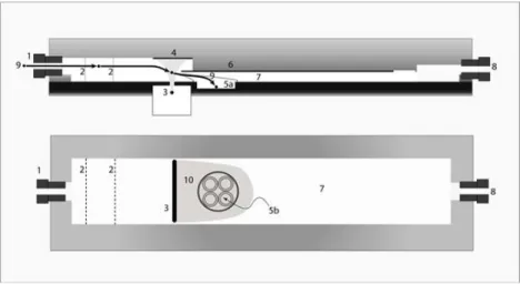
To create the EAVES, a TSI 3100 Electrostatic Aerosol Sampler (EAS) (TSI, Inc., St.
Paul, MN) (TSI Inc. 2008) was modified to accommodate in vitro exposures of cells to PM (see Figure 2-1). First, the electronics were replaced with modern solid-state devices
and cooling fans were added to help maintain the proper temperature when the device
was operated. Second, a circular well was milled into the anodized aluminum collection
plate to hold a titanium dish that contained tissue culture media during the exposure.
This allowed the sample delivered by EAVES to directly deposit on cells maintained at
air-liquid interface without significant interference from culture media, while providing
nutrients from the basolateral side. The circular well is 0.6 cm deep, 3.5 cm in diameter,
and is centered 3.75 cm from the corona-charging slot. A top-view and a side-view,
cen-terline cross-section schematic of the charging and collection area of the EAVES is
pat-25
tern of the particles collected, which facilitates relatively uniform particle deposition
over the whole cell culture surface.
For all the experiments described here, the EAVES was housed in a tissue culture
incubator held at 37˚ C. To prevent particle loss during the exposure, carbon impre
g-nated silicon tubing was used to supply the EAVES with particle-containing air mixtures.
Clean chamber air was mixed with CO2 (to achieve 5 %) using a mass flow controller
(Aalborg Instruments & Controls, Inc., Orangeburg, New York). The mixture was allowed
to flow through EAVES for one hour at 1 L/min (including CO2 at 0.05 L/min) to
equili-brate the system.
Particle-containing samples from an outdoor atmospheric reaction chamber, or
other test sources, also mixed with CO2 (to achieve 5 %), were pulled through the device
at a constant flow rate of 1 L/min; this flow was maintained by a mass flow controller
(Aalborg Instruments & Controls, Inc., Orangeburg, New York).
Growth and Exposure of Cell Cultures
A549 cells, a human epithelial lung cell line, which has retained several alveolar
type II cell characteristics, were used in this study. A549 cells were grown on collagen
coated membrane supports (Millicell R-CM; Millipore; Costar, Cambridge, MA) in
com-plete media (F12K, 10 % fetal bovine serum, with antibiotics; Invitrogen, Carlsbad, CA)
as described by Jaspers, et al. (1997). Before culturing the cells, the depth of the Milli-cell insert was shortened to 0.5 cm to allow for the upper edges of the tissue culture
in-serts to be leveled with the edge of the milled titanium dish in the EAVES. When the
26
replaced with serum-free media (F12K, 1.5 μg/mL bovine serum albumin, with antibio
t-ics). Immediately before exposure in the EAVES, media were removed from the apical
side of the membrane, while media remained in the basolateral side by contact with the
porous membrane. This arrangement facilitates direct exposure of lung epithelial cells
to the sample delivered by EAVES across an air-liquid interface without significant
inter-ference from the culture media, while providing the cells with nutrients from the serum
free media from the basolateral side.
Four tissue culture inserts (0.69 cm2 surface area each) were placed into the
tita-nium dish with 1.5 mL of serum-free media that was then placed into the milled well in
the anodized aluminum collection plate (see Figure 2-2). The titanium dish does not
in-terfere with the media because it is a noncreative metal. The cells were exposed to
vari-ous samples in the EAVES for one hour and then transferred back to individual wells of a
12-well tissue-culture plate containing 1 mL of fresh, serum-free media. In all
expo-sures, A549 cells maintained in a regular tissue-culture incubator served as controls.
Ba-solateral supernatants were collected nine hours post-exposure and stored at -20o C
un-til analysis for cytotoxicity and inflammatory mediator production could be performed.
Analysis of Cytotoxicity and Inflammatory Mediator Production
IL-8 proteins in the supernatant were measured using a commercially available
ELISA (BD Biosciences, San Diego, CA) according to the manufacturer’s instructions. For
the analysis of cytotoxicity, the basolateral supernatants were analyzed for the release
of LDH, using a coupled enzymatic assay (TAKARA), according to the supplier’s
27 Sample Stream Modification Tests
In some experiments, samples of the test atmospheres were taken upstream and
downstream of the EAVES to determine if passage through the EAVES modified the
stream’s reactive organic compound composition. The stream airflows were sampled
through an aqueous o-(2,3,4,5,6-pentafluorobenzyl) hydroxylamine (PFBHA) collection
solution, which binds to carbonyl functional groups with high specificity. The airborne
carbonyls and carbonyls on particles dissolve into the water and are derivatized by the
PFBHA (Yu 1997). Samples were analyzed on a Varian CP-3800 gas chromatograph/mass
spectrometer using both electron impaction (EI) and chemical ionization (CI) modes and
with a flame ionization detector and using a 60 m x 0.32 mm inner diameter column
with RTX-S stationary phase (Restek Corp.; Model Number 10242) with helium as the
carrier gas. Methane was used as the chemical ionization reagent.
Sampling Efficiency Tests
The collection efficiency of the EAVES was determined using 0.2 and 0.5 µm
fluo-rescent-labeled, polystyrene latex spheres (PSL), (0.20 um Yellow-Green, YG; 0.5 um
Red, polychromatic, PC, Fluoresbrite Microspheres, Polysciences, Inc., Warrington, PA).
The size and concentration of these PSL particles at the inlet and the outlet of the EAVES
were measured using a Scanning Mobility Particle Sizer (SMPS) (Model 3936L25, TSI Inc.,
St. Paul, MN). The sheath flow rate of the differential mobility analyzer (DMA) was set to
2.0 L/min, and the aerosol sample flow rate was set to 0.3 L/min. These settings allow
the instrument to measure particles with midpoint diameters ranging from 19 to 882
28
EAVES with the device powered on and off to evaluate the total collection efficiency of
the device.
Using a setup similar to that described by Sioutas, et al. (Sioutas 1994; Sioutas 1999), the PSL spheres were nebulized from diluted aqueous solutions into a flask where
they were mixed with clean, humidified air before entering a charge neutralizer (TSI Inc.,
St. Paul, MN). Once neutralized, the PSL spheres were mixed in a 20-liter glass jar to
fa-cilitate sampling.
PSL particle deposition on cell-free tissue culture membranes was visually
ana-lyzed with a black light, and quantitatively anaana-lyzed by fluorescence spectrophotometry.
Samples for quantitative analysis were prepared by carefully removing all of the
parti-cles from the walls and rim of the tissue culture inserts before rinsing the inserts with 5
mL of ethyl acetate to dissolve the PSL spheres, releasing the florescent dye into
solu-tion. The ethyl acetate rinse containing fluorescent dye from the deposited PSL spheres
was analyzed using a spectrophotometer (Hitachi-High Technology Corporation, F4500
Fluorescence Spectrophotometer, Tokyo, Japan) at optimized excitation and emission
readings. Calibration curves were created for each particle size to be used in these tests.
The fluorescence readings obtained from the cell culture inserts collections were
com-pared to the standard curve appropriate for the particle size used in the test, resulting in
a value for the total particle mass collected on each insert.
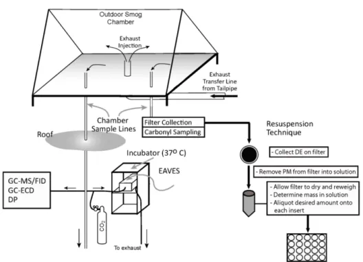
Tests of Diesel Exhaust Exposure
Diesel exhaust was collected from the tailpipe of a 1980 Mercedes-Benz model
29
normal idling conditions. Without a load, the engine was throttled up to approximately
2700 rpm, with an engine temperature of 90°C, and held there throughout the duration
of sampling. DE was collected from the vehicle in a large outdoor smog chamber
(120 m3 Teflon film chamber, see Figure 2-3) before exposure (Jeffries 1976; Jeffries
1985; Doyle 2004; Lee 2004; Doyle 2007). The vehicle’s exhaust was used to pre-heat
the delivery line until the temperature of the exhaust, measured at the chamber, was
125°F. DE was injected into the smog chamber until the particle mass concentration in
the chamber was approximately 4 mg/m3, as measured by SMPS. Size distribution and
mass concentration of the aerosol entering and leaving the EAVES device were also
monitored by SMPS.
Statistical Analysis
All data are presented as the mean + standard error from the mean (S.E.M) and
expressed as fold increase over the same measurements performed on a set of cells
maintained in the incubator throughout the exposure period. Data were analyzed using
an unpaired Student’s t-tests and differences were considered significant when p was
less than 0.05.
Results
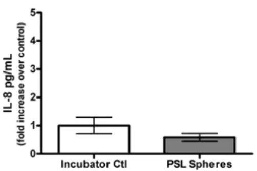
Testing the Effect of EAVES Conditions on Cell Viability
When operated with a corona current setting of 1.5 µA at a sample rate of 1
L/min, the corona wire of the EAVES, which is used to charge the particles, produces an
average of 60 ppb of ozone in the exhaust air after an hour of operation. Besides this
30
cause particles to precipitate might adversely affect cells in the device. To evaluate
these potential problems, the EAVES was tested with two sets of cells using
particle-free, clean air. In the first test, the electrical field was turned off for one hour and in the
second, was turned on for one hour. The inflammatory response and cytotoxicity of the
two sets of cells were measured following exposure. Neither LDH nor IL-8 (Figure 2-4)
release was statistically different from cells maintained in the incubator for the entire
exposure period, suggesting that the ozone produced and the precipitating field do not
cause significant adverse effects on A549 cells.
Effects of EAVES Conditions on Sample Stream Chemical Composition
As indicated above, the corona wire produces a small amount of ozone that could
conceivably react with components of the exposure air stream and alter its chemical
composition. To determine whether the chemical composition of the exposure air
stream was altered by the charging mechanism and the ozone formed in the process,
diesel exhaust (DE) was collected in a Teflon bag and allowed to flow through the EAVES
at one liter per min. As described in Methods section, PFBHAsamples were taken at the
inlet and outlet of the EAVES, and were analyzed with GC/MS to determine if there were
any changes in chemical composition of the air stream. The chromatograms in Figure
2-5 show the analysis of a DE sample before and after passing through the EAVES. The
carbonyls readily identified by GC/MS from the DE in both samples were acetaldehyde,
acetone, acrolein, methyl ethyl ketone, methyl vinyl ketone, crotonaldehyde, and
ben-zaldehyde. As is apparent in Figure 2-5, the magnitudes of the peaks are the same in