Novel genetic loci underlying human intracranial volume
identified through genome-wide association
A full list of authors and affiliations appears at the end of the article.
Abstract
Intracranial volume reflects the maximally attained brain size during development, and remains
stable with loss of tissue in late life. It is highly heritable, but the underlying genes remain largely
undetermined. In a genome-wide association study of 32,438 adults, we discovered five novel loci
for intracranial volume and confirmed two known signals. Four of the loci are also associated with
adult human stature, but these remained associated with intracranial volume after adjusting for
height. We found a high genetic correlation with child head circumference (
ρ
genetic=0.748), which
indicated a similar genetic background and allowed for the identification of four additional loci
CORRESPONDENCE: M.A. Ikram, MD, PhD, Associate Professor of Neuroepidemiology, Department of Epidemiology, Erasmus
MC University Medical Center, Wytemaweg 80, 3015 CE, Rotterdam, the Netherlands, Telephone number: +31 10 70 43930, Fax number: +31 10 70 43489, [email protected] Paul M. Thompson, PhD, Associate Dean for Research, Keck School of Medicine USC, Professor of Neurology, Psychiatry, Engineering, Radiology, Pediatrics, and Ophthalmology, Imaging Genetics Center, and Institute for Neuroimaging and Informatics, Keck School of Medicine of USC, University of Southern California, USA, 2001 N. Soto Street, SSB1-102, Los Angeles, CA 90032, Telephone number: +1 (323) 442-7246, Fax number: +1 (323) 442-0137,
246These authors contributed equally to this work. 247These authors jointly direct this work. *These authors contributed equally. **These authors contributed equally.
COMPETING FINANCIAL INTERESTS STATEMENT
The authors declare no competing financial interest related to any of the work described in this manuscript.
AUTHOR CONTRIBUTIONS
Conceived of the study and drafted the manuscript: H.H.H.A., D.P.H., V.C., J.L.S., M.E.R., S.T., A.A., P.N, V.G., G.S., M.F., B.F., S.D., S.E.M., M.A.I., P.M.T.
Performed statistical analyses: H.H.H.A., D.P.H., V.C., J.L.S., M.E.R., S.T., A.A., Sy.D., A.H.B., N.J., K.W., Lu.A., N.A., M.A., B.S.A., N.J.A., La.A., A.B., M.B., J.C.B., L.M.E.B., S.H.B., M.M.B., Ja.B., O.C., M.M.C., Ga.C., Q.C., C.R.K.C., G.C., Nh.D., St.E., Ti.G., Su.G., A.L.G., C.U.G., Ol.G., M.E.G., T.G., Jo.H., U.K.H., S.H., E.H., M.H., D.J., T.J., N.K., D.K., S.K., M.K., B.K., P.H.L., J.L., D.C.M.L., L.M.L., M.L., Ch.M., Su.M., An.M., Ma.M., M.M., Be.M., D.R.M., R.M., Y.M., R.L.M., K.N., L.M.O., J.O., Ma.P., I.P., L.P., S.P., B.P., K.B.R., A.R., J.S.R., S.L.R., R.R., Na.R., N.A.R., T.R., C.L.S., Li.S., An.J.S., L.S., J.S., A.V.S., E.S., L.T.S., Al.T., Ro.T., Di.T., R.T., D.T., Dh.V., J.V., S.J.V., D.vdM., M.M.J.V., K.R.V., D.vR., Es.W., L.T.W., A.M.W., G.W., C.W., Th.W., L.R.Y., J.Y., M.P.Z., A.M.D., I.O.F., B.M., T.E.N., J.A.T., B.X., Sa.A., A.M.B., A.dB., A.J.H., A.C.N., P.G.S., C.D.W., S.M.B., R.M.B., G.D., J.G., O.G., R.K., C.M., M.A.N., D.V., B.N.V., T.W., E.J.R.
Acquired data: P.N., Su.S., K.A.A., T.A., M.P.B., Ir.F., R.F.G., D.H., K.A.M., Em.S., B.G.W., A.Z., I.A., N.T.A., L.A., D.A., P.A., O.A.A., S.A., A.A.A., M.E.B., D.M.B., J.T.B., D.A.B., J.B., H.v.a.B., D.I.B., H.B., H.G.B., R.L.B., J.K.B., K.B.B., W.C., V.D.C., D.M.C., G.L.C., C-Y.C., C.C., S.C., M.R.C., A.C., B.C., J.E.C., M.C., G.E.D., E.J.C.D., P.L.D., G.I.D., N.D., Ch.D., A.DeS., A.D., Sr.D., W.C.D., R.D., T.D.D., S.E., T.E., D.A.E., G.F., L.F., S.E.F., D.A.F., I.F., T.M.F., P.T.F., C.F., Ma.F., D.C.G., R.L.G., H.H.H.G., H.J.G., R.C.G., S.G., N.K.H., J.H., C.A.H., R.H., K.H., An.H., S.L.e.H., D.G.H., D.J.H., B.H., P.J.H., W.H., A.H., F.H., G.H., N.H., J-J.H., H.E.H., M.I., M.K.I., C.R.J., R.J., E.G.J., J.J., R.S.K., I.K., D.S.K., P.K., J.B.K., L.J.L., S.M.L., H.L., X.L., D.L.L., W.L., O.L.L., S.L., O.M., J.M., V.S.M., A.M.M., F.J.M., K.L.M., P.M., I.M., A.M., S.M., G.W.M., D.W.M., T.H.M., T.W.M., M.N., W.J.N., M.M.N., L.N., K.O., R.L.O., R.A.O., M.P., T.P., Z.P., B.W.J.P., G.P., S.G.P., B.M.P., S.R., Ma.R., J.L.R., N.R., J.I.R., M.R., R.L.S., P.S.S., A.J.S., He.S., P.R.S., S.S., A.S., S.M.S., C.S., J.W.S., H.S., V.S., V.M.S., D.J.S., J.E.S., A.T., H.T., A.W.T., B.T., J.T., C.T., A.G.U., M.C.V., M.vdB., A.V., N.J.A.V., C.M.V., N.E.M.V., M.V., D.J.V., M.W.V., H.V., H.W., J.M.W., T.H.W., M.E.W., D.R.W., M.W.W., W.W., E.W., T.Y.W., C.B.W., R.H.Z., A.B.Z., I.J.D., C.D., R.S., N.G.M., A.J.M.D., M.J.W., V.G., G.S., M.F., B.F., S.D., S.E.M., M.A.I., P.M.T., A.Sim., Sa.A., A.M.B., A.dB., A.J.H., A.C.N., P.G.S., C.D.W., S.M.B., R.M.B., G.D., J.G., O.G., R.K., C.M., M.A.N.,
HHS Public Access
Author manuscript
Nat Neurosci
. Author manuscript; available in PMC 2017 June 01.
Published in final edited form as:
Nat Neurosci. 2016 December ; 19(12): 1569–1582. doi:10.1038/nn.4398.
A
uthor Man
uscr
ipt
A
uthor Man
uscr
ipt
A
uthor Man
uscr
ipt
A
uthor Man
uscr
through meta-analysis (N
combined= 37,345). Variants for intracranial volume were also related to
childhood and adult cognitive function, Parkinson’s disease, and enriched near genes involved in
growth pathways including PI3K–AKT signaling. These findings identify biological
underpinnings of intracranial volume and provide genetic support for theories on brain reserve and
brain overgrowth.
The intricate genetic control of the human brain, complemented by environmental factors,
leads to the observed variations in brain size in human populations
1. Intracranial volume is
closely related to brain volume in early life as the brain grows.
2,3However, it becomes
stable after the brain has fully developed and remains unaffected by later age-related
changes such as brain atrophy
4,5, thus representing the maximal attained brain size.
Discovering genetic variants that influence intracranial volume can contribute to our
understanding of brain development and related diseases, but prior studies have only
identified two influential genetic loci
6–9.
Here, we performed genome-wide association studies in populations from the Cohorts for
Heart and Aging Research in Genomic Epidemiology (CHARGE)
10and Enhancing
NeuroImaging Genetics through Meta-Analysis (ENIGMA)
11consortia on intracranial
volume measured by magnetic resonance imaging. Genotypes were imputed to the 1000
Genomes reference panel (phase 1, version 3). Meta-analysis revealed five novel loci
associated with intracranial volume. We also discovered genome-wide overlap between
intracranial volume and other key traits including height, cognitive ability, and Parkinson’s
disease. Furthermore, we found relatively enriched patterns of association for certain
functional categories of variants and near genes that are involved in specific pathways.
RESULTS
Genome-wide association studies
Detailed information on the population characteristics, image acquisition and processing,
and genetic quality control can be found in the Online Methods and Supplementary Tables
S1–3.
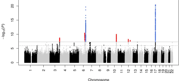
The discovery meta-analysis (N = 26,577) yielded seven genome-wide significant (p < 5 ×
10
−8) loci, five of them novel (Figures 1–2; Table 1). The quantile-quantile plot showed
inflation (
λ
= 1.092; Supplementary Figure S1), which we determined to be mainly due to
polygenicity rather than cryptic relatedness or population stratification using LD score
regression
12. Next we analyzed European samples (N = 2,362; not included in the discovery
sample) and generalization samples with African (N = 938), Asian (N = 955), and Hispanic
(N = 1,605) ancestries (Table 1). All variants had the same direction of effect in the
additional European samples (sign test, P = 0.0078), and three variants replicated, at nominal
significance. Although sample sizes were generally small for the non-Europeans, here too,
the direction of effect was generally concordant with the discovery (sign test, P = 0.039).
Five nominally significant associations were detected across all three ethnicities.
Next we were able to map the association to novel variants for two previously identified loci
at chromosome 17q21 (rs199525; P = 3.8 × 10
−21) and 6q22 (rs11759026; P = 2.2 ×
A
uthor Man
uscr
ipt
A
uthor Man
uscr
ipt
A
uthor Man
uscr
ipt
A
uthor Man
uscr
10
−20)
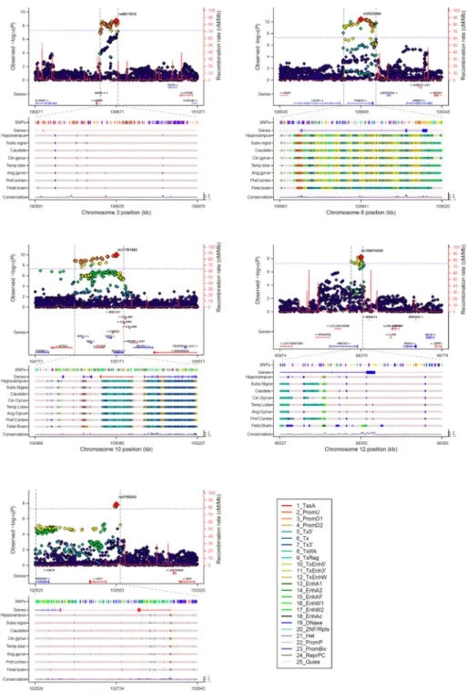
6,7. The five novel loci were on chr 6q21 (rs2022464; P = 3.7 × 10
−11), chr 10q24
(rs11191683; P = 1.1 × 10
−10), chr 3q28 (rs9811910; P = 2.0 × 10
−9), chr 12q14
(rs138074335/ rs7312464; P = 6.2 × 10
−9), and chr 12q23 (rs2195243; P = 1.5 × 10
−8).
Functional annotation of the variants and those in LD (r
2>0.8) can be found in
Supplementary Table S4.
Height-adjusted analyses
Four of the seven loci for intracranial volume were previously discovered for height (17q21,
6q22, 6q21, and 12q14), prompting us to investigate genome-wide overlap between the two
traits. As height and intracranial volume are correlated (weighted average Pearson’s r =
0.556; Supplementary Table S5) and this could drive association signals, we performed a
GWAS of intracranial volume adjusted for height in the studies that had measured height (N
= 21,875). Findings were compared to the corresponding subset of studies without
adjustment (N = 22,378). Using LD score regression (Online Methods), we found that there
is considerable genetic correlation between intracranial volume and height (
ρ
genetic= 0.241,
P = 2.4 × 10
−10), which disappears after adjusting for height (
ρ
genetic= 0.049, P = 0.21)
(Table 2). The associations of the seven intracranial volume loci, however, remained
significant after adjusting for height (Supplementary Table S6). To investigate whether more
height loci were associated with intracranial volume independently of height, we analyzed
all 697 genome-wide significant height variants
13. An additional 73 variants (10.7%; 14
variants not available) showed nominally significant associations with intracranial volume
but were not attenuated after adjustment for height, although none survived Bonferroni
correction (Supplementary Table S7). For some variants, the direction of effect was
discordant, i.e. positive for height and negative for intracranial volume. Furthermore, a
polygenic score of the 697 variants predicted intracranial volume, and this was also the case
after adjustment for height in a subset of the studies (Supplementary Table S8).
Genetic correlation
In addition to height, we examined the genome-wide genetic overlap between intracranial
volume and other anthropometric traits, cognitive function, and neurodegenerative diseases
(Table 2). We found a strong genetic correlation with child head circumference (
ρ
genetic=
0.748), which validates intracranial volume as a measure of brain growth during early
development. Since this high correlation indicates that the genetic determinants of
intracranial volume and child head circumference are largely shared, we aimed to leverage
this information by performing a meta-analysis of both traits. The meta-analysis (combined
N = 37,345) led to the identification of four novel loci (Figure 3; Supplementary Table S9).
Weaker correlations were found with birth length and weight (
ρ
genetic< 0.3), which
attenuated after adjusting for height. Additionally, intracranial volume was genetically
correlated with cognitive function in childhood (
ρ
genetic= 0.277, P = 2.2×10
−3) as well as
general cognitive function in middle-aged and older adults (
ρ
genetic= 0.202, P = 6.3×10
−4).
Furthermore, we found a positive genetic correlation with Parkinson’s disease (
ρ
genetic=
0.315, P = 6.6 × 10
−7), but there was no significant genetic overlap with Alzheimer’s
disease, white matter lesions, and psychiatric traits.
A
uthor Man
uscr
ipt
A
uthor Man
uscr
ipt
A
uthor Man
uscr
ipt
A
uthor Man
uscr
Enrichment analyses
Next, we assessed whether particular subsets of genetic variants were enriched for
association with intracranial volume using partitioned heritability and pathway analyses
(Online Methods). Overall, we found that common variants genotyped from across the
whole genome explained 25.42% (S.E. 2.73%) of the variation in intracranial volume.
Partitioning heritability by chromosome showed that chromosome 22 contributed twofold
more to variation in intracranial volume than would be expected by its size (Figure 4A),
which was not seen for any of the other complex traits from the genetic correlation analysis
(Supplementary Figure S2). Partitioning by functional elements showed an enrichment for
introns and several histone codes that are found in actively transcribed promoters (Figure
4B). The enrichment for intronic variants was specific to intracranial volume, whereas the
other functional classes were also enriched in other complex traits (Supplementary Figure
S3). We also found that loci associated with intracranial volume cluster around genes
involved in specific pathways, with 94 pathways significantly enriched (Figure 4C; full list
in Supplementary Table S10). These pathways included all cell cycle components – the M-,
G1-, S-, and G2-phases – and various growth factor signaling pathways, including PI3K–
AKT.
Head growth trajectories
Although intracranial volume reflects brain development until maturation, and we identified
influences of many growth-related processes contributing to its variation, all loci were still
discovered via cross-sectional associations in adults. Therefore, we tested whether a
polygenic score of the 7 loci could predict head growth in a longitudinal cohort of 2,824
children of European ancestry followed prenatally until 6 years of age (Online Methods).
We found that a higher polygenic score, representing a genetically larger intracranial volume
in adults, was also associated with a larger child head circumference (
β
= .031 per SD, P =
0.010). Furthermore, the effect of the polygenic score was age-dependent and more
prominent in older children (
β
= 0.0080 per SD polygenic score per year age, P
interaction=
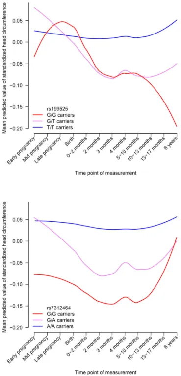
0.0091). When investigating the individual loci separately, both 17q21 and 12q14 showed
significant associations with child head circumference, but they influenced the trajectories of
head growth differently (Figure 4A–B). For 17q21, the negative impact of the G allele on
head circumference becomes apparent postnatally and increases towards six years, whereas
the 12q14 locus exerts an effect from early pregnancy to one year of age, but is less
prominent later in life.
DISCUSSION
Genes contributing to variation in the size of the human brain remain challenging to
discover. In a worldwide project of unprecedented scale, we performed the largest-ever
meta-analysis of genome-wide association studies of intracranial volume. We discovered
five novel genetic loci associated with intracranial volume, and replicated two known
signals. The discovery sample included Europeans only, but the direction of effect was
similar in other ethnicities. The genes in these loci provide intriguing links between maximal
brain size and various processes, including neural stem cell proliferation (FOXO3),
neurodegeneration (MAPT), bone mineralization (CENPW), growth signaling (IGF1,
A
uthor Man
uscr
ipt
A
uthor Man
uscr
ipt
A
uthor Man
uscr
ipt
A
uthor Man
uscr
HMGA2), DNA replication (GMNC), and rRNA maturation (PDCD). On a genome-wide
scale, we discovered evidence of genetic correlation between intracranial volume and other
key traits such as height and cognitive function, and also with Parkinson’s disease,
indicating that the genes underlying brain development have far-reaching effects well
beyond the initial years of life.
The 17q21 locus tags a 1Mb inversion that is under positive selection in Caucasians
14. It
contains multiple genes including the MAPT and KANSL1. The MAPT gene is consistently
implicated in various neurodegenerative disorders including Parkinson’s disease,
Alzheimer’s disease, and frontotemporal dementia
15,16, and microduplications have been
reported to cause microcephaly
17. KANSL1 causes the reciprocal 17q21.31 microdeletion
syndrome - a multisystem disorder with intellectual disability, hypotonia and distinctive
facial features
18. The signal at 6q22 is intergenic to CENPW and RSPO3, but now lies
172kb closer to CENPW. Interestingly, multiple variants at this locus independently
influence bone mineral density
19,20, and our signal particularly overlaps with the variant
showing high specificity for the skull
20.
The significant variants at chr 6q21 span FOXO3, a gene associated with longevity
21,
height
13, and serum IGF1 levels
22. FOXO3 regulates the proliferation of neural stem cells,
and knockout mice show larger brains resulting from increased proliferation immediately
after birth
23, followed by a decrease in adult neural stem cell renewal
23,24. The rs3800229
variant in strong LD with our top variant (r
2= 0.84) contains chromatin promoter marks in
the fetal brain (Supplementary Table S4), and regulates serum IGF1 levels in infants
25. This
provides a link to the genome-wide significant locus on chr12q23 near IGF1, pointing to a
potential mechanism through which these loci may affect brain growth. Chr12q23 lies 20Mb
from one of two loci previously detected for head circumference in children
26, but that
region was not associated with intracranial volume in our study (rs7980687; P = 0.06). The
other reported child head circumference locus, however, corresponded to our chr12q14
signal, with the top variant lying 14kb downstream of HMGA2, and already showed
suggestive association with intracranial volume in a previous report
7. It has also previously
been associated with height
13and is essential for growth
27. The chr10q24 LD-block covers
multiple genes, but an intronic variant within PDCD11 is most significant. PDCD11 encodes
an NF-kappa-B-binding protein required for rRNA maturation and generation of 18S
rRNA
28. A variant in LD (rs7894407) has recently been identified in a GWAS of cerebral
white matter hyperintensities
29. The top chr3q28 variant is located upstream of GMNC,
which codes for the geminin coiled-coil domain-containing protein essential for DNA
replication
30.
Prior efforts to identify variants affecting intracranial volume were much smaller and
critically did not adjust for height
6–9. We found that 4 out of 7 loci were already discovered
for height
13, but also that over 10% of the known ‘height loci’ actually affect intracranial
volume, even after regressing out height. Interestingly, some variants showed discordant
associations for height and intracranial volume - in line with the recent finding that different
height loci disproportionally affect either leg length or spine/head length
31and may be a
marker for pathological development
32. Also, height might thus serve as a proxy phenotype
for intracranial volume, with the tenfold larger sample of the height GWAS giving greater
A
uthor Man
uscr
ipt
A
uthor Man
uscr
ipt
A
uthor Man
uscr
ipt
A
uthor Man
uscr
power to detect associations. Neural genes are also enriched in pathway analyses of height
13.
However, to fully disentangle whether these identified genes are ‘height genes’, ‘brain
volume genes’, or ‘growth genes’ (i.e., pleiotropic), a large collaborative effort is needed that
examines the association of these variants with both intracranial volume and height under
various models.
When investigating genome-wide overlap with other traits, we found a strong correlation
with child head circumference, underlining that intracranial volume is valid measure for
maximal attained brain size. We were able to leverage this genetic link by meta-analyzing
both traits, which led to the identification of four additional loci (2q32.1, 3q23, 7p14.3,
22q13.2). The correlations with birth length and weight were weaker and decreased further
after adjusting for height, so a similar phenotypic correlation between head size and body
size at younger age may drive these correlations. Intracranial volume was also genetically
associated with cognitive function in childhood as well as general cognitive function in
middle-aged and older individuals. This indicates that variation in maximally attained brain
size during development shares a genetic basis with cognitive ability later in life and
supports intracranial volume as a measure of brain reserve
5.
The brain reserve hypothesis states that premorbid brain size can modify resilience to
age-related brain pathology
33, but there was no indication of a genome-wide overlap with
Alzheimer’s disease. However, we found a positive genetic correlation with Parkinson’s
disease that rather points to a brain “overgrowth” hypothesis. Interestingly, the IGF1 and the
PI3K–AKT pathways, key factors in both growth signaling and our current study of
intracranial volume, are neuroprotective in a model system of Parkinson’s disease
34. There
were no correlations with other neurological or psychiatric traits, indicating that this finding
might be specific to Parkinson’s disease. However, it is important to note that there is a
certain extent of variation in the sample size and power of these studies, and larger GWAS
might reveal genetic correlation with other traits as well.
It is not yet known if variance in intracranial volume, within the normal range, contributes to
disease risk or brain reserve. There is no doubt that in the pathological extremes of the
distribution, size can matter, as in disorders such as microcephaly or macrocephaly. Here we
found evidence for a shared genetic background between intracranial volume and cognitive
function, and risk of Parkinson’s disease. While not definitive, these are novel pieces of
empirical evidence in the debate on whether or not whole brain size matters.
The pathway analyses highlight cellular growth and proliferation and included all
components of the cell cycle (M-, G1-, S-, and G2-phase) and various growth factor
signaling pathways. PI3K–AKT signaling has a well described role in brain overgrowth
disorders
35,36, and was the only significant pathway using a different pathway analysis
method (Supplementary Table S11). Interestingly, AKT3 intronic variants showed
suggestive evidence for association with intracranial volume (rs7538011; P = 9.2 × 10
−7).
Deletions of AKT3 cause microcephaly syndromes
37, whereas duplications give rise to
macrocephaly
38. Similar to FOXO3, it is part of the IGF1 signaling pathway, which is
important for human longevity
39. The PI3K–AKT signaling pathway seems to have an
important role in brain growth, not only in pathological extremes, but also for normal
A
uthor Man
uscr
ipt
A
uthor Man
uscr
ipt
A
uthor Man
uscr
ipt
A
uthor Man
uscr
variation at a population level. Other pathways enriched for association with intracranial
volume highlight neuronal functions such as neurotransmission and axon guidance.
We identified novel loci all influencing intracranial volume and, at a genome-wide level,
there seem to be common pathways, but our longitudinal study reveals that their
developmental effects are complex. The loci influenced trajectories of head growth
differently; it also would be interesting to investigate whether their spatial profiles of effects
are distinct, where certain loci promote growth of particular brain regions.
Here we identified key genetic loci implicated in intracranial volume within a global
collaborative effort, followed by computational analyses to determine the important
biological pathways and functional elements. While the majority of the genetic variants are
yet to be discovered, it is clear that these will provide better insight into brain development,
but also into related neuropsychiatric traits such as cognitive functioning and even for
neurodegeneration late in life. Uncovering the remaining heritability will advance our
understanding of the brain’s complex genetic architecture.
ONLINE METHODS
Study population
This study reports data on 32,438 subjects from 52 study sites that are part of the Cohorts for
Heart and Aging Research in Genomic Epidemiology (CHARGE)
10consortium and
Enhancing NeuroImaging Genetics through Meta-Analysis (ENIGMA)
11consortium.
Briefly, the CHARGE consortium is a collaboration of predominantly population-based
cohort studies that investigate the genetic and molecular underpinnings of age-related
complex diseases, including those of the brain. The ENIGMA consortium brings together
numerous studies, mainly with a case-control design, which performed neuroimaging in a
range of neuropsychiatric or neurodegenerative diseases, as well as healthy normative
populations. Studies participated in either the discovery cohort of European ancestry, the
replication in European ancestry, or the generalization to other ethnicities. An overview of
the demographics and type of contribution for each cohort is provided in Supplementary
Table S1. Written informed consent was obtained from all participants. Each study was
approved by the respective Institutional Review Board or Local Ethics Committee.
Genetics
Genotyping was performed using a variety of commercial arrays across the contributing
sites. Both samples as well as variants underwent similar quality control procedures based
on genetic homogeneity, call rate (less than 95%), minor allele frequency (MAF < 0.01), and
Hardy-Weinberg Equilibrium (HWE p-value less than 1 × 10
−6). Good quality variants were
used as input for imputation to the 1000 Genomes reference panel (phase 1, version 3) using
validated software packages (MaCH/minimac, IMPUTE2, BEAGLE, GenABLE). Variants
that were poorly imputed (R
2< 0.5) or uncommon (MAF < 0.5%) were removed prior to
meta-analysis. Full details on the site-specific genotyping and quality control may be found
in Supplementary Table S2.
A
uthor Man
uscr
ipt
A
uthor Man
uscr
ipt
A
uthor Man
uscr
ipt
A
uthor Man
uscr
Imaging
Magnetic resonance imaging (MRI) was obtained from scanners with a diversity of
manufacturers, field strengths, and acquisition protocols. Images were used to estimate
milliliters of intracranial volume from automated segmentations generated by freely
available or in-house methods that have been described and validated earlier. Most sites
measured intracranial volume for each participant by multiplying the inverse of the
determinant of the transformation matrix required to register the subject’s MRI scan to a
common template by the template volume (1,948,105 mm3), using the FreeSurfer software.
Visual inspections were performed to identify and remove poorly segmented images. Either
all scans were visually inspected, or sites generated histogram plots to identify any outliers,
which were defined as individuals with a volume more than three standard deviations away
from the mean. Statistical outliers were only excluded if the segmentations were deemed
improper
‥
More site-specific information related to the imaging is available in
Supplementary Table S3.
Genome-wide association studies
Genome-wide association studies of intracranial volume were performed for each site
separately, controlling for age, sex, and, when applicable, age
2, population stratification
variables (MDS / principal components), study site (for multi-site studies only), diagnosis
(for case-control studies only). Studies of unrelated individuals performed a linear regression
analyses whereas studies of related individuals (ASPSFam, BrainSCALE, ERF, GeneSTAR,
GOBS, NeuroIMAGE, NTR-Adults, OATS, QTIM, SYS) used linear mixed models to
account for familial relationships. Summary statistics, including the effect estimates of the
genetic variant with intracranial volume under an additive model, were exchanged to
perform a fixed-effects meta-analysis weighting for sample size in METAL
40. After the final
meta-analysis, variants were excluded if they were only available for fewer than 5,000
individuals. Meta-analyses were stratified by race and done separately for discovery,
replication, and generalization samples. Beta coefficients were recalculated from Z-scores,
allele frequencies, and the sample, as described earlier
41Site-specific quantile-quantile plots
were generated to inspect the presence of genomic inflation. The variance explained by all
variants in the GWAS was estimated using LD score regression
12,42. Sensitivity analyses
were performed by excluding patients.
Functional annotation
All tracks of the regional association plots were taken from the UCSC Genome Browser
Human hg19 assembly. SNPs (top 5%) shows the top 5% associated variants within the
locus and are colored by their correlation to the top variant. Genes shows the gene models
from GENCODE version 19. The tracks give the predicted chromatin states based on
computational integration of ChIP-seq data for 12 chromatin marks in various human tissues
derived from the Roadmap Epigenomics Consortium
43. Additionally, we used HaploReg
version 3 for annotation of the top variants and all variants in LD (> 0.80) (
http://
www.broadinstitute.org/mammals/haploreg/haploreg_v3.php
).
A
uthor Man
uscr
ipt
A
uthor Man
uscr
ipt
A
uthor Man
uscr
ipt
A
uthor Man
uscr
Genetic correlation
The genetic correlation analyses were also performed using LD score regression. The
GWAS meta-analysis of intracranial volume, as well as the height adjusted and height subset
meta-analyses, were correlated with published GWAS of the following traits: Child head
circumference
26, birth weight
44, birth length
45, adult height
13, childhood cognitive
function
46, adult cognitive function
47, Alzheimer’s disease
48, Parkinson’s disease
49, white
matter lesions
50, psychiatric disorders
51, neuroticism
52, and extraversion
53.
Enrichment analyses
To determine whether the intracranial volume association results were enriched for certain
types of genetic variants, we employed two strategies: partitioned heritability and pathway
analyses.
Partitioned heritability was calculated using a previously described method
42. This was done
by partitioning variants by chromosome and by 28 functional classes: coding, UTR,
promoter, intron, histone marks H3K4me1, H3K4me3, H3K9ac5 and two versions of
H3K27ac, open chromatin DNase I hypersensitivity Site (DHS) regions, combined
chromHMM/Segway predictions, regions that are conserved in mammals, super-enhancers
and active enhancers from the FANTOM5 panel of samples (Finucane et al. page 4)
42.
Multiple testing thresholds were calculated accordingly: P
thresh= 0.05/(22 chromosomes) =
2.27 × 10
−3for the chromosomes and P
thresh= 0.05/(28 classes) = 1.79 × 10
−3for the
functional classes.
Pathway analyses were performed using the KGG2.5
54and MAGENTA
55software
packages. LD was calculated based with the 1000 Genomes Project European samples as a
reference (see URLs). Variants were considered to be within a gene if they were within 5 kb
of the 3’/5’ UTR based on chromosome positions (hg19) coordinates. Gene-based tests were
done with the GATES test
54without weighting P-values by predicted functional relevance.
Pathway analysis was performed using the HYST test of association
56. A multiple testing
threshold accounting for the number of pathways tested resulting in a significance threshold
of P
thresh= 0.05/(671 pathways) = 7.45 × 10
−5.
Head growth trajectories
Head growth trajectory analyses were done within the Generation R study, a longitudinal
cohort study situated in Rotterdam, the Netherlands. For this analysis we included 2,824
children of European ancestry followed prenatally until 6 years of age. Head size was
measured at the following points: prenatally (using echo) during the first, second, and third
trimester, and postnatally (measuring head circumference) at 0–2 months, 2 months, 3
months, 4 months, 5–10 months, 10–13 months, 13–17 months, and 5 years of age. We
tested whether a polygenic score of the 7 loci, as well as the 7 loci themselves separately,
were related to head growth using linear mixed models and included an interaction term
between time and the genetic score/variant (SAS software). Next, the predicted values were
calculated for each person and plotted over time, stratified by genotype (0/1/2 risk alleles)
using the R software package.
A
uthor Man
uscr
ipt
A
uthor Man
uscr
ipt
A
uthor Man
uscr
ipt
A
uthor Man
uscr
URLs
ftp://pricelab:[email protected]/LDSCORE/
http://enigma.ini.usc.edu/protocols/genetics-protocols/
http://genenetwork.nl/bloodeqtlbrowser/
http://gump.qimr.edu.au/general/gabrieC/LocusTrack/
)
Supplementary Material
Refer to Web version on PubMed Central for supplementary material.
Authors
Hieab HH Adams
1,2,246, Derrek P Hibar
3,246, Vincent Chouraki
4,5,6,246, Jason L
Stein
3,7,246, Paul A Nyquist
8,246, Miguel E Rentería
9,246, Stella Trompet
10,246,
Alejandro Arias-Vasquez
11,12,13,14,246, Sudha Seshadri
4,6, Sylvane Desrivières
15,
Ashley H Beecham
16,17, Neda Jahanshad
3, Katharina Wittfeld
18,19, Sven J Van der
Lee
1, Lucija Abramovic
20, Saud Alhusaini
21,22, Najaf Amin
1, Micael Andersson
23,
Konstantinos Arfanakis
24,25,26, Benjamin S Aribisala
27,28,29, Nicola J
Armstrong
30,31, Lavinia Athanasiu
32,33, Tomas Axelsson
34, Alexa Beiser
4,35,6,
Manon Bernard
36, Joshua C Bis
37, Laura ME Blanken
38,39, Susan H Blanton
16,17,
Marc M Bohlken
20, Marco P Boks
20, Janita Bralten
11,14, Adam M Brickman
40, Owen
Carmichael
41, M Mallar Chakravarty
42,43, Ganesh Chauhan
44, Qiang Chen
45,
Christopher RK Ching
3,46, Gabriel Cuellar-Partida
9, Anouk Den Braber
47, Nhat
Trung Doan
32, Stefan Ehrlich
48,49,50, Irina Filippi
51, Tian Ge
52,50,53,54, Sudheer
Giddaluru
55,56, Aaron L Goldman
45, Rebecca F Gottesman
57, Corina U
Greven
13,58,59, Oliver Grimm
60, Michael E Griswold
61, Tulio Guadalupe
62,63,
Johanna Hass
64, Unn K Haukvik
32,65, Saima Hilal
66,67, Edith Hofer
68,69, David
Hoehn
70, Avram J Holmes
71,49, Martine Hoogman
11,14, Deborah Janowitz
19, Tianye
Jia
15, Dalia Kasperaviciute
72,73, Sungeun Kim
74,75,76, Marieke Klein
11,14, Bernd
Kraemer
77, Phil H Lee
52,49,53,54,78, Jiemin Liao
79, David CM Liewald
80, Lorna M
Lopez
80, Michelle Luciano
80, Christine Macare
15, Andre Marquand
14,81, Mar
Matarin
72,82, Karen A Mather
30, Manuel Mattheisen
83,84,85, Bernard Mazoyer
86,
David R McKay
87,88, Rebekah McWhirter
89, Yuri Milaneschi
90, Nazanin
Mirza-Schreiber
70, Ryan L Muetzel
38,39, Susana Muñoz Maniega
27,29,80, Kwangsik
Nho
74,75,76, Allison C Nugent
91, Loes M Olde Loohuis
92, Jaap Oosterlaan
93,
Martina Papmeyer
94,95, Irene Pappa
96,38, Lukas Pirpamer
68, Sara Pudas
23, Benno
Pütz
70, Kumar B Rajan
97, Adaikalavan Ramasamy
98,82,99, Jennifer S
Richards
13,14,58, Shannon L Risacher
74,76, Roberto Roiz-Santiañez
100,101, Nanda
Rommelse
12,14,58, Emma J Rose
102, Natalie A Royle
27,80,29,103, Tatjana
Rundek
104,105, Philipp G Sämann
70, Claudia L Satizabal
4,6, Lianne
Schmaal
106,107,108, Andrew J Schork
109,110, Li Shen
74,75,76, Jean Shin
36, Elena
Shumskaya
11,14,81, Albert V Smith
111,112, Emma Sprooten
94,87,88,113, Lachlan T
Strike
9,114, Alexander Teumer
115, Russell Thomson
116, Diana
Tordesillas-A
uthor Man
uscr
ipt
A
uthor Man
uscr
ipt
A
uthor Man
uscr
ipt
A
uthor Man
uscr
Gutierrez
117,101, Roberto Toro
118, Daniah Trabzuni
82,119, Dhananjay Vaidya
120,
Jeroen Van der Grond
121, Dennis Van der Meer
122, Marjolein MJ Van
Donkelaar
11,14, Kristel R Van Eijk
123, Theo GM Van Erp
124, Daan Van
Rooij
13,14,122, Esther Walton
64, Lars T Westlye
33,125, Christopher D Whelan
3,22,
Beverly G Windham
126, Anderson M Winkler
87,127, Girma Woldehawariat
91,
Christiane Wolf
128, Thomas Wolfers
11,14, Bing Xu
15, Lisa R Yanek
120, Jingyun
Yang
25,129, Alex Zijdenbos
130, Marcel P Zwiers
14,81, Ingrid Agartz
32,65,131, Neelum
T Aggarwal
97,25,129, Laura Almasy
132,133,134, David Ames
135,136, Philippe
Amouyel
5, Ole A Andreassen
32,33, Sampath Arepalli
137, Amelia A Assareh
30,
Sandra Barral
40, Mark E Bastin
27,80,103,29, Diane M Becker
120, James T Becker
138,
David A Bennett
25,129, John Blangero
132, Hans van Bokhoven
11,14, Dorret I
Boomsma
47, Henry Brodaty
30,139, Rachel M Brouwer
20, Han G Brunner
11,14,140,
Randy L Buckner
49,141, Jan K Buitelaar
13,14,58, Kazima B Bulayeva
142, Wiepke
Cahn
20, Vince D Calhoun
143,144, Dara M Cannon
91,145, Gianpiero L Cavalleri
22,
Christopher Chen
66,67, Ching-Yu Cheng
146,79,147, Sven Cichon
148,149,150, Mark R
Cookson
137, Aiden Corvin
102, Benedicto Crespo-Facorro
100,101, Joanne E
Curran
132, Michael Czisch
70, Anders M Dale
151,152, Gareth E Davies
153, Eco JC De
Geus
47, Philip L De Jager
154,53,155, Greig I de Zubicaray
156, Norman Delanty
157,22,
Chantal Depondt
158, Anita L DeStefano
35,6, Allissa Dillman
137, Srdjan
Djurovic
55,159, Gary Donohoe
160,161, Wayne C Drevets
91,162, Ravi Duggirala
132,
Thomas D Dyer
132, Susanne Erk
163, Thomas Espeseth
125,33, Denis A Evans
97,
Iryna O Fedko
47, Guillén Fernández
13,14, Luigi Ferrucci
164, Simon E Fisher
62,14,
Debra A Fleischman
25,165, Ian Ford
166, Tatiana M Foroud
167,76, Peter T Fox
168,
Clyde Francks
62,14, Masaki Fukunaga
169, J Raphael Gibbs
137,82, David C
Glahn
87,88, Randy L Gollub
49,50,53, Harald HH Göring
132, Hans J Grabe
19, Robert
C Green
170,53, Oliver Gruber
77, Vilmundur Gudnason
111,112, Sebastian Guelfi
82,
Narelle K Hansell
9,114, John Hardy
82, Catharina A Hartman
122, Ryota
Hashimoto
171,172, Katrin Hegenscheid
173, Andreas Heinz
163, Stephanie Le
Hellard
55,56, Dena G Hernandez
137,82,174, Dirk J Heslenfeld
175, Beng-Choon Ho
176,
Pieter J Hoekstra
122, Wolfgang Hoffmann
115,18, Albert Hofman
1, Florian
Holsboer
70,177, Georg Homuth
178, Norbert Hosten
173, Jouke-Jan Hottenga
47,
Hilleke E Hulshoff Pol
20, Masashi Ikeda
179, M Kamran Ikram
146,79,67,66,1, Clifford R
Jack Jr
181, Mark Jenkinson
127, Robert Johnson
182, Erik G Jönsson
131,32, J Wouter
Jukema
10, René S Kahn
20, Ryota Kanai
183,184,185, Iwona Kloszewska
186, David S
Knopman
187, Peter Kochunov
188, John B Kwok
189,190, Stephen M Lawrie
94, Hervé
Lemaître
51, Xinmin Liu
91,191, Dan L Longo
192, WT Longstreth Jr
193, Oscar L
Lopez
194, Simon Lovestone
195,196, Oliver Martinez
197, Jean-Luc Martinot
51,
Venkata S Mattay
45,57,198, Colm McDonald
145, Andrew M McIntosh
94,80, Katie L
McMahon
199, Francis J McMahon
91, Patrizia Mecocci
200, Ingrid Melle
32,33, Andreas
Meyer-Lindenberg
60, Sebastian Mohnke
163, Grant W Montgomery
9, Derek W
Morris
160,161, Thomas H Mosley
126, Thomas W Mühleisen
150,149, Bertram
Müller-Myhsok
70,201,202, Michael A Nalls
137, Matthias Nauck
203,204, Thomas E
Nichols
205,127, Wiro J Niessen
206,2,207, Markus M Nöthen
149,208, Lars Nyberg
23,
Kazutaka Ohi
171, Rene L Olvera
168, Roel A Ophoff
92,20, Massimo Pandolfo
158,
Tomas Paus
209,210,211, Zdenka Pausova
36,212, Brenda WJH Penninx
108, G Bruce
A
uthor Man
uscr
ipt
A
uthor Man
uscr
ipt
A
uthor Man
uscr
ipt
A
uthor Man
uscr
Pike
213,214, Steven G Potkin
124, Bruce M Psaty
215, Simone Reppermund
30,216,
Marcella Rietschel
60, Joshua L Roffman
49, Nina Romanczuk-Seiferth
163, Jerome I
Rotter
217, Mina Ryten
82,98, Ralph L Sacco
17,104,105,218, Perminder S
Sachdev
30,219, Andrew J Saykin
74,76,167, Reinhold Schmidt
68, Peter R
Schofield
189,190, Sigurdur Sigurdsson
111, Andy Simmons
220,221,222, Andrew
Singleton
137, Sanjay M Sisodiya
72, Colin Smith
223, Jordan W Smoller
52,49,53,54,
Hilkka Soininen
224,225, Velandai Srikanth
226, Vidar M Steen
55,56, David J Stott
227,
Jessika E Sussmann
94, Anbupalam Thalamuthu
30, Henning Tiemeier
1,39, Arthur W
Toga
228, Bryan J Traynor
137, Juan Troncoso
229, Jessica A Turner
230, Christophe
Tzourio
231, Andre G Uitterlinden
1,232, Maria C Valdés Hernández
27,80,103,29, Marcel
Van der Brug
233, Aad Van der Lugt
2, Nic JA Van der Wee
234, Cornelia M Van
Duijn
1, Neeltje EM Van Haren
20, Dennis Van 't Ent
47, Marie-Jose Van Tol
235, Badri
N Vardarajan
40, Dick J Veltman
108, Meike W Vernooij
1,2, Henry Völzke
115, Henrik
Walter
163, Joanna M Wardlaw
27,80,103,29, Thomas H Wassink
236, Michael E
Weale
98, Daniel R Weinberger
45,237, Michael W Weiner
238, Wei Wen
30, Eric
Westman
239, Tonya White
39,2, Tien Y Wong
146,79,147, Clinton B Wright
104,105,218, H
Ronald Zielke
182, Alan B Zonderman
240, the Alzheimer’s Disease Neuroimaging
Initiative, EPIGEN, IMAGEN, SYS, Ian J Deary
80, Charles DeCarli
197, Helena
Schmidt
241, Nicholas G Martin
9, Anton JM De Craen
242, Margaret J
Wright
114,199,247, Lenore J Launer
243,247, Gunter Schumann
15,247, Myriam
Fornage
244,247, Barbara Franke
11,12,14,247, Stéphanie Debette
44,245,4,247, Sarah E
Medland
9,247, M Arfan Ikram
1,2,180,247, and Paul M Thompson
3,247Affiliations
1
Department of Epidemiology, Erasmus MC, Rotterdam, 3015 CE, the Netherlands
2
Department of Radiology and Nuclear Medicine, Erasmus MC, Rotterdam, 3015
CE, the Netherlands
3Imaging Genetics Center, USC Mark and Mary Stevens
Neuroimaging & Informatics Institute, Keck School of Medicine of University of
Southern California, Los Angeles, 90292, USA
4Department of Neurology, Boston
University School of Medicine, Boston, MA, USA
5Lille University, Inserm, CHU Lille,
Institut Pasteur de Lille, U1167 - RID-AGE - Risk factors and molecular
determinants of aging-related diseases, F-59000 Lille, France
6Framingham Heart
Study, Framingham, MA USA
7Department of Genetics & UNC Neuroscience
Center, University of North Carolina (UNC), Chapel Hill, North Carolina, USA
8
Department of Neurology, Department of Anesthesia/Critical Care Medicine,
Department of Neurosurgery, Johns Hopkins, USA600 N. Wolfe St, Baltimore
Maryland, 21287
9QIMR Berghofer Medical Research Institute, Brisbane, 4006,
Australia
10Department of Cardiology, Leiden University Medical Center, Leiden, the
Netherlands
11Department of Human Genetics, Radboud University Medical Center,
Nijmegen, the Netherlands
12Department of Psychiatry, Radboud University Medical
Center, Nijmegen, the Netherlands
13Department of Cognitive Neuroscience,
Radboud University Medical Center, Nijmegen, the Netherlands
14Donders Institute
for Brain, Cognition and Behaviour, Radboud University, Nijmegen, the Netherlands
15
MRC-SGDP Centre, Institute of Psychiatry, Psychology and Neuroscience, King’s
College London, London, SE5 8AF, UK
16Dr. John T. Macdonald Foundation
A
uthor Man
uscr
ipt
A
uthor Man
uscr
ipt
A
uthor Man
uscr
ipt
A
uthor Man
uscr
Department of Human Genetics, University of Miami, Miller School of Medicine,
Miami, FL, USA
17John P. Hussman Institute for Human Genomics, University of
Miami, Miller School of Medicine, Miami, FL, USA
18German Center for
Neurodegenerative Diseases (DZNE) Rostock/Greifswald, Greifswald, 17487,
Germany
19Department of Psychiatry, University Medicine Greifswald, Greifswald,
17489, Germany
20Brain Center Rudolf Magnus, Department of Psychiatry, UMC
Utrecht, Utrecht, 3584 CX, the Netherlands
21Department of Neurology and
Neurosurgery, Montreal Neurological Institute, McGill University, Montreal, H3A 2B4,
Canada
22The Royal College of Surgeons in Ireland, 123 St Stephen's Green,
Dublin 2, Ireland
23Department of Integrative Medical Biology and Umeå center for
Functional Brain Imaging, Umeå University, Umeå, 901 87, Sweden
24Department
of Biomedical Engineering, Illinois Institute of Technology, Chicago, Illinois, 60616,
USA
25Rush Alzheimer's Disease Center, Rush University Medical Center, Chicago,
Illinois, 60612, USA
26Department of Diagnostic Radiology and Nuclear Medicine,
Rush University Medical Center, Chicago, IL, USA
27Brain Research Imaging
Centre, University of Edinburgh, Edinburgh, EH4 2XU, UK
28Department of
Computer Science, Lagos State University, Lagos, Nigeria
29Scottish Imaging
Network, A Platform for Scientific Excellence (SINAPSE) Collaboration, Department
of Neuroimaging Sciences, University of Edinburgh, Edinburgh, EH16 4SB, UK
30
Centre for Healthy Brain Ageing, School of Psychiatry, University of New South
Wales, Sydney, 2052, Australia
31Mathematics and Statistics, Murdoch University,
Perth, Australia
32NORMENT - KG Jebsen Centre, Institute of Clinical Medicine,
University of Oslo, Oslo, 0315, Norway
33NORMENT - KG Jebsen Centre, Division
of Mental Health and Addiction, Oslo University Hospital, Oslo, 0315, Norway
34
Department of Medical Sciences, Molecular Medicine and Science for Life
Laboratory, Uppsala University, Box 1432, SE-751 44 Uppsala, Sweden
35
Department of Biostatistics, Boston University School of Public Health, Boston,
MA; 2) Framingham Heart Study, Framingham, MA
36Hospital for Sick Children,
University of Toronto, Toronto, M5G 1X8, Canada
37Cardiovascular Health Research
Unit, Department of Medicine, University of Washington, Seattle, WA USA, 1730
Minor Avenue / Suite 1360 / Seattle, WA 98101
38Generation R Study Group,
Erasmus Medical Center, Rotterdam, 3015 CE, the Netherlands
39Department of
Child and Adolescent Psychiatry/Psychology, Erasmus MC-Sophia Children's
Hospital, Rotterdam, 3015 CE, the Netherlands
40Taub Institute for Research on
Alzheimer’s Disease and the Aging Brain; G.H. Sergievsky Center; Department of
Neurology. Columbia University Medical Center. 639 West 1168th Street.New York,
NY 10032, USA
41Pennington Biomedical Research Center, Baton Rouge, LA,
70808
42Cerebral Imaging Centre, Douglas Mental Health University Institute,
Montreal, H4H 1R3, Canada
43Department of Psychiatry and Biomedical
Engineering, McGill University, Montreal, H3A 2B4, Canada
44INSERM Unit U1219,
University of Bordeaux, 33076, France
45Lieber Institute for Brain Development,
Baltimore, 21205, USA
46Interdepartmental Neuroscience Graduate Program,
UCLA School of Medicine, Los Angeles, California 90095, USA
47Biological
Psychology, Neuroscience Campus Amsterdam, Vrije Universiteit University & Vrije
A
uthor Man
uscr
ipt
A
uthor Man
uscr
ipt
A
uthor Man
uscr
ipt
A
uthor Man
uscr
Universiteit Medical Center, Amsterdam, 1081 BT, the Netherlands
48Division of
Psychological and Social Medicine and Developmental Neurosciences, Faculty of
Medicine, TU Dresden, 01307 Germany
49Department of Psychiatry, Massachusetts
General Hospital, Boston, 02114, USA
50Martinos Center for Biomedical Imaging,
Massachusetts General Hospital, Charlestown, 02129, USA
51INSERM Unit 1000
"Neuroimaging and Psychiatry", University Paris Sud, University Paris Descartes;
Maison de Solenn, Adolescent Psychopathology and Medicine Department, APHP
Hospital Cochin, 97 Bd de Port Royal, Paris; France
52Psychiatric and
Neurodevelopmental Genetics Unit, Center for Human Genetic Research,
Massachusetts General Hospital, Boston, Massachusetts, 02114, USA
53Harvard
Medical School, Boston, Massachusetts, 02115, USA
54Stanley Center for
Psychiatric Research, Broad Institute of MIT and Harvard, Boston, Massachusetts,
02141, USA
55NORMENT - KG Jebsen Centre for Psychosis Research, Department
of Clinical Science, University of Bergen, 5021, Norway
56Dr. Einar Martens
Research Group for Biological Psychiatry, Center for Medical Genetics and
Molecular Medicine, Haukeland University Hospital, Bergen, 5021, Norway
57
Department of Neurology, Johns Hopkins University School of Medicine,
Baltimore, MD, 21205, USA
58Karakter Child and Adolescent Psychiatry University
Center, Nijmegen, the Netherlands
59King's College London, Medical Research
Council Social, Genetic and Developmental Psychiatry Centre, Institute of
Psychology, Psychiatry and Neurosciene, United Kingdom
60Central Institute of
Mental Health, Medical Faculty Mannheim, University Heidelberg, Mannheim,
68159, Germany
61Center of Biostatistics and Bioinformatics, University of
Mississippi Medical Center, Jackson, MS, USA
62Language and Genetics
Department, Max Planck Institute for Psycholinguistics, Nijmegen, 6525 XD, the
Netherlands
63International Max Planck Research School for Language Sciences,
Nijmegen, 6525 XD, the Netherlands
64Department of Child and Adolescent
Psychiatry, Faculty of Medicine of the TU Dresden, Dresden, 01307 Germany
65
Department of Research and Development, Diakonhjemmet Hospital, Oslo, 0319,
Norway
66Department of Pharmacology, National University of Singapore,
Singapore
67Memory Aging & Cognition Centre (MACC), National University Health
System, Singapore
68Department of Neurology, Clinical Division of Neurogeriatrics,
Medical University Graz, Austria, Auenbruggerplatz 22, 8036 Graz, Austria
69
Institute of Medical Informatics, Statistics and Documentation, Medical University
Graz, Austria, Auenbruggerplatz 22, 8036 Graz, Austria
70Max Planck Institute of
Psychiatry, Department of Translational Research in Psychiatry, Munich, 80804,
Germany
71Department of Psychology, Yale University, New Haven, 06520, USA
72UCL Institute of Neurology, London, United Kingdom and Epilepsy Society, Bucks,
UK
73Department of Medicine, Imperial College London, London, SW7 2AZ, UK
74Center for Neuroimaging, Radiology and Imaging Sciences, Indiana University
School of Medicine, Indianapolis, Indiana, 46202, USA
75Center for Computational
Biology and Bioinformatics, Indiana University School of Medicine, Indianapolis,
Indiana, 46202, USA
76Indiana Alzheimer Disease Center, Indiana University
School of Medicine, Indianapolis, Indiana, 46202, USA
77Section for Experimental
A
uthor Man
uscr
ipt
A
uthor Man
uscr
ipt
A
uthor Man
uscr
ipt
A
uthor Man
uscr
Psychopathology and Neuroimaging, Dept. of General Psychiatry, Heidelberg
University, Heidelberg, Germany
78Lurie Center for Autism, Massachusetts General
Hospital, Harvard Medical School, Lexington
79Singapore Eye Research Institute,
Singapore National Eye Centre, Singapore
80Centre for Cognitive Ageing and
Cognitive Epidemiology, Psychology, University of Edinburgh, Edinburgh, EH8 9JZ,
UK
81Donders Centre for Cognitive Neuroimaging, Radboud University, Nijmegen,
The Netherlands
82Reta Lila Weston Institute and Department of Molecular
Neuroscience, UCL Institute of Neurology, London, WC1N 3BG, UK
83Department
of Biomedicine, Aarhus University, Aarhus, DK-8000, Denmark
84The Lundbeck
Foundation Initiative for Integrative Psychiatric Research, iPSYCH, Aarhus and
Copenhagen, DK-8000, Denmark
85Center for integrated Sequencing, iSEQ,
Aarhus University, Aarhus, DK-8000, Denmark
86UMR5296 University of Bordeaux,
CNRS, CEA, Bordeaux, France, 146 rue Leo Saignat, 33076 Bordeaux 2 cedex
87
Department of Psychiatry, Yale University, New Haven, Connecticut, 06511, USA
88
Olin Neuropsychiatric Research Center, Hartford, Connecticut, 06114, USA
89
Menzies Institute for Medical Research, University of Tasmania, Hobart, Tasmania
7000, Australia
90Department of Psychiatry, EMGO Institute for Health and Care
Research and Neuroscience Campus Amsterdam, VU University Medical
Center/GGZ inGeest, Amsterdam, 1081 HL, The Netherlands
91Experimental
Therapeutics and Pathophysiology Branch, National Institute of Mental Health
Intramural Research Program, National Institutes of Health, Bethesda, Maryland
20892, USA
92Center for Neurobehavioral Genetics, University of California, Los
Angeles, California, 90095, USA
93Department of Clinical Neuropsychology, VU
University Amsterdam, Amsterdam, the Netherlands
94Division of Psychiatry, Royal
Edinburgh Hospital, University of Edinburgh, Edinburgh, EH10 5HF, UK
95Division of
Systems Neuroscience of Psychopathology, Translational Research Center,
University Hospital of Psychiatry, University of Bern, Switzerland
96School of
Pedagogical and Educational Sciences, Erasmus University Rotterdam, Rotterdam,
3015 CE, the Netherlands
97Rush Institute for Healthy Aging, Rush University
Medical Center, Chicago, Illinois, 60612, USA
98Department of Medical and
Molecular Genetics, King’s College London, London, SE1 9RT, UK
99The Jenner
Institute Laboratories, University of Oxford, Oxford OX3 7DQ, UK
100Department of
Medicine and Psychiatry, University Hospital Marqués de Valdecilla, School of
Medicine, University of Cantabria-IDIVAL, Santander, 39008, Spain
101CIBERSAM
(Centro Investigación Biomédica en Red Salud Mental), Santander, Spain
102
Psychosis Research Group, Department of Psychiatry & Trinity Translational
Medicine Institute, Trinity College Dublin
103Centre for Clinical Brain Sciences,
University of Edinburgh, Edinburgh, EH16 4SB, UK
104Department of Neurology,
University of Miami, Miller School of Medicine, Miami, FL, USA
105Department of
Epidemiology and Public Health Sciences, University of Miami, Miller School of
Medicine, Miami, FL, USA
106Orygen, The National Centre of Excellence in Youth
Mental Health, Melbourne, VIC, Australia
107Centre for Youth Mental Health, The
University of Melbourne, Melbourne, VIC, Australia
108Department of Psychiatry,
Neuroscience Campus Amsterdam, VU University Medical Center, Amsterdam,
A
uthor Man
uscr
ipt
A
uthor Man
uscr
ipt
A
uthor Man
uscr
ipt
A
uthor Man
uscr
1007 MB, the Netherlands
109Multimodal Imaging Laboratory, Department of
Neurosciences, University of California, San Diego, 92093, USA
110Department of
Cognitive Sciences, University of California, San Diego, 92161, USA
111Icelandic
Heart Association, Kopavogur, Iceland
112Faculty of Medicine, University of Iceland,
Reykjavik, Iceland
113Department of Psychiatry, Icahn School of Medicine at Mount
Sinai, New York, NY, USA
114Queensland Brain Institute, University of Queensland,
Brisbane, 4072, Australia
115Institute for Community Medicine, University Medicine
Greifswald, Greifswald, 17489, Germany
116School of Computing Engineering and
Mathematics, Western Sydney University, Parramatta, Australia
117Neuroimaging
Unit, Technological Facilities. Valdecilla Biomedical Research Institute IDIVAL,
Santander, Cantabria, Spain
118Institut Pasteur, Paris, 75015, France
119Department
of Genetics, King Faisal Specialist Hospital and Research Centre, Riyadh, 11211,
Saudi Arabia
120GeneSTAR Research Center, Department of Medicine, Johns
Hopkins University School of Medicine, 1830 E Monument St Suite 8028, Baltimore,
MD, 21287, USA
121Department of Radiology, Leiden University Medical Center,
Leiden, the Netherlands
122Department of Psychiatry, University of Groningen,
University Medical Center Groningen, Groningen, the Netherlands
123Brain Center
Rudolf Magnus, Human Neurogenetics Unit, UMC Utrecht, Utrecht, 3584 CG, the
Netherlands
124Department of Psychiatry and Human Behavior, University of
California-Irvine, Irvine, California, 92617, USA
125NORMENT - KG Jebsen Centre,
Department of Psychology, University of Oslo, Oslo, 0373, Norway
126Department of
Medicine, University of Mississippi Medical Center, Jackson, MS, USA
127FMRIB
Centre, University of Oxford, Oxford, OX3 9DU, UK
128University of Wuerzburg,
Department of Psychiatry, Psychosomatics and Psychotherapy, Wuerzburg,
Germany
129Department of Neurological Sciences, Rush University Medical Center,
Chicago, Illinois, 60612, USA
130Biospective Inc, Montreal, Quebec, Canada, 6100
Avenue Royalmount, Montréal, Québec, Canada H4P 2R2
131Department of Clinical
Neuroscience, Centre for Psychiatric Research, Karolinska Institutet, Stockholm,
SE-171 77, Sweden
132South Texas Diabetes and Obesity Institute, University of
Texas Rio Grande Valley School of Medicine Brownsville/Edinburg/San Antonio, TX,
USA
133Department of Genetics, Perelman School of Medicine, University of
Pennsylvania, Philadelphia PA 19104 USA
134Department of Biomedical and Health
Informatics, The Children's Hospital of Philadelphia, Philadelphia PA 29104 USA
135
National Ageing Research Institute, Royal Melbourne Hospital, Melbourne, 3052,
Australia
136Academic Unit for Psychiatry of Old Age, University of Melbourne, 3101,
Australia
137Laboratory of Neurogenetics, National Institute on Aging, National
Institutes of Health, Bethesda, Maryland, 20892, USA
138Departments of Psychiatry,
Neurology, and Psychology, University of Pittsburgh, 3501 Forbes Ave., Suite 830.
Pittsburgh PA 15213
139Dementia Collaborative Research Centre - Assessment and
Better Care, UNSW, Sydney, 2052, Australia
140Department of Clinical Genetics,
Maastricht University Medical Center, Maastricht, 6200 MD, the Netherlands
141