*For correspondence:rtdame@ chem.leidenuniv.nl
Competing interests:The authors declare that no competing interests exist.
Funding:See page 15 Received:03 April 2017 Accepted:25 September 2017 Published:26 September 2017 Reviewing editor: Gisela Storz, National Institute of Child Health and Human Development, United States
Copyright van der Valk et al. This article is distributed under the terms of theCreative Commons Attribution License, which permits unrestricted use and redistribution provided that the original author and source are credited.
Mechanism of environmentally driven
conformational changes that modulate
H-NS DNA-bridging activity
Ramon A van der Valk
1, Jocelyne Vreede
2, Liang Qin
1, Geri F Moolenaar
1,
Andreas Hofmann
3, Nora Goosen
1, Remus T Dame
1,4*
1
Leiden Institute of Chemistry, Leiden University, Leiden, Netherlands;
2
Computational Chemistry, Van ‘t Hoff Institute for Molecular Sciences, University
of Amsterdam, Amsterdam, Netherlands;
3Institute for Theoretical Physics,
University of Heidelberg, Heidelberg, Germany;
4Centre for Microbial Cell Biology,
Leiden University, Leiden, Netherlands
Abstract
Bacteria frequently need to adapt to altered environmental conditions. Adaptation requires changes in gene expression, often mediated by global regulators of transcription. The nucleoid-associated protein H-NS is a key global regulator inGram-negative bacteria and is believed to be a crucial player in bacterial chromatin organization via its DNA-bridging activity. H-NS activity in vivo is modulated by physico-chemical factors (osmolarity, pH, temperature) and interaction partners. Mechanistically, it is unclear how functional modulation of H-NS by such factors is achieved. Here, we show that a diverse spectrum of H-NS modulators alter the DNA-bridging activity of H-NS. Changes in monovalent and divalent ion concentrations drive an abrupt switch between a bridging and non-bridging DNA-binding mode. Similarly, synergistic and antagonistic co-regulators modulate the DNA-bridging efficiency. Structural studies suggest a conserved mechanism: H-NS switches between a ‘closed’ and an ‘open’, bridging competent, conformation driven by environmental cues and interaction partners.DOI: https://doi.org/10.7554/eLife.27369.001
Introduction
Although the bacterial genome is compacted by a vast variety of factors, including DNA supercoil-ing, and macromolecular crowdsupercoil-ing, it owes much of its organization to nucleoid-associated proteins (Dame, 2005;Dame et al., 2011;Dillon and Dorman, 2010;Dorman, 2013;Rimsky and Travers, 2011;Travers and Muskhelishvili, 2005;Luijsterburg et al., 2008;Dame and Tark-Dame, 2016). A key protein in nucleoid organization of Gram-negative bacteria is the Histone-like Nucleoid Struc-turing protein (H-NS). Genome-wide binding studies have revealed that H-NS binds along the genome in long patches (Grainger et al., 2006;Kahramanoglou et al., 2011;Lucchini et al., 2006;
Navarre, 2006;Oshima et al., 2006), which have been proposed to mediate the formation of geno-mic loops (Noom et al., 2007;van der Valk et al., 2014). H-NS is also an important regulator of global gene expression, implied in mediating global transcriptional responses to environmental stim-uli (osmolarity, pH, temperature) (Atlung and Ingmer, 1997), and operating as xenogeneic silencer, silencing horizontally integrated DNA (Navarre et al., 2006). A large fraction ofEscherichia coliand
dimerization domain (Bloch et al., 2003;Cerdan et al., 2003;Esposito et al., 2002;Ueguchi et al., 1996) and a central dimer-dimer interaction domain responsible for multimer formation (Arold et al., 2010;Leonard et al., 2009). These two interaction domains are connected by a long
a-helix (Arold et al., 2010) (helix a3). H-NS exhibits two seemingly distinct DNA-binding modes: DNA bridging (Dame et al., 2006; Dame et al., 2000; Dame et al., 2001; Dame et al., 2002;
Schneider et al., 2001), the condensation of DNA by intra- and inter- molecular DNA binding by H-NS and DNA stiffening, the rigidification of DNA through the formation of a H-NS-DNA filament (Amit et al., 2003;Dame and Wuite, 2003;Liu et al., 2010). These modes have been attributed to the basic functional H-NS unit (a dimer) binding to DNA either in cis or in trans (Wiggins et al., 2009;Joyeux and Vreede, 2013). H-NS paralogues StpA, Sfh, Hfp, and truncated derivatives such as H-NST, have been proposed to modulate H-NS function by forming heteromers with H-NS (Ban˜os et al., 2008; Deighan et al., 2003; Mu¨ller et al., 2010; Williams et al., 1996;
Williamson and Free, 2005), with DNA-binding properties different from homomeric H-NS. Mem-bers of the Hha/YmoA family of proteins are H-NS co-regulators with limited sequence homology to H-NS (Madrid et al., 2007). At many targets along the genome, H-NS and Hha co-localize. Localiza-tion of Hha at these sites is strictly H-NS dependent, whereas the genome-wide binding pattern of H-NS is only mildly affected by Hha (Ueda et al., 2013).
Although evidence has been put forward that the concentration of divalent ions determines the binding mode of H-NS (Liu et al., 2010), a mechanistic explanation is lacking. Moreover, the possi-ble effect of co-regulators of H-NS, such as Hha, on these binding modes has remained unexplored. To obtain a better understanding of the molecular basis underlying the H-NS binding modes, it is crucial to determine the effects of ion valence and concentration, as well as the presence of helper proteins on both the stiffening and the bridging mode. Here, we investigate DNA stiffening on short DNA tethers using Tethered Particle Motion (TPM). AsintramolecularDNA bridging does not occur on short DNA tethers, DNA stiffening can be uncoupled fromDNA bridging. In addition, to accu-rately determine intermolecular DNA-bridging efficiencies in solution, we developed a sensitive quantitative bulk assay. Using these two assays, we unravel the assembly pathway of bridged DNA-H-NS-DNA complexes and the roles of mono- and divalent ions, helper proteins Hha and YdgT, and truncated H-NS derivatives. Finally, Molecular Dynamics (MD) simulations reveal that ions and inter-acting proteinsdirectlyalter H-NS structure from a ‘closed’ bridging incapable to an ‘open’ bridging
eLife digest
The genetic information every cell needs to work properly is encoded in molecules of DNA that are much longer than the cell itself. A key challenge in biology is to understand how DNA is organized to fit inside each cell, whilst still providing access to the information that it contains. Since the way DNA is organized can influence which genes are active, rearranging DNA plays an important role in controlling how cells behave.In Escherichia coli and many other bacteria, a protein called H-NS contributes to DNA reorganization by forming or rupturing loops in the DNA in response to changes in temperature, the levels of salt and other aspects of the cell’s surroundings. In controlling loop formation, it dictates whether specific genes are switched on or off. However, it remains unclear how H-NS detects the environmental changes.
To address this question, van der Valk et al. used biochemical techniques to study the activity of H-NS fromE. coliunder different environmental conditions. The experiments show that changes in the environment cause structural changes to H-NS, altering its ability to form DNA loops. A previously unnoticed region of the protein acts as a switch to control these structural changes, and ultimately affects which genes are active in the cell.
These findings shed new light on how bacteria organize their DNA and the strategies they have developed to adapt to different environments. The new protein region identified in H-NS may also be present in similar proteins found in other organisms. In the future, this knowledge may ultimately help to develop new antibiotic drugs that target H-NS proteins in bacteria.
capable conformation, thus providing a molecular understanding of the modulation of H-NS function.
Results
The role of Mg
2+and H-NS multimerization in DNA bridging and DNA
stiffening
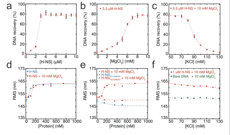
In order to dissect the role of divalent ions in the formation of bridged and stiffened complexes, we applied a novel, sensitive, andquantitativeDNA-bridging assay and carried out TPM experiments (providing a quantitative and selective readout of DNA stiffening). The DNA-bridging assay relies on immobilization of bait DNA on magnetic microparticles and the capture and detection of32P labeled prey DNA if DNA-DNA bridge formation occurs (seeFigure 1—figure supplement 4b for a sche-matic depiction of the assay). 80% of initial prey DNA is recovered at high H-NS concentrations (see
Figure 1a). In the absence of either the H-NS protein or bait DNA, no prey DNA is recovered under our experimental conditions. Next, we used this assay to quantify the DNA-bridging efficiency of H-NS as a function of the amount of Mg2+ ions (seeFigure 1b), reproducing the qualitative results ofLiu et al. (2010)and providing independent confirmation of the previously observed effects. The concentration range from 0 to 10 mM Mg2+ is considered to be physiologically relevant (Hurwitz and Rosano, 1967). Importantly, the transition from no bridging to complete bridging is abrupt between 4–6 mM Mg2+, indicating that changes in Mg2+concentration might drive a binary switch.
Earlier studies have shown that H-NS binding along a single DNA molecule results in DNA stiffen-ing (Amit et al., 2003;Liu et al., 2010). However, due to the co-occurrence of DNA bridging, previ-ous studies were incapable of measuring DNA stiffening in the presence of Mg2+. Here, we used
TPM to investigate DNA stiffening as a function of Mg2+ concentration. In TPM, the Root Mean
Square displacement (RMS) of bead movement is a direct reflection of tether stiffness and length (Figure 1—figure supplement 4a). DNA-binding proteins can affect both, but previous studies have shown that the DNA contour length is not affected by binding of H-NS (Dame et al., 2006;
Dame et al., 2001). Thus, an increase in stiffness due to H-NS binding translates into a higher RMS value of a DNA tether (Figure 1d). Here, we measured the effects of H-NS on DNA stiffness in the absence and presence of 10 mM Mg2+and confirmed that H-NS stiffens DNA (Amit et al., 2003;
Liu et al., 2010); importantly, our experiments reveal that Mg2+does not affect the stiffness of the
fully formed H-NS-DNA complexes at saturation, as in both conditions the RMS is the same (Figure 1d). Analysis of the binding characteristics using the McGhee-von Hippel equation revealed that the association constant of H-NS is somewhat reduced in the presence of Mg2+, while coopera-tivity increases under these conditions (Figure 3—figure supplement 1a,d). The reduction in DNA-binding affinity of H-NS may be attributed to shielding of the negatively charged phosphate back-bone by Mg2+.
The cooperative binding of H-NS and DNA stiffening observed by TPM suggest that H-NS multi-merizes along DNA, likely via the recently defined dimer-dimer interaction domain (Arold et al., 2010). Multimerization along DNA has been previously suggested (Esposito et al., 2002;
Williams et al., 1996) but has never been conclusively demonstrated. To test this hypothesis, we generated a mutant, H-NSY61DM64D, predicted to have disrupted dimer-dimer interaction based on
the H-NS1-83crystal structure (Arold et al., 2010). Size exclusion chromatography showed that this
H-NS mutant indeed exists solely as a dimer in solution independent of protein concentration ( Fig-ure 1—figFig-ure supplement 1b), whereas wild-type H-NS forms large multimeric structures ( Fig-ure 1—figFig-ure supplements 1aandArold et al., 2010;Leonard et al., 2009). The multimerization behavior of both proteins was unaffected by the presence of Mg2+. Electrophoretic Mobility Shift Assay confirmed that the DNA binding of the H-NS mutant is intact (Figure 1—figure supplement 2). TPM experiments reveal that H-NSY61DM64Dbinding does not lead to the formation of stiff
H-NS-DNA filaments (Figure 1e). The RMS is reduced compared to that of bare DNA, indicating not only that dimer-dimer interactions are disrupted, but also that individual H-NS dimers mildly distort DNA. DNA-bridging experiments reveal that H-NSY61DM64D is also incapable of forming DNA-H-NS-DNA
complexes (Figure 1—figure supplement 3). This indicates that individual H-NSY61DM64Ddimers do
cooperativity due to high local DNA concentration adjacent to existing bridges (Dame et al., 2006;
Dame et al., 2000), is insufficient to explain the formation of bridged DNA-H-NS-DNA complexes. The nature of the effect of Mg2+on DNA-bridging efficiency is not understood. An increase in
affin-ity would be expected if Mg2+would only facilitate interactions between the DNA phosphate
back-bone and negatively charged residues on H-NS, but this is not observed (Figure 3—figure supplement 1). As Mg2+does not affect the multimeric state of H-NS in solution (Figure 1—figure
supplement 1), a model involving an effect on H-NS multimerization can be excluded. A structural effect of Mg2+on individual units within H-NS filaments could explain the observed effects of Mg2+ on the bridging efficiency of H-NS.
a
0 20 40 60 80 1000 2 4 6 8 10
3.3 µM H-NS
DNA recovery (%)
[MgCl2] (mM)
DNA recovery (%)
[H-NS] (µM)
b
0 20 40 60 80 1000 2 4 6 8 10
c
135 145 155 165 1750 200 400 600 800 1000
RMS (nm) [Protein] (nM) 135 145 155 165 175 0
0 200 400 600 800 1000
RMS (nm) [Protein] (nM) 0 20 40 60 80 100
50 70 90 110 130
3.3 µM H-NS + 10 mM MgCl2
DN A recovery (%) [KCl] (mM) RMS (nm) [KCl] (mM)
50 70 90 110 130
135 145 155 165 175
Bare DNA + 10 mM MgCl2
1 µM H-NS + 10 mM MgCl2
d
e
f
H-NS + 10 mM MgCl2
H-NS H-NS + 10 mM MgCl2
H-NSY61DM64D
H-NSY61DM64D + 10 mM MgCl2
Figure 1.Modulation of H-NS function by ionic conditions. (a) DNA-bridging efficiency as a function of H-NS concentration in the presence of 10 mM
MgCl2. (b) DNA-bridging efficiency as a function of MgCl2concentration. (c) DNA-bridging efficiency as a function of the KCl concentration. (d) Root
Mean Square displacement (RMS) as a function of H-NS concentration, in the presence and absence of 10 mM MgCl2(N > 70, per data point). (e) RMS
of DNA as a function of H-NSY61DM64Din the presence and absence of MgCl2(N > 70, for each point). (f) Extension of DNA as a function of the KCl
concentration (N > 70, for each point). Error bars indicate standard deviation. Dashed lines are to guide the eye.
DOI: https://doi.org/10.7554/eLife.27369.003
The following figure supplements are available for figure 1:
Figure supplement 1.Multimeric state of H-NS measured using size exclusion chromatography.
DOI: https://doi.org/10.7554/eLife.27369.004
Figure supplement 2.Electrophoretic Mobility shift assay.
DOI: https://doi.org/10.7554/eLife.27369.005 Figure supplement 3.DNA recovery of H-NS and.
DOI: https://doi.org/10.7554/eLife.27369.006
Figure supplement 4.Schematic depiction of techniques used in this study.
DOI: https://doi.org/10.7554/eLife.27369.007
Figure supplement 5.Modulation of H-NS by alternative anions.
Mg
2+alters H-NS structure
To investigate the role of Mg2+on individual H-NS dimers we carried out MD simulations of an H-NS dimer in both the absence and presence of Mg2+, using our previously established model of a
full-length H-NS dimer (van der Valk et al., 2014). Visual inspection of the H-NS dimer simulations at 50 mM KCl reveals that H-NS changes from an ‘open’ extended conformation into more compact ‘closed’ shapes (seeFigure 2afor snapshots from these simulations,Figure 2—figure supplement 1for examples of the ‘closed’ conformation, orFigure 2—video 1for a movie of one such simula-tion). The three domains in H-NS interact, and these inter-domain interactions are facilitated by par-tial unfolding and buckling of the long central a helix (helixa3) connecting the dimerization and dimer-dimer interaction domains. The average distance between the donor and acceptor of all heli-cal hydrogen bonds (O-H distance) in helixa3 indicates that the buckle forms in region Glu42-Ala49 (Figure 2b). By analyzing the O-H distance between residues Ser45 and Ala49 in time, key residues at the site of buckle formation (seeFigure 2—figure supplement 3), we found that buckles can be reversible and irreversible, within the simulation time scale of 50 ns. Reversible buckles, caused by thermal fluctuations, typically last a few nanoseconds and occur several times during a single simula-tion run (see green line inFigure 2—figure supplement 3for an example). Irreversible buckles, sta-bilized by inter-domain interactions, do not return to a helical conformation during our simulations (see red line in Figure 2—figure supplement 3 for an example). To characterize the interactions that occur during the simulations, we generated contact maps that show the probability of finding interactions between residues, with a contact defined as the minimum distance between two resi-dues being 0.6 nm or less, see Materials and methods for details of this analysis (Figure 2—figure supplement 2a). These maps reveal that the DNA-binding domain interacts with other parts of the protein complex. In absence of Mg2+, the DNA-binding domain interacts with the dimerization domain, rendering the DNA-binding QGR motif (residues 112–114) (Gordon et al., 2011) of one DNA-binding domain inaccessible (see the snapshots inFigure 2a). Although the simulations were performed in absence of DNA, these observations suggest that DNA bridging is not possible in such a conformation, as the H-NS dimer can bind DNA only through its remaining/second DNA-binding domain. In this ‘closed’ conformation, interactions occur between the IRT residues at position 10–12 and the AMDEQGK residues at position 122–128. These interactions are hydrophilic in nature, sup-plemented by a salt bridge between R11 and D124 or E125. In the presence of Mg2+interactions between the DNA-binding domain and the dimerization domain no longer occur (see the snapshots inFigure 2a or the movie inFigure 2—video 2), The absence of such interactions is further illus-trated by the contact map inFigure 2—figure supplement 2band in higher detail inFigure 2—fig-ure supplement 5. The likelihood of finding Mg2+interacting with (i.e. being within 0.6 nm of) H-NS
residues, indicated by PMg2+, revealed that Mg2+ has a preference for glutamate residues in region
22–35 (Figure 2c,Figure 2—figure supplement 8), where the ions shield this region from interact-ing with the DNA-bindinteract-ing domains. The Mg2+ions transiently interact with the glutamate residues,
with residence times in the order of a few ns (as seen inFigure 2—video 2). Furthermore, Mg2+ions
interact with region 98–105 (sequence DENGE), right next to the DNA-binding QGR motif (Figure 2c). The presence of Mg2+stabilizes the ‘open’ conformation of H-NS, ensuring that DNA bridging can occur. Furthermore, we noted that Mg2+ is also located close to the buckle and may
directly stabilize helixa3 through interactions with Glu42, Glu43, Glu44, and Ser45 (Figure 2—fig-ure supplement 7), resulting in an ‘open’, bridging capable, H-NS conformation (seeFigure 2—vid-eos 1and2). These data suggest that Mg2+modulates H-NS by shielding interactions between the DNA-binding domain and dimerization domain, and by influencing the conformation of helix a3. Based on these observations, we designed an H-NS mutant predicted to bridge DNA independent of Mg2+. We therefore generated a mutant in which several of the amino acids involved in buckle
formation (E43,E44,S45) were substituted with alanines. In our DNA-bridging assay, H-NSE43A,E44A,
S45Aretains its ability to bridge DNA, but indeed achieves high DNA recovery (±50%) in the absence
of Mg2+(Figure 2—figure supplement 9a). Low concentrations of Mg2+are sufficient to reach
satu-rated DNA bridging (up to 80% DNA recovery) (Figure 2—figure supplement 9a). This observation independently confirms our model that the stretch of glutamates at the buckle is responsible for Mg2+sensing and Mg2+-dependent bridging by H-NS. Additionally, we note that the DNA-binding affinity of H-NE43A,E44A,S45Ais not significantly altered and that its DNA binding cooperativity is
MSEALKILNNIRTLRAQARECTLETLEEMLEKLEVVVNERREEESAAAAEVEERTRKLQQYREMLIADGIDPNELLNSLAAVKSGTKAKRAQRPAKYSYVDENGETKTWTGQGRTPAVIKKAMDEQGKSLDDFLIKQ | | | | | | | | | | | | | | 1 10 20 30 40 50 60 70 80 90 100 110 120 130
_1 _2 _3 _4 ` `2 _5 _6
Dimerization domain Dimer-dimer interaction domain DNA binding domain
MSEALKILNNIRTLRAQARECTLETLEEMLEKLEVVVNERREEESAAAAEVEERTRKLQQYREMLIADGIDPNELLNSLAAVKSGTKAKRAQRPAKYSYVDENGETKTWTGQGRTPAVIKKA
0 0.1 0.2 0.3 0.4
0.5 10 mM MgCl2 + 50 KCl
0 0.1 0.2 0.3 0.4
0.5 10 mM MgCl2 + 130 mM KCl
0 0.1 0.2 0.3 0.4 0.5
0 20 40 60 80 100 120
Hha + 10 mM MgCl2 + 50 mM KCl
a
b
c
10 mM MgCl
2+ 50 mM KCl
50 mM KCl
<d
O
-N
> [nm]
P
Mg
2+
Residue Index
Residue Index
0.2 0.3 0.4 0.5 0.6
0.2 0.3 0.4 0.5 0.6 0.2 0.3 0.4 0.5 0.6
`
` `2 _5 _6
30 40 50 60
22 64
50 mM KCl
10 mM MgCl2 + 50 mM KCl
130 mM KCl
10 mM MgCl2 + 130 mM KCl
Hha + 50 mM KCl
Hha + 10 mM MgCl2 + 50 mM KCl
Figure 2.Conformation of the H-NS dimer as a function of osmolarity. (a) Snapshots depicting representative conformations of H-NS in the simulations
with 50 mM KCl (top) and 10 mM MgCl2+50 mM KCl (bottom) (For the full movies depicting these effects seeFigure 2—videos 1and2, for other
examples of the ‘closed’ conformations of H-NS seeFigure 2—figure supplement 1). The buckle region is highlighted in red and the domains are
colored blue, green and brown for the dimerization domain, the dimer-dimer interface and the DNA-binding domain, respectively. The conserved motif
figure supplement 1d). Yet, in the presence of Mg2+, wild-type H-NS exhibits a far more coopera-tive DNA binding (Figure 3—figure supplement 1d). This suggests that the transition between the ‘open’ and ‘closed’ conformation of H-NS promotes lateral filament formation by H-NS.
Modulation of DNA bridging by osmotic factors
Although it has long been known that the expression of some H-NS controlled genes (such as the
proUoperon) is modulated by the osmolarity of the medium (Cairney et al., 1985), the underlying mechanism remains undetermined. Previous studies have revealed that the H-NS DNA stiffening mode is mildly sensitive to the KCl concentration (Amit et al., 2003;Liu et al., 2010). Using TPM, we were able to confirm these observations. The reduction in DNA stiffening is gradual (Dame and Wuite, 2003) and modest (Figure 1f). It is thus questionable whether the multimerization of H-NS along DNA alone is sufficient to explain its role in repression of transcription (and modulation thereof). Could the modulation of gene repression be due to ionic effects on DNA-bridging effi-ciency? Using our DNA-bridging assay, we observed complete abolishment of H-NS DNA bridging by KCl at concentrations exceeding 120 mM (Figure 1c), a binary response, similar to what we observed for the Mg2+ titration. This in vitro observation mirrors the in vivo response of the ProU operon, at which KCl concentrations exceeding 100 mM are required to alleviate H-NS-mediated repression (Cairney et al., 1985). Control experiments using K-glutamate (Figure 1—figure supple-ment 5) confirm that K+, and not the counter-ion, is responsible for the observed effects, even
though the counter ion may affect the DNA-binding affinity of the protein (Figure 1—figure supple-ments 5candLeirmo et al., 1987) This strong and abrupt effect on DNA bridging, while leaving DNA stiffening essentially unaffected, might indicate that H-NS reverts to the ‘closed’ conformation by the addition of K+. To investigate this effect at a structural level we performed MD simulations at
Figure 2 continued
involved in DNA binding is highlighted as ball-stick models. Mg2+ions are shown as dark gray orbs. The protein is shown in ribbon representation with
a transparent surface. The atomic radii in the protein were set to 3 A to smooth the surface. (b) Location of buckle in helixa3. The average distance d
O-Nbetween donor and acceptor in the helical hydrogen bond in helixa3 is plotted as a function of the residue index of the acceptor. The dashed black
line in the graphs indicates the distance threshold for forming a hydrogen bond. Time traces of these distances are given inFigure 2—figure
supplement 3. (c) Location of Mg2+on H-NS. The probability of finding Mg2+ions within 0.6 nm of an H-NS residue, P
Mg2+, is plotted as function of
the residue index for the three systems containing Mg2+.
DOI: https://doi.org/10.7554/eLife.27369.009
The following video and figure supplements are available for figure 2:
Figure supplement 1.Examples of ‘closed’ H-NS conformations in the presence of (a) 50 mM KCl, (b) 130 mM KCl, or (c) Hha.
DOI: https://doi.org/10.7554/eLife.27369.010
Figure supplement 2.Contact maps of full-length H-NS dimers simulations in different conditions.
DOI: https://doi.org/10.7554/eLife.27369.011
Figure supplement 3.Time traces of the O-H distance between residues 45 and 49.
DOI: https://doi.org/10.7554/eLife.27369.012 Figure supplement 4.Location of K+on hr-NS.
DOI: https://doi.org/10.7554/eLife.27369.013
Figure supplement 5.Contact maps of H-NS dimers in different conditions, focused on the interactions between the dimerization domain and the DNA-binding domain.
DOI: https://doi.org/10.7554/eLife.27369.014 Figure supplement 6.Location of Hha on H-NS.
DOI: https://doi.org/10.7554/eLife.27369.015
Figure supplement 7.Correlation between hydrogen bond distance and proximity of Mg2+to the buckle in helixa3.
DOI: https://doi.org/10.7554/eLife.27369.016 Figure supplement 8.Mg2+localization on H-NS.
DOI: https://doi.org/10.7554/eLife.27369.017
Figure supplement 9.Function of the H-NS derivative, H-NSE43A,E44A,S45A.
DOI: https://doi.org/10.7554/eLife.27369.018
Figure 2—video 1.Conformational flexibility of H-NS.
DOI: https://doi.org/10.7554/eLife.27369.019
Figure 2—video 2.The effect of magnesium on the conformational flexibility of H-NS.
high KCl concentrations. The presence of 130 mM KCl alters interactions between the various domains in H-NS (see contact maps inFigure 2—figure supplement 2c and d). In addition to inter-actions between the DNA-binding domain and the dimerization domain, the two DNA-binding domains in the dimer interact with each other. Moreover, the DNA-binding domains interact with helixa3. In particular regions 98–105 (KYSYVDENGE) and 123–129 (EQGKS) are involved in these interactions. The average distance between the donors and acceptors in the hydrogen bonds within helixa3 (Figure 2b) indicates that that buckles in helixa3 also occur at a high KCl concentration, at the same location as determined by low-salt conditions. These observations indicate that the ‘closed’ state can have multiple forms (seeFigure 2—figure supplement 1for snapshots), but that all these conformations block the DNA-binding motif QGR from interacting with DNA. The presence of Mg2+ does not significantly alter the occurrence of buckles (Figure 2b). Instead, Mg2+is capable of
deter-ring interactions between the DNA-binding domain and the dimerization domain by shielding resi-dues in the dimerization domain involved in these interactions (Figure 2c, Figure 2—figure supplement 8). However, the probability of interactions occurring between the DNA-binding domain and other parts of the protein is reduced significantly in the presence of Mg2+ (Figure 2— figure supplement 2andFigure 2—figure supplement 5). High K+ concentrations therefore inhibit H-NS bridging by promoting the ‘closed’ conformation of H-NS even in the presence of Mg2+ and
elucidates the modulatory and regulatory effects of KCl and osmolarity.
Modulation of DNA bridging and DNA stiffening by truncated H-NS
variants
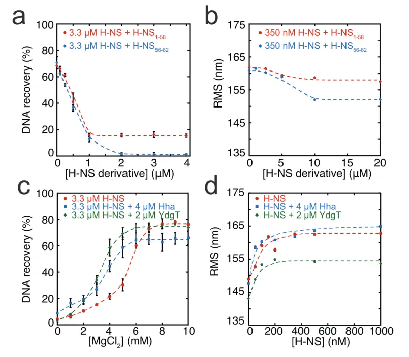
In addition to environmental factors such as osmolarity, H-NS activity is affected by interactions with other proteins in vivo. Members of the Hha/YmoA protein family, such as Hha and YdgT, are known to cooperate with H-NS in repression of genes (Ban˜os et al., 2008), while other proteins such as H-NST are capable of inhibiting H-NS function (Liu et al., 2010), likely by hampering H-NS multime-rization. To systematically investigate the latter mechanism, we designed and synthesized truncated H-NS derivatives, targeting the H-NS dimerization domain (H-NS1-58) or dimer-dimer interaction
domain (H-NS56-83). Interfering with H-NS dimerization, through the addition of H-NS1-58 in
DNA-bridging experiments, we observed a reduction in DNA recovery from 75% to 20% at ratios higher than 1: 3 H-NS1-58/ H-NS (Figure 3a). Similarly, targeting the dimer-dimer interaction domain
(through H-NS56-82) resulted in complete abolishment of DNA bridging (Figure 3a). Next, we
investi-gated the effects of H-NS derivatives on H-NS DNA stiffening using TPM. Only at very high H-NS derivative concentrations (30-fold excess) reduction of DNA stiffening was observed (Figure 3b). These experiments reveal that both H-NS dimerization and dimer-dimer interactions can be effec-tively targeted for inhibition of H-NS activity and that the respective domains are crucial to the for-mation of bridged filaments. This suggests that natural H-NS inhibitors such as H-NST operate by disrupting DNA bridging and provides clues for rational design of artificial peptide inhibitors of H-NS.
Modulation of H-NS by Hha and YdgT
Gene regulation by H-NS often occurs in conjunction with other proteins; these co-regulators are known to interact with H-NS at specific loci along the genome. Two such proteins, Hha, and YdgT, are members of the Hha/YmoA (Madrid et al., 2007) family of proteins. In order to understand the modulation of H-NS function by these proteins we investigated their influence on the H-NS DNA binding modes. We observed that Hha, when added at equimolar concentrations, enhances DNA bridging by H-NS at low Mg2+concentrations (Figure 3c). A similar enhancement of H-NS-mediated DNA bridging was observed with the Hha paralogue, YdgT. While Hha and YdgT promote DNA bridging to a similar extent, YdgT promotes DNA bridging at significantly lower concentrations, likely due to a higher affinity for H-NS. At higher concentrations of YdgT the effect on H-NS-medi-ated bridging closely resembles the bridging profile obtained for H-NSE43A,E44A,S45A(Figure
RMS (nm)
[H-NS] (nM)
DNA recovery (%)
[MgCl
2] (mM)
0
20
40
60
80
100
0
2
4
6
8
10
3.3 µM H-NS
3.3 µM H-NS + 4
ȝ
M Hha
3.3 µM H-NS + 2
ȝ
M YdgT
c
d
a
b
DNA recovery (%)
[H-NS derivative] (
ѥ
M)
0
20
40
60
80
100
0
1
2
3
4
3.3 µM H-NS + H-NS
1-583.3 µM H-NS + H-NS
56-82135
145
155
165
175
0
5
10
15
20
350 nM H-NS + H-NS
1-58350 nM H-NS + H-NS
56-82RMS (nm)
[H-NS derivative] (
ѥ
M)
135
145
155
165
175
0
200 400 600 800 1000
H-NS
H-NS + 4
ȝ
M Hha
H-NS + 2
ȝ
M YdgT
Figure 3.Modulation of H-NS function by protein cofactors. (a) DNA bridging efficiency as a function of inhibiting peptides targeting either the
dimerization domain (H-NS1-58) and multimerization domain (H-NS56-82). (b) Root Mean Square displacement (RMS) as a function of inhibiting peptides
targeting either the dimerization (H-NS1-58) and multimerization (H-NS56-82) (N > 70, for each point). (c) DNA bridging efficiency as a function of Mg2+
concentration in the presence and absence of 4mM Hha or 2mM YdgT. (d) RMS of DNA in the presence of H-NS, H-NS-Hha, and H-NS-YdgT. Dashed
lines are to guide the eye (N > 60, for each point).
DOI: https://doi.org/10.7554/eLife.27369.021
The following figure supplements are available for figure 3:
Figure supplement 1.McGhee-von Hippel analysis of H-NS DNA binding curves based on TPM data.
DOI: https://doi.org/10.7554/eLife.27369.022
Figure supplement 2.DNA-bridging efficiency of H-NS (red), H-NS + 4mM YdgT (blue) and H-NSE43A,E44A,S45A(orange) as a function of MgCl2
concentration.
presence of Mg2+and YdgT, H-NS causes ‘DNA collapse’ in TPM (data not shown), similar to earlier
observations for H-NS in the presence of Mg2+without (Liu et al., 2010;Wang et al., 2014) or with
Hha added (Wang et al., 2014). This ‘DNA collapse’ is attributed to H-NS-mediated DNA bridge formation. One possible explanation for the effects of Hha and YdgT, is that they effectively increase the DNA-binding affinity of H-NS (Ali et al., 2013). To investigate whether Hha affects H-NS confor-mation, we performed MD simulations, incorporating structural information from the recently resolved H-NS1-43-Hha co-crystal structure (Ali et al., 2013). Our MD simulations reveal that Hha
does not prevent buckles in helixa3 (seeFigure 2b). Instead Hha alters the interactions between the dimerization domain and the DNA-binding domain (Figure 2—figure supplement 2) by block-ing access to parts of dimerization domain. This hypothesis is further supported by interactions between Hha and other parts of H-NS, including the DNA-binding domain and helixa3 (Figure 2— figure supplement 6). In the presence of Mg2+ and Hha, the contacts between the DNA-binding
domain and dimerization domain are reduced even further. This shows that Hha modulates H-NS function by stabilizing the ‘open’ -bridging capable- conformation of H-NS.
Discussion
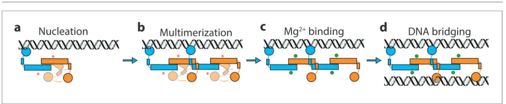
It has been known for many years that H-NS binding induces gene silencing. H-NS activity in vivo is modulated by physico-chemical factors (osmolarity, pH, temperature) and interaction partners. These findings support the hypothesis that H-NS plays a role in environmental adaptation. However, mechanistically it is unclear how functional modulation of H-NS by such factors is achieved. Based on our findings, we conclude that H-NS is incapable of bridging or stiffening DNA as dimers. H-NS dimers bind DNA in cis (Dame et al., 2006; Dame et al., 2000) and associate side-by-side along DNA, likely via the recently identified dimer-dimer interaction domain (Arold et al., 2010), resulting in DNA stiffening. This process is cooperative as H-NS dimers interact with neighbors, as well as with DNA. Our studies reveal that H-NS-DNA filaments arestructurallyvery similar, independent of the presence of Mg2+(Figure 1d). Butfunctionally,these H-NS-DNA filaments are distinct. H-NS can be ‘activated’ by Mg2+, which promotes a conformational change, rendering both DNA-binding domains of H-NS dimers accessible for DNA bridging. In our model, the assembly of bridged com-plexes proceeds in distinct steps: (1) nucleation (Lang et al., 2007) (binding of an H-NS dimer at a high affinity site), (2) lateral filament growth by H-NS dimer-dimer interactions (leading to DNA stiff-ening) and (3) bridging of the assembled filament to bare DNA provided in trans (Figure 4). Each step can potentially be modulated by osmolarity and protein interaction partners. Here, we show that these factors most effectively target DNA bridging.
What are the implications of our observations? Our observations add to the large body of evi-dence showing that regulation of transcription via H-NS is complex, and that it does not proceed via a single, simple, well-defined mechanism. The most straightforward form of repression by H-NS is via occlusion of RNA polymerase from the promoter (Lim et al., 2012; Prosseda et al., 2004;
Go¨ransson et al., 1990). Whether this mechanism of repression involves lateral filament formation or bridging is unclear. It is expected that both types of complexes assembled at a promoter site can in principle occlude RNA polymerase. A second mechanism of repression is to trap RNA polymerase
*
Mg
2+binding
Nucleation
*
* *
* *
Multimerization
DNA bridging
a
b
c
d
Figure 4.Model of H-NS complex assembly. (a) H-NS nucleates at preferred DNA sequences in the genome. (b) H-NS laterally multimerizes laterally
along the DNA in the ‘closed’ conformation. (c) In the presence of Mg2+or other H-NS modulators such as Hha, H-NS switches to the ‘open’, bridging
capable conformation. (d) H-NS forms DNA bridges in trans. The red asterisk indicates the buckle location. Mg2+ions are shown as green orbs.
at the promoter, preventing promoter escape (Schro¨der and Wagner, 2000; Shin et al., 2005). RNA polymerase trapping likely involves DNA bridging of promoter upstream and downstream ele-ments (Dame et al., 2002;Shin et al., 2005). A third mechanism of repression by H-NS is to inter-fere with RNA polymerase progression during active transcription by intragenic binding (Dole et al., 2004). In this model, both modes could interfere with transcription. However, it was suggested recently that only bridged filaments are capable of interfering with transcription (Kotlajich et al., 2015), with lateral filaments likely being disassembled as RNA polymerase encounters them. Gener-ally, the type of complex formed by H-NS is expected to depend on the type and number of ions, combined with local DNA conformation (dependent on DNA sequence or DNA topology), and the presence of modulating proteins. The interplay between these factors will determine the strength of the complex, and degree of repression. Thus, different H-NS repressed genes are expected to be subject to different types of modulation, providing a key to a coordinated response in gene expres-sion to altered conditions and selectivity for the interplay with specific co-regulators at specific tar-get regions.
Materials and methods
Construction of expression vectors
H-NS expression vector
The vector pRD18 for expression of H-NS was constructed by inserting the PCR amplifiedhnsgene into the pET3His overexpression vector using NdeI and XhoI restriction sites. Using the XhoI restric-tion site, the encoded protein does not contain a C-terminal His-tag.
H-NS
Y61DM64Dexpression vector
The vector pRD69 for expression of H-NSY61DM64Dwas constructed by inserting a PCR fragment
con-taining thehnsgene mutated to encode aspartic acid instead of tyrosine/methionine at position 61 and 64 into pET3His using NdeI and XhoI restriction sites.
Hha expression vector
The vector pRD38 for expression of N-terminally His tagged Hha was constructed by inserting a PCR fragment containing thehhagene into pET3His using XhoI and BamHI restriction sites.
YdgT expression vector
The vector pRD39 for expression of N-terminally His tagged YdgT was constructed by inserting a PCR fragment containing theydgTgene into pET3His using XhoI and BamHI restriction sites.
Protein overproduction and purification
H-NS/H-NSY61DM64D/H-NSE43A,E44A,S45A. BL21 (DE3) (RRID: WB-STRAIN: HT115(DE3)) Dhns::kan/frt
pLysE (NT201, our lab) cells transformed with plasmids expressing H-NS/H-NS mutants were grown to an OD600of 0.4, and induced for 2 hr using IPTG (500mM). For the H-NE43A,E44A,S45Aprotein, the
cells were co-transformed with pRD252, coding for LacI to help suppress leaky expression of H-NE43A,E44A,S45A. The cells were pelleted and lysed by sonication in 100 mM NH4Cl, 20 mM Tris pH
7.2, 10% glycerol, 8 mMb-mercaptoethanol, 3 mM benzamidine). The soluble fraction was loaded onto a P11 column and eluted using a 100 mM-1 M NH4Cl gradient, the protein eluted at 280 mM
NH4Cl. The peak fractions were dialysed to buffer B (identical to buffer A, but containing 130 mM
NaCl instead of NH4Cl) by overnight dialysis. The dialysate was loaded onto a heparin column (GE
H-NS
1-58BL21 (DE3) (RRID: WB-STRAIN: HT115(DE3))Dhns::kan/frt pLysE (NT210, our lab) cells transformed with plasmids expressing H-NS/H-NS mutants were grown to an OD600of 0.4, and induced for 2 hr
using IPTG (500mM). The cells were pelleted and lysed by sonication in 100 mM NaCl, 20 mM Tris pH 7.2, 10% glycerol, 8 mM b-mercaptoethanol, 3 mM benzamidine). The soluble fraction was heated to 65
˚
C for 10 min and then spun down at 10.000 RPM for 10 min. The supernatant was col-lected and a 1:1 ratio saturated ammonium sulfate (50 mM Tris pH 7.2, 4M ammonium sulfate) was gradually added to the cooled sample. The sample was spun down at 8.000 RPM for 15 min and a 1:1 ratio of 5 mM Tris pH 7.2, 15% glycerol was added to the supernatant. To remove further impuri-ties, the sample was run through a 1 ml hydrophobic interaction column and 1 ml Blue-agarose col-umn (the protein should not bind to either of these colcol-umn) before finally binding the protein to 1 ml Resource-Q column (GE Healthcare). The protein was eluted with a 25 mM 1M gradient of NaCl, the protein eluted at roughly 380 mM NaCl. The purity of the protein was verified on an SDS-PAGE gel. The protein concentration was determined using a Bicinchoninic Acid assay (Pierce BCA protein assay kit, Thermo Scientific).Hha/YdgT
BL21 (DE3) (RRID: WB-STRAIN: HT115(DE3))Dhns::frt, hha::kan, pLysE (our lab) cells transformed with plasmids pRD38/pRD39 expressing hha/ydgT were grown at 37
˚
C to an OD600 of 0.4, andinduced for two hours using IPTG (500mM). The cells were pelleted and lysed in 20 mM HEPES pH 7.9, 1 M KCl, 10% glycerol, 8 mMb-mercaptoethanol. The soluble fraction was loaded onto a Ni-col-umn. The column was first washed with buffer D (20 mM HEPES pH 7.9, 0.5 M KCl, 10% glycerol, 8 mMb-mercaptoethanol). The protein was then eluted using a 0 mM-0.5 M Imidazole gradient, the protein eluted at 300 mM Imidazole. The peak fractions were dialysed to buffer E (identical to buffer D, but containing 100 mM KCl) by overnight dialysis. The sample was then loaded onto pre-equili-brated SP Hi-Trap-column and Ni-column connected in series. After loading the samples on the col-umn, the SP Hi-Trap -column was disconnected and the protein was eluted from the Ni-column using a 0–0.5 M imidazole gradient. The purity of the protein was verified on an SDS-PAGE gel. The protein concentration was determined using a Bicinchoninic Acid assay.
Peptide production and purification
A truncated form of H-NS (H-NS56-82)was synthesized by way of automated solid phase synthesis
using standard protocols via Fmoc-strategy. Purification was performed by RP-HPLC with a Gemini 5mC18 reversed phase column. Identity of the peptides was determined via MALDI-MS. The purity was determined by means of analytical RP-HPLC. The peptide was freeze dried and dissolved in 20 mM Tris pH 7.2, 300 mM KCl, 10% glycerol, 8 mMb-mercaptoethanol. The peptide concentration was determined using a Bicinchoninic Acid assay (Pierce BCA protein assay kit, Thermo Scientific).
Size exclusion chromatography
Size exclusion chromatography was done using a Superose-12 column with a flow of 0.3 ml/min, pre-equilibrated with 10 mM Tris-HCl, 50 mM KCl, 5% glycerol containing or lacking 10 mM MgCl2.
The absorbance of the eluting fractions was measured at 215 nm. These experiments were per-formed in triplicate.
DNA substrates
DNA preparation
All experiments were performed using a random, AT-rich, 685 bp (32% GC) DNA substrate (Laurens et al., 2012). The DNA substrate was generated by PCR, and the PCR products were puri-fied using a GenElute PCR Clean-up kit (Sigma-Aldrich). If required, DNA was 32P-labeled as described previously (Wagner et al., 2011).
DNA-bridging assay
resuspended in 200mL CB containing 100 nM biotinylated DNA. Next, the bead suspensions were incubated for 30 min on a rotary shaker (1000 rpm) at 25
˚
C. After incubation, the beads were washed twice with Incubation buffer (IB: 10 mM Tris-HCl pH 8.0, 50 mM KCl, 10* mM MgCl2, 5% v/vGlycerol, 1 mM DTT and 1 mM Spermidine) before resuspension in IB and addition of±8000 cpm of radioactively labeled32P 685 bp DNA. Radioactive DNA was supplemented with unlabeled 685 bp DNA to maintain a constant (20 nM) DNA concentration. The DNA-bridging protein H-NS (concen-trations indicated in the text), and if applicable Hha or YdgT were added and the mixture was incu-bated for 30 min on a shaker (1000 rpm) at 25
˚
C. To remove unbridged prey DNA, the beads were washed with IB, before resuspension in 12mL stop buffer (10 mM Tris pH 8.0, 1 mM EDTA, 200 mM NaCl, 0.2% SDS). All samples were quantified through liquid scintillation counting over 10 min. All values recovered from the DNA-bridging assay were corrected for background signal (using a sam-ple lacking H-NS), and normalized to a reference samsam-ple containing the amount of labeled32P 685 bp DNA used in the assay. The samples were then run on a 5% 0.5x TBE gel to ensure DNA integ-rity. DNA bridging was calculated based on a reference sample containing 2mL of prey DNA. All DNA-bridging experiments were performed in triplicate. Unless indicated otherwise all DNA-bridg-ing experiments were performed in the presence of 10 mM of MgCl2 and 3,3 mM of H-NS (10%more H-NS than is required for saturation - seeFigure 1A). Each experiment contains 50 mM KCl, to which additional KCl or K-glutamate are added depending on the experimental condition tested.
Tethered particle motion experiments
Tethered particle motion experiments were performed as reported previously (Driessen et al., 2014;van der Valk et al., 2017). Flow cells were prepared as described with minor modifications (Driessen et al., 2014;van der Valk et al., 2017). Here, before flowing in protein diluted in the experimental buffer (10 mM Tris-HCl pH 8.0, 50 mM KCl, 10 mM MgCl2/EDTA, 5% v/v Glycerol, 1
mM DTT) the flow cell was washed using 4 flow cell volumes with the experimental buffer. Next, the flow cell was incubated for 10 min with protein solution before sealing the flow cell. The flow cell was maintained at a constant temperature of 25
˚
C. Measurements were started 10 min after the introduction of protein solution. TPM experiments were done at least in duplicate. The data were analyzed as previously described (Driessen et al., 2014;van der Valk et al., 2017). The RMS was first converted to persistence length(Lp) usingEquation 1(Go¨ransson et al., 1990):RMS¼233 156
1þ0:08Lp0:45 (1)
Lp values were then used to calculate the fractional coverage (Equation 2) (Go¨ransson et al.,
1990):
n#¼
ffiffiffiffiffiffiffiffiffiffiffiffiffiffi1
Lp;measured
q ffiffiffiffiffiffiffiffiffiffiffi1
Lp;naked
q
ffiffiffiffiffiffiffiffiffiffiffiffiffiffi1
Lp;saturated
q ffiffiffiffiffiffiffiffiffiffiffi1
Lp;naked
q (2)
The fractional coverage (d) was fit using the McGhee-von Hippel model for cooperative lattice binding (Equation 3-5)(McGhee and von Hippel, 1974):
#
c¼Kð1 dÞ
2!þ1
ð Þð1 dÞ þ# R
2! 1
ð Þð1 dÞ
n 1
1 ðnþ1Þ#þR
2 1ð dÞ
2
(3)
where
R¼
ffiffiffiffiffiffiffiffiffiffiffiffiffiffiffiffiffiffiffiffiffiffiffiffiffiffiffiffiffiffiffiffiffiffiffiffiffiffiffiffiffiffiffiffiffiffiffiffiffiffiffiffiffiffiffiffi
1 ðnþ1Þ#
ð Þ2þ4!#ð1 dÞ
q
(4)
and
d¼n#
To this end, weighted orthogonal distance regression (ODR) was performed to estimate the parameters of the nonlinear implicit equation describing the cooperative ligand binding. The binding site size (n) of H-NS was fixed to a value of 30 bp during regression, which corresponds to values determined previously (Dame et al., 2006;Amit et al., 2003). The association constant (K) and the cooperativity parameter (w) were assumed to be positive real numbers. A custom fitting routine was implemented in Fortran and makes use of the ODRPACK library (Boggs et al., 1992).
Molecular dynamics simulations
The starting conformation of the full-length H-NS dimer was constructed as described previously (van der Valk et al., 2014). The system was placed in a periodic dodecahedron box with a distance of at least 0.8 nm between the box edge and the most extended atom of the protein dimer, fol-lowed by the addition of water and ions. With this system we performed Molecular Dynamics (MD) simulations of full length H-NS at different concentrations of KCl, and MgCl2and with the addition
of Hha, summing up to a total of six different systems. Hha was added by aligning the crystal struc-ture containing the H-NS – Hha complex (PDB code 4ICG [Ali et al., 2013]) with the full-length struc-tural model and copying the Hha molecules. System size ranged from 513457 atoms for the Hha systems to around 1,135,000 atoms for the other four systems.
Interactions between atoms were described by the AMBER99-SB-ILDN force field ( Lindorff-Larsen et al., 2010), in combination with the TIP3P water model (Jorgensen et al., 1983). Long-range electrostatic interactions were treated via the Particle Mesh Ewald method (Darden et al., 1993; Essmann et al., 1995) with a short-range electrostatic cutoff distance at 1.1 nm. Van der Waals interactions were cut off at 1.1 nm. Preparation of the systems consisted of energy minimiza-tion equilibraminimiza-tion of the solvent. Energy minimizaminimiza-tion was performed by the conjugate gradient method. After energy minimization, the positions of water molecules and ions were equilibrated by a 1 ns molecular dynamics run at a temperature of 298 K and a pressure of 1 bar in which the heavy atoms in the protein were position-restrained with a force constant in each direction of 1000 kJ/mol nm. After preparation, we performed 16 50 ns runs for each system, varying initial conditions by assigning new random starting velocities drawn from the Maxwell-Boltzmann distribution at 298 K. All simulations were performed with GROMACS v.4.6.3 (Pronk et al., 2013) at the Dutch National Supercomputer with the leap-frog integration scheme and a time step of 2 fs, using LINCS (Hess et al., 1997) to constrain all bonds. All simulations were performed in the isothermal-isobaric ensemble at a pressure of 1 bar, using the v-rescale thermostat (Bussi et al., 2007) and the Parri-nello-Rahman barostat (Parrinello and Rahman, 1981).
Frames were stored every 10 ps. The first 10 ns of each simulation are excluded from analysis, unless stated otherwise. Analysis focused on determining contacts between domains, between H-NS and Hha, between H-NS and ions, and helical hydrogen bonds. Contact maps of interactions between residues in the H-NS dimer system were obtained by first calculating the minimum distance between each residue pair in the system. A residue pair is counted to be in contact if they are at a minimum distance of 0.6 nm or less. The probability of a contact is then calculated as the average over all 16 simulations (excluding the first 10 ns) and displayed as a contact probability matrix. We used a modified version of the g_mdmat tool in GROMACS (Pronk et al., 2013) in combination with Perl scripts to generate contact maps. To determine the location of ions with respect to the H-NS system, we calculated the minimum distance between each residue in the H-NS dimer and the ions and counted a contact if the distance between an H-NS residue and an ion is 0.6 nm or less. These contact probabilities (PMg2+, PK+and PCl-) are averaged over all 16 simulations and the two
mono-mers. A similar procedure was performed to determine the probability of contacts between H-NS and Hha, PHha, and between H-NS domains, with P1-40indicating the probability of finding the
DNA-binding domain (residues 96–137) close to the dimerization domain (residues 1–40). P96-137indicates
the probability of finding the dimerization domain close to the DNA-binding domain.
To determine the location of the buckle in helixa3, we calculated the helical hydrogen bond dis-tances dO-Nfor residues 22–67 in each monomer between the backbone carbonyl oxygen O of
Acknowledgements
We thank Bas de Mooij for his assistance in standardizing the bridging assay, Wim Jesse and Alexan-der Kros for synthesizing the H-NS56-83peptide and their assistance during its purification. We also
thank Rosalie Driessen for her assistance with data analysis and all group members for valued discus-sions. JV acknowledges the use of the Dutch National Supercomputer Cartesius for the MD simulations.
Additional information
Funding
Funder Grant reference number Author
NanonextNL of the Govern-ment of the Netherland and 130 partners
Ramon A van der Valk Geri F Moolenaar Remus T Dame
Netherlands Organisation for Scientific Research
VIDI 864.08.001 Ramon A van der Valk
Geri F Moolenaar Nora Goosen Remus T Dame
Netherlands Organisation for Scientific Research
Athena grant 700.58.802 Jocelyne Vreede
Human Frontier Science Pro-gram
RGP0014/2014 Andreas Hofmann
Remus T Dame
China Scholarship Council No. 201506880001 Liang Qin
Netherlands Organisation for Scientific Research
VICI 016.160.613 Remus T Dame
The funders had no role in study design, data collection and interpretation, or the decision to submit the work for publication.
Author contributions
Ramon A van der Valk, Conceptualization, Formal analysis, Supervision, Validation, Investigation, Visualization, Methodology, Writing—original draft, Writing—review and editing; Jocelyne Vreede, Conceptualization, Resources, Data curation, Software, Formal analysis, Funding acquisition, Valida-tion, InvestigaValida-tion, VisualizaValida-tion, Methodology, Writing—review and editing; Liang Qin, Formal anal-ysis, Validation, Investigation, Visualization; Geri F Moolenaar, Nora Goosen, Conceptualization, Resources, Validation, Investigation, Methodology, Project administration; Andreas Hofmann, Soft-ware, Formal analysis, Investigation, Methodology; Remus T Dame, Conceptualization, Data cura-tion, Formal analysis, Supervision, Funding acquisicura-tion, Visualizacura-tion, Methodology, Writing—original draft, Project administration, Writing—review and editing
Author ORCIDs
Remus T Dame, http://orcid.org/0000-0001-9863-1692
Decision letter and Author response
Decision letterhttps://doi.org/10.7554/eLife.27369.026
Author responsehttps://doi.org/10.7554/eLife.27369.027
Additional files
Supplementary files .Transparent reporting form
References
Ali SS, Whitney JC, Stevenson J, Robinson H, Howell PL, Navarre WW. 2013. Structural insights into the regulation of foreign genes in Salmonella by the Hha/H-NS complex.Journal of Biological Chemistry288: 13356–13369.DOI: https://doi.org/10.1074/jbc.M113.455378,PMID: 23515315
Amit R, Oppenheim AB, Stavans J. 2003. Increased bending rigidity of single DNA molecules by H-NS, a temperature and osmolarity sensor.Biophysical Journal84:2467–2473.DOI: https://doi.org/10.1016/S0006-3495(03)75051-6,PMID: 12668454
Arold ST, Leonard PG, Parkinson GN, Ladbury JE. 2010. H-NS forms a superhelical protein scaffold for DNA condensation.PNAS107:15728–15732.DOI: https://doi.org/10.1073/pnas.1006966107
Atlung T, Ingmer H. 1997. H-NS: a modulator of environmentally regulated gene expression.Molecular Microbiology24:7–17.DOI: https://doi.org/10.1046/j.1365-2958.1997.3151679.x,PMID: 9140961
Ban˜os RC, Pons JI, Madrid C, Jua´rez A. 2008. A global modulatory role for the Yersinia enterocolitica H-NS protein.Microbiology154:1281–1289.DOI: https://doi.org/10.1099/mic.0.2007/015610-0,PMID: 18451036
Bloch V, Yang Y, Margeat E, Chavanieu A, Auge´ MT, Robert B, Arold S, Rimsky S, Kochoyan M. 2003. The H-NS dimerization domain defines a new fold contributing to DNA recognition.Nature Structural Biology10:212– 218.DOI: https://doi.org/10.1038/nsb904,PMID: 12592399
Boggs PT, Byrd RH, Rogers JE, Schnabel RB. 1992.ODRPACK.Software for Weighted Orthogonal Distance Regression. 2.01. National Institute of Standards and Technology.DOI: https://doi.org/10.6028/NIST.IR.4834 Bussi G, Donadio D, Parrinello M. 2007. COMP 8-Canonical sampling through velocity rescaling.Abstr Pap Am
Chem S234.
Cairney J, Booth IR, Higgins CF. 1985. Osmoregulation of gene expression in Salmonella typhimurium: proU encodes an osmotically induced betaine transport system.Journal of Bacteriology164:1224–1232.PMID: 3 905768
Cerdan R, Bloch V, Yang Y, Bertin P, Dumas C, Rimsky S, Kochoyan M, Arold ST. 2003. Crystal structure of the N-terminal dimerisation domain of VicH, the H-NS-like protein of Vibrio cholerae.Journal of Molecular Biology 334:179–185.DOI: https://doi.org/10.1016/j.jmb.2003.09.051,PMID: 14607110
Ceschini S, Lupidi G, Coletta M, Pon CL, Fioretti E, Angeletti M. 2000. Multimeric self-assembly equilibria involving the histone-like protein H-NS. A thermodynamic study.Journal of Biological Chemistry275:729–734. DOI: https://doi.org/10.1074/jbc.275.2.729,PMID: 10625601
Dame RT, Kalmykowa OJ, Grainger DC. 2011. Chromosomal macrodomains and associated proteins: implications for DNA organization and replication in gram negative bacteria.PLoS Genetics7:e1002123. DOI: https://doi.org/10.1371/journal.pgen.1002123,PMID: 21698131
Dame RT, Noom MC, Wuite GJ. 2006. Bacterial chromatin organization by H-NS protein unravelled using dual DNA manipulation.Nature444:387–390.DOI: https://doi.org/10.1038/nature05283,PMID: 17108966
Dame RT, Tark-Dame M. 2016. Bacterial chromatin: converging views at different scales.Current Opinion in Cell Biology40:60–65.DOI: https://doi.org/10.1016/j.ceb.2016.02.015,PMID: 26942688
Dame RT, Wuite GJ. 2003. On the role of H-NS in the organization of bacterial chromatin: from bulk to single molecules and back.Biophysical Journal85:4146–4148.DOI: https://doi.org/10.1016/S0006-3495(03)74826-7, PMID: 14645101
Dame RT, Wyman C, Goosen N. 2000. H-NS mediated compaction of DNA visualised by atomic force
microscopy.Nucleic Acids Research28:3504–3510.DOI: https://doi.org/10.1093/nar/28.18.3504,PMID: 10982 869
Dame RT, Wyman C, Goosen N. 2001. Structural basis for preferential binding of H-NS to curved DNA. Biochimie83:231–234.DOI: https://doi.org/10.1016/S0300-9084(00)01213-X,PMID: 11278073
Dame RT, Wyman C, Wurm R, Wagner R, Goosen N. 2002. Structural basis for H-NS-mediated trapping of RNA polymerase in the open initiation complex at the rrnB P1.Journal of Biological Chemistry277:2146–2150. DOI: https://doi.org/10.1074/jbc.C100603200,PMID: 11714691
Dame RT. 2005. The role of nucleoid-associated proteins in the organization and compaction of bacterial chromatin.Molecular Microbiology56:858–870.DOI: https://doi.org/10.1111/j.1365-2958.2005.04598.x, PMID: 15853876
Darden T, York D, Pedersen L. 1993. Particle mesh Ewald: An Nlog(N) method for Ewald sums inlarge systems.The Journal of Chemical Physics98:10089–10092.DOI: https://doi.org/10.1063/1.464397
Deighan P, Beloin C, Dorman CJ. 2003. Three-way interactions among the Sfh, StpA and H-NS nucleoid-structuring proteins of Shigella flexneri 2a strain 2457T.Molecular Microbiology48:1401–1416.DOI: https:// doi.org/10.1046/j.1365-2958.2003.03515.x,PMID: 12787365
Dillon SC, Dorman CJ. 2010. Bacterial nucleoid-associated proteins, nucleoid structure and gene expression. Nature Reviews Microbiology8:185–195.DOI: https://doi.org/10.1038/nrmicro2261,PMID: 20140026
Dole S, Nagarajavel V, Schnetz K. 2004. The histone-like nucleoid structuring protein H-NS represses the Escherichia coli bgl operon downstream of the promoter.Molecular Microbiology52:589–600.DOI: https:// doi.org/10.1111/j.1365-2958.2004.04001.x,PMID: 15066043
Dorman CJ. 2013. Genome architecture and global gene regulation in bacteria: making progress towards a unified model?Nature Reviews Microbiology11:349–355.DOI: https://doi.org/10.1038/nrmicro3007, PMID: 23549066
Esposito D, Petrovic A, Harris R, Ono S, Eccleston JF, Mbabaali A, Haq I, Higgins CF, Hinton JC, Driscoll PC, Ladbury JE. 2002. H-NS oligomerization domain structure reveals the mechanism for high order self-association of the intact protein.Journal of Molecular Biology324:841–850.DOI: https://doi.org/10.1016/S0022-2836(02) 01141-5,PMID: 12460581
Essmann U, Perera L, Berkowitz ML, Darden T, Lee H, Pedersen LG. 1995. A smooth particle mesh Ewald method.The Journal of Chemical Physics103:8577–8593.DOI: https://doi.org/10.1063/1.470117
Gordon BR, Li Y, Cote A, Weirauch MT, Ding P, Hughes TR, Navarre WW, Xia B, Liu J. 2011. Structural basis for recognition of AT-rich DNA by unrelated xenogeneic silencing proteins.PNAS108:10690–10695.DOI: https:// doi.org/10.1073/pnas.1102544108,PMID: 21673140
Go¨ransson M, Sonde´n B, Nilsson P, Dagberg B, Forsman K, Emanuelsson K, Uhlin BE. 1990. Transcriptional silencing and thermoregulation of gene expression in Escherichia coli.Nature344:682–685.DOI: https://doi. org/10.1038/344682a0,PMID: 1691451
Grainger DC, Hurd D, Goldberg MD, Busby SJ. 2006. Association of nucleoid proteins with coding and non-coding segments of the Escherichia coli genome.Nucleic Acids Research34:4642–4652.DOI: https://doi.org/ 10.1093/nar/gkl542,PMID: 16963779
Hess B, Bekker H, Berendsen HJC, Fraaije JGEM. 1997. LINCS: A linear constraint solver for molecular
simulations.Journal of Computational Chemistry18:1463–1472.DOI: https://doi.org/10.1002/(SICI)1096-987X (199709)18:12<1463::AID-JCC4>3.0.CO;2-H
Hurwitz C, Rosano CL. 1967. The intracellular concentration of bound and unbound magnesium ions in Escherichia coli.The Journal of Biological Chemistry242:3719–3722.PMID: 5341484
Jorgensen WL, Chandrasekhar J, Madura JD, Impey RW, Klein ML. 1983. Comparison of simple potential functions for simulating liquid water.The Journal of Chemical Physics79:926–935.DOI: https://doi.org/10. 1063/1.445869
Joyeux M, Vreede J. 2013. A model of H-NS mediated compaction of bacterial DNA.Biophysical Journal104: 1615–1622.DOI: https://doi.org/10.1016/j.bpj.2013.02.043,PMID: 23561538
Kahramanoglou C, Seshasayee AS, Prieto AI, Ibberson D, Schmidt S, Zimmermann J, Benes V, Fraser GM, Luscombe NM. 2011. Direct and indirect effects of H-NS and Fis on global gene expression control in
Escherichia coli.Nucleic Acids Research39:2073–2091.DOI: https://doi.org/10.1093/nar/gkq934,PMID: 21097 887
Kotlajich MV, Hron DR, Boudreau BA, Sun Z, Lyubchenko YL, Landick R. 2015. Bridged filaments of histone-like nucleoid structuring protein pause RNA polymerase and aid termination in bacteria.eLife4:e04970.
DOI: https://doi.org/10.7554/eLife.04970,PMID: 25594903
Lang B, Blot N, Bouffartigues E, Buckle M, Geertz M, Gualerzi CO, Mavathur R, Muskhelishvili G, Pon CL, Rimsky S, Stella S, Babu MM, Travers A. 2007. High-affinity DNA binding sites for H-NS provide a molecular basis for selective silencing within proteobacterial genomes.Nucleic Acids Research35:6330–6337.DOI: https://doi. org/10.1093/nar/gkm712,PMID: 17881364
Laurens N, Driessen RP, Heller I, Vorselen D, Noom MC, Hol FJ, White MF, Dame RT, Wuite GJ. 2012. Alba shapes the archaeal genome using a delicate balance of bridging and stiffening the DNA.Nature Communications3:1328.DOI: https://doi.org/10.1038/ncomms2330,PMID: 23271660
Leirmo S, Harrison C, Cayley DS, Burgess RR, Record MT. 1987. Replacement of potassium chloride by potassium glutamate dramatically enhances protein-DNA interactions in vitro.Biochemistry26:2095–2101. DOI: https://doi.org/10.1021/bi00382a006,PMID: 2887198
Leonard PG, Ono S, Gor J, Perkins SJ, Ladbury JE. 2009. Investigation of the self-association and hetero-association interactions of H-NS and StpA from Enterobacteria.Molecular Microbiology73:165–179. DOI: https://doi.org/10.1111/j.1365-2958.2009.06754.x,PMID: 19508284
Lim CJ, Lee SY, Kenney LJ, Yan J. 2012. Nucleoprotein filament formation is the structural basis for bacterial protein H-NS gene silencing.Scientific Reports2.DOI: https://doi.org/10.1038/srep00509
Lindorff-Larsen K, Piana S, Palmo K, Maragakis P, Klepeis JL, Dror RO, Shaw DE. 2010. Improved side-chain torsion potentials for the Amber ff99SB protein force field.Proteins: Structure, Function, and Bioinformatics78: NA–1958.DOI: https://doi.org/10.1002/prot.22711,PMID: 20408171
Liu Y, Chen H, Kenney LJ, Yan J. 2010. A divalent switch drives H-NS/DNA-binding conformations between stiffening and bridging modes.Genes & Development24:339–344.DOI: https://doi.org/10.1101/gad.1883510, PMID: 20159954
Lucchini S, Rowley G Goldberg MD, Hurd D, Harrison M, Hinton JC. , 2006. H-NS mediates the silencing of laterally acquired genes in bacteria.PLoS Pathogens2:81–752.DOI: https://doi.org/10.1371/journal.ppat. 0020081,PMID: 16933988
Luijsterburg MS, White MF, van Driel R, Dame RT. 2008. The major architects of chromatin: architectural proteins in bacteria, archaea and eukaryotes.Critical Reviews in Biochemistry and Molecular Biology43:393– 418.DOI: https://doi.org/10.1080/10409230802528488,PMID: 19037758
Madrid C, Balsalobre C, Garcı´a J, Jua´rez A. 2007. The novel Hha/YmoA family of nucleoid-associated proteins: use of structural mimicry to modulate the activity of the H-NS family of proteins.Molecular Microbiology63:7– 14.DOI: https://doi.org/10.1111/j.1365-2958.2006.05497.x,PMID: 17116239
Mu¨ller CM, Schneider G, Dobrindt U, Emo¨dy L, Hacker J, Uhlin BE. 2010. Differential effects and interactions of endogenous and horizontally acquired H-NS-like proteins in pathogenicEscherichia coli.Molecular
Microbiology75:280–293.DOI: https://doi.org/10.1111/j.1365-2958.2009.06995.x,PMID: 19968792
Navarre WW, Porwollik S, Wang Y, McClelland M, Rosen H, Libby SJ, Fang FC. 2006. Selective silencing of foreign DNA with low GC content by the H-NS protein in Salmonella.Science313:236–238.DOI: https://doi. org/10.1126/science.1128794,PMID: 16763111
Navarre WW. 2006. Selective silencing of foreign DNA with low GC content by the H-NS protein in Salmonella. Science313:236–238.DOI: https://doi.org/10.1126/science.1128794
Noom MC, Navarre WW, Oshima T, Wuite GJ, Dame RT. 2007. H-NS promotes looped domain formation in the bacterial chromosome.Current Biology17:R913–R914.DOI: https://doi.org/10.1016/j.cub.2007.09.005, PMID: 17983565
Oshima T, Ishikawa S, Kurokawa K, Aiba H, Ogasawara N. 2006. Escherichia coli histone-like protein H-NS preferentially binds to horizontally acquired DNA in association with RNA polymerase.DNA Research13:141– 153.DOI: https://doi.org/10.1093/dnares/dsl009,PMID: 17046956
Parrinello M, Rahman A. 1981. Polymorphic transitions in alkali halides, a molecular dynamics study.Le Journal De Physique Colloques42:C6-511–C6-515.DOI: https://doi.org/10.1051/jphyscol:19816149
Pronk S, Pa´ll S, Schulz R, Larsson P, Bjelkmar P, Apostolov R, Shirts MR, Smith JC, Kasson PM, van der Spoel D, Hess B, Lindahl E. 2013. GROMACS 4.5: a high-throughput and highly parallel open source molecular simulation toolkit.Bioinformatics29:845–854.DOI: https://doi.org/10.1093/bioinformatics/btt055, PMID: 23407358
Prosseda G, Falconi M, Giangrossi M, Gualerzi CO, Micheli G, Colonna B. 2004. The virF promoter in Shigella: more than just a curved DNA stretch.Molecular Microbiology51:523–537.DOI: https://doi.org/10.1046/j. 1365-2958.2003.03848.x,PMID: 14756791
Rimsky S, Travers A. 2011. Pervasive regulation of nucleoid structure and function by nucleoid-associated proteins.Current Opinion in Microbiology14:136–141.DOI: https://doi.org/10.1016/j.mib.2011.01.003, PMID: 21288763
Schneider R, Lurz R, Lu¨der G, Tolksdorf C, Travers A, Muskhelishvili G. 2001. An architectural role of the Escherichia coli chromatin protein FIS in organising DNA.Nucleic Acids Research29:5107–5114.DOI: https:// doi.org/10.1093/nar/29.24.5107,PMID: 11812843
Schro¨der O, Wagner R. 2000. The bacterial DNA-binding protein H-NS represses ribosomal RNA transcription by trapping RNA polymerase in the initiation complex.Journal of Molecular Biology298:737–748.DOI: https:// doi.org/10.1006/jmbi.2000.3708,PMID: 10801345
Shin M, Song M, Rhee JH, Hong Y, Kim YJ, Seok YJ, Ha KS, Jung SH, Choy HE. 2005. DNA looping-mediated repression by histone-like protein H-NS: specific requirement of Esigma70 as a cofactor for looping.Genes & Development19:2388–2398.DOI: https://doi.org/10.1101/gad.1316305,PMID: 16204188
Shindo H, Iwaki T, Ieda R, Kurumizaka H, Ueguchi C, Mizuno T, Morikawa S, Nakamura H, Kuboniwa H. 1995. Solution structure of the DNA binding domain of a nucleoid-associated protein, H-NS, from Escherichia coli. FEBS Letters360:125–131.DOI: https://doi.org/10.1016/0014-5793(95)00079-O,PMID: 7875316
Spurio R, Falconi M, Brandi A, Pon CL, Gualerzi CO. 1997. The oligomeric structure of nucleoid protein H-NS is necessary for recognition of intrinsically curved DNA and for DNA bending.The EMBO Journal16:1795–1805. DOI: https://doi.org/10.1093/emboj/16.7.1795,PMID: 9130723
Stoebel DM, Free A, Dorman CJ. 2008. Anti-silencing: overcoming H-NS-mediated repression of transcription in Gram-negative enteric bacteria.Microbiology154:2533–2545.DOI: https://doi.org/10.1099/mic.0.2008/ 020693-0,PMID: 18757787
Travers A, Muskhelishvili G. 2005. Bacterial chromatin.Current Opinion in Genetics & Development15:507–514. DOI: https://doi.org/10.1016/j.gde.2005.08.006,PMID: 16099644
Ueda T, Takahashi H, Uyar E, Ishikawa S, Ogasawara N, Oshima T. 2013. Functions of the Hha and YdgT proteins in transcriptional silencing by the nucleoid proteins, H-NS and StpA, in Escherichia coli.DNA Research20:263– 271.DOI: https://doi.org/10.1093/dnares/dst008,PMID: 23543115
Ueguchi C, Suzuki T, Yoshida T, Tanaka K, Mizuno T. 1996. Systematic mutational analysis revealing the functional domain organization of Escherichia coli nucleoid protein H-NS.Journal of Molecular Biology263: 149–162.DOI: https://doi.org/10.1006/jmbi.1996.0566,PMID: 8913298
van der Valk RA, Laurens N, Dame RT. 2017. Tethered particle motion analysis of the DNA binding properties of architectural proteins.Methods in Molecular Biology1624:127–143.DOI: https://doi.org/10.1007/978-1-4939-7098-8_11,PMID: 28842881
van der Valk RA, Vreede J, Cre´mazy F, Dame RT. 2014. Genomic looping: a key principle of chromatin
organization.Journal of Molecular Microbiology and Biotechnology24:344–359.DOI: https://doi.org/10.1159/ 000368851,PMID: 25732337
Wagner K, Moolenaar GF, Goosen N. 2011. Role of the insertion domain and the zinc-finger motif of Escherichia coli UvrA in damage recognition and ATP hydrolysis.DNA Repair10:483–496.DOI: https://doi.org/10.1016/j. dnarep.2011.02.002,PMID: 21393072
Wang H, Yehoshua S, Ali SS, Navarre WW, Milstein JN. 2014. A biomechanical mechanism for initiating DNA packaging.Nucleic Acids Research42:11921–11927.DOI: https://doi.org/10.1093/nar/gku896,
PMID: 25274732
Williams RM, Rimsky S, Buc H. 1996. Probing the structure, function, and interactions of the Escherichia coli H-NS and StpA proteins by using dominant negative derivatives.Journal of Bacteriology178:4335–4343. DOI: https://doi.org/10.1128/jb.178.15.4335-4343.1996,PMID: 8755860