THE AXONAL TRANSCRIPTOME OF HUMAN EMBRYONIC STEM CELL DERIVED NEURONS AND THE CELL AUTONOMOUS TRANSCRIPTIONAL RESPONSE TO AXON INJURY
Rebecca Lea Bigler
A dissertation submitted to the faculty at the University of North Carolina at Chapel Hill in partial fulfillment of the requirements for the degree of Doctor of Philosophy in the Curriculum in Genetics and
Molecular Biology in the School of Medicine
Chapel Hill 2017
ABSTRACT
REBECCA LEA BIGLER: The Axonal Transcriptome of Human Embryonic Stem Cell Derived Neurons and the Cell Autonomous Transcriptional Response to Axon Injury
(Under the direction of Anne Marion Taylor)
During development and adulthood neurons rapidly react to isolated events sensed by and affecting the most distal portion of the neuron, the axon. Prompt local responses at axons to
extracellular signaling molecules and mechanical contacts suggest limited immediate involvement of the neuron cell body. The delayed response of the cell bodies may include altered gene expression and mRNA trafficking, which may influence the availability of mRNA at distal axons. The primed axonal transcriptome is critical for the rapid and local responses influenced by local protein synthesis.
To establish the presence of mRNA within human axons and quantify the axonal
transcriptome I evaluated human embryonic stem cell derived neurons (hESC-neurons) grown in axon-isolating microfluidic chambers. The hESC-neuron axonal transcriptome was significantly different from the somatic transcriptome suggesting functions within the axons that depend on local translation. The enriched functional categories within the axonal transcriptome of hESC-neurons were similar to those of primary rodent neurons demonstrating conservation of translation-dependent axonal functions, while features unique to hESC-neurons are also present. This evidence supports the use of hESC-neurons as a model system to further investigate mechanisms establishing and modifying the human axonal transcriptome and the function of local translation within human axons.
The axonal transcriptome encodes locally translated proteins required for axon injury signaling. Axon injury initiates a multi-phased injury response that begins locally at the injury site, is transmitted to the somata and eventually expands to other regions of the neural network. The
did induce a delayed, persistent synaptic function change in the in vitro neural network, consistent with previously reported in vivo findings. This functional change was dependent on a transcriptional
ACKNOWLEDGEMENTS
My thesis mentor, Dr. Anne M. Taylor, supported my interested and pursuits while guiding and focusing my efforts. As a scientist, businesswoman, wife, mother, daughter and sister she
demonstrated balancing these diverse and sometimes conflicting aspects of a full life. She was deeply understanding when challenges in my life took me away from the lab and I will forever be grateful.
My colleagues in the lab, Dr. Tharkika Nagendran, Dr. Mark Niedringhaus, Dr. Kent Gordon and Dr. Joyce Kamande, engaged me in daily discussion to streamline our work, understand new topics and bring a little entertainment into the workplace. As a small group we frequently relied on a cooperative environment and it was a pleasure working with them
My many friends shared the ups and downs of graduated school and the last seven years with me. I would like to particularly acknowledge the support of Dr .Aleksandra Skrajna, Dr. Joy Meserve, Dr. Diana Chong, Dr. Esteban Terzo and Dr. Anna Cliffe.
I would like to thank my in-laws, Roy C. Bigler, III and Diana M. Bigler, who are always there when needed. To my brother-in-law, Rhett C. Bigler, thank you for making sure I didn’t lose faith in the final dark hours.
My mother, Beatrice J. Baldwin, never made me stop asking ‘Why?’ She fostered my curiosity and made troubleshooting a game, which made work in the lab seem familiar. My dear sisters,
Heather J. Ford and Ellen M. Proctor, helped mold me from my earliest days. They made sure I wasn’t a delicate, proper little lady so I have a little streak of stubborn from Heather and a little streak of tough from Ellen, giving me a bit of what I needed to finish this degree.
My beloved ‘T-dog’, Terra Bigler, talked me into spending time outside each day to enjoy the fresh air, decompress and see the forest and the trees.
PREFACE
The majority of the work in Chapter 2 was previously published as a research article. The study was initiated by a former post-doctoral researcher Dr. Mark Niedringhaus who created an early version of Figure 2.1A, published in Niedringhaus et al (2015). He also performed the
immunofluorescent staining and imaging presented in Figure 2.2D. Former post-doctoral researcher Dr. Joyce Kamande performed the work presented in Figures 2.2A, B, C and E. Dr. Raluca Dumitru, formerly of the University of North Carolina Human Pluripotent Stem Cell Core Facility, performed all maintenance, differentiation and maturation of human embryonic stem cells. Dr. Anne M. Taylor selected the samples for RNA collection and performed the RNA isolation. I performed the remaining immunofluorescence, RNA-FISH, imaging and all bioinformatics processing and analysis of the microarray data. All figures are previously published except Figure 2.3C, 2.4A and 2.7A-B. Reproduced figures are used with permission under the Creative Commons CC-BY license.
Bigler, R.L., Kamande, J.W., Dumitru, R., Niedringhaus, M., Taylor, A.M. (2017)
Messenger RNAs localized to distal projections of human stem cell derived neurons. Sci. Rep. 7(1):611. doi: 10.1038/s41598-017-00676-w.
Portions of the work described in Chapter 3 has been published on the web in a prepublication journal and is accepted for publications by Nature Communications. The study was done in
collaboration with a post-doctoral researcher Dr. Tharkika Nagendran who initially observed the change in synaptic function following in vitro axotomy. The following figures are based on the work of Dr. Nagendran: 3.2C-D, 3.6A-E and 3.7A-B. I performed all remaining experiments. Previously published figures include: 3.1A-B, 3.2A (Somata before and after axotomy), 3.2C-D, 3.3A, 3.6A-E, 3.7A-B, and 3.8A-E. Reproduced figures are used with permission under the Creative Commons CC-BY-NC-ND license.
TABLE OF CONTENTS
LIST OF TABLES ... xiii
LIST OF FIGURES ... xiv
LIST OF ABBREVIATIONS ... xvi
CHAPTER ONE: INTRODUCTION ... 1
Overview ... 1
Neurons ... 2
The Mammalian Nervous System ... 4
Compartmentalized Analysis of Neurons in vitro ... 7
Campenot chamber ... 7
Modified Boyden chamber ... 8
Microfluidic chamber ... 9
Axonal Capacity for Translation ... 10
Axonal Ribosomes ... 10
Axonal protein secretion machinery... 11
Glia to axon transfer of RNA ... 12
Axonal transcriptomes ... 15
In vitro sensory neurons ... 16
In vitro sympathetic neurons ... 23
In vitro CNS neurons ... 24
In vivo CNS neurons ... 27
Intra-axonal translation ... 30
Cell viability and axon maintenance ... 30
Axonal translation in synaptic plasticity ... 33
Axonal translation in injury signaling... 39
Dissertation goals ... 42
CHAPTER TWO: MESSENGER RNAS LOCALIZED TO DISTAL PROJECTIONS OF HUMAN STEM CELL DERIVED NEURONS ... 44
INTRODUCTION ... 44
MATERIALS AND METHODS ... 45
Microfluidic chambers ... 45
Maintenance and differentiation of hESC-neurons ... 45
Maturation of hESC-neurons in microfluidic chambers ... 45
Immunocytochemistry ... 46
RNA-FISH ... 48
Modified rabies virus infection ... 49
Confocal imaging ... 50
Image analysis ... 50
Determination of differentiation efficiency ... 50
RNA isolation ... 51
RNA amplification and microarray ... 51
Data analysis ... 52
Data availability ... 53
RESULTS ... 53
Differentiation of human embryonic stem cell derived neurons in microfluidic chambers ... 53
Abundant transcripts within axons of hESC-neurons functionally resemble axonal transcripts localized
to rat cortical neurons ... 65
RNA-FISH verification of specific mRNAs within hESC-neuron distal projections ... 68
DISCUSSION ... 71
Significance ... 71
Possible improvements ... 73
CHAPTER THREE: DIFFERENTIAL GENE EXPRESSION FOLLOWING IN VITRO AXON INJURY OF RAT HIPPOCAMPAL NEURONS... 75
INTRODUCTION ... 75
MATERIALS AND METHODS ... 76
Hippocampal cultures ... 76
Microfluidic chambers ... 76
Cell viability assay... 77
Retrograde labeling... 77
Immunocytochemistry ... 78
Microgroove length analysis ... 78
RNA isolation ... 78
Microarray analysis ... 79
FM dye experiments and analysis ... 80
Drug treatments ... 81
Microscopy ... 81
Statistics ... 82
Data availability ... 82
RESULTS ... 82
Neurons with axons extending into the isolated
compartment are typically close to the microgrooves ... 88
Transcription is necessary for axotomy-induced functional changes ... 92
Microarray analysis suggests possible mechanisms of injury-induced plasticity ... 96
Differential gene expression following in vitro axotomy resembles that of young rats 24 hours after spinal cord injury ... 100
DISCUSSION ... 103
Significance ... 103
Possible improvements ... 105
CHAPTER FOUR: FUTURE DIRECTIONS ... 107
Novel microfluidic chamber to increase axon yield ... 107
Zooming in on the axonal transcriptome... 109
Stimulus driven changes in local translation ... 110
Axon-glia communication ... 111
Secondary axon injury signaling ... 112
Human stem cell derived neurons and axon function ... 113
Closing thoughts ... 114
APPENDIX A: hESC-NEURON EXPRESSION OF CELL TYPE SPECIFIC MARKERS ... 115
LIST OF TABLES
Table 1.1 Functional analysis of the axonal transcriptome in multiple models ... 16
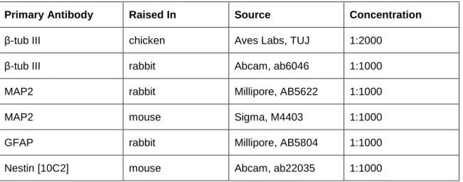
Table 2.1 Primary antibody concentrations ... 46
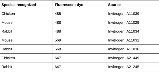
Table 2.2 Secondary antibody concentrations... 48
Table 2.3 ViewRNA ISH Cell Assay procedure ... 48
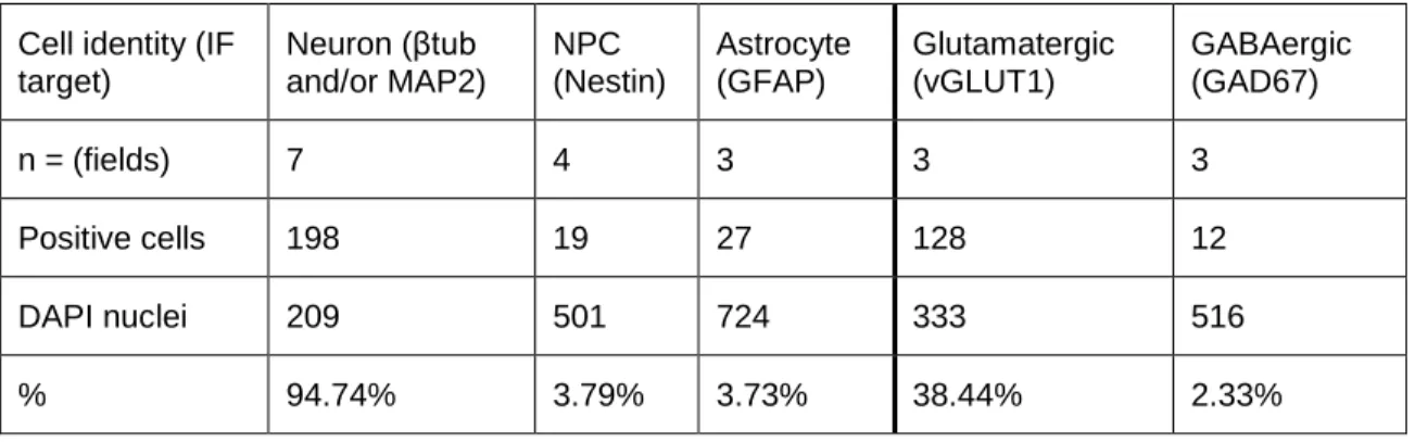
Table 2.4 Evaluation of differentiation efficiency and glutamatergic enrichment... 56
LIST OF FIGURES
Figure. 1.1. Scale representation of an adult vertebrate neuron. ... 2 Figure. 1.2. Schematic representations of compartmentalized neuron
culture chambers for isolation of axons. ... 9 Figure. 1.3. Glia-to-axon transport of the large ribosome subunit to degenerating
and regenerating axons in the sciatic nerve of the PNS... 14 Figure. 2.1. Human embryonic stem cell derived neurons (hESC-neurons)
matured in axon-isolating microfluidic chambers. ... 55 Figure. 2.2. hESC-neurons demonstrate common neuronal features. ... 57 Figure. 2.3. Evaluation of the quality of Affymetrix microarray results
generated from mRNA from hESC-neurons. ... 60 Figure. 2.4. hESC-neuron gene expression profile is enriched for
markers of mature neurons. ... 62 Figure. 2.5. Differential gene expression between the transcriptome of distal
projections and the neuronal transcriptome of hESC-neurons. ... 64 Figure. 2.6. Enriched gene ontology categories were similar between the
transcriptomes of isolated hESC-neuron axons and axons
of embryonic rat cortical neurons. ... 66 Figure. 2.7. Conserved orthologous transcripts within the axons of hESC-neurons
and embryonic rat cortical neurons are enriched for synaptic proteins. ... 68 Figure. 2.8. Multiplexed RNA-FISH verification of mRNA within the distal
projections of hESC-neurons. ... 70 Figure. 3.1. Long projecting axons of rat hippocampal neurons grown in
axon-isolating microfluidic chambers can be injured. ... 84 Figure. 3.2. Axons regenerated after axotomy and cell viability was unaffected... 86 Figure. 3.3. Rabies virus carrying a fluorescent protein gene administered to
the isolated axons allowed monitoring of the somatodendritic
arbor and axons. ... 88 Figure. 3.4. The number of cells with isolated axons depended on microgroove length. ... 90 Figure. 3.5. The distribution of cells with isolated axons depended on microgroove length. ... 92 Figure. 3.6. Axotomy-induced hyper-excitability was present 48 hours after
at the time of axotomy. ... 96 Figure. 3.8. RNA quality assessment and verification of microarray quality controls. ... 98 Figure. 3.9. Differential gene expression 24 hours after axotomy. ... 100 Figure. 3.10. Comparison of ranked gene expression fold change from
in vitro axotomy model and published in vivo
LIST OF ABBREVIATIONS
°C degrees Celsius
3H tritium
ACTB β-actin
AKT Protein kinase B
AMPAR α-amino-3-hydroxy-5-methyl-4-isoxazolepropionic acid receptor
ANOVA analysis of variance
ARC activity-regulated cytoskeleton-associated protein ATF4 activating transcription factor 4
βtub neuron-specific class III β-tubulin β-tubulin III neuron-specific class III β-tubulin BDNF brain derived neurotrophic factor
CA1 cornu ammonis 1
CA3 cornu ammonis 3
CA3-CA1 cornu ammonis 3 to cornu ammonis 1 hippocampal synapse CAMK2A calcium/calmodulin-dependent protein kinase type II alpha chain
cAMP cyclic adenosine monophosphate
Cdk5 cyclin-dependent kinase 5
cDNA complementary deoxyribonucleic acid
CMV cytomegalovirus
CNQX 6-cyano-7-nitroquinoxaline-2,3-dione disodium
CNS central nervous system
COXIV cytochrome C oxidase subunit IV CREB cAMP response element binding protein
CST corticospinal tract
d day
D-AP5 D-(-)-2-amino-5-phosphonopentanoic acid
DAVID Database of Annotation, Visualization and Integrated Discovery
DIC differential contrast
DIV days in vitro
DMSO dimethyl sulfoxide
DPO days post operation
DRB 5,6-dichloro-1-β-D-ribofuranosyl-1H-benzimidazole
DRG dorsal root ganglion
E embryonic day
eCB endocannabinoid
eCB-LTP endocannabinoid-dependent long-term depression eGFP enhanced green fluorescent protein
EM electron microscopy
ER endoplasmic reticulum
FACS fluorescence activated cell sorting
FITC fluorescein
GABA gamma-aminobutyric acid
GAD67 glutamate decarboxylase, 67 kDa
GAP43 growth associated protein 43
GFAP glial fibrillary acidic protein
GO Gene Ontology
h hour
HA human influenza hemagglutinin
HBS Hank's balanced salt solution
hESC-neuron human embryonic stem cell derived neuron
Hz Hertz
IF immunofluorescence
IMPA1 inositol monophosphatase 1
JAK Janus kinase
kDa kilodaltons
L4-eGFP ribosomal protein L4 fused to enhanced green fluorescent protein
LB2 Beta type nuclear lamin, protein
LCM laser capture microdissection
lmnb2 Beta type nuclear lamin, gene X. laevis
LTD long-term depression
LTP long-term potentiation
M7 7-methyl-guanosine-5’-triphosphate-5’-guanosine
MAG myelin-associated glycoprotein
MAP2 Microtubule-associated protein 2 MAPK mitogen-activated protein kinases
MBP myelin basic protein
microRNA micro ribonucleic acid
µA microampere
µg micrograms
µl microliter
µm micrometer
µM micromolar
mJ millijoules
mm millimeter
mRNA messenger ribonucleic acid
ms milliseconds
mtDNA mitochondrial genome
mTOR mammalian target of rapamycin
mt-rRNA mitochondrial rRNA
N2B27 media supplement N-2 with B-27
ng nanogram
NGF nerve growth factor
NLS nuclear localization sequence
nm nanometer
NMDAR N-methyl-D-aspartate receptor
NPC neural progenitor cell
nt nucleotide
NT-3 neurotrophin-3
oligo(dT) oligomer of deoxythymine
OXT oxytocin
p35 Cyclin-dependent kinase 5 activator 1 p75NTR low-affinity neurotrophin receptor
PBS phosphate buffered saline
pCREB phosphorylated CREB
PDMS poly(dimethylsiloxane)
PET polyethylene tetraphthalate
PFA paraformaldehyde
PGMEA propylene glycol methyl ether acetate
PGMEA picogram
PI3K phosphatidyl inositol-3 kinase
PNS peripheral nervous system
polyA polyadenylate
rAAV recombinant adeno-associated viral
RGC retinal ganglion cell
RINe RNA integrity number equivalent
RIP RNA immunoprecipitation
RNA ribonucleic acid
RNA-FISH ribonucleic acid fluorescent in situ hybridization
RNA-seq RNA sequencing
ROS reactive oxygen species
rpm revolutions per minute
rRNA ribosomal ribonucleic acid
RT room temperature
RT-PCR reverse transcription-polymerase chain reaction SAGE serial analysis of gene expression
SC superior colliculus of the midbrain
SCG superior cervical ganglion
SEM standard error of the mean
Sema3A semaphorin 3A
STAT signal transducer and activator of transcription
SynCAM Cell adhesion molecule 1
Trk tropomyosine receptor kinase
TrkA tropomyosine receptor kinase A
TrkB tropomyosine receptor kinase B
tRNA transfer ribonucleic acid
TTX Tetrodotoxin citrate
UTR untranslated region
UV ultraviolet light
CHAPTER ONE: INTRODUCTION
Overview
Neurons are unique among all the cells of the body in that a single cell can span up to half the length of the organism (Fig.1.1A). Throughout development and adulthood they must rapidly respond to isolated local events, such as extracellular signaling molecules or mechanical contacts. Some of these events are only sensed by and affect the most distal portions of the neuron, the dendrites and/or axon. Prompt local responses suggest a high degree of independence from the neuron cell body, the soma. Rapid quantitative and qualitative changes in the local proteome, that do not require somatic transcription or translation, demonstrate that the most distal regions are semi-autonomous. The autonomy of the axon is of particular interest as the furthest location from the soma.
There is a wide diversity of neurons, morphologically and functionally, within each organism. A broad description of the variety of neurons and components of the mammalian nervous system will be given in the first two sections of this chapter as reference for the similarities and differences between common model systems.
Historically, the dogma of axon function stated that the axon was solely dependent on the neuron cell body for all necessary protein components. This theory has been overturned within the last few decades with a growing body of evidence definitively identifying translational machinery and the organelles necessary for protein trafficking within axons. Further, the transcriptome within axons is diverse and translation-dependent axonal functions have been determined from development through adulthood. This body of work will be covered in detail in this introduction. An intriguing, still
Figure 1.1
Figure. 1.1. Scale representation of an adult vertebrate neuron.
A. The axon depicted corresponds to a length of approximately 1 cm. Several neurons have axons longer than 1 m and may form thousands of synapses in their terminal arborization. [Modified with permission from Giuditta et al (2008)].
While much seminal work has been performed in invertebrate and non-mammalian systems, such as the squid giant axon, goldfish, Aplysia californica, C. elegans, X. laevis and D. melanogaster, my focus will be mammalian systems. Significant nervous system differences exist between the branches of the animal kingdom, this includes anatomical, dendritic-axon polarity, axon myelination and chemical or electrical synaptic signaling. Literature from the body of mammalian work is more relevant to the data contained herein though I will mention seminal work in other systems for historic reference.
Neurons
significant similarities and differences. The information transfer is bidirectional within and between neurons.
Neurons are polar cells, they have unique and unequal traits at different subcellular locations within the cell. They are morphologically polar, functionally polar and the local proteome is polarized. A key morphological and functional feature of neurons are the long projections emanating from the cell body (Fig. 1.1A). These projections can be classified as dendrites, which typically extend a few millimeters to centimeters from the cell body, and axons, which can extend meters from the cell body. This polarity facilitates information transfer between neural networks through maintenance of
subcellular regions with segregated functions, this polarity also imposes significant biological constraints. The functional polarity of neurons resides within these projections, the dendrites receive information, the cell body consolidates the input and regulates whether the axon transmits information to the synaptic terminals. The local proteome is polar with many unique proteins in dendrites and axons. The polarity of the local proteome is maintained through protein transport and local protein translation (Holt and Schuman, 2013).
When an axon and dendrite are in close proximity they can form a synapse, the site of
information transfer between neurons. The presynapse is formed along the axon and the postsynapse is formed along the dendrite. Each axon and dendrite can form hundreds of synapses leading to an extremely complex network of connections. In each synapse the postsynaptic component and dendrite are many times larger than the presynaptic component and axon, creating unique technical challenges when investigating the axon and presynapse.
Rapid anterograde transmission from the soma to the synapse occurs through a well-established actively propagated wave of ions, an action potential, generated through changes in membrane potential. The result is presynaptic neurotransmitter release to the extracellular
controlling and responding to internal organ function, to the highest level brain function processes, including memory, thought and consciousness.
Retrograde signaling, from the axon to the soma, is less well established. There is evidence that some signaling occurs through active transport of protein complexes from the distal axon to the soma. In addition, there might be mechanisms of back-propagating ionic waves or biophysical methods conducted through the cytoskeleton.
Neurons can have many different dendrite and axon configurations, which provide unique signaling capabilities to each type of neuron. Multipolar neurons have many dendrites extending in multiple directions and a single axon. The broad dendritic arbor allows them to synapse with and receive information from many neurons. Bipolar neurons have clustered dendrites extending in a single direction from the soma, a more focused source of input, and a single axon. Unipolar neurons have a bifurcated single projection, one branch is the dendritic arbor and the other branch is the axon such that information bypasses the cell body. Pseudo-unipolar neurons also have a bifurcated
projections but in these cells it is only axonal in nature, with the distal branch receiving stimulation and transmitting it directly to the proximal branch.
The Mammalian Nervous System
The mammalian nervous system is classified into many branches based on function and anatomy. The neurons and support cells of each have unique features that result in significant biological differences between branches of the nervous system.
individual postsynapse, can be capable of responding to information transmitted by more than one neurotransmitter but each neuron only releases one type of neurotransmitter. Canonically axons of CNS neurons in vivo have limited capacity to regenerate and re-establish proper synaptic connections. This might be the combined effect of an inhospitable in vivo cellular milieu for regeneration, the
inability of the target neurons to release neurotrophic factors or the inability of the injured axon stump to respond to cues.
development and synaptogenesis. These preparations have been used for many studies, including axon guidance and synaptic plasticity.
The nerves and ganglia, clusters of nerve cell bodies, outside the brain and spinal cord are part of the peripheral nervous system (PNS). The PNS is divided into the somatic nervous system, which transmits sensory signals, such as taste and touch, to the CNS, and the autonomic nervous system, which controls the function of internal organs. Neurons derived from the dorsal root ganglion (DRG) of the somatic nervous system, responsible for relaying sensory information to the CNS, are a common in vitro model system. DRG neurons are pseudo-unipolar neurons, they have a single, very long axon with two functionally distinct branches, the ‘distal process’ performs dendrite-like functions while the ‘proximal process’ operates as the axon. The distal process projects to the sensing organ in the animal periphery while the proximal process projects to the spinal cord making it possible for an action potential that originated in the distal process to bypass the soma and propagate directly to the synaptic terminal with the spinal cord. The axons within the sciatic nerve are a common in vivo model system. The sciatic nerves emanate from DRGs in the lower back and innervate the legs/ hind legs, providing most of the somatic nervous system connections for the whole leg and foot. They are large and easy to identify making them an experimentally tractable system.
The autonomic nervous system is further differentiated into the sympathetic nervous system, activated during “fight or flight” situations, and the parasympathetic nervous system, functioning in “rest and digest”. Norepinephrine and epinephrine are the dominant signaling molecules of the sympathetic nervous system while acetylcholine is the primary neurotransmitter of the
parasympathetic nervous system. The superior cervical ganglion (SCG), of the sympathetic nervous system, is a common in vitro research model. These neurons are pseudo-unipolar, similar to DRGs, allowing signaling to quickly occur over long distances from the distal process to the proximal process. Axons of the SCG innervate the head and neck, including the blood vessels of the facial muscles and brain, salivary glands and thyroid gland.
have significant regenerative capability, in vivo and in vitro, such that proper functional re-innervation can occur following injury. Because of their biologically relevant regenerative mechanisms they are commonly used in to study axon injury signaling, axon-to-soma survival signaling, axon regeneration and guidance.
Compartmentalized Analysis of Neurons in vitro
The distinctive morphological and functional polarity of neurons has necessitated the development of custom cell culture environments to facilitate isolating long axons from the somata once the tissue is dissected or neurons are dissociated.
Campenot chamber
The Campenot chamber (Campenot, 1977) is a three-chamber system developed to investigate the biology of nerve growth factor (NGF) signaling in sympathetic neurons but has been applied to sensory neurons and other signaling molecules. The chamber is constructed from a collagen coated surface with multiple parallel microscratches in the coating, a three compartment Teflon piece is sealed to the surface with silicon grease. Dissociated neurons or whole tissue explants, derived from the SCG of neonatal rodents or DRG of rodents, are plated in one chamber. Guided by the microscratches as barriers the axons penetrate the fluid-impermeable barrier of silicon grease to access a separate fluidically-isolated compartment (Fig. 1.2A). If the neurons are plated in the center chamber the axons can extend into the left and right chambers (Fig. 1.2B), with a minimum axon length of about 1 mm. If the neurons are plated in a side chamber the axons can extend into the middle then the far opposite chamber allowing the manipulation of distal axons with a minimum axon length of over 3 mm (Fig. 1.2C). The fluidic environments of the three compartments can be
independently manipulated to evaluate the local effects of neurotrophic or guidance factors. Also, the cellular material within each compartment can be collected without contamination from the other compartments for downstream analysis. Optical properties of the Teflon piece and silicon grease restrict live imaging of neurons and axons but low resolution fixed imaging is possible. These
the chamber dimensions these are only appropriate for use with peripheral neurons that have very long axons, not neurons from the central nervous system.
Modified Boyden chamber
The Boyden chamber was originally developed for chemotaxis assays and has been modified to physically isolate DRG and SCG axons in explant and dissociated cell culture (Fig. 1.2D) (Zheng et al., 2001). The chamber is composed of a cell culture well with a removable insert well. The
Figure 1.2
Figure. 1.2. Schematic representations of compartmentalized neuron culture chambers for isolation of axons.
A. Top view of sympathetic or sensory neurons/ explants cultured in a Campenot chamber. A thin layer of vacuum grease seals the Teflon divider to the prepared culture surface. [Modified with permission from (Zweifel et al., 2005)]
B. Side view of a Campenot chamber with dissociated cells or ganglion explants plated in the center compartment. [Modified with permission from (Zweifel et al., 2005)]
C. Side view of a Campenot chamber with dissociated cells or ganglion explants plated in the side compartment. [Modified with permission from (Zweifel et al., 2005)]
D. Boyden chamber schematic. Dissociated cells or explants are cultured on the upper surface of a microporous membrane, with or without a glass coverslip between the neurons and membrane. Elongating axons pass through the micropores. The cell body compartment and the axon
compartment are not fluidically isolated due to diffusion but a gradient can be maintained with regular media changes. [Modified with permission from (Cox et al., 2008)]
E. The molded PDMS microfluidic chamber is affixed to a glass coverslip creating two symmetrical, fluidically isolated compartments connected by over 150 microgrooves, each compartment has two media access wells. Dissociated CNS neurons are plated in one compartment, growing axons enter the microgrooves and emerge in the parallel compartment.
Microfluidic chamber
chambers have various microgroove lengths, which regulates the minimum length necessary for an axon to reach the isolated compartment. There are two large media access points, wells, for each compartment which minimize the effect of evaporation. Sterile, replica molded poly(dimethylsiloxane) (PDMS) microfluidic chambers are placed on coated glass coverslips to create the microfluidic chamber, dissociated neurons are directly added to one of the compartments followed by media. Astrocyte feeder co-culture is not used in this culture platform, instead B-27 neuron culture
supplement (Invitrogen) is added to the media. These chambers have been optimized for visual and biochemical axon isolation and can be used for fixed or live imaging and biochemical analysis.
Axonal Capacity for Translation
For many decades dogma held that the axon of mature neurons depended exclusively on transport of proteins from the soma yet a large body of work has overturned this view and informed the establishment of a ‘local synthesis model’ of axon function (Alvarez et al., 2000). In this model a portion of the local proteome is locally synthesized from mRNA and ribosomes within the axon. Early work investigating RNA in neurons detected RNA in the adult rabbit optic nerve derived from the soma following in vivo intraocular injection of tritium labeled nucleoside, ([3H]uridine) (Gambetti et al., 1973). This study was not able to differentiate between the many different classes of RNA including, rRNA, mRNA, tRNA and microRNA, but suggested the hunt for axonal ribosomes and mRNA would be successful. Since then translational machinery has been identified within axons of many species. Further, cellular components necessary for protein trafficking and secretion, the endoplasmic reticulum and Golgi apparatus, are present in axons. Recent, controversial, work proposes transport of
ribosomes from glia to axons in the PNS.
Axonal Ribosomes
DRGs from neonatal and adult rats (Bisbal et al., 2009; Calliari et al., 2014) and in vivo by EM in adult sciatic nerves (Zheng et al., 2001). Within the central nervous system ribosomes have been
demonstrated by IF in adult rat medullary roots projecting to the spinal cord (Calliari et al., 2014), myelinated axons of spinal nerve roots from adult rabbits and rats (Koenig et al., 2000) and by EM in the axons of retinal ganglion cells within the mouse brain from embryonic, young and adult animals (Shigeoka et al., 2016). Axonally localized ribosomes have been identified within cultured hippocampal neuron by IF and EM (Taylor et al., 2013; Tcherkezian et al., 2010) and adult rat and mouse
GABAergic hippocampal neurons as well as at the presynapse, as detected by super-resolution STORM microscopy (Younts et al., 2016). Evidence suggests mechanisms to regulate the supply of axonal translational machinery to meet the local demand. Translational machinery was detected in regenerating adult rat spinal cord axons but decreased after recovery. Subsequent injury reactivated the axonal enrichment of these proteins suggesting a dynamic intra-axonal environment (Sachdeva et al., 2016). Additionally, proteomic analysis of extracted axioplasm from in vivo regenerating adult rat sciatic nerves also demonstrated in increase in translational machinery following injury (Michaelevski et al., 2010).
Axonal protein secretion machinery
Glia to axon transfer of RNA
There is a growing body of evidence demonstrating glia to axon transport of RNA. Early work investigating RNA in neurons used the peripheral nerve of the adult newt, Triturus viridescens (Singer and Green, 1968). Newts have high tissue regenerative capacity with the ability to regenerate whole functional limbs following amputation. To investigate the RNA content of axon fragments detached from the somata they locally injected [3H]uridine immediately after severing peripheral nerves. They found radioactivity in the distal peripheral nerve portions 24 h later despite the fact that these axons were severed from the somata and destined to degenerate (Fig. 1.3A and 1.3B). They concluded that the most likely source of this RNA signal was the associated Schwann cell (Singer and Green, 1968). The unusual regenerative capacity of the newt made it unclear whether this was a species specific phenomenon. Similar work identified [3H]uridine labeled RNA grains in the severed axon stump of adult rat sciatic nerves (Benech et al., 1982) suggesting transfer of RNA from glia to degenerating axon fragments occurs in mammals. Glia-to-axon RNA transfer has been definitively demonstrated in the squid giant axon in vitro. It is strongly stimulated by axon depolarization or activation of glial glutamate and acetylcholine receptors (Eyman et al., 2007) suggesting the neurotransmitters for communication between neurons might also signal to surrounding glia.
protein within glia and axons (Fig. 1.3B). All axons that demonstrated L4-eGFP signal were enveloped by L4-eGFP positive Schwann cells and uninfected Schwann cells enveloped L4-eGFP negative axons (Court et al., 2008). They showed that this transfer was relatively specific as Schwann cell cytoplasmic protein S100 was not detected in axons and neither was eGFP from a control lentivirus lacking the L4 protein. Small protrusions of the Schwann cells appeared to pierce the myelin sheath and enter the axon suggesting a model of axonal entry for the glial derived ribosomes (Court et al., 2008).
To investigate glia-to-axon ribosome transfer in the context of axon regeneration they performed similar experiments on wild-type adult mice. Degeneration of the severed axon portion is complete by day 3 after injury and regeneration begins around day 4. Seven days after injury co-localization of L4-eGFP and ribosome staining was detected in 12% ± 1% of the regenerating axons (Fig. 1.3C) (Court et al., 2011). They also investigated this phenomenon in the regenerative context of rat sciatic nerve grafts. Instead of administering the L4-eGFP lentivirus in vivo they cultured and infected Schwann cells in vitro, enriching for eGFP positive Schwann cells by fluorescence activated cell sorting (FACS). Rats received a 13 mm sciatic nerve graft then 5 x 10⁵ transduced Schwann cells were injected into the graft. Regeneration through the length of the graft was seen 16 days after surgery and co-localized L4-eGFP and ribosome staining was observed in these axons (Fig. 1.3C). There were also L4-eGFP negative ribosomes in axons potentially from the neuron soma or non-transduced Schwann cells. Co-localized L4-eGFP and ribosome staining was detected in a subpopulation of axons at 3, 6 and 8 weeks after surgery (Court et al., 2011). These long term observations could mean that the glial derived ribosomes have a very long half-life in the axon or the glial transfer of ribosomes continues after regeneration, potentially to support axon maintenance. Evidence using rat sciatic nerve explants collected 18 h after nerve injury suggests the mechanism of glia-to-axon transport of ribosomal RNA and non-ribosomal RNA molecules requires actin
Figure 1.3
Figure. 1.3. Glia-to-axon transport of the large ribosome subunit to degenerating and regenerating axons in the sciatic nerve of the PNS.
A. Mechanical injury to the sciatic nerve severs the distal portions of the axons from the somata, inhibiting soma-to-axon transport.
B. Large ribosome subunits derived from the surrounding Schwann cells can be detected in the degenerating axon fragments of the sciatic nerve after injury.
C. Large ribosome subunits derived from the surrounding Schwann cells can be detected in regenerating axons of the sciatic nerve.
following axotomy of PNS neurons and might be limited to this context. The fact that highly enriched populations of dissociated neurons, lacking significant numbers of glial cells, can be cultured in vitro for weeks demonstrates that neurons have the capacity for autonomous growth. In addition, culturing CNS neurons with glial feeder cells, compared to media supplements, does not significantly improve viability or the length of time cells can be cultured ex vivo but these feeder layers are rarely in direct contact with the neurons (Kaech and Banker, 2006; Vicario-Abejon, 2004). A deeper understanding of bidirectional communication between glia and axon, together with our understanding of axon-to-soma communication, could alter the way we approach axon biology.
Axonal transcriptomes
A critical element for axonal protein synthesis is axonally localized protein-coding mRNA. High resolution polyA mRNA in situ hybridization of cultured cortical neurons demonstrated mRNA
throughout the somata, dendrites and proximal axons after 4 d in culture (Bassell et al., 1994). With the necessary components for translation identified within axons the next logical questions were: what mRNAs are present in axons and what axonal functions are translation dependent? Unbiased
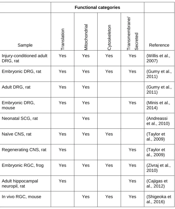
methods to evaluate large scale gene expression within the axons of a variety of neurons have revealed many conserved Gene Ontology (GO) functional groups. These include proteins involved in translation (ribosomal proteins as well as translation initiation and elongation factors), nuclear-encoded mitochondrial proteins, cytoskeletal proteins, transmembrane and secreted proteins (Table 1.1).
Table 1.1 Functional analysis of the axonal transcriptome in multiple models Functional categories Sample T ran s lat ion Mi toc h on dria l Cy tos k el eto n T ran s me m brane / S ec rete d Reference Injury-conditioned adult DRG, rat
Yes Yes Yes Yes (Willis et al.,
2007)
Embryonic DRG, rat Yes Yes Yes Yes (Gumy et al.,
2011)
Adult DRG, rat Yes Yes (Gumy et al.,
2011) Embryonic DRG,
mouse
Yes Yes Yes (Minis et al.,
2014)
Neonatal SCG, rat Yes (Andreassi
et al., 2010)
Naïve CNS, rat Yes Yes Yes (Taylor et
al., 2009)
Regenerating CNS, rat Yes Yes (Taylor et
al., 2009) Embryonic RGC, frog Yes Yes Yes Yes (Zivraj et al.,
2010) Adult hippocampal
neuropil, rat
Yes Yes (Cajigas et
al., 2012)
In vivo RGC, mouse Yes Yes Yes (Shigeoka et
al., 2016)
In vitro sensory neurons
axonal transcriptome facilitate regeneration (Twiss et al., 2000). Animals underwent sciatic nerve crush injury 7 d before primary DRGs were dissected, dissociated and cultured on Boyden chamber inserts with 8 µm diameter pores. Both the injured distal axon branch and the uninjured proximal branch of the DRG axon likely regenerated but this was not differentiated in this system. After 20 – 22 h in culture the lower surface of the membrane, coated with isolated axons, was scraped and
processed for hybridization to Atlas cDNA arrays (approximately 4000 cDNAs represented). They identified more than 200 protein encoding mRNAs, including ‘transmembrane proteins’, ‘translational machinery’, ‘cytoskeleton’, and nuclear encoded ‘mitochondrial proteins’.
To investigate the effects of axon growth-promoting and growth-inhibiting factors on the axonal transcriptome they selected a representative panel of 50 mRNAs evaluate following growth factor treatment. The growth-promoting factors selected were NGF, neurotrophin-3 (NT3) and brain derived neurotrophic factor (BDNF) which all signal through members of the tropomyosine receptor kinase (Trk) family of tyrosine receptor kinases. The growth-inhibiting factors were semaphorin 3A (Sema3A) and myelin-associated glycoprotein (MAG), selected because they induce retraction or repulsion of rat DRG axons. To prevent diffusion to the upper chamber growth factors were covalently coupled to 15 µm diameter polystyrene microspheres and the inserts were placed directly in contact with the microspheres for 4 h. An additional pharmacological manipulation in these experiments was the addition of the reversible transcription blocker, 5,6-dichloro-1-β-D-ribofuranosyl-1H-benzimidazole (DRB), to prevent any transcriptional effect of the growth factors. The ability to rule out transcription as the driving force behind changes in the axonal transcriptome allowed them to focus on targeted, stimulated trafficking of mRNA to axons though it did not rule out changes in mRNA stability. Since this form of injury-conditioned axon regeneration is transcription-independent blocking transcription did not negatively affect axon growth. Each of the 5 treatments resulted in significant increases and
decreases in specific axonal mRNAs as well as identification of transcripts that did not change (Willis et al., 2007).
response patterns included the very well characterized axonally translated β-actin mRNA; all three neurotrophins significantly increased axonal localization of this mRNA while both inhibitory factors significantly decreased its localization. Localization of growth associated protein 43 (GAP43) mRNA was increased only by NGF and remained unchanged in the presence of the other growth factors. Subsequently, this group demonstrated that the balance between axonal translation of GAP43 and β-actin regulated axon branching and axon length in this model system (Donnelly et al., 2013). In addition, vimentin mRNA had a unique response pattern, it was unchanged following NT-3 exposure but increased greater than 3.5 fold with the other growth-stimulating factors and the growth-inhibiting factors. Vimentin is a type III intermediate filament protein of the intermediate filament protein family the cytoskeleton and has a role in anchoring ER and mitochondria within the cytosol (Katsumoto et al., 1990). They showed that changes in the axonal and somatic transcriptomes were inversely related supporting the hypothesis that, in at least some cases, individual transcript changes in the axon draw from the pre-existing somatic transcriptome. Further, bath application of NGF did not stimulate the same axonal transcriptome changes as isolated axon NGF treatment suggesting the importance of the local extracellular environment in altering the local axonal transcriptome.
To determine the mechanism behind this NGF-stimulated transcriptome plasticity they demonstrated functional microtubules were critical while functional microfilaments were important but not necessary. Trk kinase activity was necessary for the NGF-stimulated increases and blocking it lead to axonal increases in the transcripts typically decreased following NGF. Lastly, individual transcripts responded differently to pharmacological inhibition of phosphatidyl inositol-3 kinase (PI3K) and mitogen-activated protein kinase (MAPK) signaling pathways downstream of Trk activation. Taken together their data suggest that the plasticity and diversity of the adult rat DRG axonal transcriptome in the integrated effect of multiple local mechanisms (Willis et al., 2007).
chambers with NGF-supplemented media for 4 d (Gumy et al., 2011). Microarray analysis of axonal mRNA revealed 2627 transcripts localized to embryonic axons and 2924 transcripts within adult axons, 1445 of these localized transcripts (55.0% and 49.4% of the embryonic and adult
transcriptomes, respectively) were present in both age samples while the remainder were age specific. The common transcripts were enriched for nuclear encoded ‘mitochondrial protein’s, proteins involved in ’translation’, and ‘neurite development and growth’. The embryonic specific transcripts were
enriched for ‘cytoskeleton’ proteins and ‘transport/trafficking’ related proteins, such as microtubule associated motor proteins, ‘vesicle’ proteins involved in membrane trafficking and synaptotagmins, synaptic vesicle proteins necessary for Ca2+-induced exocytosis. They were also exclusively enriched for ‘cell cycle’ proteins. In the adult derived DRG axons mRNAs encoding ‘inflammatory’ and ‘immune-related’ functions were enriched, specifically cytokine-cytokine receptor interactions, Toll-like receptor signaling and antigen presenting and processing functions. The enriched functions in the axonal transcriptome point toward axon growth and maintenance while those of the adult transcriptome highlight pathways that have been implicated in chronic and neuropathic pain (Gumy et al., 2011).
There are a few noteworthy details of the pharmacological manipulations in this work. First, the potent DNA cross-linker mitomycin C was included in their cultures to block the proliferation of mitotic, non-neuronal cells, such as fibroblasts and Schwann cells, present in DRG explants. This served to isolate the neuron intrinsic differences between embryonic and adult DRG axons without the potentially confounding contribution of non-neuronal cells. Secondly, as previously demonstrated by Willis et al (2007), the necessary inclusion of NGF in this culture system likely had an effect on the axonal transcriptome. This might recapitulate the in vivo environment since NGF is one of the neurotrophic factors secreted by DRG innervated tissues.
Microarrays have a reduced quantitative range and finite coverage of the genome as
‘explant’ RNA samples contained the whole DRG-spinal cord explant. Using ranked differential expression they evaluated enriched GO categories within differential expression bins spanning the expression profile from ‘absent in axons but present in explants’ to ‘highly enriched in axons’. The transcripts highly depleted in axons clustered into one functional category while the transcript highly enriched in axons clustered into 4 categories. They focused their analysis on the mRNAs that were within the extremes of differential expression (Minis et al., 2014).
The axonally depleted fraction was best represented by the GO category ‘membrane fraction’ (GO:0005624). They interpreted this as support for somatic synthesis of membrane proteins followed by transport to axonal sites. This GO term was based on experimental results of biochemical cell fractionation not a bona fide cellular compartment and was made obsolete in the EMBL-EBI database in mid-2012. They specifically look at 20 transcripts encoding receptors for neurotrophins and axon guidance cues within their RNA-seq data and found that 9 were depleted from axons, 5 were not differentially expressed and 6 were enriched in axons. While they stated this supported their conclusions I find this diverse transcript specific localization more suggestive of a variety of mechanisms regulating receptor localization.
The 4 axonally enriched categories included ‘secreted proteins’, ‘sequence specific DNA binding’, proteins involved in ‘translation’ and ‘immune response’. Beyond this broad characterization they made three unique discoveries. First, a closer analysis of the transcript encoding ‘secreted proteins’ revealed that, in particular, mRNAs encoding proteins with an N-terminal signal peptide amino acid sequence were axonally enriched. The signal peptide sequence is rapidly bound during protein translation by the signal-recognition particle which stalls translation and facilitates transport of the ribosome complex to the ER for secretion of the new protein. Another sub-category of note within ‘secreted proteins’ were related to collagen of the extracellular matrix. Transcripts encoding many collagen chain subunits and enzymes of the collagen biosynthesis pathway were highly enriched in axons. Taken together these results suggest local translation and ER-dependent protein secretion within sensory axons facilitates modification of the local extracellular environment.
expression of the 37 genes of the mitochondrial genome (mtDNA) and more than 1000 nuclear encoded mitochondrial genes (Garesse and Vallejo, 2001), events that are separated by a long physical distance in axons. Proteins encoded in both genomes form the complexes of the electron transport chain, but the enzymes of the citric acid cycle and the mitochondrial transcription machinery are all nuclear encoded. Mitochondrial translation requires mtDNA encoded tRNAs and rRNA and nuclear encoded accessory ribosomal proteins and translation factors. Mitochondrial encoded genes were within the highest expressed RNAs in both axons and explants. More than 20% of the total axonal RNAs aligned to mtDNA, while approximately 2% of the explant RNA aligned to mtDNA; mitochondrial rRNA (mt-rRNA) was extremely enriched in the axons. Transcripts from the nuclear encoded mitochondrial genes were equal expressed between the two compartments, consistent with the finding that the GO category ‘mitochondria’ was not an enriched cluster in the axons. The relative expression, not enrichment, of mtDNA genes was 1 to 2 orders of magnitude higher than the
expression of nuclear encoded genes in the axons. This novel finding of extreme differential
expression of mitochondrial genes could be a result of the smaller size of the axonal transcriptome, in terms of how many genes are represented, as compared to the explant transcriptome. Alternatively, it could point to unknown post-transcriptional events coordinating mitochondrial gene expression and function.
(UTR) of the mRNA between the stop codon and the polyA tail. They evaluated the distribution of the 5 short sequence elements within the 1888 preferentially axonal transcripts finding that the elements were equally distributed across the 5’ UTR, protein coding sequence, and 3’ UTR, after controlling for the length of each of these elements (Minis et al., 2014).
It is important to point out that proliferation of non-neuronal cells in explant tissue was not blocked in this work. Mitotic cells, particularly Schwann cells, were clearly detected in the explant cultures by immunocytochemistry. These non-neuronal cells also contributed to the quantitative gene expression profile of their explant samples to an unknown degree. Additionally, the presence of NGF in the axonal media, to stimulate axon regeneration, could affect the transcriptome (Willis et al., 2007), the methods do not specify whether the explants were also in NGF supplemented media which could have an effect on transcription.
The mitochondrial mRNA and mt-rRNA results raise a few questions. They did not consider that the extremely high mitochondrial rRNA expression could be a technical or bioinformatic artifact. Nuclear-encoded rRNA typically makes up more than 80% of the total RNA isolated from whole cell lysate and in vitro RNA amplification methods might be slightly biased toward amplifying rRNA over mRNA. Specifically, amplification methods use a pool of primers including random hexamer sequence primers and 12 – 18 nt oligo(dT) primers, the random primers equivalently amplify the whole transcript while the oligo(dT) primers amplify sequences upstream of a poly-adenylate track, such as the polyA tail. Most mRNAs have a polyA tail, co-transcriptionally added by factors associated with RNA
In vitro sympathetic neurons
Sympathetic and sensory neurons are functionally unique so the axonal transcriptome of neonatal rat SCGs were quantified by serial analysis of gene expression (SAGE) (Andreassi et al., 2010). The SAGE technique produces one short, less than 20 base pair, cDNA ‘tag’ from each RNA molecule. The library of cDNA tags are enzymatically concatenated, cloned and sequenced by traditional Sanger sequencing (Gowda et al., 2004). Theoretically the transcript to tag ratio is 1:1 so the number of unique tags mapping to a gene is a direct measure of the number of transcripts. Dissociated SCG cells from neonatal rats were plated in Campenot chambers supplemented with NGF. Regenerating axons were grown for 3 – 4 weeks. Total RNA was purified from the cell bodies and isolated axons and amplified with SAGE primers incorporating sequence elements to facilitate preparation of the SAGE library and deconvolution of the sequencing results. They sequenced 1440 clones from each library, obtaining 20082 tags from the axonal library and 20420 tags from the cell body library which they mapped to the rat UNiGene build 166 reference database. They found the somatic transcriptome contained transcripts related to ‘cytoskeletal’, ‘synaptic’, and ‘nuclear’ functions. The axonal library was enriched for transcripts of nuclear encoded ‘mitochondrial’ and ‘signal
transduction’ proteins (Andreassi et al., 2010).
The greatest difference between their somatic library and axonal library was the percentage of tags that that could not be mapped to their reference database. The UNiGene databases contain only transcribed sequences, not intergenic DNA sequences, and are filtered to omit RNAs with repeated or low-complexity sequences, RNAs derived from ribosomal RNA genes, and mtDNA genes. Tags mapping to uncharacterized but annotated genes represented 11.25% of the somatic library and 4.7% of the axonal library but tags that couldn’t be mapped comprised 30.05% of the somatic library and 39.9% of the axonal library. Comparing these unannotated axonally derived tags to a database of known noncoding RNAs suggested that approximately 1% of the total axonal transcriptome could be noncoding RNAs.
for neurons in the cell body compartment. Second, they remove NGF from the somatic compartment after a week causing cells without axons in the isolated compartment to activate programmed cell death pathways leaving a neuronal population that only had axons extending into the isolated culture compartment.
Regarding the ‘unmappable’ tags, it would be informative if these tag sequences could be mined for ribosomal RNA, mtDNA encoded proteins and mt-rRNA. This analysis could support the mitochondrial RNA findings of Minis et al (2014) in a different species and neuron type.
In vitro CNS neurons
RNA within the axons of CNS neurons is more controversial than within the axons of PNS neurons. The axonal transcriptome of CNS neurons grown in axon-isolating microfluidic chambers was evaluated in uninjured and injured regenerating axons (Taylor et al., 2009). Dissociated cortical and hippocampal neurons from embryonic rats were grown in microfluidic chambers with 450 µm microgrooves separating the somatic compartment from the axonal compartment. While these neurons have an embryonic origin they are fully differentiated ‘mature’ neurons. The media did not contain serum, limiting the proliferation of rare non-neuronal cells, or exogenous growth or guidance cues for axon elongation through the microgrooves but the media supplement B-27 (Gibco) was used.
Axonal and somatic RNA was collected on days in vitro (DIV) 13 and evaluated by microarray. Microarray data was thresholded for expression resulting in 6702 microarray probe sets in the somatic dataset and 2051 probe sets in the axonal dataset. All of the axonal probes were detected in the somatic dataset. A second threshold was set for the axonal dataset to generate a list of transcripts reliably localized to axons, 308 genes. GO categories enriched in the axonal mRNAs encoded ‘translation’, ‘mitochondrion’, ‘intracellular transport’, and ‘cytoskeleton’ proteins (Taylor et al., 2009).
Next they investigated how in vitro axon injury affected the axonal transcriptome of
axons, 480 transcripts increased and 386 transcripts decreased. Surprisingly, all but 8 of the increased transcripts had not been detected as reliably localized to axons in their initial axonal transcriptome analysis. Functional classification of the differentially expressed mRNAs suggested that the increased transcripts in regenerating axons were enriched for ‘cell-cell signaling’ molecules, ‘cell differentiation’, and ‘secreted’ proteins. The decreased transcripts were enriched for ‘mitochondrial’ and ‘cytoskeletal’ proteins and ‘intracellular transport’ proteins. Many of the enriched ‘cell differentiation’ proteins are required for nervous system development and those of the ‘cell-cell signaling’ and ‘secretion’ groups are involved in synaptogenesis, neurotransmitter release and synaptic plasticity suggesting that these regenerating axons might have an increased capacity for axon targeting and synapse formation and function. The decreased categories in regenerating axons suggests that the dynamic cytoskeleton and intracellular transport along the cytoskeleton might be unique in regenerating axons. Also, the balance between oxidative phosphorylation and glycolysis might be different in regenerating axons or transport of new mitochondria during axon outgrowth reduces the local need for mitochondrial protein synthesis. They pointed out that axonal localization of proteins involved in translation, such as ribosomal proteins and initiation and elongation factors, were not significantly changed during regeneration suggesting the local capacity for translation was not significantly altered (Taylor et al., 2009).
Growth cone turning in response to guidance cues is unequivocally dependent on local translation and the attractive or repulsive response to cues changes during the course of development (Jung et al., 2011; Jung and Holt, 2011). To determine whether this change in responsiveness was dependent on changes in the growth cone transcriptome Zivraj et al (2010) used laser capture microdissection (LCM) to isolate mRNA within growth cones. Whole-eye primordia from late stage X. laevis embryos were cultures and growth cone RNA was collected 24 h later. Microarray analysis identified 444 transcripts with known functions in the growth cones. Ingenuity Pathway Analysis (IPA) for enriched functions and signaling pathways revealed that 31% of the encoded proteins functioned in ‘protein synthesis and translation’, this included translation initiation and elongation factors, mRNA processing proteins, protein folding chaperones and 69 0f the 80 ribosomal proteins. The second largest functional category was ‘metabolic/glycolytic’ and included proteins of oxidative
included ‘cytoskeletal/motor’, ‘signaling’, ‘transmembrane/cell surface receptor’, and ‘transcription factor’. They then evaluated the mRNA within growth cones derived from earlier stage embryos grown in culture for 17 h, resulting in growth cones that were approximately 24 h younger than the ones previously evaluated. Significantly fewer mRNAs were detected in these younger growth cones, 171 genes with known function. The young growth cone transcriptome was significantly less diverse than that of the older growth cones. The majority of these transcripts could be functionally classified as involved in ‘protein synthesis’ and the major enriched pathway was ‘oxidative phosphorylation’. Of the transcripts in young growth cones 66% were also present in old growth cones, only 60 transcripts were exclusively detected in the young growth cones (Zivraj et al., 2010).
The growth cone is a unique functional subcellular compartment as compared to the axon but whether the growth cone transcriptome was distinct or reflected that of the axonal transcriptome was the next question they asked. Since the older growth cones had a more diverse transcriptome they used LCM to capture axons of similarly cultured older RCGs. They identified 5105 axonally localized mRNAs. Functionally there were many similarities between the axonal and growth cone
transcriptomes, including functional enrichment for ‘protein synthesis’, ‘metabolic/glycolytic’, and ‘cytoskeleton/motor’, as well as the ‘oxidative phosphorylation’. Categories unique to the axons included ‘protein trafficking’, ‘protein folding’ and ‘cell-mediated immune response’. They then turned to evaluate the transcripts enriched in growth cones as compared to the axons. Twenty-eight genes were enriched in the growth cones, the majority had cytoskeleton-associated functions or were involved in protein synthesis. The 523 transcripts enriched in the axons over the growth cones included the ‘metabolic/glycolytic’ functional group and ‘transcription factors’ (Zivraj et al., 2010).
To compare the RGC growth cone transcriptome across species they collected embryonic mouse RGC growth cones by LCM after 24 h in culture. They identified 1800 annotated transcripts. They found moderate conservation between species of specific transcripts within growth cones which was enriched for ‘oxidative phosphorylation’ and ‘protein synthesis’, in particular ribosomal proteins, yet the mouse growth cones included several nuclear-encoded proteins of the mitochondrial ribosome.
mRNAs. The finding that young growth cones have a smaller transcriptome than older growth cones suggests that as growth cones mature they require an increase in the diversity of the locally
synthesized proteins, potentially protein transport from the somata cannot produce a sufficiently rapid change in the local proteome of longer axons. Their work was somewhat restricted due to the limited annotation of the X. laevis genome at the time of the study. Notwithstanding, the similarities found between the X. laevis and mouse grown cone transcriptomes are consistent with previous findings.
In vivo CNS neurons
As previously pointed out cultured neurons must recover from the stress and injury associated with techniques to isolate them. Whether this has long term effects on the somatic or axonal
transcriptome is unknown making it valuable to evaluate the axonal transcriptome in vivo. The ability to collect pure axonal RNA in vivo is significantly hindered by the extreme size differential between axons and the surrounding cells. Extensive brain mapping data can suggest areas that are enriched for axons but the ability to physically isolate axons from dendrites in vivo has not been developed, though some genetic tools exist to isolate the presynapse from the postsynapse. Intra-axonal translation in mature, uninjured neurons of the adult CNS has been difficult to unequivocally prove; dendrites rely on local translation and easily mask any contribution of axonal translation. Regions enriched for
dendrites, axons and glial filaments are referred to as ‘neuropil’ and provide the best physical sites to enrich for axonal and dendritic transcripts in vivo.
Many forms of synaptic plasticity in the CA1 region of the hippocampus are translation dependent so deep sequencing was used to determine the transcripts within the neuropil of the adult rat CA1 area (Cajigas et al., 2012). The microdissected neuropil contained dendrites, axons, glia, a sparse population of interneurons and some blood vessels. They detected 8379 unique mRNAs enriched for genes classified in ‘synaptic function’, ‘myelin’ and ‘translation’. To enrich for transcripts within axons and dendrites they bioinformatically subtracted mRNAs identified in astrocytes,
oligodendrocytes, interneurons, blood vessels, mitochondria and transcripts encoding nuclear proteins. The resulting list of 2550 transcripts represents the combined axonal and dendritic
biological functions known to occur in dendrites and axons. Specific to axons they found many mRNAs encoding canonical presynaptic proteins including proteins that function to structurally organize the presynapse and regulate presynaptic vesicle release.
This work was not able to quantify or differentiate the relative contribution of transcripts localized to the axons versus the dendrites so questions regarding the differences between the transcriptomes of these subcellular compartments remain. I suspect that there is a significant amount of overlap between these transcriptomes since both regions rely on local translation for cytoskeleton dynamics, ribosomal protein expression, and mitochondrial function. Differences might include neurotrophic and endocannabinoid receptors, implicated in modulating presynaptic function, or the protein subunits of post-synaptic glutamate and GABA receptors, the major neurotransmitters in this brain region.
Recently, a group developed a method to sequence the ribosome-bound mRNAs from the axons of mouse RGCs in vivo (Shigeoka et al., 2016). The RiboTag knock-in mouse (Sanz et al., 2009) uses cre-mediated recombination to replace the endogenous ribosomal protein L22 (Rpl22) allele to a human influenza hemagglutinin (HA) tagged allele. Shigeoka et al (2016) permanently labelled RGCs by mating RiboTag mice with Pax6-alpha-Cre mice to transiently express cre in neural progenitors of the peripheral retinal primordium. The axons of these cells terminate in the superior colliculus of the midbrain (SC) so dissection of this region followed by RNA immunoprecipitation (RIP) significantly enriched for ribosome bound mRNAs within the distal axons. They also prepared samples from the retinal ganglion somata. Electron microscopy demonstrated HA-tagged ribosomes in RGC axons and presynaptic terminals of embryonic (E17.5), neonatal (P0.5), young (P7.5) and adult animals so they evaluated axonally localized mRNA at these stages. They estimated that the optimized protocol captured approximately 40% of the total HA-tagged ribosome population. They confirmed, using an in vitro ribosome run-off protocol, that 85% of these purified ribosome-mRNA complexes were actively translating so they refer to their data as capturing the ‘axonal translatome’ (Shigeoka et al., 2016).
neonatal axons and decreased with age. Axon enriched categories included ‘neuron projection’, the actin and microtubule ‘cytoskeleton’, ‘axon’, ‘synapse’, and ‘vesicle’, ‘oxidative phosphorylation’ and neurodegenerative disease pathways. Developmental changes in the translatome included categories associated with ‘morphogenesis’, ‘development’, ‘organization’, and transport at the embryonic and neonatal time points and at the young and adult time points ‘neuron remodeling’, ‘synaptic
transmission’ and ‘plasticity’, and ‘metabolic’ processes.
An interesting finding of this work was evidence in younger axons of active translation to regulate axon pruning that was absent from adult axons. This suggesting that in mature, properly targeted, uninjured RGCs axon pruning mechanisms are locally silenced through mRNA trafficking or storage in ribonucleoprotein complexes that do not contain ribosomes. Secondly, they found evidence for differential translation of isoform-specific splice variants between the somata and axon such that one variant is axonally translated while the other was only detected in the RGCs. In fact, some of these axonal splice variants contained alternative amino acid sequences, absent from the somatic isoform, potentially pointing to altered protein function in axons. Lastly, they found evidence of
ribosome-bound circular RNAs derived from three genes (Rhobt3, Ubn2, and Ankrd12) and were able to confirm this using RT-PCR without additional RNA amplification techniques.
The RiboTag mouse provides a unique genetic tool to capture and enrich for ribosome-bound mRNAs within heterogeneous material. The novel application of this allele in isolating axonally
localized ribosomes and the associated mRNAs by Shigeoka et al (2016) support existing functions for axonal translation and suggest that differential trafficking of mRNA splice variants might
mechanistically regulate some mRNA trafficking and axonal protein function. It is important to note that if glia-to-axon ribosome transport occurs in RGC axons astrocyte delivered ribosomes would not contain the HA-tagged RLP22 protein and would not be captured.
Conserved axonal transcriptome functions