REQUIREMENTS FOR MEMORY B CELL AND LONG LIVED PLASMA CELL DEVELOPMENT DURING ADAPTIVE IMMUNE RESPONSES AND
STAPHYLOCOCCUS AUREUS INFECTION
Amanda Beth Keener
A dissertation submitted to the faculty at the University of North Carolina at Chapel Hill in partial fulfillment of the requirements for the degree of Doctor of Philosophy in the Department
of Microbiology and Immunology
Chapel Hill 2014
Approved by: Barbara J. Vilen
iii
ABSTRACT
Amanda Beth Keener: Requirements for memory B cell and long lived plasma cell development during adaptive immune responses and Staphylococcus aureus infection
(Under the direction of Barbara Vilen) Memory B cells and plasma cells (PCs) are terminally differentiated B cells that
contribute to recall responses vital for protection against infections. Formation of high affinity memory B cells requires cognate T cell help, somatic hypermutation and affinity maturation within germinal centers (GCs); however, the signals that commit B cells to the GC and the memory pool remain unclear. This work identifies IgG immune complexes (ICs), FcγRs, and BAFF as vital players in the GC response and formation of memory B cells. Early secretion of IgG lead to IC-FcγR interactions that induced dendritic cells to secrete BAFF, which in turn promoted Bcl-6 expression in activated B cells. Loss of FcγRIII, hematopoietic cell-derived BAFF, or blocking Ig-Fc regions all diminished the expression of Bcl-6, the frequency of GC and memory B cells, and secondary antibody responses. This work highlights a key role for IC-FcγR interactions in B cell fate decisions.
iv
is compromised by Protein A (SpA), a surface protein that interferes with IgG-FcγR interactions and opsonization of the bacteria, and induces polyclonal expansion of VhIII B cells. Using a SpA deletion mutant, we found that GCs, antibody responses, and memory B cell formation were unaffected by the presence of SpA. However, SpA drove an enhanced short-lived extrafollicular response and reduced the pool of long-lived bone marrow resident PCs that were derived from activated memory B cells. This led to a rapid decline in antigen-specific, class-switched
antibody. Thus, failure to establish long-term protective antibody titers against S. aureus was not a consequence of diminished B cell memory formation, but a lack of long-term antibody
v
ACKNOWLEDGEMENTS
I thank my advisor, Barbara Vilen, who never let me take the easy way out and helped me learn that I can indeed learn to do anything. I am grateful for your flexibility and giving me the freedom and opportunity to explore a variety of areas within the realm of immunology. Thanks for being willing to learn and for encouraging me to think creatively, plan effectively and work smart. Special thanks to our collaborator, Anthony Richardson for providing microbiology expertise, resources and enthusiasm. Thanks for sticking it out when things did not go as
planned, and continuously bringing new creative ideas to the table.
To my other committee members: Stefanie Sarantopoulos, Glenn Matsushima, and my clinical co-mentor Robert Roubet, and to Steve Clarke, thank you for your guidance and creative thinking and for staying engaged even as my project shifted from autoimmunity into infection and immunity. Your time and input are very much appreciated.
I am grateful to have been funded and trained in part by the Howard Hughes Medical Institute Med-into-Grad translational medicine initiative and the Basic Immunology T32 training grant supplied by the National Institutes of Health.
Thanks to Lance Thurlow for your technical help and always being available to prep or run experiments, to answer questions, and talk about science. Thanks to Sun Kang who taught me how to plan and execute experiments to optimize data quality and quantity. Thanks to both of you and to other members of the Vilen and Richardson labs for your help and advice on
vi
To my friends Cara, Jessi, and Rachel, thank you for your encouragement and for helping me grow in character. “As iron sharpens iron, so one person sharpens another.” Thank you,
especially, Jessi, for the years of sharing with me a passion for learning and for coffee. Thank you Kristen, Marta, Aleeza and Fletcher for your friendship and comradery, and for helping me refine my goals inside the lab and beyond.
I am grateful to my parents, Gerald and Elizabeth Wisz for investing in my education, nurturing my interest in science and making sure I knew I could do any career I wanted to do. Thank you to my siblings, especially Melissa and Meredith, for your enduring patience and encouragement throughout my years in school.
And thank you to my husband, Justin, for your steadfast support and for challenging me to never stop short of what I am capable of. You have helped me grow in patience,
determination, responsibility, and at the same time reminded me how to have fun. Thanks for walking with me through this phase of our life together. I love you so much.
vii
TABLE OF CONTENTS
LIST OF TABLES AND FIGURES... ix
LIST OF ABBREVIATIONS ... xi
CHAPTER1: INTRODUCTION ... 1
Adaptive immune responses ... 1
The Germinal Center ... 2
Memory B cells ... 3
Plasma cells... 4
Long-lived Plasma Cells ... 4
Long-term immunity and Staphylococcus aureus infection ... 6
CHAPTER 2: IgG immune complexes promote B cell memory by inducing BAFF ... 9
Introduction ... 9
Results ... 12
Materials and Methods ... 27
CHAPTER 3: Staphylococcus aureus Protein A Disrupts Immunity Mediated by Long-Lived Plasma Cells ... 45
viii
Results ... 48
Discussion ... 59
Materials and Methods ... 64
Chapter 3 Figures ... 71
CHAPTER 4: CONCLUSION ... 87
Long-term humoral immunity in protection against disease ... 87
The Role of DCs and ICs in B cell responses ... 88
Translating IC-mediated regulation of memory to S. aureus infection ... 90
B cells in Staphylococcus aureus infection ... 91
Negative regulation of long-lived plasma cells by pathogens... 93
Determining the capacity to become a LLPC ... 94
Relevance for human vaccine design ... 96
APPENDIX 1 ... 98
APPENDIX 2 ... 99
APPENDIX 3 ... 100
ix
LIST OF TABLES AND FIGURES
Table 2. 1 Mice used for Chapter 2... 33
Figure 2. 1 BAFF-/- chimeras have defective memory and GC B cells, diminished Bcl-6 levels and decreased frequency of Tfh cells. ... 34
Figure 2. 2 Secretion of BAFF by DCs restores Bcl-6 levels and the frequency of GC B and Tfh cells. ... 36
Figure 2. 3 DC-derived BAFF regulates the expression of Bcl-6 in cultured B cells. ... 38
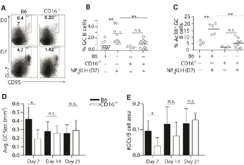
Figure 2. 4 CD16-/- mice have defective BAFF secretion, secondary antibody responses, frequency of memory B cell and Tfh cells. ... 40
Figure 2. 5 CD16-/- mice have diminished germinal center responses. ... 42
Figure 2. 6 Adoptive transfer of BAFF expressing DCs rescues GC and memory B cell populations and restores Bcl-6 levels in CD16-/- mice B cells. ... 43
Figure 2. 7 Chapter Two Model. ... 44
Table 3. 1 S. aureus strains and mutants ... 70
Figure 3. 1 Memory B cells are formed and activated in response to S. aureus. ... 71
Figure 3. 2 Protein A induces the expansion of IgM+ plasmablasts ... 73
Figure 3. 3 Protein A drives an expanded short-lived extrafollicular response ... 75
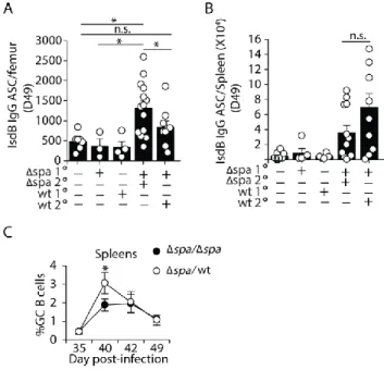
Figure 3. 4 S. aureus-specific ASCs formed after WT challenge fail to populate the bone marrow ... 77
Figure 3. 5 Protein A does not disrupt BM niche factors or the migration of ASCs to the BM ... 78
Figure 3. 6 Protein A prevents long-term maintenance of BM LLPCs and serum antibody ... 79
Supplemental Figure 3. 1 ... 80
x
Supplemental Figure 3. 3 ... 82
Supplemental Figure 3. 4 ... 83
Supplemental Figure 3. 5 ... 84
Supplemental Figure 3. 6 Chapter Three Model. ... 85
Figure 4. 1 Staphylococcus Protein A interferes with IC-induced DC BAFF in vitro but not in vivo ... 98
Figure 4. 2 BM ASCs formed in the presence of SpA express more CD19 than on those formed after Δspa challenge... 99
xi
LIST OF ABBREVIATIONS
AID Activation-induced (cytidine) deaminase APRIL A proliferation-inducing ligand
ASC Antibody secreting cell BAFF B cell activating factor BCMA B cell maturation antigen BCR B cell receptor
BM Bone marrow
BMDC Bone marrow-derived dendritic cell BMMF Bone marrow-derived macrophage CM Conditioned media
DC Dendritic cell
ELISA Enzyme-linked immunosorbent assay ELISpot Enzyme-linked immunospot
F(ab) Antigen-binding fragment (of Ig) Fc Constant fragment (of Ig)
FcγR Receptor for the constant fragment of Ig
FDC Follicular dendritic cell GC Germinal center
i.p. Intraperitoneal IFN Interferon
Ig Immunoglobulin
IV Intravenous
xii LPS Lipoploysaccharide
MF Macrophage
MRSA Methicillin-resistant Staphylococcus aureus
NP-KLH (4-hydroxy-3-nitrophenyl)-acetyl hapten conjugated to Keyhole limpet hemocyanin
OVA Ovalbumin (egg protein)
PB Plasmablast
PC Plasma cell
pDC Plasmacytoid dendritic cell s.c. Subcutaneous
SpA Staphylococcus Protein A
TACI TNFR homolog transmembrane activator and Ca2+ modulator and CAML interactor
Tfh T follicular helper
Tg Transgenic
TLR Toll-like receptor
Vh Heavy chain variable region (of Ig)
1
CHAPTER1: INTRODUCTION
Adaptive immune responses
Successful activation of the adaptive immune response results in long-term
immunological memory, which protects against re-infection. Memory B and T cells are long-lived, antigen-specific cells with the capacity to mount rapid and expanded responses upon secondary exposure to antigens or pathogens [1]. Memory responses form the basis of
vaccination and protection from re-exposure to commonly encountered pathogens. Despite this, the adaptive immune mechanisms required for memory, particularly for memory to bacterial pathogens, are not completely understood. This understanding is needed in order to design vaccines that are best suited for their target pathogens and target populations.
Antibody production by B cells is a major arm of immune memory. The selection and survival of memory B cells during a primary adaptive immune response is a carefully regulated process. Initial activation of naïve B cells via B cell receptors (BCRs) causes them to migrate to T cell-rich areas of secondary lymphoid organs in search of cognate interaction and
2
Both ASC and GC pathways may also be T cell-dependent or independent [6,7]. High affinity or avidity BCR ligation and/or Toll-like receptor (TLR) signaling may initiate T-independent B cell responses [6,8]. In general, T-T-independent responses drive short-term proliferation of ASCs in extrafollicular foci, (though extrafollicular responses may also involve T cell help), and T-dependent responses promote GC recruitment.
The Germinal Center
Early in the immune response, follicular dendritic cells (FDCs) create a reticular network that presents Ag to B and T cells and secretes chemokines such as CXCL13 to bring cells
together to form GC structures [5,9]. Within GCs, T follicular helper cells (Tfh) provide co-stimulation and survival signals to GC B cells, through interactions like CD40:CD40L and ICOS:ICOSL [10]. More recently, Tfh cells have been shown to produce local levels of BAFF within GCs to support affinity maturation of GC B cells in closest contact with Tfh cells [11].
Tfh cells and FDCs provide survival signals for GC B cells as they undergo clonal expansion, somatic hypermutation, and affinity maturation. Over time, B cell clones with BCRs of high affinity and specificity for antigen are selected for survival, while other clones die via apoptosis [9,12]. During this process, expression of the transcription factor, Bcl-6 directs B cells to maintain a GC phenotype and withstand the DNA mutations required for somatic
3 Memory B cells
The ultimate products of the GC reaction are memory B cells and affinity matured plasma cells (PCs). The mechanisms that drive one fate over the other are on an ongoing area of
investigation. Expression of the transcriptional master regulator, Blimp-1, along with XBP-1 and IRF-4, direct plasmablast (PB) and PC differentiation [15–17]. The transcriptional control of memory B cell development is less clear. Blimp-1 and Bcl-6 are mutually repressive [13,18,19], but complete differentiation of memory B cells requires that Bcl-6 ultimately be downregulated [18]. Further, Bcl-6 and GCs are dispensable for generating low-affinity, under-mutated memory B cells [18,20], although secondary responses are impaired when the primary GC reaction is disrupted [1,10].
Memory B cells are long-lived, quiescent, antigen-specific cells, capable of mounting a rapid and robust antibody response upon re-exposure to antigen [21,22]. Classically, memory B cells have been defined as having high affinity class-switched BCRs; however, in recent years, it has become clear that memory B cells can be formed without class switching or even T cell help [23,24]. Both IgM+ and IgG+ memory B cells participate in rapid secondary responses; IgM+ memory cells contribute to secondary GCs and IgG+ memory cells produce PBs/ASCs that make high affinity IgG [25–27].
Memory B cells are uniquely poised to respond quickly to their cognate antigen. Memory B cells typically localize close to T cells and constitutively express co-stimulatory molecules, which allows them quickly obtain T cell help in a secondary response [22,26,28]. Both
4
and T cell help [28]. Finally, genetic and epigenetic changes to anti-apoptotic and pro-cell cycle genes allow memory B cells to survive for long periods of time and divide rapidly when they re-engage antigen [22,29].
Plasma cells
Plasma cells are terminally-differentiated B cells that secrete antibody but do not divide. There are many paths leading to PC differentiation. First, primary T-dependent or T-independent responses can lead to short-term proliferation of antibody-secreting, dividing PBs in the
extrafollicular spaces of secondary lymphoid organs [6,30]. These cells produce an early surge of low affinity antibody and may undergo somatic hypermutation outside the GC [31]. As PBs mature, they lose surface expression of B cell markers like B220, CD19 and the BCR and cease dividing and become PCs [2,30]. Second, PCs may come out of the GC reaction. Post-GC PCs typically secrete more highly mutated, higher affinity antibody [32]. Third, during a secondary immune response, activated memory B cell clones divide and differentiate into PBs, many of which mature into PCs [32–37].
Long-lived Plasma Cells
5
or diphtheria [42]. Activation of memory B cells results in the largest proportion of LLPCs, but they can be formed after primary immune responses [12,43]. It has been speculated that
intermittent activation of memory B cells is required to repopulate the LLPC niche over time, but most evidence indicates that LLPCs persist in the absence of antigen [43] and are regulated independently of memory B cells [40,44,45].
There are no defining markers of LLPCs or of ASCs that are destined to become LLPCs. Blimp-1 is highest in BM LLPCs, and Blimp-1 expression increases in splenic ASCs as they mature, but it’s not clear if high Blimp-1 expression in the spleen is a pre-requisite for longevity
in the BM [15]. Short and long-lived PCs express the glycoprotein CD138, which binds
extracellular matrix,acts as a ligand for the PC survival factor, APRIL (A Proliferation-Inducing Ligand), and is expressed at higher levels as PCs mature [30,46,47]. T cell help and affinity maturation within the GC have both been implicated as prerequisites for LLPC differentiation [32,48]; however, it is possible for LLPCs to form without class switching or going through a GC reaction [49].
PBs and PCs express chemokine receptors that direct them to extrafollicular spaces, toward medullary cords of secondary lymphoid organs, and out of those organs into the blood [2]. These include CXCR4, which follows CXCL12 toward extrafollicular spaces and later in the response, toward the bone marrow [33,50,51]. S1P1 is also required to exit secondary lymphoid organs into the blood [52,53].
6
cells, monocytes, eosinophils, and megakaryocytes [50,54]. Niche cells secrete APRIL, BCMA, CXCL12 and TNF-alpha [50,54–56]. These survival signals activate anti-apoptotic factors like Mcl-1 [57] and proteins like XBP-1 and ATG5 to regulate endoplasmic reticulum stress caused by constitutive Ig production[58–60]. Recently, the protein ZBTB20 has been described as a vital BM PC survival factor that is regulated as a result of adjuvant present during the immune response [61]. This supports the possibility that PC longevity is determined in secondary lymphoid organs before migration to the BM.
Long-term immunity and Staphylococcus aureus infection
LLPCs play an important role in immunity to viral, parasitic, and bacterial infections [35,44,49]. LLPCs constitutively secrete protective antibody that acts as an early line of defense and enhances secondary responses [35,49,62]. Therefore, it is vital to understand the mechanisms required for LLPC persistence. In the case of Staphylocuccus aureus infection, long-term
maintenance of antigen-specific antibody is protective in humans [63–66]. However, S. aureus infections commonly recur and humoral immunity wanes unpredictably [67–71].
S. aureus is one of the most common causes of skin and soft-tissue infections and up to 65% of S. aureus infections are methicillin-resistant (MRSA), which is difficult to treat and life-threatening when it causes a systemic infection [72–74]. Therefore, there is great interest in generating a vaccine to protect vulnerable populations against S. aureus infection. However, it remains unknown whether memory B cells or LLPCs are formed in response to S. aureus infection.
7
[75,76]. The surface-expressed Staphylococcus protein A (SpA) is well known for its ability to impede B cell responses. SpA interferes with IgG-Fc receptor (FcγR) interactions by binding the Fc regions of IgG, interfering with opsonization and phagocytosis of S. aureus [77–79]. Work from our lab (Chapter 2) demonstrates that immune complex (IC) ligation of FcγRs induces B
cell activating factor (BAFF) production by dendritic cells (DCs), which in turn directs B and T lymphocyte populations to generate GCs and memory. Therefore we hypothesized that SpA blocks BAFF production by interfering with IC- FcγR interactions thereby reducing DC BAFF production and memory B cell formation. We used an isogenic Δspa mutant to examine DC
BAFF (Appendix 1), B cell memory, and GC formation (Chapter 3) in the presence of absence of SpA during S. aureus exposure or infection. We found that SpA did not interfere with any of these outputs, and characterization of the primary B cell response to S. aureus revealed that memory B cells and secondary amnestic antibody responses were not affected by SpA.
In addition to its interference with IgG- FcγR interactions, SpA can act as a super-antigen that binds naïve B cell receptors of the VhIII clan (up to 50% of naïve B cells in humans and 5-10% in mice) [80,81], and drives polyclonal B cell proliferation [75,82]. Injection of purified SpA can also induce to B cell deletion due to the absence of a second signal, like TLR
8
9
CHAPTER 2: IgG immune complexes promote B cell memory by inducing BAFF1
Introduction
Adaptive immunity requires the commitment of activated B cells to either the memory or PC compartments, the differentiation of CD4+ T cells to Tfh cells, and coordinated expression of chemoattractant receptors to position T and B cells within the follicle for cognate interactions [3,85,86]. The specialized microenvironment of the GC provides a site for rapid expansion and selection of B cell clones whose somatically mutating Ig V regions compete for a limiting amount of antigen displayed on follicular dendritic cells [9,87–90]. Although many steps in the cyclic process of somatic hypermutation and clonal selection are defined, the events required to induce memory cell responses are incompletely understood [91–94].
During the GC response, Tfh cells are critical effectors that provide help to B cells [3,95]. Tfh cells engage activated B cells at the T:B border, and their secreted cytokines promote Ig isotype switching and the selection of cells with high affinity B cell receptors in GCs
[13,85,96,97]. The expression of CXCR5, ICOS, PD-1, and the secretion of IL-21 distinguish Tfh cells from other CD4+ T cell subsets [98,99]. The formation of Tfh cells is dependent on the
1
Amanda B. Keener*, Shannon Z. Jones*, SunAh Kang*, Robert J. Benschop, Alfredo Caro-Maldonado, Jeffrey C. Rathmell, Stephen H. Clarke, Glenn K. Matsushima, Jason K. Whitmire, Barbara J. Vilen(2014) IgG-immune complexes promote B cell memory by inducing DC-derived BAFF (Unpublished work).*These authors contributed equally.
10
expression of Bcl-6, a process linked to ICOS expression on CD4+ T cells [99,100] and influenced by IL-2 [101,102]. This commits primed T cells to the Tfh pool and inhibits their differentiation to other T cell subsets [103–105]. Bcl-6 is also required for GC B cell formation [103,106,107]. In activated B cells, Bcl-6 downregulates Blimp-1; thus, directing B cells away from PC differentiation and toward the memory pathway [17,108]. Cytokines such as IL-6 and IL-21 have been shown to affect Bcl-6 expression in B and T cells [14,109]; however detailed events upstream of Bcl-6 expression are of interest in understanding B and T cell differentiation in GC responses.
BAFF plays an essential role in controlling the development and survival of B2 and marginal zone B cells [110,111], enhancing PB survival [112], and the survival of affinity-matured B cells in the GC [11]. Early studies that neutralized BAFF suggested BAFF played a role in the GC response; however, interpretations of those results were complicated by the loss of B cells associated with BAFF depletion [113–116]. Others have shown that BAFF and anti-CD40 increase ICOSL expression on B cells [117], and that TACI serves to limit the expression of ICOSL and the expansion of Tfh cells and GC B cells [118]. These studies suggest an indirect role for BAFF family members and CD40L in the formation of Tfh cells through ICOS:ICOSL interactions. Thus, BAFF has been implicated in events that contribute to GC responses; however, how BAFF is induced and where it acts in the GC response remains unclear.
11
12 Results
BAFF-/- bone marrow chimeras exhibit reduced secondary responses
Previous studies have linked BAFF with an enhanced response to vaccination, suggesting that it plays a role in adaptive immune responses [119–121]. To assess this, we generated BAFF -
bone marrow chimeras by engrafting irradiated B6 mice with B6 (BAFF+/+) or BAFF-/- bone marrow. This approach limits BAFF deficiency to hematopoietic cells, allowing other sources of BAFF to maintain the peripheral B cell population [122] (data not shown). In BAFF-/- chimeras, we found that the primary IgG response to NP14KLH (Figure 1A) was comparable to B6 control chimeras. However, 14 days after secondary immunization, the BAFF-/- bone marrow chimeras showed a 2-fold reduction in the levels of IgG compared to B6 chimeric control mice (1.4-fold lower on day 42) (Figure 1B). The reduced IgG during the secondary response could reflect diminished class switch since BAFF can induce AID expression [123,124]. However, AID mRNA levels in B cells from B6 control and BAFF-/- chimeras were not different (data not shown), suggesting that BAFF has a role other than in class switch.
BAFF-/- chimeras exhibit defects in the frequency of memory B, Tfh cells, and GC B cells Rapid, high titer secondary immune responses require the activation of memory B cells [1]. Although IgG memory B cells do not require BAFF for maintenance [125], it is not known whether BAFF is important for their formation. To determine whether BAFF affects the
13
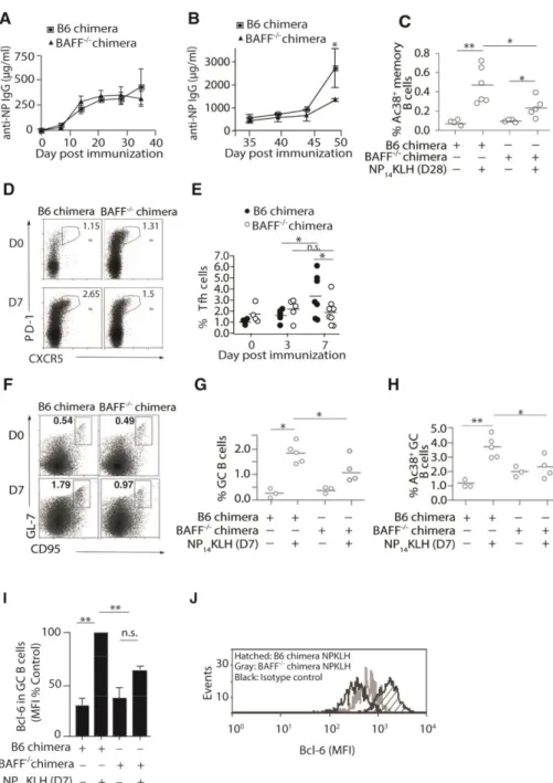
Tfh cells are critical to the early GC response and required for the differentiation of memory B cells [126–128]. It is possible that BAFF affected memory responses by influencing germinal center responses and/or Tfh cells. To assess whether BAFF affects formation and maintenance of Tfh cells, B6 and BAFF-/- chimeras were immunized and the frequencies of Tfh cells (CXCR5+PD-1+CD4+) were quantitated on days 3 and 7 post-immunization (Figure 1D and 1E). On day 3, the frequency of Tfh cells in B6 chimeras increased by 1.5-fold while in BAFF-/- chimeras it increased 1.2-fold. This suggests that BAFF does not play a significant role in the formation of Tfh cells. However on day 7 post-immunization, B6 chimeras displayed an increased frequency (1.9-fold) of Tfh cells, whereas their frequency in BAFF-/- chimeras did not change. This suggests that BAFF may stabilize Tfh cells or support their expansion.
Germinal centers are necessary for the formation of high affinity, class-switched memory B cells [129,130]. To determine whether BAFF impacted the frequency of GC B cells, we enumerated CD19+GL-7+CD95+ GC B cells 7 days after immunization. In BAFF-/- chimeras, the frequency of total (Figure 1F and 1G) and Ag-specific (Figure 1H; CD19+Ac38+IgG+ GL-7+CD95+) GC B cells in BAFF-/- chimeras was lower than the response in B6 chimeras. Thus, BAFF significantly contributes to antigen-specific GC responses.
BAFF acts at or upstream of Bcl-6 expression in B cells
Bcl-6 plays a critical role in initiating GC responses and committing activated B cells to a memory cell phenotype [106,131,132]. Thus, one possibility is that BAFF affected Bcl-6
14
acts at, or upstream of, Bcl-6 expression in B cells. Collectively, our data indicate that BAFF impacts the expansion and stabilization of the Tfh cell pool, and the formation of GC and memory B cells.
DC-derived BAFF regulates the frequency of GC B and Tfh cells
15
The binding of immune complex to FcγRs induces BAFF secretion
Previous studies showed that exogenous ICs induce BMDCs to secrete a number of cytokines, including BAFF [135,136]. In another study, mice lacking the common gamma chain of the FcγRs (Fcγc) exhibited diminished secondary immune responses [62]. Because our data
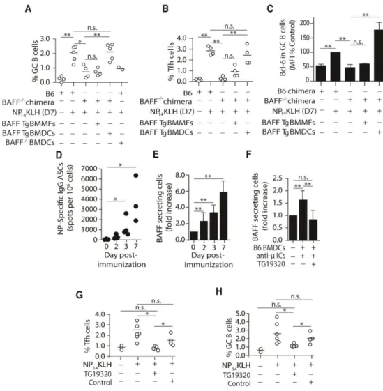
suggest that DCs may promote secondary responses via BAFF, we postulated that ICs formed by the early IgG antibody response might induce DCs to secrete BAFF. This model requires that the early IgG response occur concurrently, or precede BAFF secretion. To test this, we harvested spleens from B6 mice on days 2, 3, and 7 following NP14KLH immunization and used ELISpot to measure the numbers of antibody (IgG) secreting cells (ASCs) and BAFF-secreting DCs. NP-specific (IgG) ASCs were present in the spleen by day 2 post-immunization and expanded over the course of 7 days (Figure 2D). Similarly, the number of splenic CD11c+ DCs that secreted BAFF increased 6-fold between days 2 and 7 (Figure 2E). Thus, secretion of Ig by B cells, and production of BAFF by DCs, occurs simultaneously beginning approximately 2 days following immunization. This is consistent with the idea that IgG-ICs induce BAFF, which contributes to GC responses.
To further test the idea that ICs induce DCs to secrete BAFF, we blocked IgG-Fc:FcγRs interactions in vitro and assessed whether this impacted BAFF secretion by DCs.
Stimulation of B6 BMDCs with pre-formed IgG-ICs (IgM bound by anti-μ) induced a1.8-fold increase in BAFF secretion (Figure 2F). This was not unique to IgM/IgG ICs because
16
To assess whether blocking Fc:FcγR interactions affected the adaptive immune response
in vivo, we administered TG19320 at the time of immunization and measured the frequencies of GC B and Tfh cells. We found that co-administration of TG19320 with NP14KLH blocked an increase in GC B and Tfh cells on day 7 (Figure 2G and 2H). This indicates that the
interactions between IgG-ICs and FcγRs are necessary for optimal GC responses and for the stabilization of Tfh cells in response to immunization. Collectively, these data identify a mechanism wherein IgG-ICs formed early in the immune response ligate FcγRs on DCs to induce BAFF secretion, which in turn contributes to the GC response.
BAFF regulates the expression of Bcl-6 in activated B cells in vitro
To define whether the effects of BAFF on activated B cells was direct, we established an in vitro reconstitution system using the expression of Bcl-6 as a marker of memory B cell
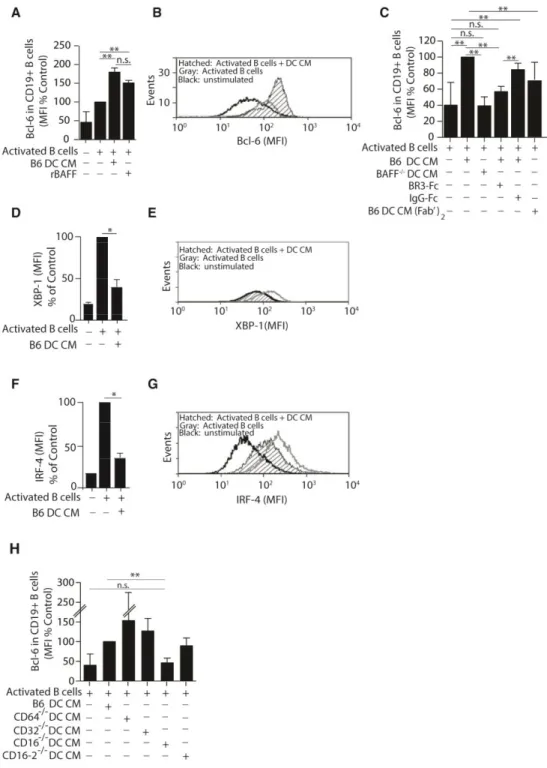
commitment. In this model, B6 B cells were stimulated with anti-μ in combination with IL-4 and IL-5 to induce Ig secretion and a low level of Bcl-6 expression (Figure 3A and B). These activated B cells were then cultured in conditioned medium (CM) prepared from DCs treated with preformed IgG1-ICs (DC CM). Addition of DC CM increased Bcl-6 expression to approximately 3-fold over unstimulated B cells (Figure 3A and 3B). These levels were comparable to those achieved with recombinant BAFF. Bcl-6 expression was dependent on Ig-Fc regions as CM from DCs stimulated with F(ab’)2-containing ICs were ineffective at inducing
Bcl-6 (Figure 3C). In this in vitro system, Bcl-6 expression was not induced in B cells cultured with DC CM where BAFF was neutralized with BR3-Fc, or where DC CM was made from BAFF-/- DCs (Figure 3C). As Bcl-6 levels become elevated, the plasma cell program is
17
XBP-1 and found that DC CM diminished the levels of both transcription factors by 2.2-fold (Figure 3D-G), indicating that BAFF may act at or upstream of Bcl-6, directing B cell differentiation away from a plasma cell fate.
IgG-ICs bind CD16 during the anti-NP response
We reasoned that if FcγR stimulation promoted BAFF secretion, loss of the FcγR(s) that
binds IgG1-anti-NP ICs would negatively affect the ability of DC CM to promote Bcl-6. To assess this, we tested whether CMs from BMDCs derived from B6 mice, or mice deficient in CD64 (FcγRI-/-), CD32 (FcγRIIb -/-), CD16 (FcγRIII
-/-), or CD16-2 (FcγRIV-/-) induced Bcl-6 in the in vitro reconstitution system described above. DC CM from CD16-/- mice failed to induce Bcl-6 expression, while CMs from all other FcγR deficient mice induced Bcl-6 to levels
comparable to, or above those induced by B6 DC CM (Figure 3H). This suggests that ligation of CD16 on DCs is required for the expression of Bcl-6 in B cells.
Impaired secondary responses in CD16-/- mice
Our data indicate that CD16 might be responsible for inducing DCs to secrete BAFF after NP14KLH immunization. To test this in vivo, we quantitated the number of splenic BAFF
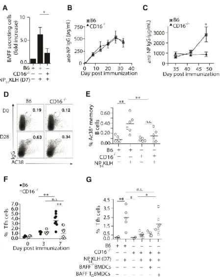
secreting DCs in B6 and CD16-/- mice 7 days after immunization (Figure 4A). In the absence of CD16, we found that the number of BAFF secreting DCs was markedly diminished compared to wild type mice. This is consistent with the idea that CD16 is the FcγR responsible for initiating
18
To test whether loss of CD16 in vivo impaired adaptive immune responses, we measured primary and secondary antibody responses in B6 and CD16-/- mice following NP14KLH
immunization. As in the BAFF-/- chimeric mice, the primary IgG response in CD16-/- mice was comparable to B6 mice (Figures 4B). In the secondary response, the levels of IgG in B6 mice increased 1.7-fold on day 42 (7 days after boost), and 4.5-fold on day 49 (14 days after boost), while IgG levels in the CD16-/- mice did not increase on days 42 or 49 (Figure 4C). This was not an indirect consequence of altered cell populations due to CD16 deficiency since the frequencies of DCs, T, and B cells in CD16-/- mice were not different than B6 mice (data not shown). This indicates that CD16 plays a role in generating memory B cells and secondary immune responses to NP-KLH.
CD16-/- mice exhibit defects in forming Tfh and memory B cells
The loss of DC-derived BAFF in CD16-/- mice supports a role for CD16 in the memory response to NP14KLH. To assess whether the diminished secondary response in CD16-/- mice reflects a reduction in memory B cells, we assessed the frequency of NP-specific memory B cells (CD19+Ac38+IgG+) in B6 and CD16-/- mice 28 days following immunization. CD16-/- mice showed a 2.4-fold decrease in the Ac38 Id+ IgG memory B cell population compared to
immunized B6 mice (Figure 4D and 4E), indicating that CD16 regulates memory responses in part through BAFF production.
19
we quantitated Tfh cells (CD4+CXCR5+PD-1+) from immunized B6 and CD16-/- mice. After 3 days, CD16-/- mice had a comparable frequency of Tfh cells compared to immunized B6 mice (Figure 4F). However on day 7, CD16-/- mice had 3-fold fewer Tfh cells, similar to the defects observed in immunized BAFF-/- chimeras. Adoptive transfer of BAFF transgenic, but not BAFF -
BMDCs into immunized CD16-/- mice, restored the frequency of Tfh cells on day 7 to levels seen in B6 mice (Figure 4G), suggesting that the defect was a consequence of reduced BAFF in CD16-/- mice. These findings demonstrate that CD16 ligation by IgG-ICs induces DCs to secrete BAFF and that CD16 is necessary for memory responses. Together with the findings from the BAFF-/- chimeras, our data suggest that IgG-IC binding to CD16 contributes to B cell memory through the effects of BAFF on Tfh cells and GC B cells.
Loss of CD16 diminishes germinal center responses
20
they are reduced in number and size early during the immune response. This suggests that stimulation of CD16 is an early event that impacts the GC response and subsequently, the memory B cell pool.
CD16 and BAFF are required for the expression of Bcl-6 and the formation of GC and memory B cells
Our data indicate that CD16 and ICs are required for DCs to make BAFF in response to NP-KLH (Figure 4A). To address whether DC-derived BAFF was sufficient to restore the GC and memory B cell pools in the CD16-/- mice, we adoptively transferred BMDCs from BAFF Tg mice at the time of immunization. We found that BAFF Tg DCs restored the frequency of GC B cells on day 7 (Figure 6A), and the frequency of Ac38 Id+ memory B cells on day 28 (Figure 6B). In contrast, BMDCs derived from BAFF-/- mice were unable to restore GC or memory B cells (Figure 6A and B), indicating that this effect was due to BAFF and not due to an increased number of DCs, or the secretion of other cytokines through activation of the transferred DCs. Since BAFF-/- DCs have intact CD16, the results also emphasizes that BAFF, induced by CD16 ligation, impacts the GC response and the frequency of memory B cells.
Our data suggest that DC-derived BAFF acts at or upstream of Bcl-6 (Figure 1I, J, 2C, 3A-C) and downstream of CD16 (Figure 4A). Thus, the absence of CD16 should also
diminished Bcl-6 during NP immunization and restoring DC-derived BAFF by BMDC transfer should restore Bcl-6 levels. To test this, we adoptively transferred BAFF Tg BMDCs into CD16 -
/-mice at the time of immunization. We found that compared to B6 controls CD16-/- GC B cells expressed low levels of Bcl-6 after immunization. Transfer of BAFF Tg BMDCs, but not BAFF -/-
21
22 Discussion
Interactions between T cells, B cells, and DCs are necessary for the proper execution of the adaptive immune response [141–143]. This study identifies a previously unappreciated mechanism for DCs, FcγRs, and BAFF in the early events of the GC response. We show that
ICs formed during the early IgG response to NP-KLH induced the production of BAFF by DCs. BAFF acted at, or upstream of, Bcl-6 to promote the optimal formation of GC B cells and in the stabilization of newly formed (day 7) Tfh cells. Loss of BAFF in hematopoietic cells, or loss of CD16, diminished the levels of Bcl-6, the frequencies of GC B cells, Tfh cells and memory cells, and the magnitude of the secondary antibody response. We found that DC-derived BAFF was sufficient to initiate these events. This series of events depends on the formation of IgG-ICs, suggesting that productive early antibody responses contribute to the optimal formation of B cell memory.
IgG-ICs and BAFF in the GC response
ICs have previously been implicated in promoting adaptive immune responses through enhanced Ag presentation [144] and stimulating cytokine production by DCs [135,136]. We observed that IC ligation of CD16 was required for BAFF production by DCs after NP-KLH immunization. Although the loss of Fcγc reduces secondary antibody responses [62], it is not clear what role IC- FcγR interactions play in GC and memory B cell formation. Our data demonstrate that GCs and memory responses are disrupted in the absence of CD16 or when IC- FcγR interactions were blocked, indicating that FcγR signaling is important for these events. We
23
likely secreted by class-switched extrafollicular PCs [140] that produce local levels of IgG early in the immune response [106,132,140].
Although at day 7 of the primary response to NP-KLH, GCs, GC B cells, memory B cells and Tfh cells were diminished in the absence of FcγR-IC signaling, they were not completely
absent and the numbers of GCs was increased on days 14 and 21. This suggests that signals other than BAFF, and perhaps other receptors for ICs are required for GC responses. We also observed incomplete loss of these populations after immunization of BAFF-/- chimeric mice, suggesting DC-derived BAFF is only one of many signals required for optimal GC formation and function.
Early in the adaptive immune response, B cells interact with cognate T cells and co-stimulatory signals induce AID activation and class switch [145]. These signals include CD40L, which is also required for GC formation [146,147]. We found that BAFF-/- chimeras had normal AID expression and primary IgG production, suggesting that cognate interactions mediated by CD40L were intact. Thus, BAFF likely acts downstream of initial T:B interactions. Our in vitro data show that recombinant BAFF directly induced Bcl-6 in activated B cells, and DC CM (elicited under conditions that induce BAFF) diminished the expression of XBP-1 and IRF-4. BAFF, therefore, may act on B cells after cognate T interaction, extinguishing the PC phenotype and either committing cells to the memory pathway or supporting the survival of GC B cell precursors. This is consistent with studies showing that DC secretion of the PC-inducing cytokine, IL-12, is dampened by IC signaling [148].
24
to promote either Blimp-1 or Bcl-6 expression, depending on the context [18], BAFF may serve as a contextual signal early in the adaptive response to direct B cells toward a GC fate. BAFF may also maintain Bcl-6 in B cells destined for the GC, allowing IL-21 from Tfh cells to take over as the response progresses.
DCs as mediators of IC signaling
Although DCs, MFs, and neutrophils are among the major producers of BAFF, [122,150– 154], our data support a role for DCs as the cells responsible for producing BAFF specifically in response to ICs after immunization. We found that transfer of BAFF-producing DCs, but not BAFF-/- DCs, restored the numbers of GC B cells in BAFF-/- chimeras and CD16-/- mice. This indicates that the defects in CD16-/- mice were mediated through the loss of BAFF. Further, BAFF-producing MFs were unable to restore the defects suggesting a unique role for DCs in this process consistent with tripartite interactions between DCs, T cells and B cells during adaptive immune responses. Other cells with Fc receptors may have roles similar to DCs that are worthy of further investigation. This current study indicates that DC-derived BAFF is sufficient to improve T-dependent GC responses and B cell memory. It also supports the use of DCs as a means to augment vaccine responses where better adjuvants are needed.
BAFF and Tfh cells
Previous studies showed that cells with a Tfh phenotype appear by day 3 after
immunization [99,155,156]. These cells migrate towards follicles where they interact with B cells at the T-B border [156–159] to promote continuous expression of Bcl-6
25
B cells and IgG-ICs are not involved in the formation of Tfh cells (day 3). This is consistent with previous studies showing that on days 1-3 post-immunization, the expression of Bcl-6 and CXCR5 in CD4+ T cells is independent of B cells [99,134,155]. Instead, BAFF acted
downstream of the formation of Tfh cells, possibly to stabilize the population or promote its expansion. Thus, previous studies showing a role for B cells in maintaining Tfh cells
[109,155,158] might reflect the need for B cell-elicited Ig, ICs, and DC-derived BAFF. The role of BAFF in maintaining the Tfh population may be indirect. One possibility is that BAFF promotes the expression of ICOSL on B cells. Previous studies showed that signaling through the BAFF-R regulates the expression of ICOSL on B cells [117,162], thereby sustaining interaction between Tfh cells and B cells at the T:B border and within GCs [99,100]. This interaction could also stabilize the expression of Bcl-6 and the downstream molecules CXCR5 and PD-1[105,163,164] to maintain the “Tfh phenotype”. This possibility is supported by studies showing that the absence of ICOSL on B cells reduces the frequency of CXCR5+ CD4+ cells after immunization [105], that ICOS:ICOSL interaction prolongs the engagement between B cells and Tfh cells [161], and that follicular bystander B cells support the formation and/or maintenance of Tfh cells by providing ICOSL in an antigen-independent manner [165].
26
Tfh maintenance. However, more work would be needed to determine whether affinity maturation is altered in these models.
Overall, our studies highlight a novel role for IgG-ICs and DC-derived BAFF in the GC response. Elucidating the events that initiate GC responses may impact our understanding of ICs and BAFF in autoimmunity. Systemic lupus erythematosus (SLE) is an autoimmune disease characterized by elevated levels of BAFF, autoantibody/autoantigen ICs, and multi-organ pathology. The formation of autoreactive memory is thought to be instrumental in driving long-lived PCs and sustaining autoantibody production [166,167]; however, the mechanisms that regulate memory formation to self-antigens are unclear. Our findings suggest that chronically high levels of ICs containing self-antigens could contribute to a break in B cell tolerance at the GC checkpoint. In SLE, elevated levels of circulating ICs may elevate BAFF and promote the GC response [137,166,168–171]. This suggests that neutralization of BAFF in patients with SLE may affect both B cell survival and GC responses that are necessary in the formation of
27 Materials and Methods
Animals
B6-Ly5.2 congenic mice were purchased from NCI, BAFF-/- mice (Schiemann et al., 2001), and CD16-2-/- (FcγRIV) mice (Nimmerjahn et al., 2010) were obtained from, Glenn Matsushima, and Charles Jennette at UNC-Chapel Hill. CD16-/- and CD64-/- mice (Hazenbos et al., 1996) were obtained from Dr. Anne Sperling at the University of Chicago, and BAFF Tg mice (Gavin et al., 2005) from Jeffrey Rathmell at Duke University. CD32-/- mice (Takai et al., 1996) were purchased from Jackson labs. Mice were used at 8-12 weeks of age and maintained in an accredited animal facility.
Reagents and Antibodies
Antibodies against mouse CD4, CD19, CD95, GL-7, ICOS, ICOSL, PD-1, and B220-647 were purchased from Biolegend, CXCR5, B220, IgG1, IgG2a, IgG2b, and IgG3 antibodies from BD Biosciences, Bcl-6, XBP-1, and IRF-4 from Santa Cruz, and BAFF (1C9) from Enzo. Anti-μ F(ab)2 was purchased from Jackson ImmunoResearch. Anti-μ (clone B7.6), anti-NP (clones
28
streptavidin-Alexa 488 and Alexa 647 from Invitrogen. Streptavidin-alkaline phosphatase and anti-IgG alkaline phosphatase were purchased from Southern Biotech. The Fc-binding TG19320 peptide was synthesized as described (Fassina et al., 1998; Marino et al., 2000).
B cell purification and bone marrow derivation of MFs and DCs
Splenic B cells were isolated from B6 mice by negative selection (StemCell
Technologies) and were 95-99% pure, as determined by flow cytometry. Splenic DCs were purified by positive selection of CD11c+ cells (Miltenyi) from enriched low-density cells (OptiPrep; Sigma). Purified cells were 80% CD11c+.
Bone marrow-derived DCs (BMDCs) and bone marrow-derived MFs (BMMFs) were prepared from single cell suspensions from the tibias and femurs of B6, CD64-/-, CD32-/-, CD16 -/-, and BAFF-/-mice. Following RBC lysis, cells were cultured 7 days in a 24 well low-cluster plate (Costar 3471) with 10 ng/ml GM-CSF and IL-4 to derive DCs and in 20 ng/ml M-CSF to derive MFs.
Cell Culture
We pre-formed immune complexes by stimulating B6 B cells (1.5 x 10⁵) with an excess of anti- (B7.6; IgG1; 30 μg/ml). The polyclonal IgM produced after seven days forms a
complex with the excess anti- resulting in IgG1-IgM ICs in the supernatant. These B cell
supernatants were used as a source of ICs in preparing DC conditioned medium (DC CM).
29
For in vitro co-cultures, 1.5 x 10⁵ purified B6 B cells were co-cultured with 1 x 10⁴ BMDCs or ex vivo DCs in a 96 well plate stimulated with IL-4 (25 ng/ml), IL-5 (25 ng/ml) and 30 μg/ml anti-μ with or without recombinant murine BAFF (5 ng/ml) or DC CM (20% of total
volume). Intracellular Bcl-6 was assessed by flow cytometry after 48 hours.
ELISAs
NP-specific IgG levels were quantitated from serum using microtiter plates coated with NP13BSA and blocked with 0.5% BSA. Serially diluted serum samples were incubated
overnight at 4oC. Anti-NP was detected using an alkaline phosphatase conjugated rabbit anti-mouse IgG antibody (1/1000 dilution) followed by phosphatase substrate. Optical density (OD) values were converted to concentration based on standard curves using the H33L (anti-NP) hybridoma.
ELISpot
For the analysis of NP-specific B cells, multiscreen ELISpot plates (Millipore) were coated with NP13BSA in PBS and blocked with 1% BSA. Single cell suspensions of spleen were prepared from immunized or naïve B6 mice. After RBC lysis, cells were plated in serial
dilutions on washed ELISpot plates. Anti-NP IgG-secreting spots were detected with anti-IgG-biotin and streptavidin-HRP (BD Biosciences). Plates were developed with 3-amino
9-ethylcarbazole.
30
(2.5 x 10⁵) were plated on ELISpot plates as above and incubated 24 hours with preformed ICs (IgM + anti-μ) prior to addition of 1C9. Anti-μ ICs were made by combining the supernatant from stimulated B cells (20 ng of IgM) with anti-μ (5 μg). In some experiments, TG19320 was added at 50 μg/ml to i
Bone Marrow Chimeras
B6-Ly5.2 congenic mice (6-8 weeks of age) were lethally irradiated (10.5 Gy; 1050 rads) and reconstituted with 8 x 10⁶ bone marrow cells from either B6 (B6 control chimeras) or BAFF-/- (BAFF-/- chimeras) mice. After 8 weeks, we monitored reconstitution by assessing the frequency of CD45.1+ and CD45.2+ splenocytes by flow cytometry.
Immunization and Adoptive Transfers of BMMF/BMDCs
B6, BAFF-/- bone marrow chimeras, and CD16-/- mice (8-10 weeks of age) were immunized by intraperitoneal (i.p.) or subcutaneous (s.c.) injection with 100 μg of NP14KLH
precipitated in an equal volume of alum (Imject® Thermoscientific). Mice were boosted by intravenous (i.v.) injection with the same dose of soluble NP14KLH at day 35. To assess the contribution of DCs or MFs in the secretion of BAFF, 8 X 10⁶ BAFF Tg or BAFF-/- BMDCs or BMMFs were injected at the time of s.c. immunization. Draining lymph nodes were harvested on day 7 for flow cytometry analysis.
TG peptide injections
B6 mice were immunized with 100 μg NP14KLH in alum (1:1) via i.p. injection and
31 Flow Cytometry
GC B cells and Tfh were analyzed on day 7 post-immunization and were defined as CD19+, GL-7+, CD95+ and CD4+, CXCR5+, PD-1+. Ac38 was used to define NP-specific GC B cells. NP-specific memory B cells were defined as Ac38+ IgG+ double positive CD19+
lymphocytes. The lymphocyte gate was determined by forward and side scatter properties. To quantitate expression of intracellular IRF-4, Bcl-6 and XBP-1, splenocytes from immunized B6, CD16-/-, B6 control chimeras and BAFF-/- chimeras mice were washed, fixed (4%
paraformaldehyde), and permeabilized with methanol for a minimum of 24 hours at -20oC. Fixed cells were washed and FcγRIIb/FcγRIII blocked with 2.4G2 before staining. Data are
expressed as fold change in MFI/isotype control MFI.
Real Time PCR
Splenic B cells from B6 and BAFF-/- chimeras were purified after NP14KLH
immunization. mRNA was isolated from 5-10 x 106 purified B cells and cDNA synthesized using Superscript VILO cDNA Synthesis Kit (Invitrogen). DNA was subsequently amplified using FastStart Universal SYBR Green Master mix (Roche). Relative values were compared using the 2-ΔΔCΤmethod. 18s rRNA was used as an internal control in all experiments. Primers included: murine Aicda forward 5’GGGAAAGTGGCATTCACCTA3’, murine Aicda reverse 5’GAACCCAATTCTGGCTGTGT3’ murine 18s rRNA forward
5’TCAAGAACGAAAGTCGGAGGTT3’, murine 18s rRNA revese
32 Germinal Center Staining and Counting
Spleens were harvested from B6 or CD16-/- mice on days 7, 14, and 21 after
immunization and flash frozen in OCT (Optimum Cutting Temperature; Fisher). Tissue sections (6 micron) were fixed in 1:1 MeOH/Acetone, blocked with 10% FBS in PBS containing 2.4G2, and stained with PNA-biotin and B220-Alexa647, and Streptavidin-Alexa488. Germinal centers were defined as PNA+ cell clusters within B220+ follicles (Han et al., 1997). The number of germinal centers per mm2 of B220+ area was determined by dividing the number of germinal centers counted in a field by the area of B220+ follicles in the same field. This accounted for follicles that were only partially represented in a given field (Holl et al., 2011). This was done for 10-30 fields per mouse, totaling 30-100 follicles per mouse at each time point.
Microscopy
Macroscope images were obtained on a Leica MX16FA fluorescence stereo microscope/macroscope (0.63x objective; numerical aperature of 1.0). Other images were obtained using an Olympus Fluoview 500 (10x objective; numerical aperature of 0.45).
Statistics
33
Table 2. 1 Mice used for Chapter 2
Mouse line Description
C57BL/6 (B6) Wild-type
B6 Chimera Lethally irradiated B6 reconstituted with
congenic B6 bone marrow
BAFF-/- Lacks BAFF
BAFF-/- Chimera Lethally irradiated B6 reconstituted with
congenic BAFF-/- bone marrow
CD16-/- Lacks FcγRIII
BAFF Tg Constitutively expresses BAFF in Myeloid
34 Chapter 2 Figures
Figure 2. 1 BAFF-/- chimeras have defective memory and GC B cells, diminished Bcl-6 levels and decreased frequency of Tfh cells.
(A) B6 and BAFF-/- chimeric mice were immunized (i.p) with 100 g NP14KLH in alum. Serum IgG anti-NP responses in B6 and BAFF-/- chimeric mice were measured by ELISA on days 7, 14, 21, 28, and 35 post immunization. n=4-6 mice per group over 3 experiments. (B)
35
of 100 g of soluble NP14KLH on day 35. n = 3-6 mice over 3 experiments. (C) CD19+Ac38+
IgG+ B cells enumerated by flow cytometry from the spleens of B6 and BAFF-/- chimeras immunized for 28 days. n = 4-6 mice over 4 experiments. (D and E) CD4+CXCR5+PD-1+ T cells from B6 and BAFF-/- chimeric mice enumerated on days 0, 3 and 7 following
immunization. n = 3-6 mice per time point over 2 experiments. (F and G) CD19+, GL-7+, CD95+ GCB cells enumerated by flow cytometry on day 7 post-immunization. n = 3-5 mice over 3 experiments. (H) CD19+Ac38+CD95+GL-7+ B cells enumerated on day 7
36
Figure 2. 2 Secretion of BAFF by DCs restores Bcl-6 levels and the frequency of GC B and Tfh
cells.
(A-C) BAFF Tg BMDCs, BMMFs, or BAFF-/- BMDCs (8 x 10⁶) were injected (s.c.) into B6 or
37
quantitated. n = 3-4 mice per time point over 3 experiments. (E) 1 x 10⁶purified CD11c+ cells from B6 mice immunized for 2, 3, and 7 days were plated on BR3-Fc coated ELISpot plates. After 60 hours, the number of BAFF secreting cells was enumerated. n = 3-6 mice per time point over 3 experiments. (F) BAFF secreting cells enumerated from B6 BMDCs (2.5 x 10⁵) cultured for 24 hrs in the presence or absence of anti-μ ICs, with or without Fc blocking peptide (TG19320; 50 μg/ml). n = 3-10 over 6 experiments. (G-H) B6 mice were immunized with
NP14KLH and dosed with 15-30 mg/kg of Fc blocking peptide (TG19320) or an unrelated scrambled peptide (Control) via i.p. injection. On day 7, the frequency of Tfh cells
38
Figure 2. 3 DC-derived BAFF regulates the expression of Bcl-6 in cultured B cells.
39
in B cells activated by DC CM as in (A). n = 3-4 mice over 4 experiments. (C) Purified B6 B cells were co-cultured with CM generated from B6 DCs treated with ICs containing intact Fc regions (B6 DC CM), F(ab’)2 (B6 DC CM (Fab’)2), BAFF-/- DC CM, or B6 DC CM neutralized
with BR3-Fc (10 μg/ml) or control IgG-Fc (10 μg/ml). n = 4-7 over 4 experiments. (D-G) B cells were co-cultured with B6 DC CM. Intracellular levels of XBP-1 (D, E) and IRF-4 (F, G)
40
Figure 2. 4 CD16-/- mice have defective BAFF secretion, secondary antibody responses, frequency of memory B cell and Tfh cells.
(A) BAFF secreting cells enumerated from purified CD11c+ cells isolated from B6 and CD16 -/-mice on day 7. n = 3 over 3 experiments. (B and C) B6 and CD16-/- mice were immunized (i.p)
with 100 g NP14KLH in alum. Serum IgG anti-NP (B) primary responses measured by ELISA
on days 0, 7, 14, 21, 28, and 35; serum IgG (C) secondary antibody levels measured on days 39,
41
frequency of CD4+CXCR5+PD-1+ T cells from immunized B6 and CD16-/- mice on days 0, 3 or 7. n = 3-8 mice per time point over 4 experiments. (G) B6, BAFF Tg, and BAFF-/- BMDCs (8 x 10⁶) were injected into CD16-/-mice that were simultaneously immunized with NP14KLH. On day 7, the frequencies of CD4+CXCR5+PD-1+ T cells from inguinal lymph nodes were
42
Figure 2. 5 CD16-/- mice have diminished germinal center responses.
(A and B) The frequency of splenic CD19+GL-7+CD95+ GC B cells was measured by flow cytometry from B6 and CD16-/- mice 7 days after immunization. n = 4-11 mice per group over 3 experiments. (C) The frequency of NP-specific GC B cells (CD19+Ac38+GL-7+CD95+)
measured in B6 and CD16-/- mice 7 days after immunization. n = 3-10 mice over 3 experiments.
43
Figure 2. 6 Adoptive transfer of BAFF expressing DCs rescues GC and memory B cell
populations and restores Bcl-6 levels in CD16-/- mice B cells.
(A) BMDCs (8 x 10⁶) from BAFF Tg or BAFF-/- mice were injected (s.c.) into CD16-/-mice and simultaneously immunized (s.c.) with NP14KLH. On day 7, the percentage of GC B cells
(CD19+GL-7+CD95+) were measured from inguinal lymph nodes. n = 3-7 mice per group over 3 experiments. (B) Same as (A) but on day 28, CD19+Ac38+IgG+ memory B cells were
44
Figure 2. 7 Chapter Two Model.
During an adaptive immune response (in this example, to NP-KLH), B cells respond to BCR ligands and co-stimulation by differentiating into short-lived plasma cells that secrete IgG. IgG binds antigen and forms IC’s that signal DCs to make BAFF by binding FcγR (in the case of NP-KLH, FcγRIII). Before and/or during the germinal center reaction, BAFF signals B cells directly
45
CHAPTER 3: Staphylococcus aureus Protein A Disrupts Immunity Mediated by Long-Lived Plasma Cells2
Introduction
Staphylococcus aureus is a major cause of hospital and community acquired infections and has become more difficult to treat as antibiotic-resistant strains spread [72–74]. S. aureus infections commonly recur without inducing long-term immunity [67,68]. Attempts to design a human vaccine have failed despite the ability of some vaccine approaches to induce short-term protection in mouse models [173–180]. Sustained serum antibody is key for conferring long-term protection against infection. Antibodies against S. aureus enhance phagocytosis of the bacteria, block adhesion, and prevent abscess formation in mice [179,181,182]. In humans, high levels of pre-existing antibody are associated with reduced incidence of infection [64–66]. However antibody levels wane unpredictably, leading to the loss of vaccine efficacy [69–71]. This suggests that S. aureus disrupts the longevity of the humoral immune response.
Memory B cells and LLPCs are two cell types that confer long-term humoral immunity. Serum antibody levels are sustained by LLPCs that are derived mainly from memory B cells activated during secondary immune responses [33–37]. Memory B cells have been identified
2
46
after infection with viral, parasitic, and bacterial pathogens [183–186], but whether these cells are formed after S. aureus infection has not been addressed. Memory B cells reside in an inactive state in the secondary lymphoid organs, and upon re-exposure to antigen, rapidly divide into daughter cells or differentiate into immature ASCs called PBs (CD138+, B220lo/neg, Ig+) [32,187].
The majority of PBs become short-lived PCs (CD138+, B220lo/neg, Ig-); however, some migrate to survival niches in the BM and mature into LLPCs [33,37,188]. LLPCs and their constitutively secreted antibody are now recognized as major contributors of protection against bacterial infection [35,44,49]. LLPCs can survive at least 100 days [43] and up to the lifetime of the organism [36,40]. In humans, LLPC survive for months to decades and are the main source of convalescent serum IgG [36,40,189,190]. Recent work has revealed a relationship between long-term antibody maintenance and resistance to recurring S. aureus infections [63], yet it remains unknown whether LLPCs are formed after S. aureus infection.
47
that the presence of SpA on S. aureus alters the B cell response to infection. However, whether SpA influences long-term B cell memory, or LLPCs has not been addressed.
In this study, we show that SpA disrupts BM accumulation of LLPC by enhancing the short-lived ASC response. Previously inoculated mice that were challenged with a SpA-deficient S. aureus mutant (Δspa), but not with WT S. aureus, formed LLPCs and maintained
serum antibody for at least 12 weeks after challenge. The lack of long-term humoral immunity to WT S. aureus was not due to defects in memory B cell formation or activation because primary infection with either WT or Δspa induced the formation of germinal centers and
functional memory B cells. Rather, SpA altered the differentiation of ASCs during the
48 Results
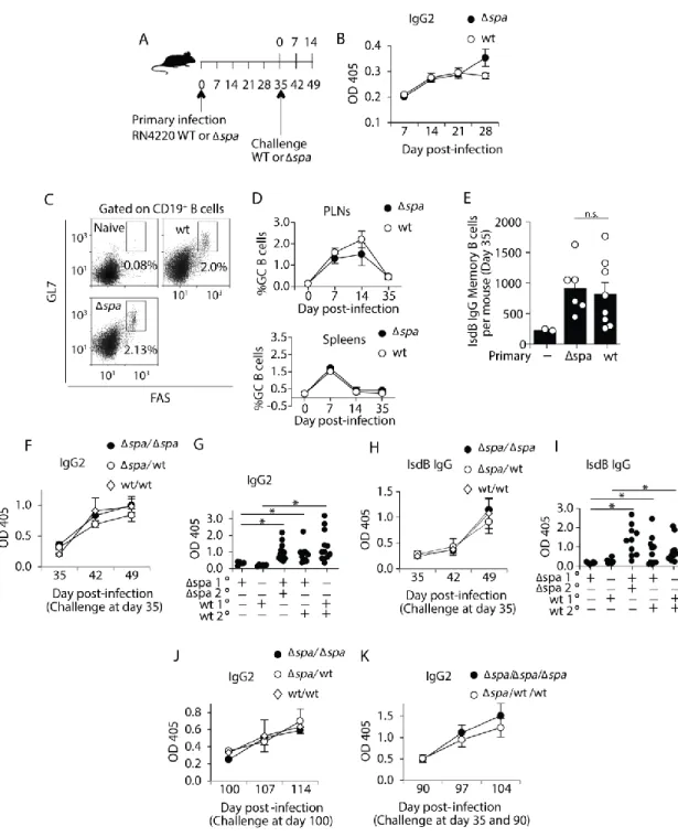
The primary and secondary antibody responses to S. aureus are unaffected by Protein A.
A successful adaptive immune response results in immunological memory that confers long-term protection during subsequent exposure. To assess whether S. aureus induces the formation of memory B cells, and whether this was impacted by Protein A (SpA) we
subcutaneously inoculated mice with strain RN4220 expressing SpA (WT), or an isogenic spa mutant (Δspa). We chose subcutaneous inoculation for the primary response because clearance
of S. aureus could be readily monitored at the injection site. We found that infection was cleared within 3 weeks and the mild virulence of RN4220 allowed inoculated mice to be analyzed over a long period of time without significant weight loss, morbidity, or depletion of B cells
(Supplemental Figure 1A). To characterize the primary immune response, we measured the
total S. aureus-specific IgG and IgG2 over time (Figure 1A). A modest level of IgG2 (Figure 1B) and total IgG (Supplemental Figure 1B) was detectable between 2 and 5 weeks after primary inoculation regardless of whether mice were inoculated with S. aureus Δspa (1.5-fold) or WT (2-fold).
The increase of S. aureus-specific IgG between the first and second weeks after primary inoculation suggested that a T-dependent immune response had induced class switch. GCs are the sites of class switching and affinity maturation, and are required for the formation of high-affinity memory B cells [9,130,195]. To define whether S. aureus induced GCs, we quantitated GC B cells over the course of the primary response. Subcutaneous inoculation with S. aureus RN4220 Δspa or WT induced GC B cells in the draining lymph nodes and spleen with similar
49
which are required for GC development [3,95] were also induced in similar frequencies after primary inoculation with S. aureus Δspa or WT (Supplemental Figure 1C).
One possible mechanism underlying the lack of long-term immunity to S. aureus is failure to promote the formation of memory B cells. We used ELISpot to indirectly detect the numbers of S. aureus antigen-specific memory B cells after in vitro polyclonal stimulation as no flow cytometry methods are available. IsdB was chosen as the target antigen since anti-IsdB IgG has been detected in infected mice, and has recently been studied in vaccination trials [84,173,196]. On day 35 post-inoculation, ex vivo lymphocytes from the spleens and lymph nodes were polyclonally activated with pokeweed mitogen. The 6-day culture period is too short to induce class-switch, thus all antigen-specific IgG-secreting cells detected by ELISpot were derived from memory B cells [183,184,189]. We detected significantly greater numbers of class-switched IsdB-specific ASCs in both Δspa and WT-inoculated mice compared to naïve mice
(Figure 1E), suggesting that memory B cells were formed as a result of infection in the presence or absence of SpA. Together these data suggest that the lack of long-term protection against S. aureus is not due to failure to form memory B cells.
Another possibility is that memory B cells develop normally, but fail to be reactivated upon re-infection. Memory B cell responses are more rapid and robust than primary responses to the same stimuli [1,22,197]. To test whether memory B cells are activated upon secondary exposure to S. aureus, we inoculated mice a second time (herein referred to as challenge) and measured antibody responses. We challenged mice intravenously (IV) with S. aureus RN4420 WT or Δspa to mimic sepsis conditions, and measured antibody responses over two weeks
50
IgG2 responses whether the primary inoculation was WT or Δspa (Figure 1F and
Supplemental 1D).
To ensure that the enhanced and rapid IgG response induced by IV challenge was
dependent on previous infection rather than initiating a new primary response, we compared IgG and IgG2 responses on day 49 (14 post-challenge) to those produced 14 days after a primary IV infection. Consistent with memory cell activation IV challenge of previously inoculated mice induced significantly higher levels of IgG2 and IgG compared to naïve mice (Figure 1G and Supplemental 1E). In addition, we found that challenge with either WT or Δspa S. aureus
induced amnestic IgG responses to IsdB (Figure 1H), that were also dependent on previous infection (Figure 1I). To ensure that the secondary response was not unique to RN4220, we subcutaneously inoculated and IV challenged mice with the clinical strains, Newman and USA300. We observed enhanced secondary responses similar to those induced by RN4220
(Supplemental Figure 1F).
A hallmark of memory B cells is their longevity [21]. It is possible that the memory B cells formed after S. aureus infection are not long-lived, thus diminishing long-term immunity. To assess this, we challenged mice 100 days after primary inoculation and found that challenge with WT or Δspa induced an amnestic S. aureus-specific antibody response (2-3 fold) (Figure
51
SpA, long-lived memory B cells are formed and activated after S. aureus inoculation and challenge.
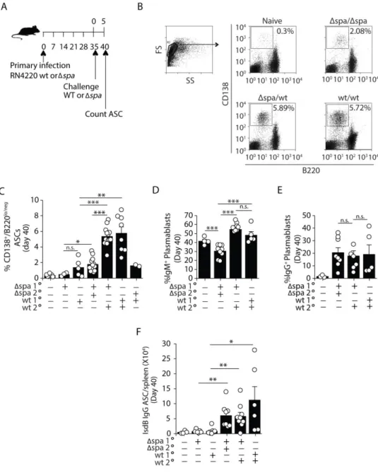
The presence of Protein A during challenge preferentially expands plasmablasts
When memory B cells are activated, they rapidly divide, forming antibody-secreting PBs (CD138+ B220lo/neg surface Ig+) that mature into plasma cells (PCs) (CD138+ B220neg surface Ig-) [22,24,32,33,198]. To assess whether SpA impacted the PC response, we enumerated PBs and PCs (collectively referred to herein as ASCs) [199]. Mice challenged (IV) with either WT or Δspa S. aureus made more total ASCs compared to naïve mice given a primary IV infection
(Figure 2A-C and Supplemental Figure 2A). However, the frequencies and numbers of total
splenic ASCs were significantly higher after WT challenge. This expansion was induced by WT challenge regardless of whether mice were initially inoculated with WT or Δspa (Figure 2B-C and Supplemental Figure 2A), and was unrelated to changes in total CD19+/B220+ B cells
(Supplemental Figure 2B).
52
2D). The frequency of ASCs retaining IgG was not significantly different between the two groups (20.5% of Δspa and 17.8% of WT) (Figure 2E). These data indicate that most of the
expanded ASCs were derived from IgM+ cells suggesting they were induced through the F(ab)-binding activity of SpA.
To assess whether expansion of IgM+ cells impacted the antigen-specific IgG response, we quantitated the class-switched IsdB-specific ASC response by ELISpot on day 40 (day 5 post-challenge) (Figure 2A). Consistent with a memory response (Figure 1E), we found that mice challenge with S. aureus RN4220 Δspa or WT, produced 2-6 times more IsdB-specific IgG ASCs than naive mice that were inoculated IV on the same day; however, the numbers of IsdB-specific ASCs did not differ between mice challenged with WT versus Δspa (Figure 2F). Collectively, these data suggest that long-lived, memory cells are formed regardless of whether mice are inoculated with WT or Δspa S. aureus. Upon reactivation, the newly formed PCs
produce comparable amounts of antigen-specific IgG antibody; however, challenge with WT S. aureus promotes a robust expansion of IgM+ PBs.
SpA is a known virulence factor of S. aureus and kidney bacterial load is reduced after infection with S. aureus Δspa compared to WT [204–206]. Thus, it is possible that the reduced virulence of Δspa limits the expansion or IgM+
PBs [207,208]. To test this, we challenged mice with S. aureus Newman WT, Δspa, or ΔsrrAB, a mutant that expresses SpA but displays a virulence defect similar to that of Δspa (Supplemental Figure 3A). Challenge with S. aureus ΔsrrAB induced expansion of IgM+
53
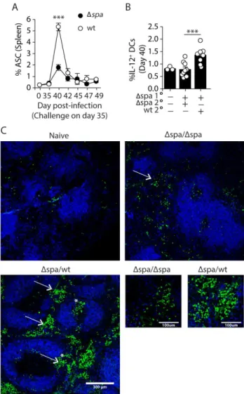
The presence of Protein A during challenge enhances the short-lived extrafollicular response
IgM+ ASC responses are typically short-lived [8,38,39]. To determine whether the expanded ASCs induced by SpA were short-lived, we monitored CD138+ B220lo/neg cells by flow cytometry over the course of two weeks. We found that the frequencies and number of ASCs peaked in the spleens of both WT and Δspa-challenged mice on day 40 (day 5 post-challenge)
and declined by day 42 (day 7 post-challenge) (Figure 3A). This is consistent with the expanded PB population consisting mainly of short-lived cells.
Short-lived responses are typically extrafollicular [30,209,210], so to examine whether the ASC expansion induced by WT challenge was extrafollicular, we first examined splenic DCs, which are key mediators of extrafollicular responses that carry intact antigen into
lymphoid organs and produce cytokines conducive to PB differentiation [201,202,211–213]. We analyzed the frequencies of classical and plasmacytoid DC subsets and their expression of
surface markers associated with activation (CD80, CD86, MHCII, CD11c and CD40). We found no significant differences in the DC populations induced by WT or Δspa challenge (data not
shown). TLR2 ligands expressed duringS. aureus infection induce DCs to make IL-12, a
cytokine that promotes ASC differentiation [197,214–217]. IL-12+ DCs are expanded in immunized Fcγ-/- mice, indicating a role for immune complexes (ICs) in regulating DC IL-12 [148]. Because IC binding to FcγRs is impeded by SpA’s Fc-binding function, we quantitated IL-12-producing CD11c+ DCs on day 40 (day 5 post-challenge). We found that WT and ΔsrrAB challenge induced 1.7-fold more IL-12+ DCs than Δspa challenge (Figure 3B and