UC San Francisco
UC San Francisco Previously Published Works
Title
Is Hip Abduction Strength Asymmetry Present in Female Runners in the Early Stages of
Patellofemoral Pain Syndrome?
Permalink
https://escholarship.org/uc/item/8c32m8hp
Journal
The American Journal of Sports Medicine, 44(1)
ISSN
0363-5465
Authors
Plastaras, C
McCormick, Z
Nguyen, C
et al.
Publication Date
2015-11-13
DOI
10.1177/0363546515611632
Peer reviewed
eScholarship.org
Powered by the California Digital Library
http://ajs.sagepub.com/
Medicine
The American Journal of Sports
http://ajs.sagepub.com/content/44/1/105
The online version of this article can be found at:
DOI: 10.1177/0363546515611632
2016 44: 105 originally published online November 13, 2015
Am J Sports Med
Carneiro, Andrew Cucchiara, Joel Press, Jim McLean and Franklin Caldera
Christopher Plastaras, Zack McCormick, Cayli Nguyen, Monica Rho, Susan Hillary Nack, Dan Roth, Ellen Casey, Kevin
Patellofemoral Pain Syndrome?
Is Hip Abduction Strength Asymmetry Present in Female Runners in the Early Stages of
Published by:
http://www.sagepublications.com
On behalf of:
American Orthopaedic Society for Sports Medicine
can be found at:
The American Journal of Sports Medicine
Additional services and information for
http://ajs.sagepub.com/cgi/alerts
Email Alerts:
http://ajs.sagepub.com/subscriptions
Subscriptions:
http://www.sagepub.com/journalsReprints.nav
Reprints:
http://www.sagepub.com/journalsPermissions.nav
Permissions:
What is This?
- Nov 13, 2015
OnlineFirst Version of Record
- Jan 4, 2016
Version of Record
Is Hip Abduction Strength Asymmetry
Present in Female Runners in the Early
Stages of Patellofemoral Pain Syndrome?
Christopher Plastaras,* MD, Zack McCormick,
zMD, Cayli Nguyen,
§DO, Monica Rho,
zMD,
Susan Hillary Nack,
zDO, Dan Roth,
||DO, Ellen Casey,
{MD, Kevin Carneiro,
#DO,
Andrew Cucchiara,* Joel Press,
zMD, Jim McLean,
yzMD, and Franklin Caldera,* ** DO, MBA
Investigation performed at the Rehabilitation Institute of Chicago, Chicago, Illinois, USA
Background:The current literature indicates that hip abduction weakness in female patients is associated with ipsilateral
patel-lofemoral pain syndrome (PFPS) as part of the weaker hip abductor complex. Thus, it has been suggested that clinicians should consider screening female athletes for hip strength asymmetry to identify those at risk of developing PFPS to prevent the con-dition. However, no study to date has demonstrated that hip strength asymmetry exists in the early stages of PFPS.
Purpose:To determine whether hip abduction strength asymmetry exists in female runners with early unilateral PFPS, defined as
symptoms of PFPS not significant enough to cause patients to seek medical attention or prevent them from running at least 10 miles per week.
Study Design:Controlled laboratory study.
Methods:This study consisted of 21 female runners (mean age, 30.5 years; range, 18-45 years) with early unilateral PFPS, who
had not yet sought medical care and who were able to run at least 10 miles per week, and 36 healthy controls comparably bal-anced for age, height, weight, and weekly running mileage (mean, 18.5 mi/wk). Study volunteers were recruited using flyers and from various local running events in the metropolitan area. Bilateral hip abduction strength in both a neutral and extended hip position was measured using a handheld dynamometer in each participant by an examiner blinded to group assignment.
Results:Patients with early unilateral PFPS demonstrated no significant side-to-side difference in hip abduction strength,
ac-cording to the Hip Strength Asymmetry Index, in both a neutral (mean, 83.5 6 10.2; P= .2272) and extended hip position (mean, 96.3621.9;P= .6671) compared with controls (mean, 87.068.3 [P= .2272] and 96.6616.2 [P= .6671], respectively). Hip abduction strength of the affected limb in patients with early unilateral PFPS (mean, 9.962.2;P= .0305) was significantly stronger than that of the weaker limb of control participants (mean, 8.961.4;P= .0305) when testing strength in a neutral hip position; however, no significant difference was found when testing the hip in an extended position (mean, 7.061.4 [P= .1406] and 6.661.5 [P=.1406], respectively).
Conclusion:The study data show that early stages of unilateral PFPS in female runners is not associated with hip abduction
strength asymmetry and that hip abduction strength tested in neutral is significantly greater in the affected limb in the early stages of PFPS compared with the unaffected limb. However, when tested in extension, no difference exists. Further studies investigat-ing the early stages of PFPS are warranted.
Clinical Relevance:Unlike patients with PFPS seeking medical care, early PFPS does not appear to be significantly associated
with hip abduction strength asymmetry.
Keywords:patellofemoral syndrome; dynamometer; muscle strength; sports injury
Patellofemoral pain syndrome (PFPS) is one of the most common orthopaedic injuries, with a reported incidence of 8.5% to 15%,1,3,5,6accounting for approximately 25% of all knee injuries.8PFPS is particularly common in women, with a female:male ratio of 2:1 to 3:1,2,4and it is character-ized by altered patellar tracking that leads to increased
forces within the patellofemoral joint during flexion. Vastus medialis weakness; decreased gastrocnemius, soleus, hamstring, quadriceps, or iliotibial band flexibility; patella alta; femoral anteversion; excessive femoral inter-nal rotation; a shallow femoral trochlear groove; excessive subtalar joint pronation; and leg-length discrepancy have all been proposed to contribute to PFPS.yy
yy
References 7, 10, 12, 16, 22, 23, 25-29, 31, 33, 35, 37. The American Journal of Sports Medicine, Vol. 44, No. 1
DOI: 10.1177/0363546515611632
Ó2015 The Author(s)
5-in-5
105
at UCSF LIBRARY & CKM on August 16, 2016
ajs.sagepub.com
Gluteal weakness has been implicated in other knee pain syndromes such as iliotibial band syndrome.14
Recently, hip abduction weakness has been implicated in PFPS. Hip abduction weakness causes excessive internal rotation of the leg,37which predisposes the patella to track more laterally in the patellofemoral groove, potentially leading to PFPS. Dierks et al9 and Ireland et al17 found that female patients with PFPS had, respectively, 12% and 26% less hip abduction strength in the affected side compared with the hip strength of asymptomatic controls. Building on this evidence, Baldon Rde et al2found a 28%
reduction in eccentric hip abduction strength in female patients with PFPS compared with healthy controls, and Souza and Powers32found a 22% decrease in hip abduction strength during running in similar groups. Training of hip abductor muscles in the early phases of the rehabilitation of PFPS has been shown to decrease symptoms more quickly than does quadriceps training.11
More relevant to clinicians, as the standard physical examination compares side-to-side differences to deter-mine muscular strength, Magalhaes et al,21 Robinson and Nee,30and Cichanowski et al5investigated hip
abduc-tion strength asymmetry in female patients and reported 12% to 22% reduction in hip strength of the leg with PFPS compared with the unaffected side. The only study to date that demonstrated no asymmetry in hip abduction strength in patients with PFPS compared with healthy controls was reported by Piva et al,25 but this study was unblinded and included a coeducational population with bilateral symptoms. Thus, the current body of literature indicates that in female patients, unilateral patellofemoral pain is associated with hip abduction weakness on the same side.
The authors of this body of literature concluded that clinicians should consider screening female athletes for hip strength weakness or asymmetry as a means of pre-venting PFPS. However, to the best of our knowledge, no study to date has demonstrated that hip strength asymme-try exists in the early stages of PFPS before the utilization of health care. Our study investigated athletes before symptoms became significant enough to require medical attention.
We investigated hip abduction strength asymmetry in a population of female athletes with patellofemoral pain who had not yet sought medical care and who were func-tionally active (running at least 10 mi/wk). Studying this population with early symptoms allowed us to determine whether hip strength asymmetry exists before developing more significant PFPS symptoms that require the
utilization of health care. Therefore, we posed the following question: Is there a role for screening female athletes for hip strength asymmetry to identify those in the early stages of PFPS? We hypothesized that female patients with early PFPS would demonstrate hip abduction strength asymmetry, which would support the consensus recommendation for clinicians to consider hip strength asymmetry as a means of identifying those in the early stages of PFPS before they require health care.
METHODS
Participants
Approval was obtained from the University of Pennsylva-nia Institutional Review Board (IRB) and the Northwest-ern University IRB for the described study protocol, and approval was obtained from the University of Pennsylva-nia IRB for subsequent statistical analysis. This was a sin-gle, blinded case-control study including 21 female runners with early unilateral PFPS and 36 healthy female runners who served as controls.
A power analysis was performed that estimated that 5 patients and 5 controls would provide 80% power to detect a clinically significant difference of 18% in side-to-side hip asymmetry in patients with PFPS compared with controls, estimated as the mean difference for the combined partici-pants reported in the reviewed literature.6,30,34Our enrolled sample of 21 patients with early unilateral PFPS and 36 controls could detect a difference in hip abduction strength asymmetry of 8% with the targeted 80% power, which is a difference less than that of the smallest difference reported in the literature at 12%.6,30,34
Procedures
Study volunteers were recruited using IRB-approved recruit-ment flyers as well as recruitrecruit-ment in the vendor section at various local road races, half marathons, triathlons, and run-ning club meetings in the metropolitan area, and each was provided US$30 reimbursement for participating. The text on the recruitment flyer read, ‘‘Are you an avid female run-ner between 18-45? Do you suffer from pain in the front knee while running? The purpose of the study is to examine the relationship between hip strength and PFPS. PFPS is pain in the front of the knee or behind the kneecap that occurs with running, prolonged sitting, or when climbing
**Address correspondence to Franklin Caldera, DO, MBA, Physical Medicine and Rehabilitation, University of Pennsylvania, 1800 Lombard Street, Phil-adelphia, PA 19146, USA (email: [email protected]).
*Physical Medicine and Rehabilitation, University of Pennsylvania, Philadelphia, Pennsylvania, USA.
y
Deceased.
z
Physical Medicine and Rehabilitation, Rehabilitation Institute of Chicago, Chicago, Illinois, USA. §Brown Hand Center, Austin, Texas, USA.
||Centers for Pain Relief, Fort Wayne, Indiana, USA.
{
Drexel University College of Medicine, Philadelphia, Pennsylvania, USA.
#Physical Medicine & Rehabilitation, University of North Carolina at Chapel Hill, Chapel Hill, North Carolina, USA.
One or more of the authors has declared the following potential conflict of interest or source of funding: This research was supported by the University of Pennsylvania Clinical and Translational Research Center (grant UL1-RR-024134) and the Rehabilitation Institute of Chicago.
stairs. Whether you have knee pain or not, you may qualify for participation in this research study.’’ An IRB-approved consent form was reviewed with potential participants orally, and written consent was obtained from the participant if she enrolled. The consent introduction read, ‘‘You are asked to take part in a research study. The purpose of the study is to determine the relationship between hip strength and patellofemoral syndrome. You are asked to take part in this study because we want to know more about the relationship between hip strength and patellofemoral syndrome.’’
Prescreening for inclusion and exclusion criteria (see below) was performed over the telephone before study vol-unteers were later tested at the Rehabilitation Institute of Chicago. After telephone prescreening, an assessment of study volunteers was performed at the Rehabilitation Institute of Chicago using 4 stations to gather further nec-essary data to either include or exclude volunteers from analysis according to the inclusion and exclusion criteria (Figure 1).
At the first station, participants filled out a biographical questionnaire, which included their age, their ball-kicking leg preference to determine leg dominance, and the Ante-rior Knee Pain Questionnaire (AKPQ),20which is reliable and sensitive to clinical change for PFPS.18,36
At the second station, participants underwent hip strength testing by a single trained examiner blinded to groups (symptomatic or control). Hip strength in neutral and extended hip positions was tested. Hip abduction in extension is considered to preferentially recruit the gluteus medius, while hip abduction in neutral recruits the tensor fasciae latae more.14The preliminary pilot studies of one of the authors (J.M.) suggested that there might be a differ-ence between the 2 positions in the female population; thus, this study examined female athletes only using these 2 hip positions. In a side-lying position, each participant’s hip abduction strength was tested in a neutral and
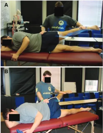
extended hip position, with a handheld dynamometer (manual muscle testing system No. 01163; Layfayette Instruments). The trunk, hips, and legs were in neutral alignment and parallel to the long axis of the table with the left and right anterior superior iliac spine aligned ver-tically. The participant’s downward-facing arm was placed underneath her head, and the participant’s upward-facing arm grasped the table (Figure 2). Three measurements were taken on each side with the hip in approximately 30°of abduction, with 5 seconds of rest between each mea-surement. The examiner stabilized the hip and placed the dynamometer 5 cm proximal to the lateral malleolus while the participant abducted the hip with maximal effort for 3 seconds. In the neutral position, the legs remained verti-cally aligned during testing, while in the extended posi-tion, the top leg was placed in 15° of extension before testing hip abduction strength. The order of testing and the first side tested were both randomized by Maple soft-ware (Cybernet Systems Co Ltd).
At the third station, an examiner blinded to the group designation measured the height, weight, and leg-length discrepancy of the participants. Leg-length discrepancy was measured with the participant in a neutral side-lying position. The distance from the anterior superior iliac spine to the medial malleolus was measured with a tape measure. Recruitment at local road races, half marathons,
triathlons, and running club meetings
72 women considered for the study
62 women accepted to be assessed
26 PFPS subjects examined, 5 excluded by physical examination:
• 2 patellar tendonopathy • 1 anterior cruciate ligament tear • 1 iliotibial band syndrome • 1 bilateral meniscal tears
21 PFPS subjects enrolled 36 control subjects enrolled 4 control subjects excluded:
1 had a patellar fracture by history 6 symptomatic subjects excluded:
• 4 bilateral knee pain • 1 no pain with running • 1 PFPS by history
Telephone prescreening for inclusion/exclusion criteria by history
Figure 1. Flowchart for enrollment of study participants.
PFPS, patellofemoral pain syndrome.
Figure 2. Hip abduction strength testing in (A) neutral hip
position and (B) extended hip position.
AJSM Vol. 44, No. 1, 2016 Hip Abduction Strength Asymmetry and Female Runners 107
at UCSF LIBRARY & CKM on August 16, 2016
ajs.sagepub.com
At the fourth station, participants underwent a focused physical examination of the knee by a different blinded examiner. The examiner performed a physical examination of the knee, which included an inspection, Lachman test, anterior drawer test, varus and valgus stress test, medial and lateral joint line palpation, McMurray test, and palpa-tion of the patellar borders. Once these data were collected, participants were grouped as those with PFPS or controls for analysis, or they were excluded from analysis based on the criteria described in the following section.
Study Inclusion and Exclusion Criteria
Participants were grouped into the early unilateral PFPS group if they met all of the following criteria: (1) female sex; (2) 18 to 45 years of age; (3) running at least 10 miles per week; (4) reporting unilateral anterior knee pain asso-ciated with running at a frequency of least once per week that had occurred for at least 6 weeks; and (5) exhibiting pain with compression of the patella into the femoral con-dyles, pain with palpation of the anterior surface of the patella, or pain with a single-leg squat (Table 1). Alterna-tively, participants were grouped into the control group if they met only the first 3 above inclusion criteria.
Participants were excluded from the early unilateral PFPS group if they met any of the following criteria: (1) pain syndrome in the knee or lower extremity other than PFPS; (2) bilateral knee pain; (3) history of patellar dislo-cations; (4) ligamentous or meniscal injuries in the knee; (5) history of surgery or trauma in either the knee or lower extremity; (6) physical examination results consistent with bilateral PFPS; (7) ligamentous injuries as evidenced by a positive Lachman test finding, anterior drawer, posterior drawer, varus instability, or valgus instability; or (8) phys-ical examination results consistent with meniscal injuries as evidenced by medial or lateral joint line tenderness or a positive McMurray test finding (Table 1). Participants were excluded from the control group if they (1) reported unilateral anterior knee pain associated with running at a frequency of at least once per week that had occurred for at least 6 weeks, (2) exhibited a physical examination finding consistent with PFPS or any other chronic overuse injury, (3) reported a history of PFPS in the past 6 months, or (4) met any of the exclusion criteria applied to the early unilateral PFPS group listed in the first half of this paragraph.
Statistical Analysis
Patients with early unilateral PFPS and controls were comparably balanced with respect to variables believed to be potential confounders, including age, height, weight, miles run per week, and leg-length discrepancy. A Student
ttest of independent variables was applied to confirm that an unacceptable balance was not observed between the 2 cohorts with respect to the aforementioned potential con-founders. APvalue\.05 was considered to be statistically significant.
To look for demographic differences between the early unilateral PFPS and control groups, we applied the Stu-dentttest for independent groups to continuous variables and the Pearson x2 test or Fisher exact test for discrete variables. Differences in hip abduction strength asymme-try in the early unilateral PFPS group versus control group were assessed using the Hip Strength Asymmetry Index (HSAI). The HSAI is equal to (weaker hip strength/
stronger hip) 3 100, with strength being determined by
the maximum force measured by a dynamometer during 3 trials of isometric contraction. Differences in the mean absolute hip abduction strength of the affected limb in the PFPS group versus the mean absolute hip abduction strength of the weaker limb in the control group were also calculated. We compared the 2 groups with the Stu-dentttest or Wilcoxon rank-sum test. We report descrip-tive statistics and 95% CIs where appropriate.
RESULTS
Analysis of baseline characteristics of the early unilateral PFPS and control groups failed to achieve statistically sig-nificant differences between the 2 groups with respect to age, height, weight, leg-length discrepancy, or weekly run-ning mileage (Table 2). As expected, the early unilateral PFPS group scored significantly lower on the AKPQ (mean, 85.7610.1;P\.0001) compared with the control group (mean, 97.6 6 5.7; P \ .0001), which indicated a greater degree of anterior knee pain.
Analysis of hip abduction strength asymmetry accord-ing to the HSAI failed to achieve a significant difference in hip abduction asymmetry between the early unilateral PFPS and control groups when tested in both a neutral and extended hip position (P = .2272 and .6671, respec-tively) (Table 3 and Figure 3). Because our study sample size was large enough to find a difference as small as 8% at a power of 80%, this finding of no significant difference between groups indicates that if a difference exists, it is less than 8%. Scatter charts of the data demonstrate the presence of outliers or any unusual data distribution (see
TABLE 1
Physical Examination Findings Used in Inclusion and Exclusion Criteriaa
Physical Examination Findings
Findings consistent with PFPS
Tenderness of the medial or lateral patellar facets
Pain with compression of the patella into the femoral condyles Anterior knee pain with single-leg squat
Findings consistent with a knee injury other than PFPS Positive Lachman test result
Positive posterior drawer test result Positive McMurray test result Medial joint line tenderness Lateral joint line tenderness Valgus or varus instability
aPFPS, patellofemoral pain syndrome.
the Appendix, available in the online version of this article at http://ajsm.sagepub.com/supplemental).
Analysis demonstrated that hip abduction strength of the affected limb in patients with early unilateral PFPS was significantly stronger than that of the weaker limb of controls when testing strength in a neutral hip position; however, no significant difference was found when testing the hip in an extended position (P = .0305 and .1406, respectively) (Tables 4 and 5).
DISCUSSION
The results of our study demonstrated that female runners with early symptoms of PFPS do not appear to have clini-cally significant hip abduction strength asymmetry with respect to the HSAI when compared with healthy controls. This finding indicates that screening for hip abduction strength asymmetry may not be a useful method for iden-tifying those at risk for developing PFPS that might require a health care provider visit, as had been postulated in previous literature.
Our finding of a lack of asymmetry in hip abduction strength between patients with early PFPS and healthy controls was unexpected. This result did not support our original hypothesis that hip strength asymmetry exists before the development of clinically significant PFPS and TABLE 2
Demographics of the PFPS and Control Cohortsa
Characteristic PFPS (n = 21) Control (n = 36) PValue
Age, y 30.566.1 30.4615.2 .512
Height, cm 164.665.8 166.466.6 .154
Weight, kg 62.169.9 62.668.0 .421
Weekly running mileage, mi/wk
18.666.8 18.566.9 .505
Leg-length discrepancy, cm
0.5760.08 0.6560.11 .277
AKPQ 85.7610.1 97.665.7 \.0001b
aResults are reported as mean
6 SD. AKPQ, Anterior Knee
Pain Questionnaire; PFPS, patellofemoral pain syndrome. bStatistically significant difference between groups (P\.05).
TABLE 3
Hip Strength Asymmetry Index of the PFPS and Control Cohortsa
Position PFPS (n = 21) Control (n = 36) PValue
Neutral 83.5610.2
(78.9-88.2)
87.068.3
(84.2-89.8)
.2272
Extension 96.3621.9
(86.3-106.3)
96.6616.2
(91.1-102.1)
.6671
a
Results are reported as mean6SD (95% CI). PFPS,
patellofe-moral pain syndrome.
0 10 20 30 40 50 60 70 80 90 100
Control PFPS
0 10 20 30 40 50 60 70 80 90 100
Control PFPS
Hip Strength Asymmetry Index
Extension Neutral
Figure 3. Mean Hip Strength Asymmetry Index of the hip
abductors for the patellofemoral pain syndrome (PFPS) and control groups, measured with the hip in neutral and in exten-sion. PFPS versus control:P= .2272 (neutral) andP= .6671 (extension). Error bars indicate 95% CI.
TABLE 4
Hip Abduction Strength of the PFPS (Affected/Unaffected Limbs) and Control (Weaker/Stronger Limbs) Cohortsa
PFPS (n = 21) Control (n = 36)
Position Affected Limb Unaffected Limb PValue Weaker Limb Stronger Limb PValue
Neutral 9.962.2 10.061.7 .8071 8.961.4 10.361.7 \.0001b
Extension 7.061.4 7.461.5 .7682 6.661.5 8.161.6 \.0001b
aResults are reported as mean
6SD. PFPS, patellofemoral pain syndrome.
bStatistically significant difference between weaker and stronger limbs (P\.05).
TABLE 5
Hip Abduction Strength of the PFPS (Affected Limb) and Control (Weaker Limb) Cohortsa
Position PFPS, Affected Limb (n = 21) Control, Weaker Limb (n = 36) PValue
Neutral 9.962.2 (8.9-11.0) 8.961.4 (8.4-9.4) .0305b
Extension 7.061.4 (6.4-7.7) 6.661.5 (6.1-7.1) .1406
aResults are reported as mean
6SD (95% CI). PFPS, patellofemoral pain syndrome.
bStatistically significant difference between affected and weaker limbs (P\.05).
AJSM Vol. 44, No. 1, 2016 Hip Abduction Strength Asymmetry and Female Runners 109
at UCSF LIBRARY & CKM on August 16, 2016
ajs.sagepub.com
likely contributes to the progression of PFPS. An explana-tion of this finding is left to conjecture and thus needs further study to understand the mechanisms and patho-physiology that are associated with the early evolution of patellofemoral pain. Perhaps the absence of asymmetry could be explained by the time that it takes to clinically detect strength differences due to chronic overcompensa-tion by the opposite hip. Runners with PFPS may favor their unaffected side to unload force on the painful knee, thus leading to increased strength in the favored side and decreased strength in the affected side. This measur-able strength imbalance would likely take time to develop and therefore may not be measurable until later stages of PFPS. This theory is supported by the current body of lit-erature that demonstrates hip strength asymmetry at clin-ically significant stages of PFPS19,24 in conjunction with
our finding of a lack of hip strength asymmetry relative to healthy controls at early stages of the condition. Another postulated mechanism is that gluteal weakness may develop later in the course of PFPS because of pain- or effusion-induced inhibition of nerve firing. The weakness of hip external rotators (gluteus maximus and the 6 deep lateral rotators) may also contribute as a risk factor of PFPS, but our study did not investigate these other muscles specifically. Knee joint effusion has been described to cause quadriceps weakness secondary to neural inhibi-tion.13,15 Potentially, a similar neural inhibitory
mecha-nism of the hip musculature could be activated by knee pain or effusion in PFPS; this connection remains to be explored.
The results of our study also demonstrated no signifi-cant difference in hip abduction strength of the hip on TABLE 6
Hip Abduction Strength of Dominant Leg Compared With Hip Abduction Strength of Both Legs in Controls
Neutral Extension
Hip Abduction Strength, kg Hip Abduction Strength, kg
Dominant Leg
Left Hip
Right Hip
Hip Stronger on Dominant Side?
Dominant Leg
Left Hip
Right Hip
Hip Stronger on Dominant Side?
Right 10.4 10.8 Yes Right 9.1 9.2 Yes
Right 8.5 8.5 Equal Right 7.6 6.3 No
Right 10.2 10.6 Yes Right 9.5 6.9 No
Right 7.7 9.9 Yes Right 6.4 5.7 No
Right 10.2 12.0 Yes Right 8.9 9.5 Yes
Right 7.4 10.4 Yes Right 6.1 10.1 Yes
Right 8.3 9.3 Yes Right 6.0 6.4 Yes
Right 10.4 9.5 No Right 7.5 6.1 No
Right 7.6 7.7 Yes Right 4.6 4.6 Equal
Left 8.2 9.8 Yes Left 6.8 7.4 Yes
Right 9.8 11.0 Yes Right 8.9 8.2 No
Right 8.5 9.2 Yes Right 8.0 4.4 No
Right 8.6 9.9 Yes Right 6.1 4.9 No
Right 9.2 9.6 Yes Right 8.1 7.1 No
Right 6.0 7.3 Yes Right 4.1 5.8 Yes
Right 8.0 10.1 Yes Right 5.3 5.9 Yes
Right 10.5 14.2 Yes Right 8.3 11.4 Yes
Right 11.3 10.7 No Right 7.7 5.5 No
Right 7.7 9.2 Yes Right 6.7 4.9 No
Right 9.2 9.2 Equal Right 7.0 5.4 No
Right 9.6 13.3 Yes Right 7.3 9.3 Yes
Right 7.9 8.9 Yes Right 6.6 6.7 Yes
Right 6.0 6.6 Yes Right 6.4 4.9 No
Right 10.0 10.7 Yes Right 8.8 6.4 No
Right 8.6 9.2 Yes Right 9.3 6.4 No
Right 11.3 9.9 No Right 9.9 6.8 No
Right 8.9 10.2 Yes Right 7.0 6.0 No
Right 8.0 10.4 Yes Right 8.6 7.1 No
Right 10.9 11.4 Yes Right 10.7 8.9 No
Right 12.1 10.1 No Right 8.1 4.7 No
Right 8.8 10.5 Yes Right 7.4 5.7 No
Right 8.1 9.4 Yes Right 8.1 8.3 Yes
Right 10.1 13.6 Yes Right 7.9 8.6 Yes
Right 7.9 10.4 Yes Right 7.4 8.2 Yes
Right 12.4 12.7 Yes Right 11.2 10.8 No
Right 8.6 7.0 No Right 7.9 7.7 No
the same side as the knee affected by PFPS in women with early PFPS compared with hip abduction strength of the weaker hip of controls when tested in an extended position. However, hip abduction strength of the hip on the same side as the knee affected by PFPS in women with early PFPS was significantly stronger than the weaker hip of controls when tested in a neutral position. Because testing hip abduction strength in a neutral position engages the tensor fasciae latae in addition to the gluteus medius com-pared with testing in an extended position, it may be the case that in early stages of PFPS, the tensor fasciae latae on the affected side is initially strengthened. It is possible that in the early stages of PFPS, the tensor fasciae latae on the affected side is engaged more significantly to unload stress on the knee affected by PFPS, thus leading to initial strengthening of this muscle.
With regard to our study methods, we elected not to use the Limb Symmetry Index (LSI) in our analysis, as done by other authors. In our opinion, the LSI is a less precise method for reporting hip strength asymmetry when com-pared with the HSAI because of 2 incorrect assumptions intrinsic to the LSI formula. First, the LSI for patients with knee pain is calculated based on the assumption that the hip on the same side as the painful knee is weaker. However, this is not always the case, as demonstrated by our data (Table 6). This assumption could bias the LSI to show less strength asymmetry than truly exists in a study population, once averaged. Consider 2 patients with a right leg that is truly 10% stronger than the left leg: one with knee pain on the right and the other with knee pain on the left. The average LSI for this group is 100%, so the mean LSI demonstrates no side-to-side hip strength asym-metry when there is truly a mean asymasym-metry of 10%. The second questionable assumption inherent to the LSI for-mula is that the stronger leg in the control group is the dominant kicking leg; however, it is not always the case that the dominant leg is stronger than the opposite limb.14,16,31
To create a more precise measure of hip strength asym-metry than the LSI, we have introduced the HSAI, which ensures that the stronger leg is correctly determined and placed in the denominator of the equation; thus, the HSAI provides the same measure of asymmetry (truly weaker/stronger side) in PFPS and control cohorts rather than assuming that the dominant kicking leg is also the stronger leg. This consistency allows for more precise com-parison between the patient and control groups; therefore, we propose that the HSAI should be used by investigators when reporting side-to-side strength asymmetry assessed with a dynamometer.
We acknowledge that this study is limited by its specific study population and the lack of a true gold standard mea-sure to objectively and clinically meamea-sure hip strength. Our data set demonstrates a lack of association between early PFPS and hip abduction strength asymmetry. How-ever, a large prospective trial would be required to further define the point during the spectrum of the disease at which hip abduction strength asymmetry does in fact develop. This would help answer the ‘‘chicken or the egg’’ question: Does hip abduction strength asymmetry develop
because of PFPS, or does it cause progression of PFPS symptoms? Moreover, we studied only female runners aged 18 to 45 years, which limits the generalizability of our results beyond female runners in this age range.
CONCLUSION
Our results indicate that, unlike patients with PFPS seek-ing medical care, early PFPS does not appear to be signif-icantly associated with hip abduction strength asymmetry.
ACKNOWLEDGMENT
The authors thank Andrew Cucchiara, PhD, Biostatistics, University of Pennsylvania (Philadelphia, Pennsylvania), and Jungwha Lee, PhD, MPH, Preventive Medicine, Bio-statistics Collaboration Center, Northwestern University (Chicago, Illinois).
REFERENCES
1. Arroll B, Ellis-Pegler E, Edwards A, Sutcliffe G. Patellofemoral pain syndrome: a critical review of the clinical trials on nonoperative ther-apy.Am J Sports Med. 1997;25(2):207-212.
2. Baldon Rde M, Nakagawa TH, Muniz TB, Amorim CF, Maciel CD, Serra˜o FV. Eccentric hip muscle function in females with and without patellofemoral pain syndrome.J Athl Train. 2009;44(5):490-496. 3. Boling M, Padua D, Marshall S, Guskiewicz K, Pyne S, Beutler A.
Gender differences in the incidence and prevalence of patellofemoral pain syndrome.Scand J Med Sci Sports. 2010;20(5):725-730. 4. Caylor D, Fites R, Worrell TW. The relationship between quadriceps
angle and anterior knee pain syndrome.J Orthop Sports Phys Ther. 1993;17(1):11-16.
5. Cichanowski HR, Schmitt JS, Johnson RJ, Niemuth PE. Hip strength in collegiate female athletes with patellofemoral pain.Med Sci Sports Exerc. 2007;39(8):1227-1232.
6. Crossley KM, Bennell KL, Cowan SM, Green S. Analysis of outcome measures for persons with patellofemoral pain: which are reliable and valid?Arch Phys Med Rehabil. 2004;85(5):815-822.
7. DeHaven KE, Lintner DM. Athletic injuries: comparison by age, sport, and gender.Am J Sports Med. 1986;14(3):218-224.
8. Devereaux MD, Lachmann SM. Patello-femoral arthralgia in athletes attending a sports injury clinic.Br J Sports Med. 1984;18(1):18-21. 9. Dierks TA, Manal KT, Hamill J, Davis IS. Proximal and distal
influen-ces on hip and knee kinematics in runners with patellofemoral pain during a prolonged run.J Orthop Sports Phys Ther. 2008;38(8):448-456.
10. Ditroilo M, Forte R, Benelli P, Gambarara D, De Vito G. Effects of age and limb dominance on upper and lower limb muscle function in healthy males and females aged 40-80 years. J Sports Sci. 2010;28(6):667-677.
11. Dolak KL, Silkman C, Medina McKeon J, Hosey RG, Lattermann C, Uhl TL. Hip strengthening prior to functional exercises reduces pain sooner than quadriceps strengthening in females with patellofemoral pain syndrome: a randomized clinical trial. J Orthop Sports Phys Ther. 2011;41(8):560-570.
12. Eckhoff DG, Brown AW, Kilcoyne RF, Stamm ER. Knee version asso-ciated with anterior knee pain.Clin Orthop Relat Res. 1997;339:152-155.
13. Fahrer H, Rentsch HU, Gerber NJ, Beyeler C, Hess CW, Gru¨nig B. Knee effusion and reflex inhibition of the quadriceps: a bar to effec-tive retraining.J Bone Joint Surg Br. 1988;70(4):635-638.
AJSM Vol. 44, No. 1, 2016 Hip Abduction Strength Asymmetry and Female Runners 111
at UCSF LIBRARY & CKM on August 16, 2016
ajs.sagepub.com
14. Fredericson M, Cookingham CL, Chaudhari AM, Dowdell BC, Oestreicher N, Sahrmann SA. Hip abductor weakness in distance runners with iliotibial band syndrome.Clin J Sport Med. 2000;10(3): 169-175.
15. Hopkins JT. Knee joint effusion and cryotherapy alter lower chain kinetics and muscle activity.J Athl Train. 2006;41(2):177-184. 16. Hvid I, Andersen LI. The quadriceps angle and its relation to femoral
torsion.Acta Orthop Scand. 1982;53(4):577-579.
17. Ireland ML, Willson JD, Ballantyne BT, Davis IM. Hip strength in females with and without patellofemoral pain.J Orthop Sports Phys Ther. 2003;33(11):671-676.
18. Kendall FP, McCreary EK, Provance PG, et al.Muscles: Testing and Function With Posture and Pain. 5th ed. Baltimore, Maryland: Lippin-cott Williams & Wilkins; 2005.
19. Kubo T, Muramatsu M, Hoshikawa Y, Kanehisa H. Profiles of trunk and thigh muscularity in youth and professional soccer players.
J Strength Cond Res. 2010;24(6):1472-1479.
20. Kujala UM, Jaakkola LH, Koskinen SK, Taimela S, Hurme M, Nelimarkka O. Scoring of patellofemoral disorders. Arthroscopy. 1993;9(2):159-163.
21. Magalhaes E, Fukuda TY, Sacramento SN, Forgas A, Cohen M, Abdalla RJ. A comparison of hip strength between sedentary females with and without patellofemoral pain syndrome.J Orthop Sports Phys Ther. 2010;40(10):641-647.
22. Maupas E, Paysant J, Datie AM, Martinet N, Andre´ JM. Functional asymmetries of the lower limbs: a comparison between clinical assessment of laterality, isokinetic evaluation and electrogoniometric monitoring of knees during walking. Gait Posture. 2002;16(3): 304-312.
23. Milgrom C, Finestone A, Eldad A, Shlamkovitch N. Patellofemoral pain caused by overactivity: a prospective study of risk factors in infantry recruits.J Bone Joint Surg Am. 1991;73(7):1041-1043. 24. Palmieri RM, Weltman A, Edwards JE, et al. Pre-synaptic modulation
of quadriceps arthrogenic muscle inhibition.Knee Surg Sports Trau-matol Arthrosc. 2005;13(5):370-376.
25. Piva SR, Goodnite EA, Childs JD. Strength around the hip and flexi-bility of soft tissues in individuals with and without patellofemoral pain syndrome.J Orthop Sports Phys Ther. 2005;35(12):793-801.
26. Plastaras CT, Rittenberg JD, Rittenberg KE, Press J, Akuthota V. Comprehensive functional evaluation of the injured runner. Phys Med Rehabil Clin N Am. 2005;16(3):623-649.
27. Powers CM. Patellar kinematics, part II: the influence of the depth of the trochlear groove in subjects with and without patellofemoral pain.
Phys Ther. 2000;80(10):965-978.
28. Powers CM, Ward SR, Fredericson M, Guillet M, Shellock FG. Patel-lofemoral kinematics during weight-bearing and non-weight-bearing knee extension in persons with lateral subluxation of the patella: a preliminary study.J Orthop Sports Phys Ther. 2003;33(11):677-685. 29. Reikera˚s O. Patellofemoral characteristics in patients with increased
femoral anteversion.Skeletal Radiol. 1992;21(5):311-313.
30. Robinson RL, Nee RJ. Analysis of hip strength in females seeking physical therapy treatment for unilateral patellofemoral pain syn-drome.J Orthop Sports Phys Ther. 2007;37(5):232-238.
31. Sanchis-Alfonso V, Rosello-Sastre E, Martinez-Sanjuan V. Pathogen-esis of anterior knee pain syndrome and functional patellofemoral instability in the active young.Am J Knee Surg. 1999;12(1):29-40. 32. Souza RB, Powers CM. Differences in hip kinematics, muscle
strength, and muscle activation between subjects with and without patellofemoral pain.J Orthop Sports Phys Ther. 2009;39(1):12-19. 33. Souza RB, Powers CM. Predictors of hip internal rotation during
run-ning: an evaluation of hip strength and femoral structure in women with and without patellofemoral pain.Am J Sports Med. 2009;37(3): 579-587.
34. Tiberio D. The effect of excessive subtalar joint pronation on patello-femoral mechanics: a theoretical model.J Orthop Sports Phys Ther. 1987;9(4):160-165.
35. Timm KE. Randomized controlled trial of Protonics on patellar pain, position, and function.Med Sci Sports Exerc. 1998;30(5):665-670. 36. Watson CJ, Propps M, Ratner J, Zeigler DL, Horton P, Smith SS.
Reliability and responsiveness of the lower extremity functional scale and the anterior knee pain scale in patients with anterior knee pain.
J Orthop Sports Phys Ther. 2005;35(3):136-146.
37. Witvrouw E, Lysens R, Bellemans J, Cambier D, Vanderstraeten G. Intrinsic risk factors for the development of anterior knee pain in an athletic population: a two-year prospective study. Am J Sports Med. 2000;28(4):480-489.
For reprints and permission queries, please visit SAGE’s Web site at http://www.sagepub.com/journalsPermissions.nav