Ultra-long-acting removable drug delivery system
for HIV treatment and prevention
Martina Kovarova
1
, S. Rahima Benhabbour
2
, Ivana Massud
3
, Rae Ann Spagnuolo
1
, Brianna Skinner
4
,
Caroline E. Baker
1
, Craig Sykes
5
, Katie R. Mollan
6
, Angela D. M. Kashuba
5
, J. Gerardo García-Lerma
3
,
Russell J. Mumper
2
& J. Victor Garcia
1
Non-adherence to medication is an important health care problem, especially in the
treat-ment of chronic conditions. Injectable long-acting (LA) formulations of antiretrovirals (ARVs)
represent a viable alternative to improve adherence to HIV/AIDS treatment and prevention.
However, the LA-ARV formulations currently in clinical trials cannot be removed after
administration even if adverse events occur. Here we show an ultra-LA removable system
that delivers drug for up to 9 months and can be safely removed to stop drug delivery. We
use two pre-clinical models for HIV transmission and treatment, non-human primates (NHP)
and humanized BLT (bone marrow/liver/thymus) mice and show a single dose of
sub-cutaneously administered ultra-LA dolutegravir effectively delivers the drug in both models
and show suppression of viremia and protection from multiple high-dose vaginal HIV
chal-lenges in BLT mice. This approach represents a potentially effective strategy for the ultra-LA
drug delivery with multiple possible therapeutic applications.
DOI: 10.1038/s41467-018-06490-w
OPEN
1Division of Infectious Diseases, Center for AIDS Research, Department of Medicine, School of Medicine, University of North Carolina at Chapel Hill, 120 Mason Farm Rd. CB 7042, Chapel Hill, NC 27599, USA.2Division of Molecular Pharmaceutics, UNC Eshelman School of Pharmacy, University of North Carolina at Chapel Hill, 125 Mason Farm Rd. 4205, Chapel Hill, NC 27599, USA.3Laboratory Branch, Division of HIV/AIDS Prevention, National Center for HIV/AIDS, Viral Hepatitis, STD, and TB Prevention, Centers for Disease Control and Prevention, 1600 Clifton Rd, MS G45, Atlanta, GA 30329, USA. 4Comparative Medicine Branch, Division of Scientific Resources, National Center for Emerging and Zoonotic Infectious Diseases, Centers for Disease Control and Prevention, Atlanta, GA 30329, USA.5Division of Pharmacotherapy and Experimental Therapeutics, UNC Eshelman School of Pharmacy, University of North Carolina at Chapel Hill, 120 Mason Farm Rd. CB#7361, Chapel Hill, NC 27599, USA.6The University of North Carolina Center for AIDS Research, 3126 McGavran-Greenberg Hall, Chapel Hill, NC 27599, USA. Correspondence and requests for materials should be addressed to
M.K. (email:[email protected]) or to J.V.G. (email:[email protected])
123456789
A
dherence to medication is essential to treatment success
1.
In most cases the extent to which patients are able to
follow prescribed treatments determines the
fi
nal
out-come. This is particularly important in the treatment of chronic
conditions like mental illnesses, hypertension, diabetes, and HIV/
AIDS
1. Sustained drug release has successfully improved
adher-ence in patients with schizophrenia
2and as contraceptives
3.
Long-acting (LA) injectable formulations of ARV can increase
adherence and effectiveness of HIV treatment and prevention
4. In
particular, LA formulations can (1) simplify dosing schedules, (2)
reduce possible side effects, (3) provide constant concentration of
drug, and (4) have a positive effect on patient
’
s overall quality of
life. LA-ARV formulations currently in clinical trials, are
for-mulated as nanosuspensions for injections every 8 weeks
5–7and it
is impossible to remove the injected nanosuspension from the
body in the case of a medical emergency.
To address this signi
fi
cant limitation and to extend the
dura-tion of treatment, an ultra-LA injectable and removable
for-mulation for HIV PrEP based on in situ forming implant
technology was developed. This allows drug administration by
subcutaneous injection followed by implant solidi
fi
cation in vivo
and subsequent biodegradation of the implant resulting in
sus-tained drug release
8,9. The ef
fi
cacy of drug delivery via ultra-LA
formulation in the context of HIV is evaluated in BLT humanized
mice, systemically reconstituted with human hematopoietic
cells
10. BLT mice have been extensively used for HIV
transmis-sion, replication, and persistence studies
11–14. Importantly,
humanized BLT mice allow evaluation of HIV treatment and
prevention strategies with a variety of transmitted/founder HIV-1
isolates via relevant routes of transmission.
Results
Preparation of ultra-LA dolutegravir
. Dolutegravir, a highly
effective second-generation HIV integrase strand transfer
inhi-bitor with extensive track record of ef
fi
cacy and safety
15, was used
for preparation of ultra-LA formulation. In addition to active
drug, the formulation consists of two relatively low cost
FDA-approved excipients: (1) poly(lactic-
co
-glycolic acid) (PLGA), a
biodegradable copolymer that eventually and safely biodegrades
and, (2)
N
-methyl-2-pyrrolidone (NMP), a water miscible and
biocompatible organic solvent
16,17. Composition of the ultra-LA
formulation was
fi
rst optimized for release kinetics in vitro and
then sustained delivery of dolutegravir in vivo (Fig.
1
). The
for-mulation contained dolutegravir/PLGA/NMP at a ratio 0.3:1:2 by
weight, had a viscosity 845 cP at 25 °C (Brook
fi
eld Cone and Plate
Digital Rheometer,
n
=
3). Formulation stability was assessed as
the ability to remain in solution (measured by dynamic light
scattering, Zetasizer Nano ZS Particle Analyzer), and to maintain
a stable dolutegravir concentration (98% of the original
b
d
c
a
Time after ultra-LA DTG administration (days)
DTG in plasma (ng/ml)
0 20 40 60 80 100 120 140 Day 7 Day 28 Day 84
104
103
102
101
DTG (ng/ml/g)
Plasma Cervix Uterus Vagina
0 50 100 150 200 250 300 350
105
104
103
102
101
100
102
101
100
Time (days)
DTG in plasma (ng/ml)
Fig. 1Implant formation and dolutegravir concentration in plasma and female reproductive tract.aSolidified ultra-LA dolutegravir surgically removed
concentration) measured by HPLC analysis. Dolutegravir was
chemically stable in this formulation at 25 °C for at least
6 months. In the aqueous environment in vitro (0.01 M PBS, pH
=
7.4, 2% solutol) solidi
fi
cation of the implant was instantaneous.
Pharmacokinetics of ultra-LA dolutegravir
. For an initial
in vivo evaluation, anesthetized female NSG and BLT mice
received a single subcutaneous injection of the ultra-LA
dolute-gravir on their back (5.5
–
7.0 mg dolutegravir in 50
–
80 µl). The
injected formulation
fi
rst formed a hard translucent globule
under the skin that turned yellowish white (within 48 h of
administration) as the formulation solidi
fi
ed. The formulation
was well tolerated by the mice and no injection site reactions or
other signs of overt toxicity, changes in behavior, movement,
water consumption or weight loss were noted. For the purpose of
observing the nature of the implant and to con
fi
rm that it could
be readily removed from the mice, 1 week after administration a
small incision was made near the location of the implant in one of
the mice allowing rapid removal of the implant from the mouse
(Fig.
1
a). The rest of the mice were used for in vivo
pharmaco-kinetic analysis of drug release (Fig.
1
b). Plasma concentrations of
dolutegravir were quantitated using a validated high-performance
liquid chromatography
–
tandem mass spectrometry LC/MS
–
MS
method
18. Non-compartmental analysis of the median composite
pharmacokinetic (PK) pro
fi
le demonstrated a biexponential
decay. After an initial 1st order decline in plasma concentrations,
the release of dolutegravir approached zero-order kinetics. Plasma
concentration of dolutegravir was ten times greater than the
protein adjusted (PA)-IC
90for at least 5 months post
tration. Even at 283 days after ultra-LA dolutegravir
adminis-tration 1/3 mice still had detectable dolutegravir in plasma. We
then used a sparse PK analysis to compare the concentration of
plasma dolutegravir between BLT humanized and
non-humanized mice. Our results demonstrated similar
pharmacoki-netics of dolutegravir in both types of mice (Supplementary
Fig. 1).
Concentration of dolutegravir was also evaluated in the female
reproductive tract (FRT) in 14 NSG mice receiving a single
subcutaneous injection of ultra-LA dolutegravir. Vagina, cervix,
uterus, and plasma from treated mice were collected 1, 4, and
12 weeks post administration and dolutegravir concentrations
determined (Fig.
1
c). One week after ultra-LA dolutegravir
administration, the median concentration of dolutegravir in
plasma, vagina, cervix, and uterus were 1350 ng/ml, 196 ng/mg,
158 ng/mg, and 272 ng/ml, respectively. One month post
administration, the median dolutegravir concentrations were
958 ng/ml, 233 ng/mg, 262 ng/mg, and 303, ng/mg, respectively,
and 12 weeks post administration the median concentrations
were 1200 ng/ml, 356 ng/mg, 170 ng/mg, and 284 ng/mg,
respec-tively (Fig.
1
c, Table
1
). Differences in DTG concentrations
within each compartment (vagina, cervix, uterus, and plasma)
comparing 1 week, 4 weeks, and 12 weeks (
n
=
6 per group) did
not reach statistical signi
fi
cance (Kruskal
–
Wallis test plasma
p
=
0.21, cervix
p
=
0.09, uterus
p
=
0.70, vagina
p
=
0.17).
Observa-tions were combined over weeks 1, 4, and 12 to evaluate whether
plasma concentrations were higher than tissue concentrations for
each tissue type separately. Dolutegravir concentrations in plasma
were higher than in tissue for each of the three tissue types for
every animal (Wilcoxon signed-rank
p
< 0.001 for cervix, uterus,
and vagina analyses, respectively). Together, these results
demonstrate the sustained in vivo release of dolutegravir into
plasma and its ef
fi
cient penetration into tissues of the female
reproductive tract.
To assess safety and drug release pro
fi
le of the ultra-long-acting
formulation of dolutegravir in a large animal model, two rhesus
macaques were subcutaneously administered the ultra-LA
dolutegravir formulation (100 mg). Animals were monitored for
signs of toxicity and skin reactions at the injection site, including
erythema, edema, and hematoma formation, presence of
indura-tion, and any other lesions such as abscesses, necrosis, dehiscence,
or local in
fl
ammation twice a week. The implants were well
tolerated with little or no sign of toxicity for 5 months (last point
analyzed). Administration of the ultra-long-acting formulation of
dolutegravir resulted in sustained dolutegravir concentration in
plasma for more than 140 days (Fig.
1
d). These results
demonstrate the feasibility of ultra-long-acting dolutegravir
delivery system for sustained delivery in rhesus macaques.
However, this formulation originally developed for BLT mice
will have to be further optimized prior to ef
fi
cacy studies in
macaques and its potential future applications in humans.
Inhibition of HIV-1 replication ex vivo
. Having demonstrated
sustained concentration of dolutegravir in plasma of both mice
and macaques and in tissues from mice we proceeded to evaluate
its antiviral activity. Serum obtained from the ultra-LA
dolute-gravir-treated mice in Fig.
1
c demonstrated strong, concentration
dependent antiviral activity (Fig.
2
a). Speci
fi
cally, a one
hundredth-fold dilution of serum was able to block in vitro viral
infection by >86% when collected 7 or 28 days post
administra-tion (median 89%, range 86.4%
–
90.8% and median 86%, range
74.1%
–
87.9% for 7 and 28 days, respectively). A 100-fold dilution
of serum was also able to block HIV infection by 66% when
collected 84 days post ultra-LA dolutegravir administration
(range 63.5%
–
67.0%) (Fig.
2
b). Statistical analysis of the antiviral
activity present in serum demonstrated a strong correlation with
dolutegravir concentrations (Kendall rank correlation coef
fi
cient
0.75; 95% CI: 0.65
–
0.85) (Fig.
2
c).
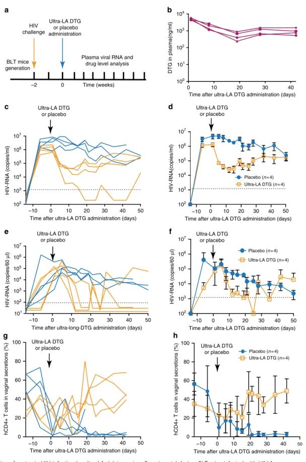
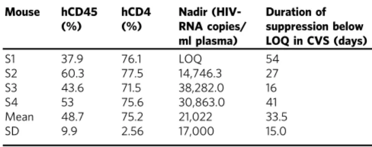
Inhibition of HIV-1 replication in vivo
. To establish the in vivo
inhibitory effect of dolutegravir administered via the ultra-LA
dolutegravir formulation on HIV replication, eight BLT
huma-nized mice were
fi
rst infected intravenously with HIV-1
JR-CSF(3 × 10
4TCIU) (Fig.
3
a, Table
2
). HIV-RNA was readily detected
in plasma from all exposed mice 2 weeks after exposure (median
1.97 × 10
6copies/ml, range 0.65
–
8.50 × 10
6copies/ml). Two
weeks post infection, mice received a single dose of ultra-LA
dolutegravir- or placebo administered via subcutaneous injection
(
n
=
4 for each group). Dolutegravir plasma concentrations in
treated mice were sustained throughout the entire experiment
(Fig.
3
b). Placebo-treated mice maintained high concentration of
plasma HIV-RNA. In contrast, strong suppression of HIV
replication (~1
–
2 log) was noted in all mice treated with the
ultra-LA dolutegravir formulation (Fig.
3
c, d). In one mouse, viral load
fell below the level of quantitation (LOQ, 1375 copies of
HIV-RNA/ml) as early as 2 weeks post administration (Fig.
3
c). Plasma
viral load AUC was smaller in the ultra-long-acting dolutegravir
group compared to the placebo group,
p
=
0.03 (exact
Table 1 Tissue/plasma ratio of dolutegravir in female
reproductive tract
Day 7 Day 28 Day 84
Cervix 0.136 0.277 0.184
Uterus 0.191 0.345 0.208
Vagina 0.160 0.354 0.252
Mean ± SD 0.162 ± 0.03 0.326 ± 0.04 0.215 ± 0.03
Wilcoxon
–
Mann
–
Whitney test,
n
=
8). Virus RNA from plasma
of mice treated with ultra-LA dolutegravir for 19 and 50 days
were sequenced to determine whether mutations associated with
drug resistance were acquired during this course of dolutegravir
monotherapy. At day 19 bulk RNA sequences were obtained from
the three mice with detectable viral loads. All three had a single
nucleotide substitution resulting in an amino acid change at
position 157 (E
→
K). At day 50, viral RNA from all four-treated
mice was analyzed. At this time point, all three mice analyzed at
day 19 had mutated from 157K to 157Q. In addition, in one
mouse a mutation was also detected at position 263 (R
→
K). On
day 50, the viral RNA from the mouse that was below detection at
day 19 and thus could not be analyzed, had a mutation at position
157 (E
→
K). Sequencing of individual clones from all viral RNAs
obtained at days 19 and 50 revealed the presence of several other
mutations (Table
3
). The majority of them were naturally
occurring polymorphic substitutions. Interestingly, both the
R263K and E157Q mutations were found in 2/8 clones from one
mouse. Together, these two mutations have been shown to
increase the resistance of HIV to dolutegravir in vitro
19.
To determine the effect of ultra-LA dolutegravir on the
concentration of HIV-RNA in the female reproductive tract mice
were lavaged at the indicated time points and samples analyzed
for the presence of HIV-RNA and dolutegravir concentrations.
Within 2
–
3 weeks of treatment the concentration of HIV RNA in
cervico-vaginal secretions (CVS) rapidly decreased below LOQ
(81 HIV-RNA copies per 60 µl) with transient low-level viral
increases in two mice (Fig.
3
e, f). These results indicate that
despite the observed lower concentration of dolutegravir in the
FRT compared to plasma (Table
1
), the dolutegravir
concentra-tion in FRT was suf
fi
cient to ef
fi
ciently suppress viral replication
(Table
2
). A slow decrease in CVS HIV-RNA was also observed in
the placebo-treated mice, most likely as a result of the dramatic
reduction in CD4 T-cell numbers occurring during HIV infection
in the FRT (Fig.
3
h)
11. In contrast to the dramatic depletion of
CD4
+T cells noted in CVS from control (i.e., not treated) mice,
in ultra-LA dolutegravir-treated mice, CD4 T-cell numbers
progressively increased to the pre-exposure levels. CD4
+T-cell
percentage AUC was larger for all mice in the ultra-long-acting
dolutegravir group compared to the control group,
p
=
0.03
(exact Wilcoxon
–
Mann
–
Whitney test,
n
=
8) further
demonstrat-ing the ef
fi
cacy of ultra-LA dolutegravir treatment on systemic
HIV infection (Fig.
3
g, h).
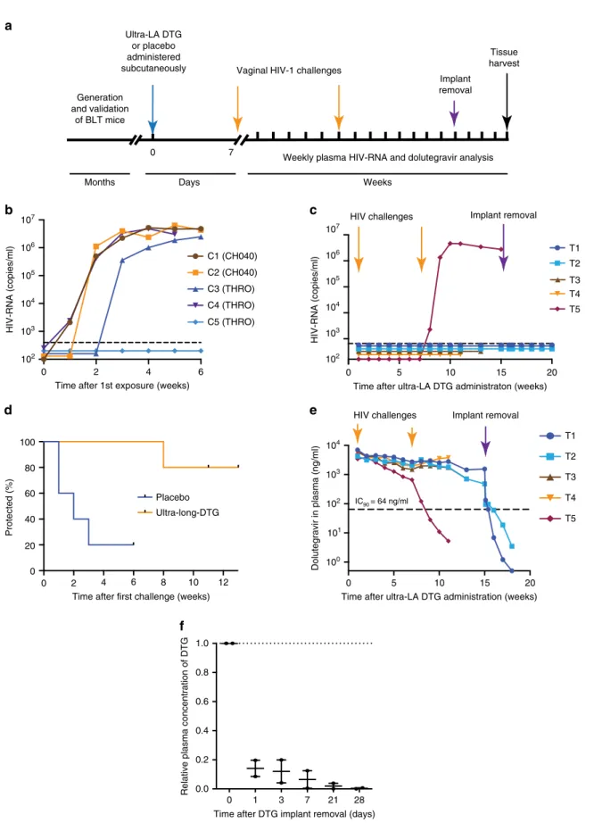
Protection from vaginal HIV acquisition
. To evaluate the
potential of the ultra-LA dolutegravir to prevent vaginal HIV
transmission, BLT mice (
n
=
5 per group) were subcutaneously
administered ultra-LA dolutegravir (treated mice) or placebo
(control mice) (Fig.
4
a). Seven days later, mice were challenged
vaginally with a high dose of one of two transmitted/founder
viruses HIV-1
CH040(3.0 × 10
5TCIU, 2 controls, three treated
mice) or HIV-1
THRO(3.5 × 10
5, three controls, two treated mice).
The two control mice exposed to HIV-1
CH040became plasma
HIV-RNA positive within 2 weeks after challenge and 2/3 control
mice challenged with HIV-1
THRObecame plasma HIV-RNA
positive within 3 weeks after the challenge (Fig.
4
b). All ultra-LA
dolutegravir-treated mice remained HIV negative after the
fi
rst
exposure. Six weeks after
fi
rst challenge, ultra-LA
dolutegravir-treated mice were challenged vaginally a second time with a high
dose of HIV-1
CH040(3.0 × 10
5TCIU). One-treated mouse became
HIV-RNA positive 1 week after the second challenge, and
sequence analysis identi
fi
ed the breakthrough virus as
HIV-1
CH040with no mutations in the HIV integrase gene. All other
treated mice (4/5) remained negative for plasma HIV-RNA (Fig.
4
d, Table
4
). Two different but complementary approaches were
a
c
–1 0 1
0 20 40 60 80 100
Log [DTG concentration (ng/ml)]
Inhibition (%)
Kendall’s : 0.75
(95% CI: 0.65 – 0.85)
Day 7 Day 28 Day 84
0 20 40 60 80 100
Inhibition (%)
b
–4.5 –4.0 –3.5 –3.0 –2.5 –2.0 –1.5
0 20 40 60 80 100
Log [relative serum concentration]
Inhibition (%)
Day 7
Day 28
Day 84
Fig. 2In vitro inhibition of HIV-1 infection with serum from ultra-LA
dolutegravir-treated mice. Serum from female NSG mice administered with ultra-LA dolutegravir (250 mg/kg) collected at days 7 (n=3), 28 (n=4), and 84 (n=2) was used for a TZM-bl cell-based assay (measured in duplicates). aInhibition of HIV-1 infection (%) with various dilution of serum. Solid lines indicate nonlinear curvefit for the data.bComparison of in vitro inhibitory activity of 1% serum collected from ultra-LA dolutegravir-treated NSG mice at the indicated time points (means ± s.e.m).cComparison of in vitro inhibitory activity (%) of serum from ultra-LA dolutegravir-treated mice and log10
a
b
c
d
e
f
g
BLT mice generationPlasma viral RNA and drug level analysis HIV
challenge
Ultra-LA DTG or placebo administration
Time (weeks)
–2 0
h
–10 0 10 20 30 40 50
Time after ultra-LA DTG administration (days)
HIV-RNA (copies/ml)
Ultra-LA DTG or placebo
–10 0 10 20 30 40 50
Time after ultra-LA DTG administration (days)
HIV-RNA(copies/ml)
Placebo (n= 4) Ultra-LA DTG (n= 4)
Ultra-LA DTG or placebo
–10 0 10 20 30 40 50
Time after ultra-long-DTG administration (days)
HIV-RNA (copies/60
µ
l)
Ultra-LA DTG or placebo
–10 0 10 20 30 40 50
Time after ultra-LA DTG administration (days)
HIV-RNA (copies/60
µ
l) Placebo (n= 4)
Ultra-LA DTG (n= 4)
Ultra-LA DTG or placebo
–10 0 10 20 30 40 50
0 20 40 60 80 100
Time after ultra-LA DTG administration (days)
hCD4+ T cells in vaginal secretions (%)
Ultra-LA DTG or placebo
–10 0 10 20 30 40 50
0 20 40 60 80 100
Time after ultra-LA DTG administration (days)
Placebo (n= 4) Ultra-LA DTG (n= 4)
Ultra-LA DTG or placebo
0 10 20 30 40
104
103
102
101
100
107 10
7
106
105
104
103
102
107
106
105
104
103
102
101
106
105
104
103
102
107
106
105
104
103
102
DTG in plasma(ng/ml)
Time after ultra-LA DTG administration (days)
hCD4+ T cells in vaginal secretions (%)
Fig. 3Suppression of systemic HIV infection by ultra-LA dolutegravir.aExperimental design. BLT mice infected with HIV-1JRC-SFwere subcutaneously
used to exclude the possibility that HIV-1 was transmitted but
suppressed due to the continuous presence of dolutegravir. First,
two mice without evidence of HIV-RNA in plasma were collected
11 and 13 weeks post ultra-LA dolutegravir administration for
tissue HIV-DNA analysis. No evidence of HIV DNA was noted in
any of the tissues analyzed con
fi
rming systemic protection from
infection. Second, the ultra-LA dolutegravir implant was
surgi-cally removed in the remaining two HIV-1-negative mice
15 weeks post-implantation. Implant removal resulted in a rapid
decrease of plasma dolutegravir concentrations (>1 log within
fi
rst 3 days after the removal) (Fig.
4
f). After implant removal, no
HIV-RNA was detected in plasma (Fig.
4
c). Four weeks
post-removal of the ultra-LA dolutegravir mice were killed, tissues
were collected and analyzed for the presence of cell-associated
HIV-DNA. No evidence of HIV-DNA was found in any of the
tissues analyzed con
fi
rming that the lack of viremia after ultra-LA
dolutegravir removal was not due to an occult infection masked
by the continuous presence of high concentration of drug
(Table
4
). Rather these results strongly indicate that these four
mice were fully protected from two high dose vaginal exposures
to HIV 6 weeks apart.
In order to establish a possible cause for the single
break-through infection noted above, plasma drug concentrations
throughout the course of the experiment were analyzed. The
concentration of dolutegravir in plasma of 4/5 treated mice was
maintained at 2805 ng/ml (median, range 1510
–
4510 ng/ml) for
11 weeks. In the mouse with the breakthrough infection, plasma
dolutegravir concentration began to slowly decrease 3 weeks after
injection. At the time of the second viral challenge, the plasma
dolutegravir concentration was 659 ng/ml. Plasma dolutegravir
concentration continued to drop and by 11 weeks post ultra-LA
dolutegravir administration it was below 10 ng/ml (Fig.
4
e). These
results suggest that the breakthrough infection is likely due to the
lower concentrations of dolutegravir in this mouse.
Discussion
The HIV epidemic continues to be a signi
fi
cant health concern
worldwide. In 2016, ~36.7 million people were living with HIV
20.
Among promising preventive interventions is pre-exposure
pro-phylaxis (PrEP), in which ARVs are taken by HIV-negative
people before potential exposure to the virus. Clinical studies
established that daily oral PrEP with Truvada
®can prevent HIV
infection among high-risk populations
21–25. However, recent
clinical trials demonstrate that lack of adherence to the indicated
drug regimen is common, resulting in lack of protection from
HIV infection
26,27.
In the speci
fi
c case of HIV prevention, the lack of adherence by
clinical trial participants has served to highlight the urgent need
for drug delivery systems capable of offering long-term protection
from HIV infection
26,27. Two LA-ARV formulations for HIV
PrEP are in clinical trials. Cabotegravir, an integrase strand
transfer inhibitor (INSTI), is in phase IIa and IIb/III clinical
trials
28and rilpivirine, a non-nucleoside reverse transcriptase
inhibitor (NNRTI), is in a phase II clinical trial
29. Both drugs are
formulated as nanosuspensions for LA intramuscular injections
every 8 weeks
5–7. Major limitations of this type of drug
for-mulations are the complete inability to remove the injected
nanosuspension from the body in the case of a medical
emer-gency and the inability of co-formulation of two or more drugs.
Removal of LA formulations is essential to circumvent adverse
reactions or to prevent long-term sub-therapeutic drug exposure
after PrEP discontinuation. These concerns are currently being
partially addressed by an oral regimen of drug for 5 weeks prior
to the administration of the LA formulation and by daily oral
administration of Truvada for up to 1 year at the end of a dosing
regimen
7. Given the documented evidence of lack of compliance
with this approach, these options are likely not appropriate for
the targeted populations.
Humanized mouse models were used for pre-clinical ef
fi
cacy
assessment of LA formulations of ARVs. A nanosuspension of
crystalline rilpivirine, administered intramuscularly, protected
BLT mice from a single vaginal high-dose HIV-1 challenge
1 week after drug administration and provide partial protection
4 weeks after drug administration
12. Similarly, single
sub-cutaneous dose of LA raltegravir signi
fi
cantly protected BLT mice
from vaginal HIV acquisition 4 weeks after drug
administra-tion
13. Recently, a lipophilic modi
fi
ed DTG prodrug encapsulated
into poloxamer nanoformulations signi
fi
cantly protected CD34
+
humanized mice from the HIV-1 for 2 weeks
30. These results
Table 2 HIV-1-RNA suppression by ultra-LA dolutegravir
Mouse hCD45
(%)
hCD4 (%)
Nadir (HIV-RNA copies/ ml plasma)
Duration of suppression below LOQ in CVS (days)
S1 37.9 76.1 LOQ 54
S2 60.3 77.5 14,746.3 27
S3 43.6 71.5 38,282.0 16
S4 53 75.6 30,863.0 41
Mean 48.7 75.2 21,022 33.5
SD 9.9 2.56 17,000 15.0
BLT mice with indicated amount of human CD45+(hCD45) cells and human CD3+CD4+ (hCD4) in peripheral blood were intravenously infected with HIV-1JRC-SFand treated with
ultra-LA dolutegravir formulation (250 mg/kg) 3 weeks later. Limit of quantitation (LOQ) was 1375 copies/ml in plasma and 81 copies/60µl in CVS
CVS cervico-vaginal lavage, SD standard deviation
Table 3 DTG resistance development during ultra-LA-DTG
monotherapy
Mouse Dolutegravir
exposure (days)
Identified
mutations
Clones with the mutation/ Clones analyzed
S1 19 NA NA
50 E157K 5/5
S2 19 E157K 1/9
E157K+Q35K +Q85K+R269G
1/9
V260A 1/9
50 R269K 1/9
W243stop 1/9
E157K 2/6
E157Q 1/6
R263K 1/6
S3 19 E157K 2/5
K159N 1/5
D3R+G4E 1/5
50 E157Q 1/5
E157Q+R166K +D41N+V281M
1/5
S4 19 D229G 1/9
50 G70K 1/8
D6E 1/8
E157Q 1/8
R263K 1/8
R263K+E157Q 2/8
BLT humanized mice infected intravenously with HIV-1JR-CSF(3 × 10⁴TCIU) received a single
0 2 4 6 107
106
105
104
103
102
107
106
105
104
103
102
104
103
102
101
100 Time after 1st exposure (weeks)
HIV-RNA (copies/ml)
C1 (CH040) C2 (CH040) C3 (THRO) C4 (THRO) C5 (THRO)
0 5 10 15 20
Time after ultra-LA DTG administraton (weeks)
HIV-RNA (copies/ml)
T1 T2
T4 T5 T3 Implant removal HIV challenges
0 2 4 6 8 10 12 0
20 40 60 80 100
Time after first challenge (weeks)
Protected (%)
Placebo Ultra-long-DTG
b
c
d
e
a
Months Days Weeks
Generation and validation
of BLT mice
Ultra-LA DTG or placebo administered
subcutaneously Vaginal HIV-1 challenges
Weekly plasma HIV-RNA and dolutegravir analysis Tissue harvest
0 7
Implant removal
f
Dolutegravir in plasma (ng/ml)
Time after ultra-LA DTG administration (weeks) T1 T2 T3 T4 T5
0 5 10 15 20
IC90 = 64 ng/ml
Implant removal HIV challenges
0 1 3 7 21 28 0.0
0.2 0.4 0.6 0.8 1.0
Time after DTG implant removal (days)
Relative plasma concentration of DTG
Fig. 4A single dose of ultra-LA dolutegravir protects against multiple high dose HIV-1 challenges.aExperimental design. BLT mice were treated with
ultra-LA dolutegravir (n=5) or placebo (n=5) and vaginally exposed to HIVCH040or HIVTHRO1 week, and 7 weeks later. The implant was removed from two
suggest utility of humanized mouse models for pre-clinical
eva-luation of LA formulation of AVR.
Optimally, a LA delivery system for HIV prophylaxis should be
effective, safe, easy to apply, and affordable so that it can be used
in resource-poor and constrained settings. The data in this paper
indicate that ultra-LA dolutegravir can be readily prepared and
sterilized by
fi
ltration. It can ef
fi
ciently deliver dolutegravir for up
to 9 months in mice and 140 days in NHP (last time point
analyzed). A single administration of ultra-LA dolutegravir
strongly inhibited acute HIV replication, and effectively protected
against repeated high-dose vaginal HIV challenges using highly
relevant primary transmitted/founder viruses. An important
aspect of the ultra-LA dolutegravir formulation is the fact that it
can be easily removed resulting in rapid decrease of drug
con-centration providing a measure of safety that is not afforded by
any of the current LA drug delivery systems in clinical trials for
HIV prevention. However, when immediate removal is not
necessary, the formulation is biodegradable and does not require
surgical removal. In addition, the formulations offer the
fl
exibility
to include multiple drugs and the future potential to be reloadable
in situ
31.
In this stage of formulation development, it is dif
fi
cult to
predict how much ultra-LA dolutegravir will be needed to achieve
same ef
fi
cacy of prevention from HIV transmission as seen in
BLT mice. However, we can compare this formulation to existing
LA formulations in clinical trials. Cabotegravir nanoformulation
consists of two 2-ml injections of product containing 200 mg/ml
drug administered every 2 months. Our goal is to develop a
formulation that can accommodate 250 mg/ml dolutegravir. As
the IC90 for dolutegravir (64 ng/ml) is 2.5-fold lower that the
IC90 for cabotegravir (166 ng/ml) we consider this to be an
appropriate target. PK analysis shown in Fig.
1
also suggests
longer sustained release of dolutegravir in vivo compared to the
current cabotegravir formulation. Therefore, using the same
parameters currently used for cabotegravir, our ultra-LA
dolute-gravir is likely scalable to humans.
This technology was inspired from the Atrigel product that is
commercially available as Eligard (please see citation 8 and 9).
This is a palliative treatment for advanced prostate cancer.
However, there are several differences between Eligard and our
ultra-LA delivery system. (1) Eligard is packaged as a 2-syringe
system due to the limited stability of leuprolide acetate in the
PLGA/NMP formulation and administered as a dispersion rather
than a solution
32. (2) The ultra-LA dolutegravir formulation
presented in this manuscript uses an antiretroviral drug instead of
a synthetic hormone, (3) the formulation is prepared as a solution
that is stable for more than 6 months at room temperature. Since
the Atrigel technology has already been approved by the FDA for
treatment of prostate cancer we anticipate that it is also applicable
for HIV PrEP.
Several key observations are worth highlighting. Dolutegravir
monotherapy resulted in strong and sustained suppression of
HIV replication. However, as early as 19 days post therapy
initiation, resistance mutations begun to appear in the replicating
viruses. By day 50 post therapy initiation, both drug resistance
and compensatory mutations were found. Consistent with studies
in women
18, the concentration of dolutegravir in FRT tissues
from BLT mice was 3
–
7 times lower than in plasma. However,
even under these conditions signi
fi
cant protection from high dose
vaginal infection was noted. The one case of breakthrough
infection could be attributed to the lower drug concentration
present in this mouse at or near the time of exposure (659 ng/ml).
Of note, the dolutegravir plasma concentrations sustained in the
mice protected from high dose HIV infection (~2800 ng/ml) are
directly comparable to those of cabotegravir reported to be
pro-tective in a non-human primate model of low dose repeated
exposure vaginal SHIV infection
33. In summary, the results
demonstrate the in vivo effectiveness of an ultra-LA formulation
of dolutegravir to deliver drug for up to 9 months that results in
sustained viral suppression and prolonged protection from high
dose HIV vaginal challenges. With its ultra-long duration, low
cost of production, ease of administration and the ability to be
removed this represents a signi
fi
cant advance in drug delivery for
HIV pre-exposure prophylaxis.
Methods
Chemicals. 50:50 Poly(DL-lactide-co-glycolide) was purchased from LACTEL
(Birmingham, AL; Cat. No. B6010–1P, MW 27 kDa).N-methyl-2-pyrrolidone (NMP) was purchased from ASHLAND (Wilmington, DE, Product Code 851263, 100% NMP). Dolutegravir (DTG) was purchased from Selleckchem (Houston, TX; Cat. No. S2667, 99.8% DTG).
Preparation of ultra-LA dolutegravir formulations. In a 7 mL scintillation vial back-filled with argon, 50:50 Poly(DL-lactide-co-glycolide) (PLGA) was mixed with N-methyl-2-pyrrolidone (NMP) at a 1:2 PLGA/NMP weight ratio (w/w) and allowed to dissolve by continuous mixing at room temperature (Placebo). For-mulation was sterilized byfiltration (0.2 µmfilter). Dolutegravir (DTG) was
Table 4 BLT mice protection with ultra-LA-DTG from high dose vaginal HIV-1 challenges
Mouse hCD45 (%) hCD4 (%) Treatment Virus for 1st
challenge
Virus for 2nd challenge
Infecting virus
Presence of viral DNA in tissues
Lymph nodes
Spleen Liver Lung Bone
marrow Org.
C1 49.6 85.9 Vehicle CH040 None CH040 + + + + + +
C2 84.0 92.4 Vehicle CH040 None CH040 + + + + + +
C3 74.0 86.9 Vehicle THRO None THRO + + + + + +
C4 84.3 87.2 Vehicle THRO None THRO + + + + + +
C5 94.3 82.0 Vehicle THRO None THRO + + + + + +
T1 74.6 87.5 LA-DTG CH040 CH040 None − − − − − −
T2 84.4 88.4 LA-DTG CH040 CH040 None − − − − − −
T3 73.2 89.8 LA-DTG THRO CH040 None − − − − − −
T4 80.2 91.9 LA-DTG THRO CH040 None − − − − − −
T5 67.2 93.0 LA-DTG CH040 CH040 CH040 + + + + + +
Mean 76.6 88.5
SD 21.0 12.2
BLT mice with indicated amount of human CD45+(hCD45) cells and human CD3+CD4+(hCD4) were administered with ultra-long-acting dolutegravir formulation (250 mg/kg) or placebo subcutaneously. One week after treatment mice were vaginally exposed to the indicated HIV-1 isolate. Six weeks later mice were re-exposed to HIV-1CH040. DTG-dolutegravir. Cell-associated DNA was
analyzed in indicated tissue
subsequently added to the PLGA/NMP solution and stirred at 40 °C for 2 h to dissolve DTG and produce an ultra-LA dolutegravir formulation with afinal concentration of dolutegravir 93 mg/mL (ultra-LA dolutegravir; DTG/PLGA/NMP =0.3:1:2 w/w/w). The viscosity of the ultra-LA dolutegravir (845 cP) was measured using a Brookfield Cone and Plate Digital Rheometer (Model: LVDV-III+CP) and the reading was recorded at 25 °C and 1 rpm. The chemical stability of DTG in the ultra-LA dolutegravir formulation (DTG/PLGA/NMP 0.3:1:2 w/w/w) was mea-sured at 25 °C over 90 days. The ultra-LA dolutegravir formulation remained as a solution for 90 days as measured by dynamic light scattering (DLS, a Zetasizer Nano ZS Particle Analyzer (Malvern Instruments, Inc.). In addition, DTG con-centration at day 90 was ~98% the original concon-centration (91 ± 0.6 mg/mL) as measured by HPLC analysis using a Finnigan Surveyor HPLC system (Thermo Finnigan, San Jose, CA) with a Photodiode Array (PDA) Plus Detector, auto-sampler, and LC Pump Plus. The stationary phase utilized for the analysis was a Inertsil ODS-3 column (5μm, 4.6 × 150 mm, [GL Sciences, Torrance, CA]) maintained at 40 °C. Chromatographic separation was achieved by gradient elution using a mobile phase consisting of 0.1% trifluoroacetic acid in water and acet-onitrile (ACN) (H2O/ACN 95:5 v/v). Theflow rate was 1.0 mL/min and the total run time was 25 min for each 25μL injection. Implant formation in vitro was instantaneous upon injection of the formulation in the aqueous release medium (0.01 M PBS, pH=7.4, 2% solutol).
BLT humanized mice and non-human primates. BLT humanized mice were prepared by implanting a sandwich of human thymus-liver-thymus tissue under the kidney capsule of irradiated (200 rads) 8–12 weeks old female NOD/SCID/γc−/−
(NSG) mice (The Jackson Laboratory, Bar Harbor, ME). Following tissue implan-tation, mice received autologous CD34+hematopoietic stem cells via tail vein injection. Starting at 8 weeks post transplantation, human immune cell reconstitution was monitored in the peripheral blood of BLT mice byflow cytometry longitudinally. Specifically, concentration of human cells (human CD45+cells, BD Pharmingen™ APC Mouse Anti-Human CD45, cat. #555485, 1:100), human T cells (human CD45 +CD3+cells, BD Pharmingen™FITC Mouse Anti-Human CD3, cat. #555339, 1:100) and human CD4 T cells (human CD45+CD3+CD4+cells, BD Pharmingen™ PE Mouse Anti-Human CD4, cat. #555347, 1:100) were determined and mice with
≥40% human CD45+cells (~3 months after humanization) were used for
experi-ments12,34–37. Due to the focus on prevention of vaginal HIV transmission, only female mice were used for the experiments. Mice were maintained by the Division of Comparative Medicine at UNC-Chapel Hill according to protocol approved by the Institutional Use and Care Committee under AAALAC guidelines. Two rhesus macaques (Macaca mulatta) were used for the in vivo evaluation of the ultra-LA dolutegravir. Animals were maintained at the CDC Roybal Campus animal facility in Atlanta, Georgia, all procedures were performed by the Comparative Medicine Branch (CMB) with certified attending DVMs and trained staff. All animal protocols have been approved by the onsite IACUC under AAALAC guidelines.
Administration of ultra-LA-DTG and sample collection. Liquid ultra-LA dolutegravir (250 mg/kg in 50–80 µl) or a similar volume of placebo formulations were administered subcutaneously with a 19 G needle on the shaved back of anesthetized BLT or NSG mice. Peripheral blood was collected from mice into capillary tubes coated with or without EDTA to isolate plasma or serum, respec-tively. CVS were obtained by lavage with sterile PBS (three washes of 20μl each, ~60μl total volume). All samples were stored at−80 °C until analysis. Similarly, ultra-LA dolutegravir formulation was administered to two rhesus macaques via subcutaneous injection. The administration site was clipped of hair and aseptically treated with povidone iodine or chlorhexiderm and isopropanol on the day of implantation. Formulation was administered subcutaneously under anesthesia using a 19G needle. Injections were done above the thoracic vertebrae T1/T216. We used a custom-made primate jacket and a T-shirt under to avoid animal scratching. Animals went through an acclimation period for the jackets prior to the implan-tation. The jackets are not restrictive and do not interfere with movement or eating. Peripheral blood samples were collected and analyzed for drug concentrations essentially as indicated above.
Implant removal from BLT mice. Mice were anesthetized and their back was shaved to visualize the location of the implant. Implant was removed under sterile conditions via a small cutaneous incision adjacent to the implant and removing the implant using forceps.
Analysis of dolutegravir in plasma and tissues. Quantification of DTG plasma
concentrations was performed by protein precipitation and LC–MS/MS analysis. Calibration curves were obtained using a 1/concentration2weighted linear regression of analyte:internal standard peak area ratio versus nominal concentra-tion. Compilation of concentration results and descriptive statistical analyses were performed using Sciex Analyst version 1.6.1. Ten microliters of each stored plasma sample was mixed with 30μL of acetonitrile containing the isotopically labeled internal standard, dolutegravir-d715N (DTG-IS). Following vigorous mixing and centrifugation, a portion of the supernatant was diluted with 50:50 methanol:water prior to LC–MS/MS analysis. DTG was eluted from a Varian (Agilent) Pursuit Diphenyl (2.1 × 50 mm, 3μm particle size) analytical column. An API- 5000 triple
quadrupole mass spectrometer (AB Sciex, Foster City, CA) was used to detect the analyte. For DTG, the precursor ion was 420 m/z and the product ion was 277 m/z. For DTG-IS, the precursor ion was 428 m/z and the product ion was 283 m/z. The data were collected using AB Sciex Analyst Chromatography Software. The dynamic range of the assay was 1–10,000 ng/mL. All calibrators and quality control (QC) samples were within 15% of the nominal value for both within-day and between-day runs. The high, medium, and low QC values used for plasma were 3.00, 300, and 3000 ng/mL. Within-day and between-day precision calculations were <15%. The recovery range for DTG in plasma was 98.0–103%, and the recovery of DTG-IS was 104%.
Thirty microliters of each vaginal wash sample was mixed with 270μL of acetonitrile containing DTG-IS. Following vortex and centrifugation, a portion of the supernatant was diluted with 50:50 methanol:water. DTG was detected using identical conditions to those described for plasma. The dynamic range of the assay was 1–1000 ng/mL. The recovery range for DTG in CVF was 99.4–105%, and the recovery of DTG-IS was 101%. All calibrators and quality controls samples were within 15% of the nominal value. The low, medium, and high QC values used for CVF were 3.00, 30.0, and 800 ng/mL. Within-day and between-day precision was <15%.
In order to extract DTG from tissue samples, they were initially homogenized in 1 mL of 80:20 water:acetonitrile. Fifty microliters of the resulting homogenate was extracted by protein precipitation with acetonitrile containing DTG-IS. Following vortex and centrifugation, a portion of the supernatant was diluted with water. DTG was detected on an LC–MS/MS system using identical conditions as described above. During method validation, calibration standards were prepared in mouse tissue homogenate. QC samples were prepared in mouse genital tract tissue homogenates. The dynamic range of the assay was 0.2–200 ng/mL homogenate. The recovery range of DTG in tissue homogenate was 50.7–63.4%, and the recovery of DTG-IS was 72.0%. All calibrators and quality controls samples were within 15% of the nominal value with precision values <15%. The low, medium, and high QC values used for tissues were 0.600, 6.00, and 160 ng/mL, respectively.
Preparation of HIV-1 stocks for infection. Viral stocks of HIV-1JR-CSF,
HIV-1CH040, and HIV-1THROwere generated by transfecting proviral DNA38–42into 293T cells (American Tissue Culture Collection, catalog number CRL-3216, pas-sage <10) using Lipofectamine 2000 (Invitrogen). Viral particles were collected from tissue culture medium, pre-cleared by centrifugation (3000 RPM for 20 min at 4 °C) and concentrated by ultracentrifugation (31,000 RPM for 70 min at 4 °C). Tissue culture infectious units (TCID)/ml of HIV was determined by titration on TZM-bl cells (NIH AIDS Research and Reference Reagent Program, catalog number 8129, passage number 2–6), infected TZM-bl cells were visualized by with staining solution (4 µM potassium ferrocyanide, 4 µM potassium ferricyanide, 2 µM magnesium chloride, 0.4 mg/ml X-gal)12,43. TZM-bl and 293T cells were cul-tured at 37 °C, 10% CO2in Dulbecco’s Modified Eagle Medium (Sigma) supple-mented with 10% fetal bovine serum, 25 mM HEPES, 500 units/ml penicillin, 50 µg/ml streptomycin and 2 mML-glutamine (Cellgro), cells were regularly checked for morphology by microscope.
In vitro HIV-1 inhibition. TZM-bl cells were maintained in TZM-bl medium as described above and cultured at 37 °C and 5% CO2. TZM-bl cells were plated in 96-well plates at a density of 1 × 10⁵cells per well in TZM-bl medium. The next day, the medium was removed and 100μl of diluted serum from dolutegravir-treated or placebo-treated mice collected 7 day, 28 days, or 84 days post administration was added. Serum was diluted 1:20, 1:100, 1:500, 1:2500, 1:12,500, and 1:62,500 and incubated with cells for 30 min. The cells were infected with 100μl of TZM-bl medium containing 40 µg/ml of DEAE-dextran and 3 × 10³TCIU of HIV-1
JR-CSF per well was added. Approximately 48 h later, the medium was removed and the luciferase substrate One-Glo reagent (Promega, Madison, WI) supplemented with 0.2% Triton X-100 was added to allow for the measurement of luciferase activity and to inactivate virus. Luciferase was measured with a SpectraMax M3 Spectro-meter (Molecular Devices, Sunnyvale, CA) and the results normalized to the luciferase activity of cells infected with HIV in the absence of serum and expressed as a percentage of decrease in luciferase activity. The TZM-bl cells were obtained through the NIH AIDS Reagent Program (catalog number 8129, passage number 2–6), cells were regularly checked for morphology by microscope.
HIV exposure of BLT mice. For the analysis of the effect of dolutegravir mono-therapy on HIV replication, BLT mice were exposed intravenously to 3 × 10⁴TCIU of HIV-1JRC-SF. Two weeks after infection mice were subcutaneously administered the ultra-LA dolutegravir (or placebo) formulation. To evaluate the ability of the ultra-LA dolutegravir (or placebo) preparation to prevent vaginal HIV transmis-sion following multiple virus challenges, treated BLT mice were exposed vaginally to HIV-1CHO40or HIV-1THRO1 and 7 weeks after ultra-LA dolutegravir admin-istration. Anesthetized BLT mice were exposed vaginally to 3.0 × 10⁵TCIU of HIV by pipetting virus (20 µl) directly into the vaginal cavity.
CATGTTTTCAGCATTATCAGAAGGA, TGCTTGATGTCCCCCCACT, and the MGB-probe carboxyfluorescein (FAM)-CCACCCCACAAGATTTAAACACCAT GCTAA-Q (non-fluorescent quencher) (Applied Biosystems) (assay sensitivity of 1375 HIV-RNA copies/ml). The presence of HIV-DNA in tissues (spleen, lymph nodes, bone marrow, human thymus, liver, lung, and FRT) collected from BLT mice at necropsy was determined by real-time PCR analysis of DNA extracted from isolated mononuclear cells (assay limit of detection: 28 copies) with same primers and HIV-RNA (Supplementary Table 1). As a control for the presence of DNA extracted from human cells, all samples were tested for the presence of human gamma globin DNA by real-time PCR using primers: CGCTTCTGGAACGTCT-GAGATT (forward), CCTTGTCCTCCTCCTCTGTGAAATGA (reverse) (Sup-plementary table 1). Concentration of human cells (human CD45+cells, BD Pharmingen™APC Mouse Anti-Human CD45, cat. #555485, 1:100), human T cells (human CD45+CD3+cells, BD Pharmingen™FITC Mouse Anti-Human CD3, cat. #555339, 1:100) and human CD4 T cells (human CD45+CD3+CD4+cells, BD Pharmingen™PE Mouse Anti-Human CD4, cat. #555347, 1:100) were determined using a BD FACSCantoTMflow cytometer and analyzed using FlowJo software (FLOWJO, LLC, version 10.0.8).
Sequence analysis of infecting viruses. The identity of the infecting viruses was
determined by sequence analysis. Viral RNA was isolated from plasma using QIAamp viral RNA columns (Qiagen, Hilden, Germany) according to the man-ufacturer’s protocol, and cDNA was generated using Superscript III Reverse Transcriptase (Invitrogen, Carlsbad, CA) with the primer 5′-CTTCCAATTATGT TGACAGGTGTAGG-3′. cDNA was amplified by nested PCR using the Expand High Fidelity PCR System (Roche, Mannheim, Germany). PCR primers were designed to anneal in regions with the fewest possible primer mismatches to HIV-1CH040, HIV-1JR-CSF, and HIV-1THROsequences. Primer sequences were as follows: outer forward primer, 5′-CTCAATAAAGCTTGCCTTGAGTGC-3′; outer reverse prime 5′-CTTCCAATTATGTTGACAGGTGTAGG-3′; inner forward primer, 5′ -GTGTGGAAAATCTCTAGCAGTGGC-3′; inner reverse primer 5′-CTGTATCAT CTGCTCCTGTATCTAATAGAGC-3′(Supplementary Table 1). Amplified viral DNA ofgagwas sequenced and compared to the sequences of challenge viruses. To identify the emergence of drug-resistant mutations in the breakthrough viruses from BLT mice receiving ultra-LA dolutegravir PrEP or BLT mice suppressed with ultra-LA dolutegravir monotherapy, viral RNA was extracted from plasma using QIAamp viral RNA kit (Qiagen, Hilden, Germany). cDNA was generated from RNA with Superscript III RT (Invitrogen, Carlsbad, CA) with primer 5′-GGTCAGGGTCTACTTGTGTGC-3′followed by nested PCR with Expand High Fidelity Kit (Roche, Mannheim, Germany) to amplify 1.5 kb region of reverse transcriptase. The following primers were used: forward outer primer 5′-C CTGAGTGGGAGTTTGTCAATAC-3′, reverse outer primer 5′-GGTCAGGGT CTACTTGTGTGC-3′, forward inner primer 5′-AGCACACAAAGGAATTGGA G-3′reverse inner primer 5′-GTGGGATTTGTACTTCTGAAC-3′(Supplementary Table 1). Amplified viral DNA of integrase was sequenced and compared to the sequence of the original HIV-1 used for infection. To further characterize the mutations found, cDNA were cloned (6–10 clones), sequenced and analyzed. FinchTV software (Geospiza, Seattle, WA) was used to analyze sequence chromatograms, NCBI BLAST to identify sequence, and ClustalW to align sequences.
Statistical analysis. All data were graphed using GraphPad Prism (version 6.0). The statistical tests used are indicated in thefigure legends and/or text.
For PK analysis in plasma and tissues, dolutegravir concentrations (ng/ml or ng/g) in plasma, and tissue from the cervix, uterus, and vagina, (Fig.1c) were measured in 18 mice with six mice observed at each time point (days 7, 28, and 84). Different mice contributed to each time point because the animals were collected to measure tissue concentrations; the same design was used for plasma analysis. Dolutegravir levels were compared between the three time points using an exact Kruskal–Wallis test to test for any statistically significant difference, with each sample type (plasma, cervix, uterus, vagina) analyzed separately. Ratios of plasma: tissue concentration within mouse were evaluated using an exact Wilcoxon signed-rank test [Ho: plasma: tissue ratio=1, i.e., log(plasma DTG)–log(tissue DTG)=0]; observations were combined over the three time points and each tissue type was analyzed separately. A 0.05 statistical significance level was used with no adjustment for multiple testing; two-sided tests were used throughout. Seven (7) null hypothesis significance tests were conducted in this report.
A non-parametric rank-based correlation analysis accounting for clustered observations (Kendall’s tau) was used to compare in vitro inhibitory activity (%) of serum from ultra-LA dolutegravir-treated mice and log10concentration of dolutegravir (Fig.2c)44.
For the suppression experiment (Fig.3) the outcomes of plasma viral load, (CVS) cervical vaginal secretion viral load, and hCD4+T-cell concentration, area under the curve (AUC) was calculated using the trapezoid method over 11 longitudinal measurements (taken between days 1 and 42) and compared between the control and dolutegravir-treated groups. A generalization of the exact Wilcoxon–Mann–Whitney test was used to account for interval-censored observations arising from HIV RNA results below the limit of detection (LOQ)45. Lower and upper bounds for the AUC were computed using zero and the LOQ, respectively, for results below the limit. In the absence of censoring (e.g., hCD4%)
this test reduces to the exact Wilcoxon rank-sum test. Notably, one mouse in the control group died after six longitudinal measurements and was assigned the worst ranked AUC for analysis (i.e., the largest viral load AUCs and lowest hCD4 % AUC). Analyses were conducted in SAS version 9.4 (Cary, NC) and R-project version 3.4.0.
An exact log rank test was used to compare protection between placebo and ultra-LA dolutegravir-treated BLT mice (Fig.4d). If not otherwise specified, standard error of the mean was used to estimate variation within each group. Variance is similar between the groups that are being statistically compared. No statistical methods were used to pre-determine the sample size. No randomization was used to determine how samples/animals were allocated to experimental groups. Mice representing the same human donor were allocated to each experimental group. Each experimental group had animals with similar levels of humanization. Blinding was used to analyze plasma concentration of HIV-RNA and
concentration of cell-associated HIV-RNA and HIV-DNA. Investigators were given numbered samples, but not the allocation of the samples to groups (treated, controls). No other blinding was used.
Code availability. Following commercially available software were used for data collection and analysis. For data collection (FACS), BD FACSDiva software (ver-sion 6.1.3) was used. The data were analyzed using FlowJo software (ver(ver-sion 10), Microsoft Excel (2016), GraphPad Prism (versions 6), SAS version 9.4 and R-project version 3.4.0.
Data availability
Sequence data that support thefindings in Table 3 of this study have been deposited in GenBank with the accession codes: MH425196-MH425242. The authors declare that all other data supporting thefindings of this study are available within the article and its Supplementary Informationfiles, or are available from the authors upon request.
Received: 19 April 2018 Accepted: 29 August 2018
References
1. Osterberg, L. & Blaschke, T. Adherence to medication.N. Engl. J. Med.353, 487–497 (2005).
2. McEvoy, J. P. Risks versus benefits of different types of long-acting injectable antipsychotics.J. Clin. Psychiatry67, 15–18 (2006).
3. Winner, B. et al. Effectiveness of long-acting reversible contraception.N. Engl. J. Med.366, 1998–2007 (2012).
4. Meyers, K. et al. High interest in a long-acting injectable formulation of pre-exposure prophylaxis for HIV in young men who have sex with men in NYC: a P18 cohort substudy.PLoS ONE9,https://doi.org/10.1371/journal. pone.0114700(2014).
5. PATH. HPTN 076 - Phase II safety and acceptability of an investigational injectable product, TMC278 LA, for pre-exposure prophylaxis (PrEP). ClinicalTrials.gov,clinicaltrials.gov/ct2/show/NCT02165202?term=HPTN +076&rank=1 (2014).
6. National Institute of Allergy and Infectious Diseases. A phase IIa study to evaluate the safety, tolerability and pharmacokinetics of the investigational injectable HIV integrase inhibitor, GSK1265744, in HIV-uninfected men and women.ClinicalTrials.gov,https://www.clinicaltrials.gov/ct2/show/
NCT02178800?term=HPTN077&rank=1(2014).
7. National Institute of Allergy and Infectious Diseases. A phase 2b/3 double blind safety and efficacy study of injectable cabotegravir compared to daily oral tenofovir disoproxil fumarate/emtricitabine (TDF/FTC), for pre-exposure prophylaxis in HIV-uninfected cisgender men and transgender women who have sex with men.ClinicalTrials.gov,https://www.clinicaltrials.gov/ct2/show/ NCT02720094?term=NCT02720094&rank=1(2016).
8. Dunn, R. L., English, J. P., Cowsar, D. R. & Vanderbult, D. P. Biodegradable in-situ forming implants and methods of producing the same.USA Pat.5, 194 (1999).
9. Dunn, R. L. & Tipton, A. J. Polymeric compositions useful as controlled release implants.USA Pat.5, 716 (1997).
10. Melkus, M. W. et al. Humanized mice mount specific adaptive and innate immune responses to EBV and TSST-1.Nat. Med.12, 1316–1322 (2006). 11. Olesen, R. et al. ART influences HIV persistence in the female reproductive
tract and cervicovaginal secretions.J. Clin. Invest.126, 892–904 (2016). 12. Kovarova, M. et al. Nanoformulations of rilpivirine for topical pericoital and
systemic coitus-independent administration efficiently prevent HIV transmission. PLoS Pathog.11,https://doi.org/10.1371/journal.ppat.1005075(2015). 13. Kovarova, M. et al. A long-acting formulation of the integrase inhibitor
14. Denton, P. W. et al. Generation of HIV latency in BLT humanized mice.J. Virol.86, 630–634 (2012).
15. Cottrell, M. L., Hadzic, T. & Kashuba, A. D. Clinical pharmacokinetic, pharmacodynamic and drug-interaction profile of the integrase inhibitor dolutegravir.Clin. Pharmacokinet.52, 981–994 (2013).
16. Royals, M. A. et al. Biocompatibility of a biodegradable in situ forming implant system in rhesus monkeys.J. Biomed. Mater. Res.45, 231–239 (1999). 17. Jouyban, A., Fakhree, M. A. & Shayanfar, A. Review of pharmaceutical
applications of N-methyl-2-pyrrolidone.J. Pharm. Pharm. Sci.13, 524–535 (2010).
18. Adams, J. L. et al. Single and multiple dose pharmacokinetics of dolutegravir in the genital tract of HIV-negative women.Antivir. Ther.18, 1005–1013 (2013).
19. Anstett, K., Cutillas, V., Fusco, R., Mesplede, T. & Wainberg, M. A. Polymorphic substitution E157Q in HIV-1 integrase increases R263K-mediated dolutegravir resistance and decreases DNA binding activity.J. Antimicrob. Chemother.71, 2083–2088 (2016).
20. UNAIDS.UNAIDS Fact Sheet 2018(UN, 2018).
21. Food and Drug Administration. FDA approvesfirst drug for reducing the risk of sexually acquired HIV infection.AIDS info,https://aidsinfo.nih.gov/news/ 1254/fda-approves-fi rst-drug-for-reducing-the-risk-of-sexually-acquired-hiv-infection(2012).
22. Grant, R. M. et al. Preexposure chemoprophylaxis for HIV prevention in men who have sex with men.N. Engl. J. Med.363, 2587–2599 (2010).
23. Baeten, J. M. et al. Antiretroviral prophylaxis for HIV prevention in heterosexual men and women.N. Engl. J. Med.367, 399–410 (2012). 24. Thigpen, M. C. et al. Antiretroviral preexposure prophylaxis for heterosexual
HIV transmission in Botswana.N. Engl. J. Med.367, 423–434 (2012). 25. Choopanya, K. et al. Antiretroviral prophylaxis for HIV infection in injecting
drug users in Bangkok, Thailand (the Bangkok Tenofovir Study): a randomised, double-blind, placebo-controlled phase 3 trial.Lancet381, 2083–2090 (2013).
26. Van Damme, L. et al. Preexposure prophylaxis for HIV infection among African women.N. Engl. J. Med.367, 411–422 (2012).
27. Marrazzo, J. M. et al. Tenofovir-based preexposure prophylaxis for HIV infection among African women.N. Engl. J. Med.372, 509–518 (2015). 28. Trezza, C., Ford, S. L., Spreen, W., Pan, R. & Piscitelli, S. Formulation and
pharmacology of long-acting cabotegravir.Curr. Opin. HIV AIDS10, 239–245 (2015).
29. Jackson, A. & McGowan, I. Long-acting rilpivirine for HIV prevention.Curr. Opin. HIV AIDS10, 253–257 (2015).
30. Sillman, B. et al. Creation of a long-acting nanoformulated dolutegravir.Nat. Commun.9, 443 (2018).
31. Brudno, Y. et al. Refilling drug delivery depots through the blood.Proc. Natl Acad. Sci. USA111, 12722–12727 (2014).
32. Ravivarapu, H. B., Moyer, K. L. & Dunn, R. L. Parameters affecting the efficacy of a sustained release polymeric implant of leuprolide.Int. J. Pharm.194, 181–191 (2000).
33. Radzio, J. et al. The long-acting integrase inhibitor GSK744 protects macaques from repeated intravaginal SHIV challenge.Sci. Transl. Med.7, 270ra275 (2015).
34. Denton, P. W. et al. Antiretroviral pre-exposure prophylaxis prevents vaginal transmission of HIV-1 in humanized BLT mice.PLoS Med.5,https://doi.org/ 10.1371/journal.pmed.0050016(2008).
35. Denton, P. W. et al. One percent tenofovir applied topically to humanized BLT mice and used according to the CAPRISA 004 experimental design demonstrates partial protection from vaginal HIV infection, validating the BLT model for evaluation of new microbicide candidates.J. Virol.85, 7582–7593 (2011).
36. Olesen, R., Wahl, A., Denton, P. W. & Garcia, J. V. Immune reconstitution of the female reproductive tract of humanized BLT mice and their susceptibility to human immunodeficiency virus infection.J. Reprod. Immunol.88, 195–203 (2011).
37. Council, O. D., Swanson, M. D., Spagnuolo, R. A., Wahl, A. & Garcia, J. V. Role of semen on vaginal HIV-1 transmission and maraviroc protection. Antimicrob. Agents Chemother.59, 7847–7851 (2015).
38. Permar, S. R. et al. Clonal amplification and maternal-infant transmission of nevirapine-resistant HIV-1 variants in breast milk following single-dose nevirapine prophylaxis.Retrovirology10, https://doi.org/10.1186/1742-4690-10-88(2013).
39. Keele, B. F. et al. Identification and characterization of transmitted and early founder virus envelopes in primary HIV-1 infection.Proc. Natl Acad. Sci. USA
105, 7552–7557 (2008).
40. Ochsenbauer, C. et al. Generation of transmitted/founder HIV-1 infectious molecular clones and characterization of their replication capacity in CD4 T lymphocytes and monocyte-derived macrophages.J. Virol.86, 2715–2728 (2012).
41. Salazar-Gonzalez, J. F. et al. Genetic identity, biological phenotype, and evolutionary pathways of transmitted/founder viruses in acute and early HIV-1 infection.J. Exp. Med.206, 1273–1289 (2009).
42. Koyanagi, Y. et al. Dual infection of the central nervous system by AIDS viruses with distinct cellular tropisms.Science236, 819–822 (1987). 43. Wahl, A. et al. Human breast milk and antiretrovirals dramatically reduce oral
HIV-1 transmission in BLT humanized mice.PLoS Pathog.8,https://doi.org/ 10.1371/journal.ppat.1002732(2012).
44. Lorenz, D. J., Datta, S. & Harkema, S. J. Marginal association measures for clustered data.Stat. Med.30, 3181–3191 (2011).
45. Fay, M. P. & Shaw, P. A. Exact and asymptotic weighted logrank tests for interval censored data: the interval R package.J. Stat. Softw.36, i02 (2010).
Acknowledgements
We thank Dr. John Kappes for HIV-1 CHO40 and THRO which were obtained via the AIDS Research and Reagent Repository Program and current lab members and veter-inary technicians at UNC Division of Comparative Medicine for their assistance with various technical aspects of this work. This work was supported by the National Institute of Allergy and Infectious Diseases (grant numbers R01AI131430 to M.K. and S.R.B., KL2TR001109–04 to S.R.B., R01AI073146, R01AI096138, R01AI111899 to J.V.G., and grant number P30AI50410 to UNC Center for AIDS Research). Thefindings and con-clusions in this report are those of the authors and do not necessarily represent the views of the Centers for Disease Control and Prevention (CDC).
Author contributions
Conceived and designed the experiments: J.V.G., R.J.M., J.G.G.-L., M.K., and S.R.B. Performed the experiments: M.K., S.R.B., I.M., R.A.S., B.S., C.E.B., and C.S. Analyzed the data: M.K., S.R.B., I.M., R.A.S., C.E.B., C.S., K.R.M., and A.D.M.K. Contributed reagents/ materials/analysis tools: S.R.B., C.S., K.R.M., and A.D.M.K. Wrote the paper: M.K., S.R.B., J.V.G., R.J.M., and J.G.G.-L.
Additional information
Supplementary Informationaccompanies this paper at
https://doi.org/10.1038/s41467-018-06490-w.
Competing interests:J.G.G.-L. is named in a US Government patent on“Inhibition of
HIV Infection through Chemoprophylaxis”. The remaining authors declare no competing interests.
Reprints and permissioninformation is available online athttp://npg.nature.com/
reprintsandpermissions/
Publisher's note:Springer Nature remains neutral with regard to jurisdictional claims in
published maps and institutional affiliations.
Open Access This article is licensed under a Creative Commons
Attribution 4.0 International License, which permits use, sharing, adaptation, distribution and reproduction in any medium or format, as long as you give appropriate credit to the original author(s) and the source, provide a link to the Creative Commons license, and indicate if changes were made. The images or other third party material in this article are included in the article’s Creative Commons license, unless indicated otherwise in a credit line to the material. If material is not included in the article’s Creative Commons license and your intended use is not permitted by statutory regulation or exceeds the permitted use, you will need to obtain permission directly from the copyright holder. To view a copy of this license, visithttp://creativecommons.org/ licenses/by/4.0/.