0 Abstract
Atherosclerosis is a chronic inflammatory disease that results in the build-up of lipid filled macrophages, called foam cells, which contribute to the formation of arterial plaque. These plaques can obstruct blood flow which can lead to serious complications, such as stroke, heart attack, and aneurisms. CXCL5 is a circulating chemokine involved in the recruitment of monocytes in acute inflammatory conditions, however, the role that CXCL5 plays in chronic cardiovascular inflammatory diseases is not defined. Our previous clinical studies demonstrated that circulating CXCL5 levels and atherosclerosis severity are inversely proportional, suggesting a possible protective role for CXCL5. We hypothesize that lower levels of CXCL5 will lead to more severe disease pathology and that CXCL5 plays an atheroprotective role by altering macrophage metabolism and polarization. To investigate the in vivo repercussions of CXCL5 depletion, we challenged wild-type and Cxcl5-/- mice with high-fat diet for 16-weeks. Given these mice were not on an atheroprone
background, as expected, we did not see any evidence of atherosclerosis in isolated aortas from mice of either genotype. Surprisingly, the Western diet did not increase cholesterol levels to the same extent in Cxcl5-/- mice compared to wild-type mice, however,
cardiac function was decreased in Cxcl5-/- mice after 16 weeks of the diet. These
1 Abbreviation Key
Common Abbreviations
CXCL5 CXC Motif Chemokine Ligand 5 LDL Low-density Lipoprotein
HDL High-density Lipoprotein ApoE Apolipoprotein E
Blood and Serum Abbreviations
RBC (M/uL) Red blood cell count. HGB (g/dL) Hemoglobin value.
HCT (%) Hematocrit value: how much of total blood count is made up of red blood cells.
MCV (fL) Mean corpuscular volume: average size of the red blood cells. MCH (pg) Mean corpuscular
hemoglobin: average amount of hemoglobin in red blood cells. MCHC (g/dL) Mean corpuscular hemoglobin concentration:
average concentration of hemoglobin in red blood cells.
RDW-SD (fL) Red cell distribution width: standard deviation of volume and size of red blood cells.
RDW-CV (%) Red cell distribution width: coefficient of variation of volume and size of red blood cells.
RET# (K/uL) Absolute number of reticulocytes.
RET% (%) How much of total blood is made of reticulocytes.
IRF (%) Immature reticulocyte fraction: ratio of immature reticulocytes to the total number of reticulocytes.
LFR (%) Low-fluorescence
reticulocytes: measure of reticulocyte maturity, least mature.
MFR (%) Medium-fluorescence reticulocytes: measure of reticulocyte maturity, middle maturity.
HFR (%) High-fluorescence
reticulocytes: measure of reticulocyte maturity, most mature.
RET-He (pg) Hemoglobin content of reticulocytes (used to measure iron deficiency).
PLT (K/uL) Platelet count.
PDW (fL) Platelet distribution width. MPV (fL) Mean platelet volume. P-LCR (%) Platelet large cell ratio. PCT (%) Plateletcrit: volume occupied by platelets in the blood.
WBC (K/uL) White blood cell count. NEUT# (K/uL) Neutrophil count. NEUT% (%) Neutrophil ratio.
LYMPH# (K/uL) Lymphocyte count. LYMPH% (%) Lymphocyte ratio. MONO# (K/uL) Monocyte count. MONO% (%) Monocyte ratio. EO# (K/uL) Eosinophil count. EO% (%) Eosinophil ratio. BASO# (K/uL) Basophil count. BASO% (%) Basophil ratio.
Echocardiography Abbreviations
EF (%) Ejection Fraction FS (%) Fractional Shortening IVS;d (mm) Interventricular septum thickness at end -diastole
IVS;s (mm) Interventricular septum thickness at end -systole
LV Mass Left ventricle mass
LV Mass (corrected) Left ventricle mass corrected for body weight
LV Vol;d Left ventricle volume at end -diastole
LV Vol;s Left ventricle volume at end -systole
LVID;d (mm) Left ventricular internal dimension at end -diastole
LVID;s (mm) Left ventricular internal dimension at end -systole
LVPW;d (mm) Left ventricular posterior wall thickness at end -diastole
1 Introduction
Cardiovascular diseases are the leading cause of death in the United States (Heron, 2018). Atherosclerosis is a chronic inflammatory disease and is the underlying cause of many cardiovascular events (Benjamin, 2018). Chronic inflammation plays a large role in atherosclerosis, and the recruitment and accumulation of immune cells modulates the disease pathology and progression. Macrophages mediate the uptake of native and modified LDL. Macrophages have few receptors for native LDL, so they are gathered via mainly endocytosis. On the other hand, macrophages have many receptors (scavenger receptors) for modified LDL. Therefore, these modified LDL, acetylated LDL (acLDL), oxidized LDL (oxLDL), and aggregated LDL (agLDL) are taken up via receptor mediated endocytosis. (Schrijvers, 2007). The presence of low-density lipoprotein (LDL) within the walls of the arteries trigger activated macrophages to engulf the LDL. When there is excessive amounts of LDL present, the amount of lipid engulfed by the macrophages becomes toxic, and the cells transition into foam cells. These foam cells are a primary component of the necrotic core of the arterial lesion and, therefore, represent potential targets in developing therapies for atherosclerosis.
2 atherosclerosis lesion progression and foam cell formation remains unknown, however, data from our lab suggests that CXCL5 may play a protective role in atherosclerosis due to a negative correlation between CXCL5 levels in the blood and coronary artery disease (CAD) severity (Ravi, 2017). Additionally, immunoneutralization of CXCL5 in mouse models lead to worse atherosclerosis due to an increase in foam cell formation (Rousselle, 2013). The inflammatory phenotype of the macrophage (M1 or M2) impacts atherosclerosis pathology, and the effect of CXCL5 on macrophage polarization is not known. The homeostasis of cholesterol and LDL levels in the blood is maintained by cells such as macrophages (Chistiakov, 2017), therefore, CXCL5’s effect on macrophages could be influencing the lipid metabolism of these macrophages.
In this study, mice lacking CXCL5 expression (Cxcl5-/-) and wild-type mice were placed
on a high-fat, Western diet for 16 weeks. Throughout the study, blood was analyzed for both complete cell counts and lipid levels in addition to conscious echocardiography to determine the effect of the genotype or diet on cardiac function. Mice are inherently resistant to atherosclerosis development, therefore, several preclinical mouse models of atherosclerosis have been developed. The atherosclerosis-prone apolipoprotein E-deficient mouse (ApoE-/-) is a well-established model for studying atherosclerosis (Lo
Sasso, 2016). We hypothesize is that athero-prone mice deficient in CXCL5 (Cxcl5-/- / ApoE-/-) will be more prone to atherosclerosis and generate lesions with higher levels of
-3
/- mice as these mice are not on an atheroprone background. Furthermore, in the absence
of atherosclerosis, hypothesized that there would not be variation in the blood and serum profile or cardiac function between the Cxcl5-/- and wild-type mice. Surprisingly we found that Cxcl5-/- mice had altered lipid homeostasis and decreased heart function.
Materials and Methods
Mice
C57BL/6 (WT) mice were purchased from by the Jackson Laboratories and the Cxcl5
-/-mice were established and provided by the Nouailles Lab. The knockout was established as described by (Nouailles, 2014) and is a complete knock out. All mice were housed in Division of Comparative Medicine (DCM) facilities and handled in accordance to the approved protocol (IACUC #17191).
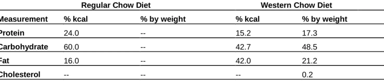
Table 1. Comparison of Western Diet Chow and Regular Diet Chow.
Regular Chow Diet Western Chow Diet
Measurement % kcal % by weight % kcal % by weight
Protein 24.0 -- 15.2 17.3
Carbohydrate 60.0 -- 42.7 48.5
Fat 16.0 -- 42.0 21.2
Cholesterol -- -- -- 0.2
Western Diet Feeding Study
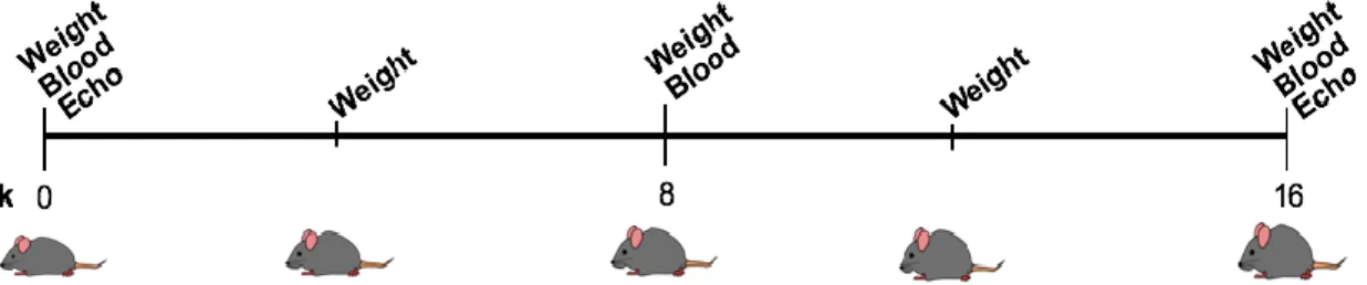
4 blood cell type composition. Two-dimensional guided M- mode echocardiography were taken using a Vevo 770 ultrasound biomicroscopy system (VisualSonics) in the parasternal long-axis view at the level of the papillary muscle. These echocardiographs and data acquisition were performed by the MHI Animal Surgery Core Lab. Weight was taken every 4 weeks. The aorta and organs (liver, kidneys, brain, gastrocnemius, and soleus) were harvested at 16 weeks.
Harvest
Blood was drawn via submandibular bleed and collected in BD Microtainer Lithium Heparin Tubes (REF #365965) BD Microtainer Serum Separator Tubes (REF #365967). The mice were then euthanized via CO2, and the aorta identified at the iliac bifurcation
and extracted up to the aortic arch. In the mice where the en face isolation was performed, the liver, brain, kidneys, gastrocnemius and soleus were removed and flash frozen with liquid nitrogen and stored at -80°C, and the rest of the organs were discarded. In the mice where the cryo isolation was performed, the heart, liver, and brain were fixed in 10% neutral buffer formalin overnight, rinsed with PBS twice and stored at 4°C in 70% EtOH, and the rest of the organs were discarded.
En face isolation
5 X-100 in PBS (2.5 g Triton X-100 in 500 mL 1X PBS) at Room temp for 1 hr with agitation. Three 5 minute PBS washes were repeated and stored at 4°C until ready for staining.
En face staining
Oil Red-O stock was prepared by adding 0.5mg Oil Red-O powder (Sigma Aldrich,O0625) into 100 mL isopropanol. A working solution was made by adding 6.0mL stock to 4.0mL diH20 and then filtered through one layer of Whatman paper x 2. The aorta was added to 60% isopropyl alcohol for 5 min at room temperature and blotted with a KimWipe. The aorta was then added to Oil Red-O for 10 min at room temperature and blotted, then moved to 60% isopropyl ethanol for 2 min at room temperature and blotted, and finally rinsed with dH2O and blotted. Aortas were dipped in hematoxylin for 10 seconds and blotted, rinsed with tap water, and stored at 4°C in PBS until imaged.
Cryo isolation
6 OCT, snap frozen on dry ice for 3-5 minutes, frozen on ice block for 30 minutes, and stored at -80°C.
Peritoneal Macrophage Isolation and Treatment
Cxcl5-/- and Cxcl5+/+ mice were injected with 2 mL 3% thioglycolate. The peritoneal macrophages were harvested 72 hrs post injection. Mice were sacrificed and injected with 8 mL of ice cold plain RPMI (Gibco, 11875-093) media into the peritoneal cavity via a catheter. The cavity was massaged and the media collected. The cells were passed through a cell strainer and centrifuged for 5 min at 23°C at 1000 rpm. The supernatant was aspirated and the pellet was re-suspended in 10% RPMI (10% Fetal Bovine Serum (FBS), 1% Penicillin Streptomycin (pen strep) by volume). 24 hours post isolation, cells were washed 3 times with 2.5% RPMI (2.5% FBS and 1% pen strep by volume). 48 hours post isolation, CXCL5 test wells were treated with 200 ng/mL CXCL5 chemokine (Peprotech 300-22B) in 2.5% RPMI, rest of wells were washed once with 2.5% RPMI. 72 hours post isolation control wells were washed once with 2.5% RPMI media, CXCL5 wells were washed once with 2.5% RPMI media, acetylated Low Density Lipoprotein (acLDL) wells were treated with 20 ug/mL acLDL (Alfa Aesar J65029) in 2.5% RPMI, M1 wells were treated with 20 ng/mL IFNg (R&D Systems, 285-IF) and 20 ng/mL lipopolysaccharides (LPS) (Sigma Aldeich, L4391) in 2.5% RPMI, and M2 wells were treated with 20 ng/mL IL4 (R&D Systems, 204-IL-010) and 20 ng/mL IL13(R&D Systems, 213-ILB) in 2.5% RPMI. 96 hours post isolation the cells were stained with Oil Red-O.
7 An Oil Red-O working solution was prepared as described previously. Oil Red-O stock was created by adding 0.5mg powder into 100 mL isopropanol. A working solution was made by adding 6.0mL stock to 4.0mL diH20 and then filtered through one layer of Whatman paper x 2. A fixative solution of 4% formaldehyde was prepared (1.6 mL 37% formaldehyde for 13.4 mL PBS). RPMI media was removed from cells and then cells were rinsed with PBS. PBS was aspirated and 4% formaldehyde was added for 7 min at room temperature then removed. They were washed twice with PBS, then PBS was removed. Oil Red-O was added for 4 min at room temperature. The Oil Red-O was removed and cells were washed 3 times with PBS and then imaged.
Analysis
JMP 14 Pro, a statistical analysis software from SAS, was utilized to perform univariate split-model ANOVA and multiple comparisons of blood and echocardiography data. Univariate analysis was performed on all of the blood and echocardiograph data and the p value was determined using a Tukey HSD All Pairwise Comparisons test. All p values less than 0.05 were considered significant.
Results
WT and Cxcl5-/- mice were placed on a high-fat western diet for 16 weeks. Incremental
8 Figure 1. Timeline of feeding study procedures. Mice began the Western Diet at 8 weeks old, weight was taken every 4 weeks, blood every 8 weeks, and echocardiography at the baseline (week 0) and end (week 16).
Figure 2. Aorta isolation and processing. Aortas were collected at the end of the 16 week Western diet feeding study and processed via either cryo or en face.
Lack of atherosclerosis plaque formation
9 once the ApoE gene is knocked out (Nakashima, 1994), and these studies are currently underway.
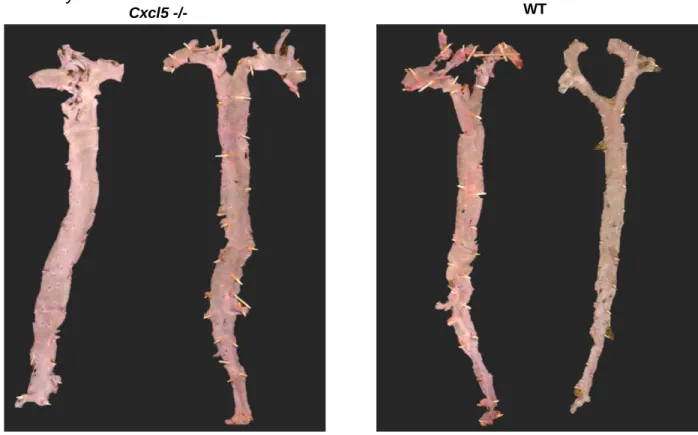
Figure 3. WT and Cxcl5-/- mice aortas post en face processing. WT and Cxcl5-/- mouse
aortas were isolated post Western diet, fixed, and stained to determine atherosclerotic lesion formation.
The loss of CXCL5 expression results in lower lipid levels in response to Western diet
Plasma obtained from Cxcl5-/- and Cxcl5+/+ mice at 0, 8, and 16 weeks of the Western diet
was tested for a complete lipid panel and glucose. Glucose and triglyceride levels increased in both Cxcl5-/- and Cxcl5+/+ mice in response to the diet, however, there was
no significant difference between the genotypes (Fig. 4D-E). The lipid levels observed were consistent with normal, non-atheroprone mice fed on a Western diet (Hoffler, 2009). Since neither genotype was on an athero-prone background, and did not develop (Fig. 3), we did not expect to see differences in cholesterol levels between the genotypes.
10 However, surprisingly, we found that Cxcl5-/- mice had significantly lower total cholesterol
at the end of the 16 week Western diet (Fig. 4A); both HDL and LDL were significantly decreased in the Cxcl5-/- mice compared to the wild-type mice (Fig. 4B-C). These data
suggest that CXCL5 effects lipid homeostasis and that the loss of CXCL5 expression leads to lower circulating cholesterol levels when on a Western diet.
Figure 4. Blood Plasma Data From Cxcl5+/+ and Cxcl5-/- Mice. Blood was drawn via
submandibular bleed from fasted (8 hr) Cxcl5+/+ and Cxcl5-/- mice and was analyzed by the UNC Animal Clinical Chemistry Core at week 0, week 8, and week 16. A. Total cholesterol levels (mg/dL). B. Low-density lipoprotein levels (mg/dL). C. High-density lipoprotein levels (mg/dL). D. Blood glucose levels (mg/dL). E. Triglyceride levels (mg/dL). (* - differences between genotypes, † - differences between 8 weeks or 16 weeks to baseline weeks, ‡ - differences between 8 weeks and 16 weeks, Variance bar = 90% confidence interval and n = approx 30)
Blood Cell Composition
We measured blood composition at 0, 8, and 16 weeks and looked at the varying levels of cell types and the % composition. Among the cell types we investigated were white blood cells; white blood cells are the components of the blood that are essential to the protection of the body against pathogens and diseases. Monocytes are a subset of white
0 8 16
0 200 400 600
Time on HFD (w)
To ta l c h o le st er o l ( m g /d L ) Cxcl5+/+ Cxcl5-/-*** *** †††, ‡‡‡ ††† ††† ‡‡‡ †††
0 8 16
0 50 100 150 200 250
Time on HFD (w)
Tr ig ly ce ri d es ( m g /d L ) †††, ‡‡‡ Cxcl5+/+ Cxcl5-/-*** ††† ‡ † †††
0 8 16
0 50 100 150 200
Time on HFD (w)
H D L ( m g /d L ) Cxcl5+/+ Cxcl5-/-†††, ‡‡‡ *** ††† *** *
0 8 16
0 50 100 150
Time on HFD (w)
L D L ( m g /d L ) Cxcl5+/+ Cxcl5-/-†††, ‡‡‡ *** add stats ††† ††† ‡‡‡ *** *
0 8 16
0 100 200 300 400
Time on HFD (w)
G lu co se ( m g /d L ) Cxcl5+/+ Cxcl5-/-†††, ‡‡‡ *** ‡‡‡ †
A
B
C
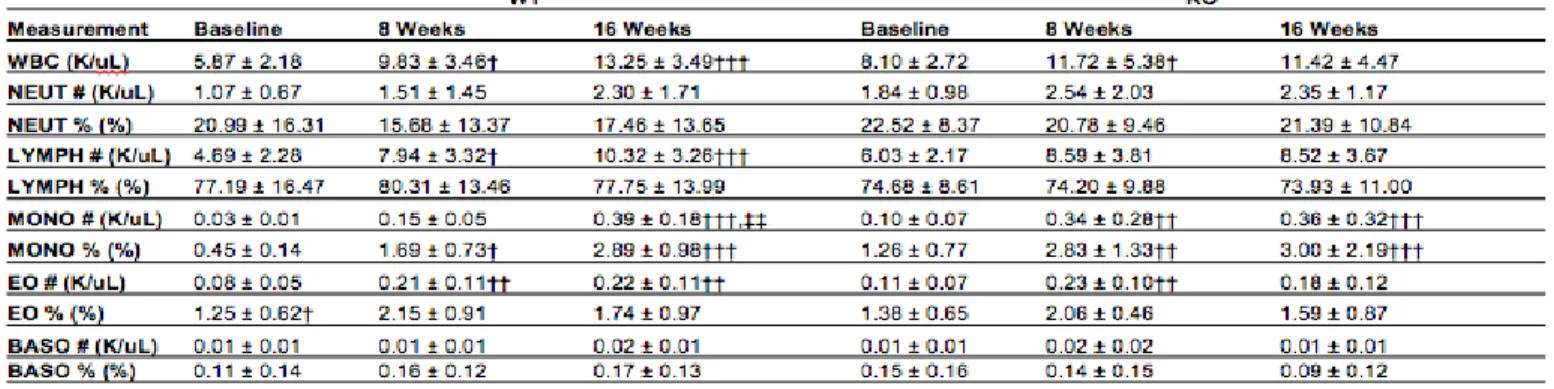
11 blood cells that are capable of differentiating into macrophage and dendritic cells. These cells are essential to the immune system response and increased levels and recruitment of monocytes are seen in infections and inflammatory diseases. We saw an increase in monocyte count within both Cxcl5-/- and Cxcl5+/+ mice (Fig. 5B). This increase in
monocytes could be indicative of inflammation occurring within the mice (Shi, 2011). Additionally, we saw a decrease in mature reticulocyte (HFR%) within both Cxcl5-/- and
Cxcl5+/+ mice (Table 2). There were no other significant disparities between the genotypes
in white blood cell, red blood cell, or platelet composition. (Table 2,3,4).
Figure 5. White Blood Cell composition of mice on Western diet. A. White blood cell percentage. B. Percentage of white blood cells that are monocytes. (* - differences between genotypes, † - differences between 8 weeks or 16 weeks to baseline weeks, ‡ - differences between 8 weeks and 16 weeks, Variance bar = 90% confidence interval and n = approx 30)
12 Table 2. Red Blood Cell Composition Data. Blood was drawn and whole blood and plasma isolated at Week 0, Week 8, and Week 16. Results are displayed in the table above. (* - differences between genotypes, † - differences between 8 weeks or 16 weeks to baseline weeks, ‡ - differences between 8 weeks and 16 weeks, * p < 0.05, ** p < 0.01, *** p < 0.001)
Table 3. White Blood Cell Composition Data. Blood was drawn and whole blood and plasma isolated at Week 0, Week 8, and Week 16. Results are displayed in the table above. (* - differences between genotypes, † - differences between 8 weeks or 16 weeks to baseline weeks, ‡ - differences between 8 weeks and 16 weeks, * p < 0.05, ** p < 0.01, *** p < 0.001)
13 Effect of CXCL5 on Heart Function
Echocardiographs of each mice were performed and data compiled by MHI Animal Surgery Core Lab at baseline (week 0) and 16 of the Western diet. Interestingly, Cxcl5-/-
mice had lower fractional shortening (FS) and ejection fraction (EF) percentages (Fig. 6B,D). Fractional shortening and ejection fraction are measures cardiac function (Gao, 2011). More specifically, the fractional shortening measures ventricle contractility and the ejection fraction measures the amount of blood that the left ventricle pumps out after each contraction. Additionally, we found that both Cxcl5-/- and Cxcl5+/+ mice experienced an
increase in left ventricle mass (LV Mass), which could be indicative of hypertrophy due to the Western diet (Fig. 6C).
Figure 6. CXCL5 is necessary to maintain cardiac function. Echocardiographs were taken at 0 and 16 weeks of the feeding study. A. Sample echocardiograph image from a conscious mouse. B. Ejection fraction (%) values. C. Left ventricle mass corrected for weight. D. Fractional shortening (%) values. (* - difference between 16 weeks and
A
B
14 baseline, * p < 0.05, ** p < 0.01, *** p < 0.001, Variance bar = 90% confidence interval and n = approx 30)
CXCL5 does not have an effect on weight gain on mice on a Western diet
Cxcl5-/- and Cxcl5+/+ mice both gained weight throughout the feeding study, with no
significant difference in weight by week 16 on the Western diet (Fig. 7).
Figure 7. Weight measurements of Cxcl5+/+ and Cxcl5-/- mice. Weight measurements were taken every 4 weeks throughout the feeding study. (* - difference between 16 weeks and baseline, † - differences between genotypes, * p < 0.05, ** p < 0.01, *** p < 0.001, Variance bar = 90% confidence interval and n = approx 30)
Peritoneal Macrophages
In order to further investigate what affect CXCL5 plays on atherosclerosis progression and lipid homeostasis, we looked to macrophages. Peritoneal macrophages were harvested, treated with various conditions and stained with Oil Red-O to determine foam cell formation. More trials need to be run and lipid uptake will be quantified via FIJI image analysis but has not been performed yet; therefore, currently no quantitative analysis can be done with this data gathered. However, based on qualitative observations the Cxcl5-/-
15 Figure 8. CXCL5 -/- and CXCL5 +/+ Peritoneal Macrophage Staining. Oil Red O stain of peritoneal macrophages from Cxcl5-/- and Cxcl5+/+ mouse. The PM were treated with the following conditions: 200 ng/mL CXCL5 (CXCL5), 20 ug/mL acLDL (LDL), 20 ng/mL) and 20 ng/mL lipopolysaccharides (LPS) (M1), and 20 ng/mL IL4 and 20 ng/mL IL13 (M2).
Discussion
Our data demonstrate that Cxcl5-/- and Cxcl5+/+ mice respond differently to a Western diet.
Surprisingly, we found that mice lacking CXCL5 had overall lower total cholesterol, HDL, and LDL (Fig. 4A-C). Mice lacking CXCL5 also had worsened cardiac function, showing lower ejection fraction and fractional shortening values (Fig. 6B, D). Additionally, preliminary peritoneal macrophage findings demonstrated that Cxcl5-/- macrophages had a higher lipid content (Fig. 8).
Based on our findings, we hypothesize that CXCL5 acts on the macrophage level to reduce atherosclerotic plaques and as well as plays a role in the lipid metabolism. The increase in total cholesterol, HDL, and LDL was an unexpected phenotype from the Cxcl5 -/- mice, as we did not expect to see differences between the genotypes (Fig. 4). One
possible suggestion for the disparity in the blood lipid levels could be that the Cxcl5-/- mice
C
o
n
tro
l
C
X
C
L
5
Cxcl5-/- Cxcl5+/+
LDL
C
X
C
L
5
+
L
D
L
M1
M2
16 may not have eaten as much of the Western diet, but this is unlikely as the weight gain for both genotypes was consistent (Fig. 7). These findings suggest that CXCL5 is having an effect on the lipid homeostasis in mice on a Western diet. It should be noted that despite the high levels of LDL in the blood at week 16, the WT and Cxcl5-/- mice aortas did not show any atherosclerotic plaques (Fig. 3). This pathology is expected to change in our future studies using mice that have the apolipoprotein E (ApoE) gene knocked out (Nakashima, 1994). Blood composition analysis showed that both WT and Cxcl5-/- mice
had increased WBC % and monocyte % (Fig. 5). This suggests that both mouse models are exhibiting an immune response, possibly due to inflammation (Shi, 2011). Our findings also demonstrated that CXCL5 may play a role in cardiac function. Cxcl5-/- mice exhibited worsened ejection fraction as well as fractional shortening, suggesting that CXCL5 may play a protective role in cardiac function.
In order to look into the effect of CXCL5 on macrophages in the development of atherosclerotic lesions, we looked at macrophages isolated from WT and Cxcl5-/- mice.
The peritoneal macrophages isolated and treated demonstrated lower lipid levels in Cxcl5-/- macrophages. CXCL5 has been shown to upregulate ATP-binding cassette
transporter 1 (ABCA1), a cholesterol transporter. Deletion of ABCA1 lead to hypocholesterolemia in the peripheral blood, while upregulation of ABCA1 lead to increased lipid deposits. CXCL5’s upregulation of ABCA1 is thought to lead to a decrease
17 To determine how CXCL5 affects the development and progression of atherosclerosis, future directions include repeating the feeding study with Cxcl5-/-/ApoE-/- mice.
Additionally, we will repeat the study with Cxcl5+/+/ApoE-/- mice in order to identify the effect that are coming from the ApoE KO. In order to more closely model the clinical cases of reduced CXCL5 in patients, we will also repeat the feeding study with Cxcl5+/-/ApoE-/- mice. Another experiment includes Cxcl5+/+/ApoE-/- mice on a feeding study with
implanted mini pumps to measure the effect of excess CXCL5 on atherosclerosis progression, to gauge whether additional CXCL5 will play a protective role. Regarding the effect of CXCL5 on macrophage polarization and lipid homeostasis, future experiments further exploring this concept include treating the M1 and M2 polarized macrophages with CXCL5 and LDL in order to measure foam cell formation in the presence of various concentrations of CXCL5, as well as looking at the levels of important cholesterol scavenger receptors and cholesterol efflux transporters in response to varying levels of CXCL5.
Acknowledgements
Acknowledgements to Becky Sanchez-Hodge, Mustafa Rauf, and Jonathan Schisler for assistance in data collection and analysis; to the UNC Animal Clinical Chemistry Core
18 References
Benjamin, E. J., Virani, S. S., Callaway, C. W., Chamberlain, A. M., Chang, A. R., Cheng, S., . . . Muntner, P. (2018). Heart Disease and Stroke Statistics—2018 Update: A Report From the American Heart Association. Circulation, 137(12). doi:10.1161/cir.0000000000000558
Chistiakov, D. A., Bobryshev, Y. V., & Orekhov, A. N. (2015). Macrophage-mediated cholesterol handling in atherosclerosis. Journal of Cellular and Molecular Medicine,20(1), 17-28. doi:10.1111/jcmm.12689
Chistiakov, D. A., Melnichenko, A. A., Myasoedova, V. A., Grechko, A. V., & Orekhov, A. N. (2017). Mechanisms of foam cell formation in atherosclerosis. Journal of Molecular Medicine,95(11), 1153-1165. doi:10.1007/s00109-017-1575-8
Gao, S., Ho, D., Vatner, D. E., & Vatner, S. F. (2011). Echocardiography in Mice. Current protocols in mouse biology, 1, 71-83.
Heron, M. (2018). Deaths: Leading Causes for 2016. National Vital Statistics Reports,67(6). Retrieved from
https://www.cdc.gov/nchs/data/nvsr/nvsr67/nvsr67_06.pdf.
Hoffler, U., Hobbie, K., Wilson, R., Bai, R., Rahman, A., Malarkey, D., Travlos, G., … Ghanayem, B. I. (2009). Diet-induced obesity is associated with hyperleptinemia, hyperinsulinemia, hepatic steatosis, and glomerulopathy in C57Bl/6J mice.
Endocrine, 36(2), 311-25.
Lo Sasso G, Schlage WK, Boué S, Veljkovic E, Peitsch MC, Hoeng J. The Apoe(-/-) mouse model: a suitable model to study cardiovascular and respiratory diseases in the context of cigarette smoke exposure and harm reduction. J Transl Med. 2016;14(1):146. Published 2016 May 20. doi:10.1186/s12967-016-0901-1
19 arterial tree. Arteriosclerosis and Thrombosis: A Journal of Vascular
Biology,14(1), 133-140. doi:10.1161/01.atv.14.1.133
Nouailles, G., Dorhoi, A., Koch, M., Zerrahn, J., Weiner, J., Faé, K. C., . . . Kaufmann, S. H. (2014). CXCL5-secreting pulmonary epithelial cells drive destructive
neutrophilic inflammation in tuberculosis. Journal of Clinical Investigation,124(3), 1268-1282. doi:10.1172/jci72030
Ravi, S., Schuck, R. N., Hilliard, E., Lee, C. R., Dai, X., Lenhart, K., . . . Schisler, J. C. (2017). Clinical Evidence Supports a Protective Role for CXCL5 in Coronary Artery Disease. The American Journal of Pathology,187(12), 2895-2911. doi:10.1016/j.ajpath.2017.08.006
Rousselle, A., Qadri, F., Leukel, L., Yilmaz, R., Fontaine, J., Sihn, G., . . . Duchene, J. (2013). CXCL5 limits macrophage foam cell formation in atherosclerosis. Journal of Clinical Investigation,123(3), 1343-1347. doi:10.1172/jci66580
Schrijvers, D., Demeyer, G., Herman, A., & Martinet, W. (2007). Phagocytosis in atherosclerosis: Molecular mechanisms and implications for plaque progression and stability. Cardiovascular Research,73(3), 470-480. doi:10.1016/j.cardiores.2006.09.005