ENDOCRINE PRACTICE Vol 17 No. 3 May/June 2011 e1
ATA/AACE Guidelines
HYPERTHYROIDISM AND OTHER CAUSES OF THYROTOXICOSIS:
MANAGEMENT GUIDELINES OF THE
AMERICAN THYROID ASSOCIATION AND
AMERICAN ASSOCIATION OF CLINICAL ENDOCRINOLOGISTS
Rebecca S. Bahn (Chair), MD1,*; Henry B. Burch, MD2; David S. Cooper, MD3; Jeffrey R. Garber, MD, FACP, FACE4; M. Carol Greenlee,MD 5; Irwin Klein, MD6;Peter Laurberg, MD7; I. Ross McDougall, MD8; Victor M. Montori, MD1; Scott A. Rivkees, MD9; Douglas S. Ross, MD10;
Julie Ann Sosa, MD11; Marius N. Stan, MD1
American Association of Clinical Endocrinologists Medical Guidelines for Clinical Practice are systematically developed statements to assist health-care professionals in medical decision making for specific clinical conditions. Most of the content herein is based on literature reviews. In areas of uncertainty, professional judgment was applied.
These guidelines are a working document that reflects the state of the field at the time of publication. Because rapid changes in this area are expected, periodic revisions are inevitable. We encourage medical professionals to use this information in conjunction with their best clinical judgment. The presented recommendations may not be appropri-ate in all situations. Any decision by practitioners to apply these guidelines must be made in light of local resources and individual patient circumstances.
HYPERTHYROIDISM AND OTHER CAUSES OF THYROTOXICOSIS:
MANAGEMENT GUIDELINES OF THE
AMERICAN THYROID ASSOCIATION AND
AMERICAN ASSOCIATION OF CLINICAL ENDOCRINOLOGISTS
Rebecca S. Bahn (Chair), MD1,*; Henry B. Burch, MD2; David S. Cooper, MD3; Jeffrey R. Garber, MD, FACP, FACE4; M. Carol Greenlee,MD 5; Irwin Klein, MD6;Peter Laurberg, MD7; I. Ross McDougall, MD8; Victor M. Montori, MD1; Scott A. Rivkees, MD9; Douglas S. Ross, MD10;
Julie Ann Sosa, MD11; Marius N. Stan, MD1
From the 1Division of Endocrinology, Metabolism, and Nutrition, Mayo Clinic, Rochester, Minnesota; 2Endocrinology and Metabolism Division, Walter Reed Army Medical Center, Washington, District of Columbia; 3Division of Endocrinology, The Johns Hopkins University School of Medicine, Baltimore, Maryland; 4Endocrine Division, Harvard Vanguard Medical Associates, Boston, Massachusetts; 5Western Slope Endocrinology, Grand Junction, Colorado; 6The Thyroid Unit, North Shore University Hospital, Manhassett, New York; 7Department of Endocrinology, Aarhus University Hospital, Aalborg, Denmark; 8Division of Nuclear Medicine, Department of Radiology and Division of Endocrinology, Department of Medicine, Stanford University School of Medicine, Stanford, California; 9Department of Pediatrics, Yale Pediatric Thyroid Center, New Haven, Connecticut; 10Massachusetts General Hospital, Boston, Massachusetts;11Divisions of Endocrine Surgery and Surgical Oncology, Yale University School of Medicine, New Haven, Connecticut.
By mutual agreement among the authors and editors of their respective jour-nals, this work is being published jointly in Thyroid and Endocrine Practice. Copyright © 2011 AACE.
ABSTRACT
Objective: Thyrotoxicosis has multiple etiologies,
manifestations, and potential therapies. Appropriate treat-ment requires an accurate diagnosis and is influenced by coexisting medical conditions and patient preference. This article describes evidence-based clinical guidelines for the management of thyrotoxicosis that would be useful to gen-eralist and subspeciality physicians and others providing care for patients with this condition.
Methods: The development of these guidelines was
commissioned by the American Thyroid Association in association with the American Association of Clinical Endocrinologists. The American Thyroid Association and American Association of Clinical Endocrinologists assem-bled a task force of expert clinicians who authored this
report. The task force examined relevant literature using a systematic PubMed search supplemented with addi-tional published materials. An evidence-based medicine approach that incorporated the knowledge and experience of the panel was used to develop the text and a series of specific recommendations. The strength of the recom-mendations and the quality of evidence supporting each was rated according to the approach recommended by the Grading of Recommendations, Assessment, Development, and Evaluation Group.
Results: Clinical topics addressed include the initial
evaluation and management of thyrotoxicosis; manage-ment of Graves’ hyperthyroidism using radioactive iodine, antithyroid drugs, or surgery; management of toxic multi-nodular goiter or toxic adenoma using radioactive iodine or surgery; Graves’ disease in children, adolescents, or preg-nant patients; subclinical hyperthyroidism; hyperthyroid-ism in patients with Graves’ ophthalmopathy; and manage-ment of other miscellaneous causes of thyrotoxicosis.
Conclusions: One hundred evidence-based
recom-mendations were developed to aid in the care of patients with thyrotoxicosis and to share what the task force believes is current, rational, and optimal medical practice. INTRODUCTION
Thyrotoxicosis is a condition having multiple eti-ologies, manifestations, and potential therapies. The term “thyrotoxicosis” refers to a clinical state that results from inappropriately high thyroid hormone action in tissues gen-erally due to inappropriately high tissue thyroid hormone levels. The term “hyperthyroidism,” as used in these guide-lines, is a form of thyrotoxicosis due to inappropriately high synthesis and secretion of thyroid hormone(s) by the thyroid. Appropriate treatment of thyrotoxicosis requires an accurate diagnosis. For example, thyroidectomy is an appropriate treatment for some forms of thyrotoxicosis
and not for others. Additionally, beta blockers may be used in almost all forms of thyrotoxicosis, whereas antithyroid drugs are useful in only some.
In the United States, the prevalence of hyperthyroid-ism is approximately 1.2% (0.5% overt and 0.7% sub-clinical); the most common causes include Graves’ disease (GD), toxic multinodular goiter (TMNG), and toxic ade-noma (TA) (1). Scientific advances relevant to this topic are reported in a wide range of literature, including subspe-ciality publications in endocrinology, pediatrics, nuclear medicine, and surgery, making it challenging for clinicians to keep abreast of new developments. Although guide-lines for the diagnosis and management of patients with hyperthyroidism have been published previously by both the American Thyroid Association (ATA) and American Association of Clinical Endocrinologists (AACE), in con-junction with guidelines for the treatment of hypothyroid-ism (1,2), both associations determined that thyrotoxicosis represents a priority area in need of updated evidence-based practice guidelines.
The target audience for these guidelines includes gen-eral and subspeciality physicians and others providing care for patients with thyrotoxicosis. In this document, we outline what we believe is current, rational, and optimal medical practice. It is not the intent of these guidelines to replace clinical judgment, individual decision making, or the wishes of the patient or family. Rather, each recom-mendation should be evaluated in light of these elements in order that optimal patient care is delivered. In some circumstances, it may be apparent that the level of care required may be best provided in centers where there is specific expertise, and that referral to such centers should be considered.
METHODS OF DEVELOPMENT OF EVIDENCE-BASED GUIDELINES
Administration
The ATA Executive Council and the Executive Committee of AACE forged an agreement outlining the working relationship between the two groups surround-ing the development and dissemination of management guidelines for the treatment of patients with thyrotoxicosis. A chairperson was selected to lead the task force and this individual (R.S.B.) identified the other 11 members of the panel in consultation with the ATA and the AACE boards of directors. Membership on the panel was based on clini-cal expertise, scholarly approach, and representation of adult and pediatric endocrinology, nuclear medicine, and surgery. The task force included individuals from both North America and Europe. In addition, the group recruited an expert on the development of evidence-based guidelines (V.M.M.) to serve in an advisory capacity. Panel members declared whether they had any potential conflict of interest
at the initial meeting of the group and periodically during the course of deliberations. Funding for the guidelines was derived solely from the general funds of the ATA and thus the task force functioned without commercial support.
To develop a scholarly and useful document, the task force first developed a list of the most common causes of thyrotoxicosis and the most important questions that a practitioner might pose when caring for a patient with a particular form of thyrotoxicosis or special clinical condi-tion. Two task force members were assigned to review the literature relevant to each of the topics, using a systematic PubMed search for primary references and reviews sup-plemented with additional published materials available before June 2010, and develop recommendations based on the literature and expert opinion where appropriate. A pre-liminary document and a series of recommendations con-cerning all of the topics were generated by each subgroup and then critically reviewed by the task force at large. The panel agreed recommendations would be based on consen-sus of the panel and that voting would be used if agreement could not be reached. Two recommendations were not unanimous and the dissenting position is noted. Task force deliberations took place during several lengthy committee meetings, multiple telephone conference calls, and through electronic communication.
Rating of the recommendations
These guidelines were developed to combine the best scientific evidence with the experience of seasoned clinicians and the pragmatic realities inherent in implementation. The task force elected to rate the recommendations according to the system developed by the Grading of Recommendations, Assessment, Development, and Evaluation Group (3), with a modification in the grading of evidence (4). Although the rating system we chose differs from those used in previous ATA and AACE clinical practice guidelines, the approach conforms with the recently updated AACE protocol for standardized production of clinical practice guidelines (5). The balance between benefits and risks, quality of evidence, applicability, and certainty of the baseline risk are all considered in judgments about the strength of recommendations (6). Grading the quality of the evidence takes into account study design, study quality, consistency of results, and directness of the evidence. The strength of a recommendation is indicated by the number 1 or 2. Grade 1 indicates a strong recommendation (for or against) that applies to most patients in most circumstances with benefits of action clearly outweighing the risks and burdens (or vice versa). In contrast, Grade 2 indicates a weak recommendation or a suggestion that may not be appropriate for every patient, depending on context, patient values, and preferences. The risks and benefits or burdens associated with a weak recommendation are closely balanced or uncertain and the statement is
generally associated with the phrase “we suggest” or “should be considered.” The quality of the evidence is indicated by plus signs, such that + denotes low quality evidence; ++, moderate quality evidence; and +++, high quality evidence, based on consistency of results between studies and study design, limitations, and the directness of the evidence. Table 1 describes the criteria to be met for each rating category. Each recommendation is preceded by a description of the evidence and, in some cases, followed by a remarks section including technical suggestions on issues such as dosing and monitoring.
Presentation and endorsement of recommendations
The organization of the task force’s recommendations is presented in Table 2. The page numbers and the location key can be used to locate specific topics and recommen-dations. Specific recommendations are presented within boxes in the main body of the text. Location keys can be copied into the Find or Search function in a file or Web page to rapidly navigate to a particular section. A listing of the recommendations without text is provided as Appendix A.
The final document was approved by the ATA and AACE on March 15, 2011, and officially endorsed (in alphabeti-cal order) by American Academy of Otolaryngology–Head and Neck Surgery, Associazione Medici Endocrinologi, British Association of Endocrine and Thyroid Surgeons, Canadian Paediatric Endocrine Group–Groupe Canadien d’Endocrinologie Pédiatrique (endorsement of pedi-atric section only), European Association of Nuclear Medicine, The Endocrine Society, European Society of Endocrinology, European Society of Endocrine Surgeons, European Thyroid Association, International Association of Endocrine Surgeons, Latin American Thyroid Society,
Pediatric Endocrine Society, Italian Endocrine Society, and Society of Nuclear Medicine.
RESULTS
[A] Background
In general, thyrotoxicosis can occur if (i) the thyroid is inappropriately stimulated by trophic factors; (ii) there is constituitive activation of thyroid hormone synthesis and secretion leading to autonomous release of excess thyroid hormone; (iii) thyroid stores of preformed hormone are passively released in excessive amounts owing to autoim-mune, infectious, chemical, or mechanical insult; or (iv) there is exposure to extra-thyroidal sources of thyroid hormone, which may be either endogenous (struma ova-rii, metastatic differentiated thyroid cancer) or exogenous (factitious thyrotoxicosis).
Subclinical hyperthyroidism (SH) is most often caused by release of excess thyroid hormone by the gland. This condition is defined as a low or undetectable serum thyroid-stimulating hormone (TSH) with values within the normal reference range for both triiodothyronine (T3) and free thyroxine (T4) estimates. Both overt and subclinical disease may lead to characteristic signs and symptoms.
GD is an autoimmune disorder in which thyrotropin receptor antibodies (TRAbs) stimulate the TSH receptor, increasing thyroid hormone production. The natural his-tory of nodular thyroid disease includes growth of estab-lished nodules, new nodule formation, and development of autonomy over time (7). In TAs, autonomous hormone production can be caused by somatic activating mutations of genes regulating thyroid hormone systhesis. Germline mutations in the gene encoding the TSH receptor can cause sporadic or familial nonautoimmune hyperthyroidism
Table 1. GradinG of recommendaTions, assessmenT, developmenT, and evaluaTion sysTem
Type of grading Definition of grades
Strength of the recommendation 1 = strong recommendation (for or against) Applies to most patients in most circumstances Benefits clearly outweigh the risk (or vice versa) 2 = weak recommendation (for or against)
Best action may differ depending on circumstances or patient values Benefits and risks or burdens are closely balanced, or uncertain Quality of the evidence +++ = High quality; evidence at low risk of bias, such as high quality
randomized trials showing consistent results directly applicable to the recommendation
++ = Moderate quality; studies with methodological flaws, showing inconsistent or indirect evidence
Table 2. orGanizaTionofThe Task force’s recommendaTions
Location key Description Page
[A] Background e4
[B] How should clinically or incidentally discovered thyrotoxicosis be evaluated and initially managed? e7
[B1] Assessment of disease severity e7
[B2] Biochemical evaluation e7
[B3] Determination of etiology e8
[B4] Symptomatic management e9
[C] How should overt hyperthyroidism due to GD be managed? e10 [D] If 131I therapy is chosen as treatment for GD, how should it be accomplished? e11 [D1] Preparation of patients with GD for 131I therapy e11 [D2] Administration of 131I in the treatment of GD e12 [D3] Patient follow-up after 131I therapy for GD e13 [D4] Treatment of persistent Graves’ hyperthyroidism following radioactive iodine therapy e13 [E] If antithyroid drugs are chosen as initial management of GD, how should the therapy be managed? e13 [E1] Initiation of antithyroid drug therapy for the treatment of GD e14 [E2] Monitoring of patients taking antithyroid drugs e14
[E3] Management of allergic reactions e15
[E4] Duration of antithyroid drug therapy for GD e15
[F] If thyroidectomy is chosen for treatment of GD, how should it be accomplished? e16 [F1] Preparation of patients with GD for thyroidectomy e16
[F2] The surgical procedure and choice of surgeon e16
[F3] Postoperative care e17
[G] How should thyroid nodules be managed in patients with GD? e17
[H] How should thyroid storm be managed? e18
[I] How should overt hyperthyroidism due to TMNG or TA be treated? e19 [J] If 131I therapy is chosen as treatment for TMNG or TA, how should it be accomplished? e20 [J1] Preparation of patients with TMNG or TA for 131I therapy e20 [J2] Evaluation of thyroid nodules prior to radioioactive iodine therapy e21 [J3] Administration of radioactive iodine in the treatment of TMNG or TA e21 [J4] Patient follow-up after 131I therapy for TMNG or TA e22 [J5] Treatment of persistent or recurrent hyperthyroidism following for TMNG or TA 131I therapy e22 [K] If surgery is chosen as treatment for TMNG or TA, how should it be accomplished? e22 [K1] Preparation of patients with TMNG or TA for surgery e22
[K2] The surgical procedure and choice of surgeon e23
[K3] Postoperative care e23
[K4] Treatment of persistent or recurrent disease following surgery for TMNG or TA e24 [L] Is there a role for antithyroid drug therapy in patients with TMNG or TA? e24 [M] Is there a role for radiofrequency, thermal or alcohol ablation in the management of TA or TMNG? e24 [N] How should GD be managed in children and adolescents? e24
[N1] General approach e24
[O] If antithyroid drugs are chosen as initial management of GD in children, how should the therapy be managed? e25 [O1] Initiation of antithyroid drug therapy for the treatment of GD in children e25 [O2] Symptomatic management of Graves’ hyperthyroidism in children e26
[O3] Monitoring of children taking methimazole e26
[O4] Monitoring of children taking propylthiouracil e26 [O5] Management of allergic reactions in children taking methimazole e26
associated with a diffuse enlargement of the thyroid gland (8). Autonomous hormone production is caused by somatic, activating mutations of genes regulating follicu-lar cell activities. Hormone production may progress from subclinical to overt hyperthyroidism, and the administra-tion of pharmacologic amounts of iodine to such patients
may result in iodine-induced hyperthyroidism (9). GD is overall the most common cause of hyperthyroidism in the United States (10,11). Although toxic nodular goiter is less common than GD, its prevalence increases with age and in the presence of iodine deficiency. Therefore, toxic nodu-lar goiter may actually be more common than GD in older [O6] Duration of methimazole therapy in children with GD e27 [P] If radioactive iodine is chosen as treatment for GD in children, how should it be accomplished? e27 [P1] Preparation of pediatric patients with GD for 131I therapy e27 [P2] Administration of 131I in the treatment of GD in children e28
[P3] Side-effects of 131I therapy in children e28
[Q] If thyroidectomy is chosen as treatment for GD in children, how should it be accomplished? e30 [Q1] Preparation of children with GD for thyroidectomy e30
[R] How should SH be managed? e30
[R1] Frequency and causes of subclinical hyperthyroidism e30 [R2] Clinical significance of subclinical hyperthyroidism e30
[R3] When to treat subclinical hyperthyroidism e31
[R4] How to treat subclinical hyperthyroidism e31
[R5] End points to be assessed to determine effective therapy of subclinical hyperthyroidism e32 [S] How should hyperthyroidism in pregnancy be managed? e32
[S1] Diagnosis of hyperthyroidism in pregnancy e32
[S2] Management of hyperthyroidism in pregnancy e33
[S3] The role of TRAb levels measurement in pregnancy e35
[S4] Postpartum thyroiditis e36
[T] How should hyperthyroidism be managed in patients with Graves’ ophthalmopathy? e37
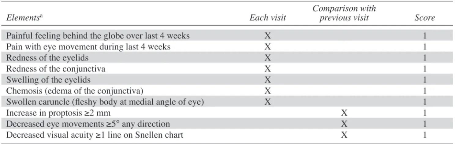
[T1] Assessment of disease activity and severity e37
[T2] Prevention of GO e38
[T3] Treatment of hyperthyroidism in patients with active GO of mild severity e39 [T4] Treatment of hyperthyroidism in patients with active and moderate-to-severe or sight-threatening GO e40 [T5] Treatment of GD in patients with inactive GO e40 [U] How should overt drug-induced thyrotoxicosis be managed? e41
[U1] Iodine-induced thyrotoxicosis e41
[U2] Cytokine-induced thyrotoxicosis e42
[U3] Amiodarone-induced thyrotoxicosis e42
[V] How should thyrotoxicosis due to destructive thyroiditis be managed? e43
[V1] Subacute thyroiditis e43
[V2] Painless thyroiditis e43
[V3] Acute thyroiditis e44
[W] How should thyrotoxicosis due to unusual causes be managed? e44
[W1] TSH-secreting pituitary tumors e44
[W2] Struma ovarii e45
[W3] Choriocarcinoma e45
[W4] Thyrotoxicosis factitia e45
[W5] Functional thyroid cancer metastases e45
GD, Graves’ disease; GO, Graves’ ophthalmopathy; SH, subclinical hyperthyroidism; TA, toxic adenoma; TMNG, toxic multinodular goiter; TRAb, thyrotropin receptor antibody; TSH, thyroid-stimulating hormone.
Location key Description Page
patients from regions of iodine deficiency (12). Unlike toxic nodular goiter, which is progressive (unless triggered by excessive iodine intake), remission of GD has been reported in up to 30% of patients without treatment (13). The mechanism of hyperthyroidism in painless and subacute thyroiditis is inflammation of thyroid tissue with release of preformed hormone into the circulation. Painless thyroiditis is the etiology of hyperthyroidism in about 10% of patients (14), occurring in the postpartum period (postpartum thyroiditis) (15), during lithium (16), or cyto-kine (e.g., interferon-alpha) (17) therapy, and in 5–10% of amiodarone-treated patients (18). Subacute thyroiditis is thought to be caused by viral infection and is characterized by fever and thyroid pain (19).
Thyroid hormone influences almost every tissue and organ system in the body. It increases tissue thermogen-esis and basal metabolic rate (BMR) and reduces serum cholesterol levels and systemic vascular resistance. Some of the most profound effects of increased thyroid hormone levels are on the cardiovascular system (20). The compli-cations of untreated thyrotoxicosis include loss of weight, osteoporosis, atrial fibrillation, embolic events, and even cardiovascular collapse and death (21,22).
The cellular actions of thyroid hormone are mediated by T3, the active form of thyroid hormone. T3 binds to nuclear receptor proteins that function as transcription fac-tors to regulate the expression of many genes. Nongenomic actions of thyroid hormone also regulate important physi-ologic parameters.
The signs and symptoms of overt and mild, or sub-clinical, thyrotoxicosis are similar, but differ in magnitude. Overt thyrotoxicosis, whether endogenous or exogenous, is characterized by excess thyroid hormones in serum and suppressed TSH (<0.01 mU/L). There are also mea-surable changes in basal metabolic rate, cardiovascular hemodynamics, and psychiatric and neuropsychological function (23). There is only moderate correlation between the elevation in thyroid hormone concentration and clini-cal signs and symptoms. Symptoms and signs that result from increased adrenergic stimulation include tachycardia and anxiety and appear to be more pronounced in younger patients and those with larger goiters (24).
[B] How should clinically or incidentally discovered thyrotoxicosis be evaluated and initially managed?
[B1] Assessment of disease severity
The assessment of thyrotoxic manifestations, and especially potential cardiovascular and neuromuscular complications, is essential to formulating an appropriate treatment plan. While it might be anticipated that the sever-ity of thyrotoxic symptoms is proportional to the elevation in the serum levels of free T4 and T3 estimates, in one study of 25 patients with GD, the Hyperthyroid Symptom Scale
did not strongly correlate with free T4 or T3 estimates and was inversely correlated with age (24). The importance of age as a determinant of the prevalence and severity of hyperthyroid symptoms has been recently confirmed (25). Cardiac evaluation may be necessary, especially in the older patient, and may require an echocardiogram, electro-cardiogram, Holter monitor, or myocardial perfusion stud-ies. In addition to the administration of beta-blockers (26), specific cardiovascular treatment may be directed toward concomitant myocardial ischemia, congestive heart fail-ure, or atrial arrhythmias (20), and anticoagulation may be necessary in patients in atrial fibrillation (27). Goiter size, obstructive symptoms, and the severity of Graves’ ophthal-mopathy (GO; the inflammatory disease that develops in the orbit in association with autoimmune thyroid disorders can be discordant with the degree of hyperthyroidism or hyperthyroid symptoms.
All patients with known or suspected hyperthyroid-ism should undergo a comprehensive history and physical examination, including measurement of pulse rate, blood pressure, respiratory rate, and body weight. In addition, thyroid size; presence or absence of thyroid tenderness, symmetry, and nodularity; pulmonary, cardiac, and neuro-muscular function (23,26,28); and presence or absence of peripheral edema, eye signs, or pretibial myxedema should be assessed.
[B2] Biochemical evaluation
Serum TSH measurement has the highest sensitivity
and specificity of any single blood test used in the evalua-tion of suspected hyperthyroidism and should be used as an initial screening test (29). However, when hyperthyroidism is strongly suspected, diagnostic accuracy improves when both a serum TSH and free T4 are assessed at the time of the initial evaluation. The relationship between free T4 and TSH (when the pituitary-thyroid axis is intact) is an inverse log-linear relationship; therefore, small changes in free T4 result in large changes in serum TSH concentra-tions. Serum TSH levels are considerably more sensitive than direct thyroid hormone measurements for assessing thyroid hormone excess (30). In overt hyperthyroidism, usually both serum free T4 and T3 estimates are elevated, and serum TSH is undetectable; however, in milder hyper-thyroidism, serum T4 and free T4 estimates can be normal, only serum T3 may be elevated, and serum TSH will be <0.01 mU/L (or undectable). These laboratory findings have been called “T3-toxicosis” and may represent the ear-liest stages of disease or that caused by an autonomously functioning thyroid nodule. As is the case with T4, total T3 measurements are impacted by protein binding. Assays for estimating free T3 are less widely validated than those for free T4, and therefore measurement of total T3 is frequently preferred in clinical practice. Subclincial hyperthyroidism is defined as a normal serum-free T4 estimate and normal
total T3 or free T3 estimate, with subnormal serum TSH concentration. Laboratory protocols that automatically add free T4 estimate and T3 measurements when screen-ing serum TSH concentrations are low avoid the need for subsequent blood draws.
In the absence of a TSH-producing pituitary adenoma or thyroid hormone resistance, if the serum TSH is normal, the patient is almost never hyperthyroid. The term “euthy-roid hyperthyroxinemia” has been used to describe a num-ber of entities, mostly thyroid hormone-binding protein disorders, that cause elevated total serum T4 concentrations (and frequently elevated total serum T3 concentrations) in the absence of hyperthyroidism (31). These conditions include elevations in T4 binding globulin (TBG) or trans-thyretin (TTR) (32), the presence of an abnormal albumin which binds T4 with high capacity (familial hyperthyroxin-emic dysalbuminia), a similarly abnormal TTR, and, rarely, immunoglobulins which directly bind T4 or T3. TBG excess may occur as a hereditary X-linked trait, or be acquired as a result of pregnancy or estrogen administration, hepati-tis, acute intermittent porphyuria, or during treatment with 5-flourouracil, perphenazine, or some narcotics. Other causes of euthyroid hyperthyroxinemia include those drugs that inhibit T4 to T3 conversion, such as amiodarone (18) or high-dose propranolol (26), acute psychosis, extreme high altitude, and amphetamine abuse. Estimates of free thyroid hormone concentrations frequently also give erroneous results in these disorders. Spurious free T4 elevations may occur in the setting of heparin therapy. When free thyroid hormone concentrations are elevated and TSH is normal or elevated, further evaluation is necessary.
After excluding euthyroid hyperthyroxinemia, TSH-mediated hyperthyroidism should be considered.
A pituitary lesion on MRI and a disproportionately high serum level of the alpha-subunit of the pituitary glycopro-tein hormones support the diagnosis of a TSH-producing pituitary adenoma (33). A family history and positive result of genetic testing for mutations in the T3-receptor support a diagnosis of thyroid hormone resistance (34). Rare prob-lems with TSH assays caused by heterophilic antibodies can cause spuriously high TSH values.
[B3] Determination of etiology
■ RECOMMENDATION 1
A radioactive iodine uptake should be performed when the clinical presentation of thyrotoxicosis is not diagnostic of GD; a thyroid scan should be added in the presence of thyroid nodularity. 1/+00
In a patient with a symmetrically enlarged thyroid, recent onset of ophthalmopathy, and moderate to severe hyperthyroidism, the diagnosis of GD is sufficiently likely that further evaluation of hyperthyroidism causa-tion is unnecessary. A radioactive iodine uptake (RAIU) is indicated when the diagnosis is in question (except dur-ing pregnancy) and distdur-inguishes causes of thyrotoxicosis having elevated or normal uptake over the thyroid gland from those with near-absent uptake (Table 3). It is usually elevated in patients with GD and normal or high in toxic nodular goiter, unless there has been a recent exposure to iodine (e.g., radiocontrast). The pattern of RAIU in GD is diffuse unless there are coexistent nodules or fibrosis. The pattern of uptake in a patient with a single TA gener-ally shows focal uptake in the adenoma with suppressed uptake in the surrounding and contralateral thyroid tissue.
Table 3. causes of ThyroToxicosis
Thyrotoxicosis associated with a normal or elevated radioiodine uptake over the necka GD
TA or TMNG Trophoblastic disease
TSH-producing pituitary adenomas
Resistance to thyroid hormone (T3 receptor mutation)b
Thyrotoxicosis associated with a near-absent radioiodine uptake over the neck Painless (silent) thyroiditis
Amiodarone-induced thyroiditis
Subacute (granulomatous, de Quervain’s) thyroiditis Iatrogenic thyrotoxicosis
Factitious ingestion of thyroid hormone Struma ovarii
Acute thyroiditis
Extensive metastases from follicular thyroid cancer
aIn iodine-induced or iodine exposed hyperthyroidism (including amiodarone type 1), the uptake may be low. bPatients are not uniformly clinically hyperthyroid.
The image in TMNG demonstrates multiple areas of focal increased and suppressed uptake, and if autonomy is exten-sive, the image may be difficult to distinguish from that of GD (35).
The RAIU will be near zero in patients with painless, postpartum, or subacute thyroiditis, or in those with facti-tious ingestion of thyroid hormone or recent excess iodine intake. The radioiodine uptake may be low after exposure to iodinated contrast in the preceeding 1–2 months or with ingestion of a diet unusually rich in iodine such as sea-weed soup or kelp. However, it is rarely zero unless the iodine exposure is reoccurring as during treatment with amiodarone. When exposure to excess iodine is suspected (e.g., when the RAIU is lower than expected), but not well established from the history, assessment of urinary iodine concentration may be helpful.
Technetium scintigraphy (TcO4) utilizes pertechnetate that is trapped by the thyroid, but not organified. While this results in a low range of normal uptake and high background activity, total body radiation exposure is less than for 123I scintiscans; either type of scan can be use-ful in determining the etiology of hyperthyroidism in the presence of thyroid nodularity. Ultrasonography does not generally contribute to the differential diagnosis of thyro-toxicosis. When radioactive iodine is contraindicated, such as during pregnancy or breastfeeding, or not useful, such as following recent iodine exposure, ultrasound showing increased color Doppler flow may be helpful in confirm-ing a diagnosis of thyroid hyperactivity (36). Doppler flow has also been used to distinguish between subtypes of amiodarone-induced thyrotoxicosis (see Section [U3], and between GD and destructive thyroiditis (see Section [V1]). An alternative way to diagnose GD is by measurement of TRAb. This approach is utilized when a thyroid scan and uptake are unavailable or contraindicated (e.g., during pregnancy and nursing). The ratio of total T3 to total T4 can also be useful in assessing the etiology of thyrotoxico-sis when scintigraphy is contraindicated. Since relatively more T3 is synthesized than T4 in a hyperactive gland, the ratio (ng/mcg) is usually >20 in GD and toxic nodular goi-ter, and <20 in painless or postpartum thyroiditis (37).
In most patients, the distinction between subacute and painless thyroiditis is not difficult. Subacute thyroiditis is generally painful, the gland is firm to hard on palpation, and the erythrocyte sedimentation rate (ESR) is almost always >50 and sometimes over 100 mm/h. Patients with painless thyroiditis may present in the postpartum period, often have a personal or family history of autoimmune thy-roid disease, and typically have low to moderate concentra-tions of antithyroid peroxidase antibodies (38).
Thyroglobulin is released along with thyroid hormone in subacute, painless, and palpation thyroiditis, whereas its release is suppressed in the setting of exogenous thyroid
hormone administration. Therefore, if not elucidated by the history, factitious ingestion of thyroid hormone can be distinguished from other causes of thyrotoxicosis by a low serum thyroglobulin level and a near-zero RAIU (39). In patients with antithyroglobulin antibodies, which interfere with thyroglobulin measurement, an alternative but not widely available approach is measurement of fecal T4 (40).
Technical remarks: Most TRAb assays are specific
for GD, but thyroid-stimulating immunoglobulins (TSI) and first-generation thyrotropin-binding inhibitor immu-noglobulin (TBII) assays are less sensitive (41,42). For example, one study found a second-generation TBII assay, which utilizes human recombinant TSH receptors, to have a specificity of 99% and a sensitivity of 95% compared to a sensitivity of 68% for a first-generation assay (43).
[B4] Symptomatic management
■ RECOMMENDATION 2
Beta-adrenergic blockade should be given to elderly patients with symptomatic thyrotoxicosis and to other thyrotoxic patients with resting heart rates in excess of 90 bpm or coexistent cardiovascular disease. 1/++0
■ RECOMMENDATION 3
Beta-adrenergic blockade should be considered in all patients with symptomatic thyrotoxicosis. 1/+00 In patients in whom the diagnosis of thyrotoxicosis is strongly suspected or confirmed, treatment with pro-pranolol, atenolol, metoprolol, or other beta-blockers leads to a decrease in heart rate, systolic blood pressure, muscle weakness, and tremor, as well as improvement in the degree of irritability, emotional lability, and exercise intolerance (24).
Technical remarks: Since there is not sufficient beta-1
selectivity of the available beta-blockers at the recom-mended doses, these drugs are generally contraindicated in patients with bronchospastic asthma. However, in patients with quiescent bronchospastic asthma in whom heart rate control is essential, or in patients with mild obstructive airway disease or symptomatic Raynaud’s phenomenon, a nonselective beta-blocker such as nadolol can be used cautiously, with careful monitoring of pulmonary status. Occasionally, very high doses of beta-blockers are required to manage symptoms of thyrotoxicosis and to reduce the heart rate to near the upper limit of normal (Table 4) (26). Calcium channel blockers, both verapamil and diltiazem, when administered orally and not intravenously, have been shown to effect rate control in patients who do not tolerate or are not candidates for beta-adrenergic blocking agents.
[C] How should overt hyperthyroidism due to GD be managed?
■ RECOMMENDATION 4
Patients with overt Graves’ hyperthyroidism should be treated with any of the following modalities: 131I ther-apy, antithyroid medication, or thyroidectomy. 1/++0 Once it has been established that the patient is hyper-thyroid and the cause is GD, the patient and physician must choose between three effective and relatively safe initial treatment options: 131I therapy (radioactive iodine), anti-thyroid drugs (ATD), or anti-thyroidectomy (44). In the United States, radioactive iodine has been the therapy most pre-ferred by physicians. In Europe and Japan, there has been a greater physician preference for ATDs and/or surgery (45). The long-term quality of life (QoL) following treatment for GD was found to be the same in patients randomly allo-cated to one of the three treatment options (46).
Technical remarks: Once the diagnosis has been made,
the treating physician and patient should discuss each of the treatment options, including the logistics, benefits, expected speed of recovery, drawbacks, potential side effects, and cost. This sets the stage for the physician to make recommendations based on best clinical judgment and allows the final decision to incorporate the personal values and preferences of the patient.
Factors that favor a particular modality as treatment for Graves’ hyperthyroidism:
a. 131I: Females planning a pregnancy in the future (in more than 4–6 months following radioiodine therapy, provided thyroid hormone levels are normal), individuals with comorbidities increas-ing surgical risk, and patients with previously operated or externally irradiated necks, or lack of access to a high-volume thyroid surgeon or con-traindications to ATD use.
b. ATDs: Patients with high likelihood of remission (patients, especially females, with mild disease, small goiters, and negative or low-titer TRAb); the elderly or others with comorbidities increasing surgical risk or with limited life expectancy; indi-viduals in nursing homes or other care facilities who may have limited longevity and are unable to follow radiation safety regulations; patients with previously operated or irradiated necks; patients with lack of access to a high-volume thyroid sur-geon; and patients with moderate to severe active GO.
c. Surgery: Symptomatic compression or large goi-ters (≥80 g); relatively low uptake of radioactive iodine; when thyroid malignancy is documented or suspected (e.g., suspicious or indeterminate cytology); large nonfunctioning, photopenic, or
Table 4. beTa-adrenerGic recepTor blockade in The TreaTmenT of ThyroToxicosisa
Drug Dosage Frequency Considerations
Propanololb 10–40 mg TID-QID Nonselective beta-adrenergic receptor blockade Longest experience
May block T4 to T3 conversion at high doses Preferred agent for nursing mothers
Atenolol 25–100 mg QD or BID Relative beta -1 selectivity Increased compliance Metoprololb 25–50 mg QID Relative beta -1 selectivity
Nadolol 40–160 mg QD Nonselective beta-adrenergic receptor blockade Once daily
Least experience to date
May block T4 to T3 conversion at high doses Esmolol IV pump 50–
100 µg/kg/min In intensive care unit setting of severe thyrotoxicosis or storm
aEach of these drugs has been approved for treatment of cardiovascular diseases, but to date none has been approved for the
treatment of thyrotoxicosis.
bAlso available in once daily preparations.
hypofunctioning nodule; coexisting hyperpara-thyroidism requiring surgery; females planning a pregnancy in <4–6 months (i.e., before thy-roid hormone levels would be normal if radioac-tive iodine were chosen as therapy), especially if TRAb levels are particularly high; and patients with moderate to severe active GO.
Contraindications to a particular modality as treatment for Graves’ hyperthyroidism:
a. 131I therapy: Definite contraindications include pregnancy, lactation, coexisting thyroid cancer, or suspicion of thyroid cancer, individuals unable to comply with radiation safety guidelines and females planning a pregnancy within 4–6 months. b. ATDs: Definite contraindications to long-term
ATD therapy include previous known major adverse reactions to ATDs.
c. Surgery: Factors that may mitigate against the choice of surgery include substantial comorbid-ity such as cardiopulmonary disease, end-stage cancer, or other debilitating disorders. Pregnancy is a relative contraindication and should only be used in this circumstance, when rapid control of hyperthyroidism is required and antithyroid med-ications cannot be used. Thyroidectomy is best avoided in the first and third trimesters of preg-nancy because of teratogenic effects associated with anesthetic agents and increased risk of fetal loss in the first trimester and increased risk of pre-term labor in the third. Optimally, thyroidectomy is performed in the latter portion of the second trimester. Although it is the safest time, it is not without risk (4.5%–5.5% risk of preterm labor) (47,48).
Factors that may impact patient preference:
a. 131I therapy: Patients choosing 131I therapy as treatment for GD would likely place relatively higher value on definitive control of hyperthy-roidism, the avoidance of surgery, and the poten-tial side effects of antithyroid medications, as well as a relatively lower value on the need for lifelong thyroid hormone replacement, rapid resolution of hyperthyroidism, and potential worsening or development of GO (49).
b. ATDs: Patients choosing antithyroid drug ther-apy as treatment for GD would place relatively higher value on the possibility of remission and the avoidance of lifelong thyroid hormone treat-ment, the avoidance of surgery, and exposure to radioactivity and a relatively lower value on the
avoidance of ATD side effects (see section E), the need for continued monitoring and the possibility of disease recurrence.
c. Surgery: Patients choosing surgery as treatment for GD would likely place a relatively higher value on prompt and definitive control of hyperthyroid-ism, avoidance of exposure to radioactivity, and the potential side effects of ATDs and a relatively lower value on potential surgical risks and need for lifelong thyroid hormone replacement.
[D] If 131I therapy is chosen, how should it be
accomplished?
[D1] Preparation of patients with GD for 131I therapy
■ RECOMMENDATION 5
Patients with GD who are at increased risk for com-plications due to worsening of hyperthyroidism (i.e., those who are extremely symptomatic or have free T4 estimates 2–3 times the upper limit of normal) should be treated with beta-adrenergic blockade prior to radioactive iodine therapy. 1/+00
■ RECOMMENDATION 6
Pretreatment with methimazole prior to radioac-tive iodine therapy for GD should be considered in patients who are at increased risk for complications due to worsening of hyperthyroidism (i.e., those who are extremely symptomatic or have free T4 estimate 2–3 times the upper limit of normal). 2/+00
Task force opinion was not unanimous; one person held the opinion that pretreatment with methimazole is not necessary in this setting.
■ RECOMMENDATION 7
Medical therapy of any comorbid conditions should be optimized prior to administering radioactive iodine. 1/+00
131I has been used to treat hyperthyroidism for six decades. This therapy is well tolerated and complications are rare, except for those related to ophthalmopathy (see section [T].) Thyroid storm occurs only rarely following the administration of radioactive iodine (50,51). In one study of patients with thyrotoxic cardiac disease treated with radioactive iodine as the sole modality, no clinical worsening in any of the cardinal symptoms of thyrotoxico-sis was seen (52). The frequency of short-term worsening of hyperthyroidism following pretreatment with ATD ther-apy is not known. However, the use of methimazole (MMI) or carbimazole, the latter of which is not marketed in the United States, before and after 131I treatment may be con-sidered in patients with severe thyrotoxicosis (i.e., those
who are extremely symptomatic or have free T4 estimates 2–3 times the upper limit of normal), the elderly, and those with substantial comorbidity that puts them at greater risk for complications of worsening hyperthyroidism (53,54). The latter includes patients with cardiovascular compli-cations such as atrial fibrillation, heart failure, or pulmo-nary hypertension and those with renal failure, infection, trauma, poorly controlled diabetes mellitus, and cerebro-vascular or pulmonary disease (50). These comorbid con-ditions should be addressed with standard medical care and the patient rendered medically stable before the adminis-tration of radioactive iodine. In addition, beta-adrenergic blocking drugs should be used judiciously in these patients in preparation for radioiodine therapy (20,55).
One committee member felt that MMI use is not nec-essary in preparation, as there is insufficient evidence for radioactive iodine worsening either the clinical or bio-chemical aspects of hyperthyroidism, and it only delays treatment with radioactive iodine. In addition, there is evi-dence that MMI pretreatment may reduce the efficacy of subsequent radioactive iodine therapy (6,52,56).
Technical remarks: If given as pretreatment, MMI
should be discontinued 3–5 days before the administration of radioactive iodine, restarted 3–7 days later, and gener-ally tapered over 4–6 weeks as thyroid function normal-izes. Over several decades, there have been reports that pretreatment with lithium reduces the activity of 131I neces-sary for cure of Graves’ hyperthyroidism and may prevent the thyroid hormone increase seen upon ATD withdrawal (57–59). However, this is not used widely, and there is insufficient evidence to recommend the practice.
[D2] Administration of 131I in the treatment of GD
■ RECOMMENDATION 8
Sufficient radiation should be administered in a single dose (typically 10–15 mCi) to render the patient with GD hypothyroid. 1/++0
■ RECOMMENDATION 9
A pregnancy test should be obtained within 48 hours prior to treatment in any female with childbearing potential who is to be treated with radioactive iodine. The treating physician should obtain this test and ver-ify a negative result prior to administering radioactive iodine. 1/+00
The goal of 131I is to control hyperthyroidism by rendering the patient hypothyroid; this treatment is very effective, provided sufficient radiation is deposited in the thyroid. This can be accomplished equally well by either administering a fixed activity or by calculating the activity based on the size of the thyroid and its ability to trap iodine (44). The first method is simple, and there is evidence
that 10 mCi (370 MBq) results in hypothyroidism in 69% (representing cure) at 1 year (60) and 15 mCi (450 MBq) results in hypothyroidism in 75% at 6 months (61). The second method requires three unknowns to be determined: the uptake of radioactive iodine, the size of the thyroid, and the quantity of radiation (µCi or Bq) to be deposited per gram (or cc) of thyroid (e.g., activity (µCi) = gland weight (g) × 150 µCi/g × [1/24 hour uptake on% of dose]). The activity in µCi is converted to mCi by dividing the result by 1000. The most frequently used uptake is calculated at 24 hours, and the size of the thyroid is determined by pal-pation or ultrasound. One study found that this estimate by experienced physicians is accurate compared with ana-tomic imaging (62); however, other investigators have not confirmed this observation (63). There is wide variation in the recommended quantity of 131I that should be deposited (i.e., between 50 and 200 µCi/g). Historically, activities at the low end of the spectrum have led to a higher proportion of treatment failures (41).
Alternately, a more detailed calculation can be made to deposit a specific number of radiation absorbed dose (rad) or Gy to the thyroid. Using this approach, it is also neces-sary to know the effective half-life of the 131I (44). This requires additional time and computation and, because the outcome is not better, this method is seldom used in the United States. Evidence shows that to achieve a hypothy-roid state, >150 µCi/g needs to be delivered (61,64,65). Patients who are on dialysis or who have jejunostomy or gastric feeding tubes require special care when being administered therapeutic doses of radioiodine (66).
Propylthiouracil (PTU) treatment before 131I increases the radioresistance of the thyroid (51,67). Whether MMI may have the same effect is unclear (51). Use of higher activities of 131I may offset the reduced effectiveness of 131I therapy following antithyroid medication (53,54). A spe-cial diet is not required before radioactive iodine therapy, but excessive amounts of iodine, including iodine-contain-ing multivitamins, should be avoided for at least 7 days. A low-iodine diet may be useful for those with relatively low RAIU to increase the proportion of radioactive iodine trapped.
A long-term increase in cardiovascular and cerebro-vascular deaths has been reported after 131I therapy, likely due to the hyperthyroidism rather than the treatment (56). While this study also found a small increase in cancer mortality, long-term studies of larger numbers of patients have not shown a statistically significant increase in cancer deaths following this treatment (68–74). In some men, there is a modest fall in the testosterone to luteinizing hormone (LH) ratio after 131I therapy that is subclinical and revers-ible (75). Conception should be delayed for 4–6 months in women to assure stable euthyroidism (on thyroid hormone replacement following successful thyroid ablation) and 3–4 months in men to allow for turnover of sperm produc-tion. However, once the patient (both genders) is euthyroid,
there is no evidence of reduced fertility and offspring of treated patients show no congenital anomalies compared to the population at large.
Technical remarks: Rendering the patient hypothyroid
can be accomplished equally well by administering either a sufficient fixed activity or calculating an activity based on the size of the thyroid and its ability to trap iodine. Fetuses exposed to 131I after the 10th to 11th week of gestation may be born athyreotic (76,77) and are also at a theoreti-cal increased risk for reduced intelligence and/or cancer. In breast-feeding women, radioactive iodine therapy should not be administered for at least 6 weeks after lactation stops to ensure that the radioactivity will no longer be actively concentrated in the breast tissues.
■ RECOMMENDATION 10
The physician administering the radioactive iodine should provide written advice concerning radiation safety precautions following treatment. If the precau-tions cannot be followed, alternative therapy should be selected. 1/+00
All national and regional radiation protection rules regarding radioactive iodine treatment should be fol-lowed (78). In the United States, the treating physician must ensure and document that no adult member of the public is exposed to 0.5 mSv (500 milli-roentgen equiva-lent in man [mrem]) when the patient is discharged with a retained activity of 33 mCi (1.22 GBq) or greater, or emits ≥7 mrem/h (70 µSv/h) at 1 m.
Technical remarks: Continuity of follow-up should
be provided and can be facilitated by written communi-cation between the referring physician and the treating physician, including a request for therapy from the former and a statement from the latter that the treatment has been administered.
[D3] Patient follow-up after 131I therapy for GD
■ RECOMMENDATION 11
Follow-up within the first 1–2 months after radioac-tive iodine therapy for GD should include an assess-ment of free T4 and total T3. If the patient remains thy-rotoxic, biochemical monitoring should be continued at 4–6 week intervals. 1/+00
Most patients respond to radioactive iodine therapy with a normalization of thyroid function tests and clini-cal symptoms within 4–8 weeks. Hypothyroidism may occur from 4 weeks on, but more commonly between 2 and 6 months, and the timing of thyroid hormone replacement therapy should be determined by results of thyroid function tests, clinical symptoms, and physical
examination. Transient hypothyroidism following radioactive iodine therapy can rarely occur, with subse-quent complete recovery of thyroid function or recurrent hyperthyroidism (79). When thyroid hormone replace-ment is initiated, the dose should be adjusted based on an assessment of free T4. The required dose may be less than the typical full replacement, and careful titration is necessary owing to nonsuppressible residual thyroid function. Overt hypothyroidism should be avoided, especially in patients with active GO (see section T2). Once euthyroidism is achieved, lifelong annual thyroid function testing is recommended.
Technical remarks: Since TSH levels may remain
suppressed for a month or longer after hyperthyroidism resolves, the levels should be interpreted cautiously and only in concert with free T4 and T3 estimates.
[D4] Treatment of persistent Graves’ hyperthyroidism following radioactive iodine therapy
■ RECOMMENDATION 12
When hyperthyroidism due to GD persists after 6 months following 131I therapy, or if there is minimal response 3 months after therapy, retreatment with 131I is suggested. 2/+00
Technical remarks: Response to radioactive iodine
can be assessed by monitoring the size of the gland, thyroid function, and clinical signs and symptoms. The goal of retreatment is to control hyperthyroidism with certainty by rendering the patient hypothyroid. Patients who have persistent, suppressed TSH with normal total T3 and free T4 estimates may not require immediate retreatment but should be monitored closely for either relapse or development of hypothyroidism. In the small percentage of patients with hyperthyroidism refractory to several applications of 131I, surgery could be consid-ered (80).
[E] If antithyroid drugs are chosen as initial management of GD, how should the
therapy be managed?
ATDs have been employed for six decades (81). The goal of the therapy is to render the patient euthyroid as quickly and safely as possible. These medications do not cure Graves’ hyperthyroidism. However, when given in adequate doses, they are very effective in controlling the hyperthyroidism; when they fail to achieve euthyroidism, the usual cause is nonadherence (82). The treatment might have a beneficial immunosuppressive role, but the major effect is to reduce the production of thyroid hormones and maintain a euthyroid state while awaiting a spontaneous remission.
[E1] Initiation of antithyroid drug therapy for the treat-ment of GD
■ RECOMMENDATION 13
Methimazole should be used in virtually every patient who chooses antithyroid drug therapy for GD, except during the first trimester of pregnancy when propyl-thiouracil is preferred, in the treatment of thyroid storm, and in patients with minor reactions to methim-azole who refuse radioactive iodine therapy or sur-gery. 1/++0
■ RECOMMENDATION 14
Patients should be informed of side effects of antithy-roid drugs and the necessity of informing the physi-cian promptly if they should develop pruritic rash, jaundice, acolic stools or dark urine, arthralgias, abdominal pain, nausea, fatigue, fever, or pharyngitis. Before starting antithyroid drugs and at each subse-quent visit, the patient should be alerted to stop the medication immediately and call their physician when there are symptoms suggestive of agranulocytosis or hepatic injury. 1/+00
■ RECOMMENDATION 15
Prior to initiating antithyroid drug therapy for GD, we suggest that patients have a baseline complete blood count, including white count with differential, and a liver profile including bilirubin and transaminases. 2/+00
In the United States, MMI and PTU are available, and in some countries, carbimazole, a precursor of MMI, is widely used. MMI and carbimazole, which is rapidly converted to MMI in the serum (10 mg of carbimazole is metabolized to approximately 6 mg of MMI), work in a virtually identical fashion and will both be referred to as MMI in this text. Both are effective as a single daily dose. At the start of MMI therapy, higher doses are advised (10–20 mg daily) to restore euthyroidism, following which the dose can be titrated to a maintenance level (generally 5–10 mg daily) (81,83). MMI has the benefit of once-a-day administration and a reduced risk of major side effects compared to PTU. PTU has a shorter duration of action and is usually administered two or three times daily, start-ing with 50–150 mg three times daily, dependstart-ing on the severity of the hyperthyroidism. As the clinical findings and thyroid function tests return to normal, reduction to a maintenance PTU dose of 50 mg two or three times daily is usually possible. Higher doses of antithyroid medication are sometimes administered continuously and combined with l-thyroxine in doses to maintain euthyroid levels
(so-called block and replace therapy). However, this approach is not generally recommended, as it has been shown to result in a higher rate of ATD side effects (81,84).
PTU may rarely cause agranulocytosis, whereas low doses of MMI may be less likely to do so (85,86). PTU very infrequently causes antineutrophil cytoplasmic anti-body (ANCA)-positive small vessel vasculitis (87,88), with a risk that appears to increase with time as opposed to other adverse effects seen with ATDs that typically occur early in the course of treatment (89,90). PTU can cause ful-minant hepatic necrosis that may be fatal; liver transplanta-tion has been necessary in some patients taking PTU (91). It is for this reason that the FDA recently issued a safety alert regarding the use of PTU, noting that 32 (22 adult and 10 pediatric) cases of serious liver injury have been associ-ated with PTU use (92,93).
MMI hepatotoxicity is typically cholestatic, but hepa-tocellular disease may rarely be seen (94,95). Aplasia cutis of the scalp is rarely found in babies born to mothers tak-ing MMI (96). MMI taken by the mother in the first tri-mester is also associated with a syndrome of MMI embry-opathy, including choanal and esophageal atresia (97,98). Arthropathy and a lupus-like syndrome rarely can occur with either MMI or PTU.
Technical remarks: Baseline blood tests to aid in the
interpretation of future laboratory values should be con-sidered before initiating antithyroid drug therapy. This is suggested in part because low white cell counts are com-mon in patients with autoimmune diseases and in African Americans, and abnormal liver enzymes are frequently seen in patients with thyrotoxicosis. In addition, a base-line absolute neutrophil count <500/mm3 or liver transami-nase enzyme levels elevated more than fivefold the upper limit of normal are contraindications to initiating antithy-roid drug therapy. It is advisable to provide information concerning side effects of ATDs to the patient both ver-bally and in writing to assure their comprehension, and document that this has been done. This information can be found on the UpToDate Web site (99).
[E2] Monitoring of patients taking antithyroid drugs
There is a need for periodic clinical and biochemical evaluation of thyroid status in patients taking ATDs, and it is essential that the patient understand its importance. An assessment of serum free T4 should be obtained about 4 weeks after initiation of therapy, and the dose of medi-cation adjusted accordingly. Serum T3 also may be moni-tored, since the estimated serum free T4 levels may nor-malize with persistent elevation of serum T3. Appropriate monitoring intervals are every 4–8 weeks until euthyroid levels are achieved with the minimal dose of medica-tion. Once the patient is euthyroid, biochemical testing and clinical evaluation can be undertaken at intervals of 2–3 months. An assessment of serum free T4 and TSH are required before treatment and at intervals after starting the treatment. Serum TSH may remain suppressed for several
months after starting therapy and is therefore not a good parameter to monitor therapy early in the course.
■ RECOMMENDATION 16
A differential white blood cell count should be obtained during febrile illness and at the onset of phar-yngitis in all patients taking antithyroid medication. Routine monitoring of white blood counts is not rec-ommended. 1/+00
There is no consensus concerning the utility of peri-odic monitoring of white blood cell counts and liver func-tion tests in predicting early onset of adverse reacfunc-tion to the medication (100). While routine monitoring of white blood cell counts may detect early agranulocytosis, this practice is not likely to identify cases, as the frequency is quite low (0.2%–0.5%) and the condition sudden in onset. Because patients are typically symptomatic, measuring white blood cell counts during febrile illnesses and at the onset of phar-yngitis has been the standard approach to monitoring. In a patient developing agranulocytosis or other serious side effects while taking either MMI or PTU, use of the other medication is absolutely contraindicated owing to risk of cross-reactivity between the two medications (101).
■ RECOMMENDATION 17
Liver function and hepatocellular integrity should be assessed in patients taking propylthiouracil who experience pruritic rash, jaundice, light-colored stool or dark urine, joint pain, abdominal pain or bloating, anorexia, nausea, or fatigue. 1/+00
Hyperthyroidism can itself cause mildly abnormal liver function tests, and PTU may cause transient eleva-tions of serum transaminases in up to one-third of patients. Significant elevations to threefold above the upper limit of normal are seen in up to 4% of patients taking PTU (102), a prevalence higher than with MMI. As noted above, PTU can also cause fatal hepatic necrosis, leading to the sug-gestion by some that patients taking this ATD have routine monitoring of their liver function, especially during the first 6 months of therapy. It is difficult to distinguish these abnormalities from the effect of persistent thyrotoxicosis unless they are followed prospectively. In patients with improving thyrotoxicosis, a rising alkaline phosphatase with normalization of other liver function does not indicate worsening hepatic toxicity. The onset of PTU-induced hep-atotoxicity may be acute, difficult to appreciate clinically, and rapidly progressive. If not recognized, it can lead to liver failure and death (92,103–105). Routine monitoring of liver function in all patients taking antithyroid medica-tion has not been found to prevent severe hepatotoxicity.
Technical remarks: PTU should be discontinued if
transaminase levels (either elevated at onset of therapy,
found incidentally or measured as clinically indicated) reach 2–3 times the upper limit of normal and fail to improve within 1 week with repeat testing. After discon-tinuing the drug, liver function tests should be monitored weekly until there is evidence of resolution. If resolution is not evident, prompt referral to a gastroenterologist or hepatologist is warranted. Except in cases of severe PTU-induced hepatotoxicity, MMI can be used to control the thyrotoxicosis without ill effect (106,107).
[E3] Management of allergic reactions
■ RECOMMENDATION 18
Minor cutaneous reactions may be managed with con-current antihistamine therapy without stopping the antithyroid drug. Persistent minor side effects of anti-thyroid medication should be managed by cessation of the medication and changing to radioactive iodine or surgery, or switching to the other antithyroid drug when radioactive iodine or surgery are not options. In the case of a serious allergic reaction, prescribing the alternative drug is not recommended. 1/+00
Minor allergic side effects, such as a limited, minor rash, may occur in up to 5% of patients taking either MMI or PTU (81).
[E4] Duration of antithyroid drug therapy for GD
■ RECOMMENDATION 19
If methimazole is chosen as the primary therapy for GD, the medication should be continued for approxi-mately 12–18 months, then tapered or discontinued if the TSH is normal at that time. 1/+++
■ RECOMMENDATION 20
Measurement of TRAb levels prior to stopping anti-thyroid drug therapy is suggested, as it aids in predict-ing which patients can be weaned from the medica-tion, with normal levels indicating greater chance for remission. 2/+00
■ RECOMMENDATION 21
If a patient with GD becomes hyperthyroid after com-pleting a course of methimazole, consideration should be given to treatment with radioactive iodine or thy-roidectomy. Low-dose methimazole treatment for lon-ger than 12–18 months may be considered in patients not in remission who prefer this approach. 2/+00 A patient is considered to be in remission if they have had a normal serum TSH, FT4, and T3 for 1 year after dis-continuation of ATD therapy. The remission rate varies considerably between geographical areas. In the United States, about 20%–30% of patients will have a lasting