ARTIGO ORIGINAL
Comparing the Application of Hema-Obs RSS to
250 Pregnancies from Obstetrics/Hematology
Consultation in Centro Hospitalar São João,
Portugal with the Application of Galit Sarig RSS
to 90 Pregnancies from Rambam Health Care
Campus, Israel
Comparação da Aplicação da EER Hema-Obs a 250 Gestações da Consulta de
Hematologia-Obstetrícia do Centro Hospitalar São João, Portugal
Versus
EER
Galit Sarig a 90 Gestações no Rambam Health Care Campus, Israel
1. Serviço de Imuno-Hemoterapia. Centro Hospitalar de S. João. Porto. Portugal. 2. Serviço de Ginecologia e Obstetrícia. Centro Hospitalar de S. João. Porto. Portugal. 3. Serviço de Hematologia Clínica. Centro Hospitalar de S. João. Porto. Portugal.
Recebido: 23 de Janeiro de 2014 - Aceite: 02 de Outubro de 2014 | Copyright © Ordem dos Médicos 2015
Ana SALSELAS1, Inês PESTANA2, Francisco BISCHOFF1, Mariana GUIMARÃES2, Joaquim Aguiar ANDRADE3
Acta Med Port 2015 Mar-Apr;28(2):189-193
RESUMO
Introdução: Na consulta de Hematologia-Obstetrícia do Centro Hospitalar São João EPE, as grávidas com trombofilias, episódios trombóticos anteriores e/ou história familiar de trombofilia são avaliadas e medicadas, recorrendo a uma escala de estratificação de risco (EER Hema-Obs).
Objectivos: Pretende-se, com este trabalho, avaliar a eficácia desta escala de estratificação de risco, comparando-a com uma escala semelhante, desenvolvida e publicada por Sarig (escala de estratificação de risco Sarig).
Material e Métodos: Procedeu-se às seguintes comparações: distribuição, por grupos de risco, obtida pela aplicação das duas es-calas, em simultâneo, a 250 grávidas seguidas, na consulta do Centro Hospitalar São João EPE; sensibilidade e especificidade para cada uma das escalas (teste DeLong aplicado às curvas Receiver Operating Characteristic); desfechos nas gestações seguidas no Centro Hospitalar São João EPE e pelo grupo de Sarig no Rambam Health Care Campus (Israel).
Resultados: A estratificação de risco nas 250 grávidas foi: a) com a escala de estratificação de risco Hema-Obs - Risco Materno (29%), Alto Risco Materno-Fetal (47%), Muito Alto Risco Materno-Fetal (24%); b) com a escala de estratificação de risco Sarig - Baixo (24%), Intermédio (53%), Alto (16%), Muito Alto (7%). Aplicando as curvas Receiver Operating Characteristic a ambas as escalas, resultam áreas calculadas de 58,8% para a escala de estratificação de risco Hema-Obs e de 38,7% para a escala de estratificação de risco de Sarig, correspondendo a uma diferença estatisticamente significativa (p = 0,0006 pelo teste de DeLong). Nas gestações acompanhadas no Centro Hospitalar São João EPE verificaram-se 91% de gestações bem-sucedidas e 9% de abortamentos; nas gestações acompanhadas por Sarig verificaram-se 82% de gestações bem-sucedidas e 18% de abortamentos.
Conclusões: Conclui-se que a escala de estratificação de risco Hema-Obs constitui um suporte eficaz para estratégias terapêuticas de acompanhamento clínico.
Palavras-chave: Estudo Comparativo; Gravidez; Israel; Medição de Risco; Portugal; Trombofilia. ABSTRACT
Introduction: Pregnant women with thromboembolic diseases, previous thrombotic episodes or thrombophilia family history were su-pervised in a multidisciplinary Obstetrics/Hematology consultation in Centro Hospitalar São João EPE, Porto, Portugal. For the evalu-ation and medicevalu-ation of these women, a risk stratificevalu-ation scale was used.
Purposes: The aim of this study was to validate a Risk Stratification Scale and thromboprophylaxis protocol by means of comparing it with a similar scale, developed and published by Sarig.
Material and Methods: We have compared: The distribution, by risk groups, obtained through the application of the two scales on pregnant women followed at Centro Hospitalar São João, Porto, Portugal, consultation; the sensibility and specificity for each one of the scales (DeLong scale, applied to Receiver Operating Characteristic) curves; the outcomes in pregnancies followed in Hospital São João, Porto, Portugal
Results: According to our Hema-Obs risk stratification scale, 29% were allocated to low-risk, 47% to high-risk and 24% to very-high-risk groups. According to Galit Sarig risk stratification scale, 24% were considered low-risk, 53% moderate, 16% high-risk and 7% as very high-risk group. In our study we observed 9% of spontaneous abortions, in comparison with 18% in the Galit Sarig cohort. From the application of Receiver Operating Characteristic curve to both risk stratification scales, the results of the calculated areas were 58,8% to our Hema-Obs risk stratification scale and 38,7% to Galit Sarig risk stratification scale, with a Delong test significancie of p = 0.0006. Conclusions: We concluded that Hema-Obs risk stratification scale is an effective support for clinical monitoring of therapeutic strate-gies.
ARTIGO ORIGINAL INTRODUCTIONPregnancy represents a physiological hypercoagulabil-ity state related to the increased concentration of some co-agulation factors accompanied by a reduced concentration of inhibitors, mainly protein S.1,2 In addition there is reduced fibrinolytic activity associated to the reduction of plasmino-gen activator tissue levels and an increase in plasminoplasmino-gen activator inhibitor (PAI) levels.2,5-7
Venous stasis is also increased starting from the initial stage of pregnancy. This is due to the action of progesterone on the venous wall and mechanical factors opposing venous return, in turn due to the growth of the pregnant uterus.3 Pregnancy is therefore associated to an increased risk of venous and pulmonary thromboembolic disease is the major direct cause of maternal mortality.9-11 Nevertheless,
the absolute risk remains low, with an incidence of 1 per 1,000 pregnancies.1,6,7,9-12
Several studies also suggest the presence of an association between the risk for venous thromboembolism and gestational complications namely preeclampsia, recurrent pregnancy loss, intrauterine growth restriction (IUGR) and placental perfusion defects.4,13
As such, the presence of risk factors for venous thromboembolism (VTE) is a strong clinical indication for prophylactic anticoagulation with low-molecular weight heparin (LMWH) in pregnancy,9-14 efficient and safe for the
foetus (as these do not cross the placental barrier) and for the mother, with minimal haemorrhagic and heparin-induced thrombocytopenia (HIT) and very low risk of an osteoporotic fracture (0.04%). The anticoagulant effect of LMWH is predictable and reliable with exceptional requirements for laboratory monitoring.3,10
The multidisciplinary Haematology-Obstetrics Outpatient Department at the Centro Hospitalar São João
EPE (CHSJ) is a referral centre for pregnant mothers
with haematological and thromboembolic pathology. Sixty percent of patients correspond to women with haematological disorders related to coagulation factor deficiency. The remaining 40% correspond to women with thrombophilia, prior thrombotic episodes (whether or not associated to previous pregnancies and/or with a family history of thrombophilia) further to clinical research due to previous obstetric adverse outcome events. These women were subject to risk stratification, using the systematic application of a Risk Stratification Scale (Hema-Obs) during the medical examination, in order to determine treatment and prevention of maternal thrombotic episodes and/ or maternal or foetal adverse outcomes associated to a vascular cause of placental insufficiency.
OBJECTIVES
Our study aimed to assess the efficacy of the risk-stratification scale applied at the Haematology-Obstetrics Outpatient Department - (Hema-Obs) in order to obtain a sustained improvement of healthcare quality provided to pregnant mothers with the risk for VTE. In more detail, we aimed to assess the level of adequacy of this diagnostic
tool through relating the classification of the risk groups with the therapeutic strategies, the final results of the obstetric follow-up and a comparative analysis with the results of the application of another risk-stratification scale (Galit Sarig, 2009).1
MATERIAL AND METHODS
A three-stage methodology was used:I) both the Hema-Obs scale (Table 1) and the scale by Galit Sarig et al.
(2009)1 were simultaneously applied to 250 consecutive pregnant mothers observed at the Haematology-Obstetrics Outpatient Department over a 10-year period (2000-2010). A comparative analysis of risk group distribution was obtained for each scale; II) the Receiver Operating Characteristic (ROC) curve15 and DeLong’s test15 were applied to both
scales, III) pregnancy outcomes were compared: a) CHSJ (250 pregnancies), Porto, Portugal vs. b) Galit Sarig (90 pregnancies), at the Rambam Health Care Campus, Israel.1
RESULTS
I) Risk-stratification scale application to 250 consecutive
pregnant mothers observed at the Haematology-Obstetrics Outpatient Department of CHSJ
In the scale proposed by Galit Sarig (2009),1 the risk factors were grouped into four categories: obstetric history, type of thrombophilia, previous thromboembolic episode and family history of thromboembolism. A score corresponded to each risk factor and the sum generated a final score and one from four possible risk levels was assigned to each pregnant mother: low, intermediate, high and extremely high risk. There is no specific reference to therapy on the Galit Sarig scale and no reference to dosage optimization for each risk group.1
The foetal risk and whether or not the pregnant mother completed any previous anticoagulant therapy, beyond clinical and laboratorial factors, were also included in the Hema-Obs scale, allowing for the definition of three risk groups, with well-defined therapeutic approaches: a) maternal high risk: postpartum (first six weeks upon delivery) LMWH in a prophylactic dose;b) foetal and maternal high risk: pregnancy and postpartum LMWH prophylaxis; c) foetal and maternal extremely high risk: pregnancy and postpartum LMWH in therapeutic dose.
The results of risk stratification upon Hema-Obs scale application to 250 pregnant mothers showed that 24% (61 pregnancies) were considered as foetal and maternal extremely high risk; 47% (117 pregnancies) as foetal and maternal high risk and 29% (72 pregnancies) as maternal high risk (Fig.1).
The application of the Galit Sarig scale showed 7% (18 pregnancies) extremely high risk pregnancies, 16% (40 pregnancies) high risk; 53% (131 pregnancies) intermediate risk and 24% (61 patients) low risk, (Fig. 2).
ARTIGO ORIGINAL Table 1 – Hema – Obs scale [J. Aguiar Andrade (JAA) & Mariana Guimarães (MG)]
Classification of risk Risk factors
Maternal and Foetal extremely high risk pregnancy
Any of the following: Anticoagulant therapy Antithrombin III < 0.70
Thromboembolic episode in current or in previous pregnancy Antiphospholipid syndrome (APS)
Homozygosity / double heterozygosity for factor V Leiden and/or PT G20210A mutations
Therapy Enoxaparin 1 mg/Kg 12/12h sc injection or dalteparin 100 IU/ kg 12/12h sc or tinzaparin: 175 IU/ kg 1x day sc in pregnancy and postpartum
Maternal and foetal
high risk pregnancy Any of the following: Previous thromboembolic event outside pregnancy, without any thrombotic risk factor * and with Protein C < 0.75 or Protein S < 0.30 or increased activated protein C resistance
Obstetric risk history**
Recurrent miscarriage before the 10th week of gestation (≥ 2 consecutive; ≥ 3 interspersed) ≥ 1 foetal death with normal morphology by direct or ultrasound examination
Isolated APS laboratorial criteria (with no clinical criteria or in primiparous mothers) Thromboembolic direct family history
Heterozygosity for the factor V Leiden or PT G20210A mutations associated to Protein C < 0.75 or Protein S < 0.30 or ≥ 3 thrombotic risk factors
Therapy Enoxaparin: 40 mg/ day sc or dalteparin: 5,000 IU/ day or 4,500 IU/ day sc in pregnancy and postpartum
Maternal high risk
pregnancy Any of the following: Thromboembolic episode outside pregnancy with maintained risk factor Thromboembolic episode outside pregnancy with 0.30 < Protein S <0.50 1-2 thrombotic risk factors with 0.30 < Protein S < 0.50
Simple heterozygosity for factor V Leiden or PT G20210A mutations
Therapy Enoxaparin: 40 mg/ day sc or dalteparin: 5,000 IU/ day or tinzaparin 4,500 IU/ day sc in postpartum
* Thrombotic risk factors: obesity (BMI > 30, hyperlipidaemia, extensive varicose veins, age > 35 years; smoking habit, multiparity > 3)
** Obstetric risk history: post 20th week foetal death; severe pre-eclampsia; HELLP syndrome; abruption placenta; intrauterine growth restriction (IUGR) with foetal biometry below 5th percentile)
Figure 1 – Risk stratification according to the Hema–Obs scale Figure 2 – Risk stratification according to the Galit Sarig scale
whilst the Galit Sarig scale showed a higher prevalence of intermediate risk pregnancies.
In addition, a smaller percentage (7% vs. 24%) of extremely high risk pregnancies was found with the Galit Sarig scale (corresponding to the group of pregnancies with maternal and foetal extremely high risk in Hema-Obs scale). A more detailed comparison between extremely high risk
pregnancies showed that 7% of the pregnancies submitted to Galit Sarig scale have a similar ranking to the Hema-Obs scale. The latter scale added 43 pregnancies (17%) to this group of pregnancies, almost all associated to foetal risks. In short, there is a risk of overvaluation with the application of the Hema-Obs scale, when the risk is ranked in high and extremely high levels, while in the Galit Sarig
Hema-Obs scale Galit Sarig scale
Maternal high risk Low
Maternal and foetal high risk
Intermediate
Maternal and foetal extremely high risk
High
Extremely high 24%
15%
7%
29% 24%
ARTIGO ORIGINAL
Figure 3 – ROC curve applied to Hema-Obs scale Figure 4 – ROC curve applied to Galit Sarig scale
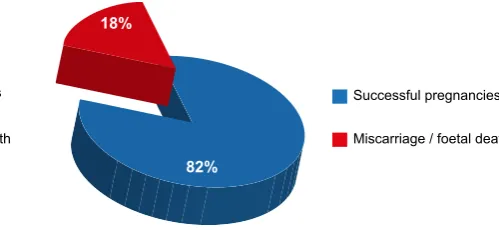
Figure 5 – Pregnancy outcome (pregnant mothers observed at the
CHSJ) Figure 6Rambam Health Care Campus, Israel) – Pregnancy outcome (pregnant mothers observed at the
scale, the risk is somewhat more undervalued.
II) ROC curve and DeLong’s test application to the Hema-Obs and Galit Sarig scales
When the ROC curve was applied to the Hema-Obs scale (Fig. 3) the AUC was 58.8%, compared to the ROC curve applied to the Galit Sarig scale (Fig. 4), where the AUC was 38.7%. There was a highly significant difference between both methods (p = 0.0006175, according to the DeLong’s test).
ROC curves were applied to the same population (250 pregnant mothers attended at the CHSJ).
III) Outcome assessment of the attended pregnancies
In our group of 250 pregnant mothers attended at the CHSJ, 91% (227 pregnant mothers) had successful pregnancies and 9% of miscarriages/foetal death (23 pregnancies) occurred (Fig. 5).
An 82% rate of successful pregnancies (74 pregnancies) and 18% miscarriages / foetal death (16 pregnancies) were found in the 90 pregnancies included in the study by Galit Sarig (Fig. 6).1
DISCUSSION
When compared to the Galit Sarig scale, the Hema-Obs scale showed an overvaluation of the risk as a significantly
higher rate of extremely high risk pregnancies was found (24% vs. 7%).
However, this situation is due to the fact that the risks associated to pregnancy – which are significant - were ignored in the Galit Sarig scale. Therefore, the therapeutic intervention associated to this assessment, milder and above all involving a lower number of pregnant mothers, seems to have had an impact on the miscarriage/foetal death rate, which doubled in the Galit Sarig scale (18%) when compared to the Hema-Obs scale (9%).
The logistic model of our study does not have enough discriminatory power to allow us to claim that the Hema-Obs scale has higher adequacy to risk factors stratification in pregnant mothers with thromboembolic risk. The fact that this scale was applied to a higher number of pregnant mothers may have influenced the study results.
However, we may infer that had the Galit Sarig scale been applied to a group of pregnant mothers attended at the CHSJ, less women would have been treated. Conversely, had the Hema-Obs scale been applied to the group of pregnant mothers at the Rambam Health Care Campus, the treatment group would have increased.
CONCLUSION
Our study allowed for the conclusion that the Hema-Obs scale is an efficient support to therapeutic strategies,
Successful pregnancies Successful pregnancies
Miscarriage / foetal death Miscarriage / foetal death
9% 18%
91% 82%
Specificity (%) Specificity (%)
AUC: 58.8% (95% IC: 48.8% - 68.7%)
AUC: 38.7% (95% IC: 28.6% - 48.8%)
100 100
100 100
80 80
60 60
40 40
20 20
0 0
80 60 40 20 0 80 60 40 20 0
ARTIGO ORIGINAL
improving the outcomes of gestations and in the end the quality of healthcare provided to pregnant mothers and newborns.
CONFLICTS OF INTEREST
The authors declare they had no conflicts of interest in writing this manuscript.
FINANCIAL SUPPORT
The authors declare there was no financial support in writing this manuscript.
REFERENCES
1. Sarig G, Vidergor G, Brenner B. Assessment and management of high-risk pregnancies in women with thrombophilia. Blood Rev. 2009;23:143-7.
2. Bremme K. Haemostasis in normal pregnancy, woman´s issues in thrombosis and hemostasis. London: Martin Dunitz Ldt.; 2002. 3. Gray G, Nelson-Piercy C. Thromboembolic disorders in obstetrics. Best
Pract Res Clin Obstet Gynaecol. 2012;26:53-64.
4. Pabinger I. Thrombophilia and its impact on pregnancy. Hamostaseologie. 2008;28:130-4.
5. Lindqvist PG, Hellgren M. Obstetric thromboprophylaxis: the Swedish guidelines. Adv Hematol. 2011;2011:157483.
6. Macklon NS, Greer IA. Venous thromboembolic disease and gynaecology: the Scottish experience. Scott Med J. 1996;41:83-6. 7. Andersen BS, Steffensen FH, Sorensen HT, Nielsen GL, Olsen J. The
cumulative incidence of venous thromboembolism during pregnancy andpuerperium – an 11 year Danish population – based study of 63,000 pregnancies. Acta Obstet Gynecol Scand. 1998;77:170–3.
8. Lewis G. Why Mothers Die 2000-2002. Sixth Report of the Confidential Enquiries into Maternal Death. London: RCOG Press; 2004.
9. Greer IA. Thrombosis in pregnancy: updates in diagnosis and management, Hematology Am Soc Hematol Educ Program. 2012;2012:203-7.
10. Jacobsen AF, Sandset PM. Venous thromboembolism associated with pregnancy and hormonal therapy. Best Pract Res Clin Haematol. 2012;25319-32.
11. Lindqvist PG, Hellgren M. Obstetric thromboprophylaxis: the Swedish guidelines. Adv Hematol. 2011;2011:157483.
12. Wu P, Poole TC, Pickett JA, Bhat A, Lees CC. Current obstetric guidelines on thromboprophylaxis in the United Kingdom: evidence based medicine? Eur J Obstet Gynecol Reprod Biol. 2013;168:7-11. 13. Martinelli I, Ruggenenti P, Cetin I, Pardi G, Perna A, Vergani P, et al.
Heparin in pregnant woman with previous placenta-mediated pregnancy complications: a prospective, randomized, multicenter, controlled clinical trial. Blood. 2012;119:3269-75.
14. Bennett SA, Bagot CN, Arya R. Pregnancy and trombophilia: the elusive link. Br J Haematol. 2012;157:529-42.
Ana SALSELAS, Inês PESTANA, Francisco BISCHOFF, Mariana GUIMARÃES, Joaquim Aguiar ANDRADE
![Table 1 – Hema – Obs scale [J. Aguiar Andrade (JAA) & Mariana Guimarães (MG)]](https://thumb-us.123doks.com/thumbv2/123dok_us/7814140.2086567/3.567.279.489.497.617/table-hema-obs-scale-aguiar-andrade-mariana-guimaraes.webp)