311
EFFECT OF ORAL ADMINISTRATION OF LACTOBACILLUS PARACASEI L9 ON MOUSE
SYSTEMIC IMMUNITY AND THE IMMUNE RESPONSE IN THE INTESTINE
Yuanbo Zhu1, Jun Zhu2, Liang Zhao2, Ming Zhang3, Huiyuan Guo2 and Fazheng Ren1,*
1College of Food Science and Nutritional Engineering, China Agricultural University, Beijing 100083, China
2Key Laboratory of Functional Dairy, Beijing 100083, China
3School of Food and Chemical Engineering, Beijing Technology and Business University, Beijing 100083, China
*Corresponding author: [email protected]
Received: February 16, 2015; Revised: August 13, 2015; Accepted: September 1, 2015; Published online: March 10, 2016
Abstract: A probiotic strain Lactobacillus paracasei L9, which was isolated from human intestine, was investigated for its immunomodulatory activity in vivo. Results showed that L9 improved systemic immunity by enhancing the phagocytic activity of peritoneal macrophages, the proliferation ratio of splenocytes, the IgG level in the serum and the level of IgA in the mucosa. Further, L9 induced the Th1-polarized immune response by elevating the IFN-γ/IL-4 ratio in the mucosa. This effect was confirmed by the enhanced IL-12-inducing activity of macrophages after in vitro stimulation of L9. Also detected was increased expression of TLR-2 mRNA in the mucosa. We predict that L9 could enhance innate immunity by activating TLR-2 in the mucosa, and enhance acquired immunity by promoting Th1 polarization through induced produc-tion of IL-12 by macrophages.
Key words: Lactobacillus paracasei L9; immunomodulatory activity; systemic immunity; Th1 immunity; TLR-2
INTRODUCTION
Immunomodulatory activity is an important criterion for selecting and evaluating probiotics as it is thought to be responsible for many probiotic effects, such as their anti-inflammatory, anti-infection and antitumor activities [1]. The immunomodulatory capacities of probiotic lactobacilli are strain-specific [2]. Therefore, it has been suggested that the immunomodulatory activity of a specific strain must be evaluated before development and application [3].
Systemic immunity, consisting of innate immu-nity (macrophage phagocytosis, etc.) and acquired immunity, have been widely used to evaluate the im-munomodulatory effect of probiotics [1,4-6]. Gut mucosa is the first line of immune defense [7], where probiotics act to induce innate immunity and acquired immunity [8]. Activation of innate immunity is re-ported to be mediated by Toll-like receptors (TLRs) and is a critical step for the development of acquired
immunity. T helper cells are key components of ac-quired immunity responses [9], which can be polar-ized into T helper type-1 (Th1), T helper type-2 (Th2) or regular T (Treg) cells by specific lactic acid bacteria (LAB) strains [10]. Some LAB strains have been dem-onstrated to induce interleukin (IL)-12 production from dendritic cells (DCs) and macrophages, convert-ing a Th2 response into a Th1-dominated response and suppressing antigen-specific IgE production in mice [11,12], whereas some strains are capable of controlling inflammatory diseases by inducing the development of CD4+ Forkhead box P3 (Foxp3)+ Tregs cells [13].
oral administration of L9 on the systemic immunity and the innate and acquired immunity in the mucosa.
MATERIALS AND METHODS
Bacterial strains and bacteria preparation
The Lactobacillus paracasei L9 (CGMCC NO. 9800) used in this study was isolated from human intestine and stored in our lab. L9 was inoculated in Man, Rog-osa, and Sharpe (MRS) broth under aerobic condition at 37°C for 12 h. Centrifugation (4000 rpm, 15 min) was used to separate the bacterial strain. The bacterial culture was washed twice with saline, and resuspend-ed in saline to prepare the requirresuspend-ed concentration.
Animal groups used in in vivo experiments
For the in vivo experiments, six-week-old male BALB/c mice were kept in a temperature-controlled envi-ronment (22±2°C) with a 12 h light-dark cycle and with free access to water and standard rodent chow throughout the experiment. All animals were treated in accordance with the guidelines on the care and use of animals and with the approval of the Animal Ethics Committee of the China Agricultural University.
After a 5 day acclimation period, the mice were randomly divided into 4 groups, with each group conta-ing 8 mice. The L9(L), L9(M), and L9(H) groups were orally administered a low dose (106 CFU/mL), a me-dium dose (108 CFU/mL), and a high dose (1010 CFU/ mL) of L9, respectively, at a volume of 0.2 mL once a day. The control group received the same volume of sterile saline at the same time. After feeding for 7 weeks, the mice were euthanized by cervical dislocation.
Measurement of serum IgG
Blood was collected from the orbital cavity before eu-thanasia. Samples were allowed to clot at room tem-perature for 3 h and then at 4°C overnight. The sam-ples were centrifuged at 1000 rpm for 10 min to isolate the supernatants. The supernatants were then stored at -20°C until use. IgG concentrations were determined
by enzyme-linked immunosorbent assay (ELISA) kits (USCN Life Science, Wuhan, China) according to the manufacturer’s instructions.
Phagocytosis of peritoneal macrophages
Macrophage phagocytosis of chicken red blood cells (CRBC) was measured as previously described [16]. After the mice were killed, the macrophage suspen-sion was prepared and mixed with equal volumes of 1% CRBC solution. Five hundred μL of the mixture was spread onto glass slides to make a smear for each mouse. The smears were incubated at 37°C for 30 min in a wet box, fixed with methanol for 1 min and then stained by 12.5% (v/v) Giemsa-phosphoric acid dye (Sigma-Aldrich, Shanghai, China) for 15 min. The number of macrophage ingesting CRBC out of a total of at least 100 cells was calculated by direct visual enu-meration using a light microscope. The phagocytosis ratio of macrophages was defined as in Eq.1.
× 100%
Splenic lymphocyte proliferation assay
= (ODConA-ODcontrol) / (ODConA--OD
control) × 100%, where ODConA, ODcontrol and ODConA- represent the OD value of wells containing cells, CCK-8 and ConA, wells containing CCK-8 and wells containing cells and CCK-8, respectively.
Secretory IgA (sIgA) concentration in the intestinal mucus
The concentration of SIgA in the intestinal mucus was measured as described in a previous report, with modifications [18]. A 5-cm length of intestine tissue of the ileum was obtained from each animal, dis-sected and washed with saline. Intestinal mucus was collected and centrifuged at 700 g for 10 min after being dissolved in 1 mL of phosphate-buffered saline (PBS). The supernatant was removed and centrifuged at 12000 g for 15 min. The supernatant was taken for measurement of sIgA by commercial ELISA kit (USCN Life Science, Wuhan, China) following the manufacturer’s instruction. The total protein content of intestinal mucus was assayed by bicinchoninic acid (BCA) kit (Takara, Beijing, China) against a bovine serum albumin (BSA) standard curve, simultaneously. The concentration of sIgA was expressed as micro-gram per millimicro-gram of protein.
Gene expression in mucosa
Real-time reverse transcription polymerase chain reac-tion (RT-PCR) assays were used to evaluate mRNA ex-pression of TLR-2, interferon (IFN)-γ, IL-4, Foxp3 and transforming growth factor (TGF)-β. After collecting mucus, the intestinal segment was scraped with a cov-erslip to obtain the mucosa [19]. The collected mucosa was stored by freezing at -80°C. Total RNA was extract-ed from the mucosa using TRIzol reagent (Life Tech-nologies, Carlsbad, CA, USA) and reverse transcribed using MMLV reverse transcriptase (Promega, Madison, WI, USA) to generate cDNA for use in RT-PCR. Each cDNA sample was stored at -20°C until further real-time PCR analysis. The specific oligonucleotide primers were synthesized by Invitrogen. The primer sequences for each gene are shown in Table 1. Real-time PCR
reac-tions were performed using the Roche Light Cycler In-strument 1.5 using Light Cycler Fast Start DNA Master PLUSSYBR Green I kit (Roche, Castle Hill, Australia). Briefly, the 20-μL reaction mixtures contained 10 μL of Master Mix, 0.4 μL of 0.75 μM forward primer and reverse primer, 2 μL of the cDNA sample and 7.6 μL of Milli Q water. Each sample was run in duplicate. The RT-PCR program was at 95°C for 30 s, 40 cycles at 95°C for 15 s, 60°C for 30 s and 72°C for 30 s. At the end of the program, melt curve analysis was performed. The fold expression or repression of the target gene relative to the internal control gene (GAPDH) in each sample was calculated by the formula:
2-∆∆crossing point (CP),
where ∆Cp = Cp target gene – Cp internal control and ∆∆Cp = ∆Cp test sample - ∆Cp control sample.
Assay for IL-12-inducing activity in vitro
Mouse peritoneal macrophages were obtained from male BALB/c mice as reported before [16]. Mouse macrophages (2×10⁵ cells) were stimulated with L9 at three doses (2×10⁴ CFU, 2×10⁵ CFU and 2×10⁶ CFU) for 24 h. The highest value of IL-12 production was extrapolated as the IL-12-inducing activity. Culture supernatants were collected and IL-12 levels measured by ELISA kits (USCN Life Science, Wuhan, China) according to the manufacturer’s instructions.
Table 1. Primer sequences used for RT-PCR.
Gene Primer sequence Reference
IL-4 Forward:5'-GGTCTCAACCCCCAGCTAGT-3'
[26] Reverse:5'-GCCGATGATCTCTCTCAAGTGAT-3'
IFN-γ Forward:5'-AGCGGCTGACTGAACTCAGATTGTAG-3'
[27] Reverse:5'-GTCACAGTTTTCAGCTGTATAGGG-3'
TGF-β Forward:5'-GGCGGTGCTCGCTTTGTA-3'
[28] Reverse:5'-GTTGTTGCGGTCCACCATTAG-3'
Foxp3 Forward:5'-CTCATGATAGTGCCTGTGTCCTCAA-3' [29]
Reverse:5'-AGGGCCAGCATAGGTGCAAG-3'
TLR-2 Forward:5'-TCTAAAGTCGATCCGCGACAT-3'
This study Reverse:5'-CTACGGGCAGTGGTGAAAACT-3’
GAPDH Forward:5'-GTGTTCCTACCCCCAATGTGT-3’
Statistical analysis
The results were expressed as means±standard devia-tion (SD). Data analysis was carried out using SPSS software, version 20.0. Differences among groups were compared using one-way ANOVA tests followed by Duncan’s post hoc test. Values of P<0.05 were con-sidered significant.
RESULTS
Effects of oral administration of L9 on systemic immunity in mice
The phagocytosis ratio of peritoneal macrophage to CRBC was determined to evaluate the innate immune response. As shown in Fig. 1, the phagocytosis ratio of mice fed with L9(M) and L9(H) were significantly higher than the control group (P<0.05). The L9(M) group had a slightly but not significantly higher phagocytosis ratio than L9(H) (P>0.05).
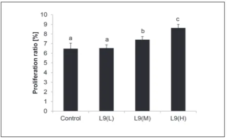
As an important index of cellular immunity in the acquired immune response, the proliferation ratio of splenic lymphocytes was detected. Viability of splenic lymphocytes exceeded 95% of all cell populations in all cases. No significant difference in splenocyte prolif-eration ratio was found between the L9(L) group and the control group (P>0.05; Fig. 2). The proliferation ratio of mice fed with L9(M) and L9(H) was signifi-cantly higher than that of the control mice (P<0.05).
B-cell function, a symbol of humoral immunity in the acquired immune response, was investigated by measuring the concentration of IgG in serum. Results showed that IgG production was significantly increased in mice fed with high doses of L9 (P<0.05; Fig. 3), while no significant changes were observed in mice fed with the medium and low doses of L9 (P>0.05).
The concentration of sIgA in the intestinal mucus was measured by ELISA (Fig. 4). In comparison to the control mice, sIgA concentrations were signifi-cantly increased in mice fed with the high dose of L9 (P<0.05), but not in mice fed with a medium or a low doses of L9 (P>0.05).
Effects of oral administration of L9 on the expression level of receptor TLR-2 mRNA in the mucosa
The expression of TLR-2 mRNA in the mucosa was measured to investigate whether the LAB strain could initiate the immune response through this receptor. As shown in Table 2, significant increases in the mRNA
Fig. 2. Proliferation of splenic lymphocytes from untreated
con-trols and mice treated with L9(L), L9(M) and L9(H) in response to ConA. Splenocytes were prepared and cultured with or without ConA for 72 h. Splenic lymphocyte proliferation was measured by the modified MTT method as described in the text, and is shown as a proliferation ratio. The values are presented as means±SD. a,b,c – data marked with different letters are significantly different at P<0.05
Fig. 1. Effect of administration of L9(L), L9(M), L9(H) and saline
expression of TLR-2 were found in L9-treated mice (P<0.05). Moreover, the L9(H) group had a signifi-cantly higher level of TLR-2 mRNA expression than the L9(L) and L9(M) groups (P<0.05).
Effects of oral administration of L9 on T helper cell polarization in the mucosa
To determine the influence of the tested LAB strain, we examined the expression of mRNAs for IFN-γ, IL-4, and TGF-β and Foxp3, representative proteins of Th1-, Th2- and Treg-type immune responses, re-spectively. As shown in Table 2, the L9(M) and L9(H) groups exhibited a relatively high ability of enhancing IFN-γ mRNA expression (P<0.05), unlike the L9(L) group (P>0.05), as compared to the control group. The L9(H) and L9(M) groups had equivalent IFN-γ mRNA levels (P>0.05). The expression of IL-4 mRNA exhibited an opposite tendency to that of IFN-γ.
Spe-cifically, the L9(L), L9(M) and L9(H) groups displayed a slightly but not significantly lower level of IL-4 mRNA than the control group (P>0.05). The IFN-γ/ IL-4 ratios in the L9(M) and L9(H) groups were sig-nificantly higher than the control group (P<0.05). The mRNA expression of TGF-β and Foxp3 were not sig-nificantly different (P>0.05) in all of the tested groups (Table 2).
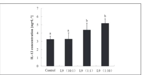
Effect of L9 on the IL-12-induced activity of macrophages in vitro
Judging by the elevated IFN-γ/IL-4 ratio, the L9 strain could induce a Th1-polarized immune response. To confirm the induction of Th1-type immunity by the L9 strain, the IL-12-inducing activity of macrophages was examined in vitro. The results in Fig. 5 show that the concentration of IL-12 was significantly increased after the macrophages were stimulated with the L9
Fig. 3. Production of IgG antibodies from mice fed with L9(L),
L9(M), L9(H) and saline. The concentration of IgG was deter-mined using ELISA. All data are presented as means±SD. a,b – data marked with different letters are significantly different at P<0.05
Fig. 4. Effect of oral administration of LGG, L9(L), L9(M), L9(H)
and saline on sIgA production in the intestines. sIgA concentra-tion in the intestinal mucus was detected by ELISA. The values are presented as means±SD. a,b – data marked with different letters are significantly different at P<0.05
Table 2. Effects of LAB on the expression of mRNAs for TLR-2, Foxp3, TGF-β, IFN-γ, and IL-4, and on the IFN-γ/IL-4 ratio in the mucosa.
mRNA expression
TLR-2 Foxp3 TGF-β IFN-γ IL-4 IFN-γ/IL-4
Control 1±0.24a 1.00±0.32 1.00±0.42 1±0.43a 1±0.22 1±0.11 a
L9(L) 1.49±0.23b 0.87±0.28 0.87±0.35 1.03±0.24a 0.91±0.28 1.02±0.13 a
L9(M) 1.43±0.30b 0.93±0.24 0.87±0.28 2.31±0.46b 0.76±0.16 2.17±0.35 b
L9(H) 1.87±0.27c 0.85±0.26 0.94±0.41 1.70±0.57b 0.75±0.16 2.15±0.58 b
strain, at 1:1 and 1:10 macrophage cell : L9 strain ra-tios (P<0.05). Meanwhile, the IL-12 concentration rose with the increment in L9 dose. As described in the Materials and Methods, the highest value of IL-12 production was extrapolated as the IL-12-inducing activity. Therefore, the IL-12-inducing activity of the macrophages was significantly increased after they were stimulated with the L9 strain (P<0.05).
DISCUSSION
To evaluate the immunomodulatory activity of L9, we administered different doses of L9 to mice and detect-ed the effect of L9 on the systemic immune response
in vivo. Our results show that L9 enhanced innate immunity by elevating phagocytosis of macrophages, strengthening the acquired immunity by improving splenic lymphocyte proliferation and increasing the serum IgG concentration and the concentration of sIgA in the mucus. It was suggested that strains of LAB that are capable of affecting a wider array of im-mune functions are likely to be more beneficial to human health [6]. L9 was shown to be a beneficial strain, capable of enhancing many aspects of systemic immunity.
Probiotics are known to be not only involved in activating the systemic immune response, but also the mucosal immune response. Gut epithelial and immune cells are continually sampling gut microbes, and
bac-terial strains can signal through pattern-recognition receptors, resulting in the modulation of various in-tracellular signaling pathways [20]. Our results showed L9-treated mice to have a higher IFN-γ/IL-4 ratio than the control group and the mRNA expression of TGF-β and Foxp3 did not significantly changed in the mucosa, which suggested that L9 tends to shift towards a Th1 cytokine profile, namely the Th1 polarized strain. It is speculated that probiotics induce the IL-12 produc-tion of gut macrophages; thereafter, IL-12 promotes the differentiation of naive CD4+ T cell into Th1 cells. We demonstrated that the IL-12-producing activity of macrophages was enhanced by L9 in vitro. According to the above results, we arrived at the conclusion that L9 can promote Th1 polarization by inducing IL-12 production by macrophages in mucosa.
Th1 cells and cytokines, including IL-12 and IFN-γ, play crucial roles in host health. Enhancement of their activities could protect against viral infection, alleviate diseases and suppress tumors [21]. We also suggest that enhanced systemic immune effects, which include lymphocyte proliferation and phagocytosis of macrophages can be attributed to the enhancement of Th1-type immune activity, since the Th1-type cy-tokines, IL-12 and IFN-γ, could promote lymphocyte proliferation and macrophage activity, respectively. In addition to the beneficial effects of a Th1-polar-ized immune response, constitutive Th1 activation is associated with autoimmune diseases. However, previous reports revealed that oral administration of probiotic LAB did not induce pathological inflamma-tion in murine models [22,23]. While the high dose of L9 (1010cfu/mL) used in this study was much higher than the dose recommended for human consump-tion (106cfu/mL), it did not cause overregulation of the Th1-type immune response. More importantly, the safety assessment of L9 done by CTC supports its safety in oral administration.
TLR-2s are known to be a family of receptors that play crucial roles in the innate immune system, where some LAB can lead to the activation of the Th1 im-mune response [24]. Our results showed that L9 could enhance TLR-2 mRNA expression, suggesting that L9 could initiate the immune response of the host
Fig. 5. Effect of different doses of L9 strain on IL-12-producing
through TLR-2. Furthermore, it was shown that Lacto-bacillus delbrueckii TUA4408L and its extracellular polysaccharides exerted immunomodulatory effect via TLR-2 and 4 [25]. We observed that L9 could produce a large amount exopolysaccharides (EPS) when com-pared with other LABs; thus, L9 could be recognized by TLR-2 through EPS, initiating the subsequent im-mune response.
In summary, we demonstrated that L9 can en-hance systemic immunity, as characterized by in-creased phagocytosis and splenocyte proliferation ratios, and increased concentrations of IgG in the serum and sIgA in the mucus. L9 induced Th1 po-larization, which led us to conclude that L9 might pro-mote Th1 polarization by inducing IL-12 production by macrophages via TLR-2 activation in the mucosa. The presented data demonstrate that L9 is an immu-nomodulating strain that can enhance systemic im-munity, and it is very effective in activating the Th1 immune response in the mucosa.
Acknowledgments:This study was supported by Beijing
Mu-nicipal Commission of Education Co-constructed program, and Beijing Science and Technology Project (D141100004814001)
Authors’ contributions: Fazheng Ren conceived the experiment.
Yunabo Zhu designed and performed the experiment, analyzed data and wrote paper together with Jun Zhu. Liang Zhao and Ming Zhang modified and polished the manuscript. Huiyuan Guo provided essential materials and was involved in experi-ment discussion
Conflict of interest disclosure: The authors have declared that
no conflict of interests exists.
REFERENCES
1. Tsai YT, Cheng PC, Fan CK, Pan TM. Time-dependent per-sistence of enhanced immune response by a potential probi-otic strain Lactobacillus paracasei subsp. paracasei NTU 101. Int J Food Microbiol. 2008;128(2):219-25.
2. Klaenhammer TR, Kleerebezem M, Kopp MV, Rescigno M. The impact of probiotics and prebiotics on the immune sys-tem. Nat Rev Immunol. 2012;12(10):728-34.
3. Ya T, Zhang Q, Chu F, Merritt J, Bilige M, Sun T, Du R, Zhang H. Immunological evaluation of Lactobacillus casei
Zhang: a newly isolated strain from koumiss in Inner Mon-golia, China. BMC Immunol. 2008;9(1):68.
4. Roller M, Rechkemmer G, Watzl B. Prebiotic inulin enriched with oligofructose in combination with the probiotics Lac-tobacillus rhamnosus and Bifidobacterium lactis modulates intestinal immune functions in rats. J Nutr. 2004;134(1):153-6.
5. Wei H, Xu Y, Cheng B, Xiong Y. Synergistic effects of Lacto-bacillus rhamnosus ZDY114 and bovine colostrums on the immunological function of mouse in vivo and in vitro. Appl Microbiol Biotechnol. 2007;75(2):427-34.
6. Gill HS, Rutherfurd KJ, Prasad J, Gopal PK. Enhancement of natural and acquired immunity by Lactobacillus rhamnosus
(HN001), Lactobacillus acidophilus (HN017) and Bifidobac-terium lactis (HN019). Br J Nutr. 2000;83(2):167-76. 7. Tsai YT, Cheng PC, Liao JW, Pan TM. Effect of the
adminis-tration of Lactobacillus paracasei subsp. paracasei NTU 101 on Peyer’s patch-mediated mucosal immunity. Int Immuno-pharmacol. 2010;10(7):791-8.
8. Shida K, Nanno M. Probiotics and immunology: separating the wheat from the chaff.Trends Immunol. 2008;29(11):565-73.
9. Kawashima T, Hayashi K, Kosaka A, Kawashima M, Igarashi T, Tsutsui H, Tsuji NM, Nishimura I, Hayashi T, Obata A.
Lactobacillus plantarum strain YU from fermented foods activates Th1 and protective immune responses. Int Immu-nopharmacol. 2011;11(12):2017-24.
10. Baken KA, Ezendam J, Gremmer ER, de Klerk A, Pennings JL, Matthee B, Peijnenburg AA, van Loveren H. Evaluation of immunomodulation by Lactobacillus casei Shirota: immune function, autoimmunity and gene expression. Int J Food Microbiol. 2006;112(1):8-18.
11. Nagao F, Nakayama M, Muto T, Okumura K. Effects of a fermented milk drink containing Lactobacillus casei strain Shirota on the immune system in healthy human subjects. Biosci Biotechnol Biochem. 2000;64(12):2706-8.
12. Pochard P, Gosset P, Grangette C, Andre C, Tonnel A-B, Pes-tel J, Mercenier A. Lactic acid bacteria inhibit TH2 cytokine production by mononuclear cells from allergic patients. J Allergy Clin Immunol. 2002;110(4):617-23.
13. Kwon HK, Lee CG, So JS, Chae CS, Hwang JS, Sahoo A, Nam JH, Rhee JH, Hwang KC, Im SH. Generation of regu-latory dendritic cells and CD4+Foxp3+ T cells by probiot-ics administration suppresses immune disorders. Proc Natl Acad Sci U S A. 2010;107(5):2159-64.
14. Pan T, Guo HY, Zhang H, Liu AP, Wang XX, Ren FZ. Oral administration of Lactobacillus paracasei alleviates clinical symptoms of colitis induced by dextran sulphate sodium salt in BALB/c mice. Benef Microbes. 2014;5(3):315-22. 15. Zhou XD, Liu AP, zhang M, Guo HY, Ren FZ. Laxative effect
of Lactobacillus paracasei subsp. paracasei LC-01 in consti-pated mice.J Dairy Sci Technol. 2012;35(5):7-11.
17. Bujalance C, Moreno E, Jimenez-Valera M, Ruiz-Bravo A. A probiotic strain of Lactobacillus plantarum stimulates lym-phocyte responses in immunologically intact and immuno-compromised mice. Int J Food Microbiol. 2007;113(1):28-34. 18. Zhong Y, Cai D, Cai W, Geng S, Chen L, Han T. Protective
effect of galactooligosaccharide-supplemented enteral nutri-tion on intestinal barrier funcnutri-tion in rats with severe acute pancreatitis. Clin Nutr. 2009;28(5):575-80.
19. Godinez-Victoria M, Drago-Serrano ME, Reyna-Garfias H, Viloria M, Lara-Padilla E, Resendiz-Albor AA, Sánchez-Torres LE, Cruz-Hernández TR, Campos-Rodriguez R. Effects on secretory IgA levels in small intestine of mice that underwent moderate exercise training followed by a bout of strenuous swimming exercise. Brain Behav Immun. 2012;26(8):1300-9.
20. Wallace TC, Guarner F, Madsen K, Cabana MD, Gibson G, Hentges E, Sanders ME. Human gut microbiota and its rela-tionship to health and disease. Nutr Rev. 2011;69(7):392-403. 21. Takeda S, Kawahara S, Hidaka M, Yoshida H, Watanabe W,
Takeshita M, Kikuchi Y, Bumbein D, Muguruma M, Kuro-kawa M. Effects of oral administration of probiotics from Mongolian dairy products on the Th1 immune response in mice. Biosci Biotechnol Biochem. 2013;77(7):1372-8. 22. Zhou JS, Gill HS. Immunostimulatory probiotic
Lactobacil-lus rhamnosus HN001 and Bifidobacterium lactis HN019 do not induce pathological inflammation in mouse model of experimental autoimmune thyroiditis. Int J Food Microbiol. 2005;103(1):97-104.
23. Kobayashi T, Kato I, Nanno M, Shida K, Shibuya K, Mat-suoka Y, Onoue M. Oral administration of probiotic bacte-ria, Lactobacillus casei and Bifidobacterium breve, does not exacerbate neurological symptoms in experimental autoim-mune encephalomyelitis. Immunopharmacol Immunotoxi-col. 2010;32(1):116-24.
24. Gomez-Llorente C, Munoz S, Gil A. Role of Toll-like recep-tors in the development of immunotolerance mediated by probiotics. Proc Nutr Soc. 2010;69(3):381-9.
25. Wachi S, Kanmani P, Tomosada Y, Kobayashi H, Yuri T, Egusa S, Shimazu T, Suda Y, Aso H, Sugawara M, Saito T, Mishima T, Villena J, Kitazawa H. Lactobacillus delbrueckii
TUA4408L and its extracellular polysaccharides attenu-ate enterotoxigenic Escherichia coli-induced inflammatory response in porcine intestinal epitheliocytes via Toll-like receptor-2 and 4. Mol Nutr Food Res. 2014;58(10):2080-93. 26. Ydens E, Cauwels A, Asselbergh B, Goethals S, Peeraer L,
Lornet G, Almeida-Souza L, Van Ginderachter JA, Timmer-man V, Janssens S. Acute injury in the peripheral nervous system triggers an alternative macrophage response. J Neu-roinflammation. 2012;9:176.
27. Kolls JK, Habetz S, Shean MK, Vazquez C, Brown JA, Lei D, Schwarzenberger P, Ye P, Nelson S, Summer WR, Shellito JE. IFN-γ and CD8+ T cells restore host defenses against Pneu-mocystis carinii in mice depleted of CD4+ T cells. J Immunol. 1999;162(5):2890-4.
28. Xie F, Sakwiwatkul K, Zhang C, Wang Y, Zhai L, Hu S. Atrac-tylodis macrocephalae Koidz. polysaccharides enhance both serum IgG response and gut mucosal immunity. Carbohydr Polym. 2013;91(1):68-73.
29. Zhong Y, Wang X, Ji Q, Mao X, Tang H, Yi G, Meng K, Yang X, Zeng Q. CD4+LAP + and CD4 +CD25 +Foxp3 + regulatory T cells induced by nasal oxidized low-density lipoprotein suppress effector T cells response and attenu-ate atherosclerosis in ApoE-/- mice. J Clin Immunol. 2012;32(5):1104-17.