Original Research Article
Solid malignant pediatric tumors: a histopathological study
Neelu Gupta
1, Monika Choudhary
1*, Sandeep Pachar
2,
Jai prakash Dhaka
1, Roshan Verma
1INTRODUCTION
Cancer is essentially a disease of adults, yet it is one of the common killers in childhood. In western countries cancer is next only to trauma as a cause of mortality in children under 15 years of age. In India, although infections and malnutrition are the major factors contributing to morbidity and mortality, with the development of preventive and curative measures of treatment, as well as fight against the malnutrition, malignant tumors in children have become the second
biggest killer.1-3 In general, the features of malignancies
in children differ biologically and histologically from those of adults with respect to incidence, type of tumor, underlying familial or genetic aberration and tendency to regress spontaneously or cytodifferantiate.4 In recent
years, identification of specific genes, oncogenes, tumor markers and other biological and pathological factors have played an important role in staging and classifying risk categorization of specific tumors as low, intermediate and high-risk lesions. This concept uses risk factors as predictors of outcomes. Risk-based management allows
ABSTRACT
Background: In general, the features of malignancies in children differ biologically and histologically from those of adults with respect to incidence, type of tumor, underlying familial or genetic aberration and tendency to regress spontaneously or cytodifferentiation. In recent years, identification of specific genes, oncogenes, tumor markers and other biological and pathological factors have played an important role in staging and classifying risk categorization of specific tumors as low, intermediate and high-risk lesions.
Methods: This study is under taken to evaluate the incidence and morphological features of solid malignant tumors in children of fifteen years and below. The material for present study is obtained from S. P. Medical College, Bikaner and referred cases. The histopathology slides and paraffin blocks are reviewed.
Results: The sections 3-5 μ thick, are cut and stained by haematoxylin and eosin in all cases and special stains like PAS, MTS, RS done where ever feasible. An analysis of 60 cases of solid malignant tumors of childhood over a period of 5years are made. The early onset and the embryonal nature of the major pediatric tumors, suggest a prenatal origin and role of genetic factors. Infections, exposure to drugs and chemicals during pregnancy are other contributory factors.
Conclusions: Accurate incidence of data is important in the planning and evaluation of clinical trials. Documentation of cases, advanced diagnostic methods like IHC, cytogenetic studies and treatment modalities with close follow up is needed to achieve better statistical evaluation of the problem.
Keywords: Histopathology, Malignant, Pediatric, Solid
1Department of Pathology, 2Department of Respiratory Medicine, Sardar Patel Medical College, Bikaner, Rajasthan,
India
Received: 22 January 2017 Accepted: 17 February 2017
*Correspondence: Dr. Monika Choudhary,
E-mail: [email protected]
Copyright: © the author(s), publisher and licensee Medip Academy. This is an open-access article distributed under the terms of the Creative Commons Attribution Non-Commercial License, which permits unrestricted non-commercial use, distribution, and reproduction in any medium, provided the original work is properly cited.
the pediatric medical and surgical oncologist to weigh the risks and benefits of treatment for each patient in an effort to maximize survival, minimize morbidity, and improve the quality of life. Hence there is need for accurate histopathological reporting in conjugation with ancillary methods.5-8
METHODS
This study is under taken to evaluate the incidence and morphological features of solid malignant tumors in children of fifteen years and below. The material for present study is obtained from S. P. Medical College, Bikaner and referred cases for a period of five years from 2012-2016. The clinical history regarding duration of the disease, mode of presentation, symptoms and signs are recorded from the case papers, request forms, patient’s history, clinical data along with relevant details obtained from available hospital and departmental records. The histopathology slides and paraffin blocks are reviewed. Gross examination is done carefully noting the size, shape, extent and configuration, nodularity, consistency (solid, cystic or mixed) and torsion. A minimum of 4-5
bits are selected from the representative areas of tumor. The tissue for routine microscopy is preserved and fixed in 10% neutral buffered formalin for 24 hours and processed in automatic tissue processor (Histokinette) and embedded in paraffin. The sections 3-5 μ thick, are cut and stained by haematoxylin and eosin in all cases and special stains like PAS, MTS, and RT done where ever feasible.
RESULTS
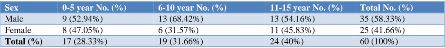
An analysis of 60 cases of solid malignant tumors of childhood over a period of 5 years is made. The following observations are made: The youngest patient at the time of diagnosis is three days old and diagnosed as immature teratoma. The majority of the tumors in this study occurred between 11-15 years (41.66%), followed by 6-10 years age group (30%) and in 0-5 years age group (28.33%) (Table 1). In the present study 35 (58.33%) malignant tumors are seen in boys and 25 (41.66%) in girls under the age of 15 years, showing male preponderance. Male to female ratio of the incidence of malignant tumors is 1.5:1.
Table 1: Incidence of malignant tumors in different age groups in relation to sex.
Sex 0-5 year No. (%) 6-10 year No. (%) 11-15 year No. (%) Total No. (%)
Male 9 (52.94%) 13 (68.42%) 13 (54.16%) 35 (58.33%)
Female 8 (47.05%) 6 (31.57%) 11 (45.83%) 25 (41.66%)
Total (%) 17 (28.33%) 19 (31.66%) 24 (40%) 60 (100%)
Table 2: Histological subtypes of tumor with relation to sex.
Histological subtypes Male (%) Female (%) Total (%) M:F ratio
Lymphoma 17 6 23 (38.33%) 2.8:1
Hodgkin lymphoma Non-Hodgkin’s lymphoma
11 6
5 1
16 (26.66%) 7 (11.66%)
Bone tumors 3 6 9 (15%)
Osteosarcoma
Ewing’s sarcoma/PNET 3 0
2 4
5 (8.33%) 4 (6.66%)
Gonadal tumors 4 3 7 (11.66%) 1.33:1
CNS tumors 1 4 5 (8.33%) 1:4
Retinoblastoma 0 2 2 (3.33%) -
Wilm’s tumor 5 0 5 (8.33%) -
Soft tissue tumors 2 3 5 (8.33%) 2:3
Neuroblastoma 2 0 2 (3.33%) -
Squamous cell carcinoma eye 1 0 1 (1.66%) -
Malignant melanoma 0 1 1 (1.66%) -
Total 35 (58.33%) 25 (41.66%) 60 (100%) 1.5:1
All the cases of present study are sporadic. No associated congenital anomalies are noted in the present study. Histological typing of tumours are depicted in Table 2. Table 2 shows the histological types of the solid
(15%). This is followed by Gonadal tumors which accounted for 7 cases (11.66%), CNS tumors, soft tissue tumors and Wilm’s tumor (5,8.33% each). The relative frequency of other malignant neoplasms is as shown in Table 2.
Lymphoma
Twenty-three cases of lymphoma are diagnosed during the study period. These accounts for 38.66% of all the pediatric malignant solid neoplasms seen in this study. Seventeen cases (73.91%) are seen in males while 6 cases (26.08%) are seen in females. This give a male: female ratio of 2.8:1. Two cases (8.69%) are seen in the 0-5-year age group, 12 cases (52.17%) are seen in the 6-10 year age group while 9 cases (39.13%) are seen in the 11-15 year age group (Table 3).
Hodgkin’s lymphoma accounts for 16 cases (69.56% of all lymphomas) while other non-Hodgkin’s lymphomas account for 7 cases. (30.43% of all lymphomas) (Figure 1-4).
Table 3: Age and sex wise distribution of lymphoma.
Age group (in years) Male Female Total (%)
0-5 0 2 2 (8.69%)
6-10 10 2 12 (52.17%)
11-15 7 2 9 (39.13%)
Total 17 2 23 (38.33%)
Figure 1: Lymph node- mixed cellularity Hodgkin lymphoma. High power view (H and E staining) showing several Hodgkin cells with polymorphic
lymphoid infiltrate rich in eosinophils.
Figure 2: Lymph node- nodular sclerosis Hodgkin lymphoma. Scanner view (H and E staining) showing
lymphoid nodules in dense fibro hyaline tissue.
Figure 3: Classical reed sternberg cell high power view (H and E staining).
Figure 4: Lymphocytic-histiocytic reed sternberg cell. High power view (H and E staining).
DISCUSSION
The various malignant tumors of childhood encountered in the present study are compared with similar studies conducted in India and abroad. The malignant tumors of all types are being reported during early life but their common site of origin differ sharply from those of adults, for example Leukemias, CNS tumors, soft tissue tumors, bone and kidney tumors are common sites of origin of malignant tumors in infants and children.
Accurate diagnosis of pediatric small-round cell tumors is important, as disparate approaches to therapy are taken for distinct tumor types. In addition, therapy is also tailored according to patient risk. It has become important to further classify tumors biologically, using cytogenetic
or molecular studies to identify chromosome
translocations, gene amplification, gene expression patterns, and/or mutations.9
The overall incidence of malignant tumors of childhood is more in the males (M: F: 1.5: 1). This observation has been made uniformly in literature by many authors. Male preponderance is noted in all age groups and female preponderance in germ cell tumors by Lee and Lee et al, Miller et al.10,11 In the present study, the peak occurrence
of tumors is found in the 10-15 years age group (40%) similar to the observations of KK Jain (35.5 %).3,11,12
Whereas Dewani et al and Jussawala and Yeole observed peak occurrence in birth-5 age group as shown in Table 4.13 The frequency of neuroblastoma, retinoblastoma and
5 years of age. An increased frequency with age is seen in
Non-Hodgkin’s lymphoma, Hodgkin’s lymphoma,
osteosarcoma and Ewing’s sarcoma. The early onset and the embryonal nature of many pediatric tumors suggest a prenatal origin, as also seen in our studies.14
Table 4: Comparison of age group distribution of tumors in various studies.
Series 0-5 years 6-10 years 11-15 years
Jain KK 4.2% 22.2% 35.5%
Dewani 47.2% 40.9% 11.9%
Jussawala 42% 29% 29%
Present study 28.33% 31.66% 40%
Various types of solid malignant tumors are observed in the pediatric age group in the present study, the
lymphomas being the commonest type, followed by soft tissue tumor, germ cell tumor, bone tumors, Wilms’ tumor, brain tumor, retinoblastoma and others.
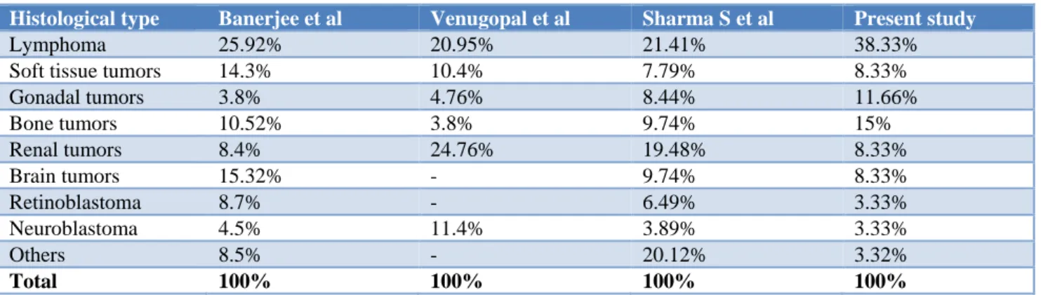
In the present study, the commonest tumours comprised Hodgkin’s (11, 16.66%) and non-Hodgkin’s lymphomas (10, 15.15%). Similar results were reported by Sonal Sharma et al, and Baneerjee and Walia.1,15
The general pattern of pediatric tumors in our center seems to resemble the distribution of pediatric cancers reported from other centers in our country and abroad. Leukemia was the leading type of cancer and malignant tumors were the second most common cancer in several countries. The same findings are noted in our series and Young et al, Pearson et al, Teppo et al and Kusumakumary P et al.14,16-18
Table 5: Histological types of tumor in different series.
Histological type Banerjee et al Venugopal et al Sharma S et al Present study
Lymphoma 25.92% 20.95% 21.41% 38.33%
Soft tissue tumors 14.3% 10.4% 7.79% 8.33%
Gonadal tumors 3.8% 4.76% 8.44% 11.66%
Bone tumors 10.52% 3.8% 9.74% 15%
Renal tumors 8.4% 24.76% 19.48% 8.33%
Brain tumors 15.32% - 9.74% 8.33%
Retinoblastoma 8.7% - 6.49% 3.33%
Neuroblastoma 4.5% 11.4% 3.89% 3.33%
Others 8.5% - 20.12% 3.32%
Total 100% 100% 100% 100%
Predominance of Hodgkin’s lymphoma over non-Hodgkin’s lymphoma correlates with studies made by Mankodi et al, Ramkumar et al and Desai et al.20-22
Majority of our cases are in 10-15 years. This is similar to observation by SEER, Mankodi et al, and Baneerjee and
Walia.1,20,23 Nodular sclerosis subtype was the
commonest subtype in SEER study and age group was 1-5 years in Venugopal et al study.19,23 Mixed cellularity is
the predominant histological subtype in present study and other studies from India and abroad.
Five cases (8.33%) of Wilms’ tumor are studied. This is in comparison to study done by Sanyal et al and Sunitha Sharma et al.24,25 The tumor stage at diagnosis,
histological features and patient age are the most important prognostic determinants which have impact on treatment selection and outcome. The Loss of Heterozygosity at chromosome 1p and 16q was associated with increased risk of relapse and death.26
In retinoblastoma, no bilateralism or family history is noted in present study. Optic nerve spread indicating bad prognosis was noted by Sang and Albert and Dhir SP.27,28
Optic nerve extension and calcification is noted in one of our case.
Neuroblastoma is the commonest tumor of early childhood and is rare beyond the age of ten years.29
Neuroblastoma, a highly aggressive tumor, was reported to have lower incidence among the Indian children by various authors like Dewani GP, Jussawala DJ and Mangal N.3,12,30 In the present study the tumor constituted
3.03% of total cases. The neuroblastic tumors NB, GNB, and GN are a spectrum of sympathetic tissue tumors ranging from the very immature and malignant NB to the mature and benign GN. It is the interplay of histologic maturity, genetic composition, and patient features that make NB, GNB and GN an enigmatic group of tumors.
CONCLUSION
of genetic factors. Infections, exposure to drugs and chemicals during pregnancy are other contributory factors. Accurate incidence of data is important in the planning and evaluation of clinical trials. Documentation of cases, advanced diagnostic methods like IHC, cytogenetic studies and treatment modalities with close follow up is needed to achieve better statistical evaluation of the problem.
Funding: No funding sources Conflict of interest: None declared Ethical approval: Not required
REFERENCES
1. Baneerjee CK, Walia BNS. Pattern of neoplasms in
childhood. Indian J Paediatr. 1986;53:93-7.
2. Atlman AJ, Schwartz AD. Malignant disease of
infancy, childhood and adolescence. In Major Problems in Clinical Paediatrics. WB Saunders, Philadelphia. 1978;18:1-102.
3. Dawani GP, Tandon PL, Ghooi AM, Jain PK.
Malignant tumours of infancy and childhood. Indian J Surg. 1972;34;460-8.
4. Kumar V, Fausto N, Abbas A. Pathologic basis of disease, Elsevier; 7th edition; 2004.
5. Variend S. Small cell tumors in childhood a review.
J Pathol. 1985;45:1-25.
6. Kliegman RM, Behrman RE, Jenson HB, Stanton
BF. Nelson Textbook of Paediatrics. 18th edition, WB Saunders; 2007.
7. Anderson WAD, Kissane JM. Pathology. 7th
edition, Volume 1 and 2, C.V. Mosby co. sty. Louis; 1977.
8. Boyd WC. Textbook of Pathology: Structure and
Function in Diseases. 9th edition, Lea and Febiger, Philadelphia; 1979.
9. Cohn SL. Diagnosis and classification of the small round-cell tumors of childhood. Am J Pathol. 1999;155(1):11-4.
10. Lee CK, Lee SK. Malignant solid tumors of infancy
and childhood in Korea. GANN Monograph on cancer research, University of Tokyo Press; 1976.
11. Miller RW, Young JL, Novakovic B. Childhood
cancer. Cancer. 1994;75:395-405.
12. Jain KK, Mathur GP, Srivastava JR. Malignancy in
childhood. A clinicopathological study of 45 cases. Ind J Paediatrics. 1975;42(326):61-6.
13. Jussawala DJ, Yeole BB. Childhood cancer in
greater Bombay. 1973-84. Indian J Cancer. 1988;25:197-206.
14. Kusumakumary P, Jocob R, Jothirmayi R, Nair MK.
Profile of paediatric malignancies: a ten year study. Indian Paediatr. 2000;37:1234-8.
15. Sharma S, Mishra K, Agarwal S, Khanna G. Solid
tumors of childhood: Ind J Paediatrics.
2004;71(6):501-4.
16. Young G, Miller RW. Incidence of malignant
tumors in US children. J Paediatr. 1975;86:254-8.
17. Pearson D, Stearward JK. Malignant tumors in
juveniles. Proc R Soc Med. 1969;62:685-8.
18. Teppo S, Salonen J, Hatailinan T. Incidence of childhood cancer in Finland. J Ntl Canc Int. 1975;55:1065-7.
19. Venugopal KV, Joseph TP, Verma KK. Solid
malignant tumor of infancy and childhood: a
clinicopathological study. Ind Pediatr.
1981;18(6):365-8.
20. Monkodi RC, Mehata. The patteren of neoplasm in
children. Indian J Pathol Microbiol. 1969;12:4-8.
21. Ramakumar L, Sood C, Divan SP. Childhood
malignancies a statistical survey. Indian J Child Health. 1963;12:190-5.
22. Desai MP, Merchant SM. Lymphosarcoma in
childhood. Indian Paediatr. 1949;7:507.
23. Ries LAG, Smith MA, Gurney JG, Linet M, Tamra
T, Young JL et al. Cancer incidence and survival among children and adolescents: National Cancer Institute, SEER Program. NIH Pub. No, Bethesda, MD; 1999:99-4649.
24. Sanyal B, Pant GC, Singhal GD, Tripathi VN,
Ambasta SS, Gupta S, et al. Renal tumours- a
review of 54 cases. Indian J Cancer.
1976;13(2):177-82.
25. Sharma S, Nath P, Srivastava AN, Singh KM.
Wilms' tumour: a clinicopathologic study with special reference to its morphological variants. Indian J Pathol Microbiol. 1995;38(1):55-62.
26. Grundy PE, Breslow NE, Li S, Perlman E, Beckwith
JB, Ritchey ML et al. Loss of heterozygosity for chromosomes 1p and 16q is an adverse prognostic factor in favorable-histology Wilms tumor: a report from the National Wilms Tumor Study Group. J Clin Oncol. 2005;23(29):7312-12.
27. Sang DN, Albert DM. Retinoblastoma- clinical and
histopathologic features. Hum Pathol.
1982;13(2):133-47.
28. Dhir SP, Jain IS, Dar GR, Gupta HD. Survival of retinoblastoma cases in North India. Indian J Ophthalmol. 1980;28(2):97-100.
29. Marwaha RK, Choudary VP. Neuroblastoma in
Indian children. Ind J Paediat. 1982;49:811-3.
30. Mangal N, Miglani N. Patteren of paediatric
malignancies in Rajasthan. Ind Paediatr.
1991;28(6):673-5.