Delayed Untwisting
The Mechanistic Link Between Dynamic Obstruction and Exercise
Tolerance in Patients With Hypertrophic Obstructive Cardiomyopathy
Jianwen Wang, PHD, MD, John M. Buergler, MD, Kumuthavally Veerasamy, RDCS,
Yelena P. Ashton, MBA, Sherif F. Nagueh, MD
Houston, Texas
Objectives We sought to determine the mechanisms linking dynamic obstruction and exercise tolerance in patients with hypertrophic obstructive cardiomyopathy (HOCM).
Background Patients with HOCM have reduced exercise tolerance. However, the association between dynamic obstruction and peak oxygen consumption (VO2max) is weak.
Methods We enrolled 25 patients with HOCM, 20 with hypertrophic cardiomyopathy (HCM), and 20 normal control pa-tients. Two-dimensional, Doppler, strain, and left ventricular (LV) twist mechanics by speckle tracking echocardi-ography were obtained. The 25 HOCM patients had left heart catheterization, and 16 were re-examined after septal reduction.
Results Deformation measurements were the lowest in HOCM patients and increased (p⬍0.05) after septal reduction. Twist and untwisting rate were not different between patients with HCM and control patients, but untwisting was significantly delayed in HCM patients and longest in HOCM patients. The delay related well with LV end-diastolic pressure (r⫽0.76) and volume (r⫽ ⫺0.73), and VO2max(r⫽ ⫺0.75, all p⬍0.01). After septal reduction, untwisting occurred earlier and accounted for the increase in end-diastolic volume (r⫽0.65), and VO2max (r⫽0.74, both p⬍0.05).
Conclusions Dynamic obstruction leads to delayed untwisting in HOCM, which accounts well for the increased LV filling pres-sures, the reduced LV volumes, and VO2max. After septal reduction, untwisting occurs earlier and leads to an im-provement in LV filling and exercise tolerance. (J Am Coll Cardiol 2009;54:1326–34) © 2009 by the American College of Cardiology Foundation
Patients with hypertrophic obstructive cardiomyopathy (HOCM) experience exertional dyspnea and exercise intolerance that are linked to dynamic obstruction (1). Furthermore, the authors of an observational study (2) have shown a worse symptomatic status and clinical outcome for patients with dynamic obstruction in com-parison with patients without obstruction. However, the exact mechanisms by which obstruction contributes to a patient’s clinical status remain poorly defined because there is a weak relation between dynamic gradients per se and exercise tolerance. Significant, albeit modest, rela-tions were also reported between left atrial (LA) volume (3) and tissue Doppler velocities (4 – 8) and exercise tolerance in patients, but the mechanisms linking these measurements with dynamic obstruction remain poorly defined.
Myocardial imaging has further refined the assessment of left ventricular (LV) systolic and diastolic function in this disease (9,10). We hypothesized that in patients with dynamic obstruction, delayed untwisting accounts for abnormal LV filling and diastolic dysfunction, which in turn leads to the reduced exercise tolerance. To examine this hypothesis, hypertrophic cardiomyopathy (HCM) patients with and without dynamic obstruction were compared with respect to cardiac mechanics and exercise tolerance before and after septal reduction therapy and in the absence of cardiac medications. The follow-up stud-ies were performed at 4 months after septal reduction therapy and not acutely to exclude the effects of ischemia and stunning on myocardial function.
Methods
Study subjects. Forty-five consecutive patients with the clinical diagnosis (1) of HCM were enrolled in the study. All studies were performed after patients had discontin-ued their medications for 48 h. There were 25 patients From Methodist DeBakey Heart and Vascular Center, The Methodist Hospital,
Houston, Texas.
Manuscript received March 17, 2009; revised manuscript received May 14, 2009, accepted May 25, 2009.
who had dynamic obstruction due to systolic anterior motion of the mitral valve at rest with a gradient ⱖ30 mm Hg. From these 25 HCM patients with dynamic obstruction, 16 underwent repeat imaging and stress testing 4 months after septal reduction procedures (alco-hol ablation in 14, and surgical myectomy in 2). The other 20 HCM patients had no obstruction at rest, or with Valsalva, or upright exercise. Three had diabetes mellitus. Twenty healthy normal subjects with a similar age were included as a control group. They had no evidence of cardiovascular disease and were referred to the echocardiography laboratory for the evaluation of a cardiac murmur or left ventricular ejection fraction (LVEF).
Transthoracic echocardiography. All patients were im-aged by the use of a GE Vivid 7 ultrasound system (GE Healthcare Clinical System, Wauwatosa, Wisconsin). Two-dimensional grayscale images were acquired in the standard parasternal and apical (apical 4, apical 2, and apical long) views at a frame rate of 80 to 100 frames/s, and 3 cardiac cycles were recorded. Parasternal short-axis views were acquired at 3 levels: basal (circular cross section at mitral valve level), midpapillary, and apical (minimum cavity distal to papillary muscle level). In the apical 4-chamber view, mitral inflow and mitral annulus tissue Doppler velocities were recorded as previously described by pulse Doppler (11) at end expiration. The peak velocity of the tricuspid regurgitation jet was re-corded from multiple windows by the use of continuous-wave Doppler and used to calculate pulmonary artery (PA) systolic pressure (11). All images were stored digitally for subsequent offline analysis.
Echocardiographic analysis. The analysis was performed offline by the use of EchoPac workstation without knowl-edge of any other data. Quantification of LV and LA volumes and LVEF were performed according to the recommendations of the American Society of Echocar-diography (12). Maximum wall thickness and Maron-Spirito scores were determined from short axis views (13). Mitral inflow, tissue Doppler mitral annulus veloc-ities, and ratio of mitral peak E velocity to annular early diastolic velocity (E/e=) were analyzed as previously described (11). After septal reduction therapy, LV peak systolic pressure was derived as the sum of systolic blood pressure and left ventricular outflow tract (LVOT) gra-dient by continuous-wave Doppler.
Myocardial deformation measurements were per-formed by the use of speckle tracking (14). In each of the apical views (4, 2, and long-axis), a global longitudinal strain curve was obtained, with all LV myocardial seg-ments as the region of interest (15). The average value of peak systolic longitudinal strain from the 3 apical views was then calculated as global LV longitudinal strain. From each of the 3 short-axis views, a circumferential strain curve was obtained. The average of peak global circumferential strain from the 3 short-axis views was
calculated as global LV circum-ferential strain. Radial strain was measured in all 16 seg-ments at the 3 short-axis views and averaged for use in statisti-cal analysis.
Cardiac rotation was com-puted by speckle tracking (16). Counterclockwise rotation was marked as a positive value and clockwise rotation as a negative value when viewed from the apex. The basal and apical ro-tation data were exported into MATLAB program (Math-works, Natick, Massachusetts). The difference between apical and basal rotations at each cor-responding time point was cal-culated as LV twist, and the
time derivatives of rotation were derived. Aortic valve closure was used to define the end of systole. Timing of untwisting was expressed as a percentage of systolic duration by the use of cardiac cycles with matched RR intervals. Interobserver reproducibility was assessed in 14 cases with previously acquired images, and a significant correlation was present (r ⫽ 0.85, p ⬍ 0.01), without a trend for over or underestimation.
Exercise test. All 45 patients with HCM performed symptom-limited treadmill exercise testing according to mod-ified Bruce protocol, with simultaneous respiratory gas analysis. The 16 patients who underwent septal reduction therapy exercised before and 4 months after the procedure.
Left heart catheterization. Left heart catheterization was performed in the 25 HOCM patients with dynamic obstruction. A 7-F pigtail catheter was used for LV pressure measurements. Medex transducers were bal-anced before acquisition of hemodynamic data with zero
Clinical Status of Patients With HCMTable 1 Clinical Status of Patients With HCM
HCM (nⴝ20) HOCM (nⴝ25) Dyspnea
IA—NYHA functional class II 16 2*
IB—NYHA functional class III/IV 4 23 Number with angina (%) 5 (25) 12 (48)
IIA—CCS class II 4 9
IIB—CCS class III 1 3
Syncope 1 2
Number with AICD 3 5
VO2max(ml/kg/min) 21⫾8 15⫾4*
Exercise duration (s) 650⫾296 373⫾257*
METs achieved 8.5⫾4 4.3⫾2.6*
*p⬍0.01 versus HCM.
AICD⫽automatic implantable cardioverter-defibrillator; CCS⫽Canadian Cardiovascular Soci-ety; HCM⫽hypertrophic cardiomyopathy; HOCM⫽hypertrophic obstructive cardiomyopathy; METs ⫽metabolic equivalents; NYHA⫽New York Heart Association; VO2max⫽peak oxygen consumption.
Abbreviations and Acronyms E/e=ⴝratio of mitral peak E velocity to annular early diastolic velocity HCMⴝhypertrophic cardiomyopathy HOCMⴝhypertrophic obstructive cardiomyopathy LAⴝleft atrial LVⴝleft ventricular
LVEFⴝleft ventricular ejection fraction
LVOTⴝleft ventricular outflow tract
PAⴝpulmonary artery
VO2maxⴝpeak oxygen
level at midaxillary line. Pressure measurements were performed before coronary angiography, and none of the patients had ventriculography. Left ventricular pre-A and LV end-diastolic pressure were recorded. The pre-A pressure was measured before the pressure increase due to atrial contraction, and LV end-diastolic pressure was determined before the increase in systolic pressure at end-expiration. The average of 3 cycles was used for analysis. Left ventricular peak and end systolic pressures were measured by the use of the simultaneously recorded LV and aortic pressures (by dual catheters).
Statistical analysis. Continuous data are presented as
mean ⫾ SD and dichotomous data as number and
percentage. Comparisons were performed with 1-way analysis of variance if the data were normally distributed. Pairwise multiple comparison procedures were performed by use of the Holm-Sidak test. Differences in proportions were compared with chi-square tests. Paired t tests and McNemar tests were applied for the comparison of clinical status and LV function before and after septal reduction therapy. The Kolmogorov-Smirnov test was applied to evaluate normality of the variables that were correlated to one another in the regression analysis, and this test was passed. The relationship between continuous variables was analyzed by the use of regression analysis. Selection of independent predictors of exercise tolerance was performed with multiple linear regression analysis. The variables entered were deformation measurements, twist, delay in untwisting, and E/e= ratio. A p value ⱕ0.05 was used to define a significant result.
Results
Patients with HCM were all symptomatic with dyspnea, angina, or syncope. Patients with dynamic obstruction had a more advanced New York Heart Association functional dys-pnea class and a greater incidence of angina (Table 1). Likewise, exercise tolerance was more limited in patients with dynamic obstruction. Three patients in the nonobstructive group had an automatic implantable cardioverter-defibrillator; 2 for primary prevention and the other after an episode of sudden cardiac death due to ventricular tachycardia. Five
LV Structure in the Control Group and in Patients With HCM
Table 2 LV Structure in the Control Group
and in Patients With HCM
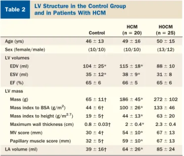
Control HCM (nⴝ20) HOCM (nⴝ25) Age (yrs) 46⫾13 49⫾16 50⫾15 Sex (female/male) (10/10) (10/10) (13/12) LV volumes EDV (ml) 104⫾25* 115⫾18* 88⫾10 ESV (ml) 35⫾12* 38⫾9* 31⫾8 EF (%) 65⫾6 66⫾5 65⫾6 LV mass Mass (g) 65⫾11† 186⫾45* 272⫾102
Mass index to BSA (g/m2) 44⫾6† 100⫾26* 133⫾46
Mass index to height (g/m2.7) 19⫾5† 44⫾13* 63⫾20 Maximum wall thickness (cm) 0.8⫾0.03† 2⫾0.4* 2.3⫾0.4 MV score (mm) 30⫾4† 54⫾10* 67⫾13 Papillary muscle score (mm) 32⫾5† 59⫾10* 67⫾13 LA volume (ml) 39⫾16† 64⫾26* 85⫾24 *p⬍0.05 versus HOCM. †p⬍0.01 versus HCM and HOCM.
BSA⫽body surface area; EDV⫽end-diastolic volume; EF⫽ejection fraction; ESV⫽end-systolic volume; LA⫽left atrial; LV⫽left ventricular; MV⫽mitral valve; other abbreviations as inTable 1.
LV Function in the Control Group and in Patients With HCMTable 3 LV Function in the Control Group and in Patients With HCM
Control HCM (nⴝ20) HOCM (nⴝ25)
Myocardial deformation (%)
Global longitudinal strain ⫺19⫾2.4* ⫺12.6⫾3.4 ⫺11.7⫾3
Global circumferential strain ⫺22⫾2.5† ⫺18.6⫾5.9 ⫺16.9⫾4.7
Global radial strain 45⫾4.4* 33⫾10.5 32⫾8.4
Twist mechanics
Twist (°) 13.6⫾4.5 14⫾6.5 14.5⫾7
Untwisting rate (°/s) ⫺99⫾26 ⫺91⫾36 ⫺108⫾30
Time to untwisting/systolic duration (%) 111⫾6* 123⫾8‡ 153⫾25
LV diastolic function
Lateral e=(cm/s) 11⫾2* 7.4⫾3‡ 5.8⫾1.3
E/e= 8.5⫾3* 10⫾4‡ 15⫾6
PA systolic pressure (mm Hg) 22⫾2.5* 30⫾11‡ 39⫾10
LVEDP invasively measured (mm Hg)§ — 25⫾6
LV pre-A pressure invasively measured (mm Hg)§ — 19⫾5
LV systolic pressure and gradient
Systolic blood pressure (mm Hg) 125⫾8 123⫾8 113⫾10
LV peak systolic pressure (mm Hg) 125⫾8‡ 123⫾8‡ 250⫾35
LV end systolic pressure (mm Hg) 111⫾11‡ 114⫾10‡ 186⫾32
LVOT gradient (mm Hg) — — 76⫾23
*p⬍0.01 versus HCM and HOCM. †p⬍0.05 versus HCM and HOCM. ‡p⬍0.01 versus HOCM. §Invasive measurements performed in 25 patients with HOCM.
LVEDP⫽left ventricular end-diastolic pressure; LVOT⫽left ventricular outflow tract; PA⫽pulmonary artery; pre-A⫽before the pressure increase due to atrial contraction; other abbreviations as inTables 1and2.
patients in the group with dynamic obstruction had an auto-matic implantable cardioverter-defibrillator implanted for the primary prevention of sudden cardiac death.
Cardiac structure in HCM. All patients with HCM had asymmetric LV hypertrophy and a dilated LA. However, patients with dynamic obstruction had a significantly greater
A
D
B
C
E
F
H
I
-5 0 5 10 15 20 0 0.2 0.4 0.6 0.8 1 1.2 -5 0 5 10 15 20 0 0.2 0.4 0.6 0.8 1 1.2 -5 0 5 10 15 20 0 0.2 0.4 0.6 0.8 1J
K
L
G
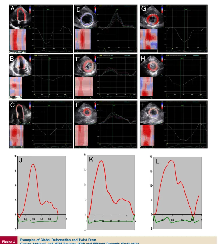
Figure 1 Examples of Global Deformation and Twist From
Control Subjects and HCM Patients With and Without Dynamic Obstruction
(Top)Global longitudinal(A to C), radial(D to F), and circumferential(G to I)strain are shown: a normal control inA, D, and G, hypertrophic obstructive cardiomyopathy (HOCM) inB, E, and H, and hypertrophic cardiomyopathy (HCM) inC, F, and I. Global longitudinal (Avs.BandC) and radial (Dvs.EandF) strain were greater in normal control patients compared with those with either obstructive or nonobstructive HCM. There were smaller differences in circumferential strain (Gvs.HandI).(Bottom)
LV mass when quantitative and semiquantitative scores were applied, as well as a greater LA maximum volume (Table 2). Left ventricular volumes were significantly smaller in patients with HOCM when compared with HCM patients and the control groups (p⬍0.01).
Myocardial deformation in HCM. In comparison with the control group, patients with HCM had significantly less deformation in the longitudinal, circumferential, and radial directions (Table 3). Notwithstanding, there were individual patients in whom 1 or more of the aforementioned deforma-tion measurements were similar to the normal group. Myocar-dial strain was rather similar in HCM and HOCM patients (Fig. 1).
Twist mechanics. Twist and untwisting rate were similar between patients with HCM and normal control patients (Fig. 1). However, the onset of LV untwisting was significantly delayed in patients with HOCM compared with the normal group and patients with HCM (Table 3). A significant correlation was present in between the delay in untwisting velocity and flow propagation velocity (r⫽ ⫺0.43, p⬍0.05), annular e= velocity (r⫽ ⫺0.41, p⬍ 0.05), and longitudinal strain (r⫽0.58, r⬍0.01).
LV diastolic function. Mitral annulus e=velocity was signif-icantly lower, whereas E/e= ratio and estimated pulmonary artery systolic pressure were significantly greater in patients with HCM versus control patients. There were significant differences between HCM and HOCM patients, such that those with dynamic obstruction had evidence of more ad-vanced diastolic dysfunction (Table 3). The delay in untwist-ing (as a percent of systolic duration) was significantly related to LVOT gradient, peak systolic pressure (r ⫽ 0.6, p ⫽ 0.01), and LV end-diastolic pressure as mea-sured during left heart catheterization (Fig. 2A) in patients with dynamic obstruction. Likewise, it was significantly related to E/e= ratio (Fig. 2B), LV end-diastolic volume (Fig. 3), and stroke volume (r⫽ ⫺0.58, p⫽ 0.02) in all 45 patients. Similar results were noted when the delay in untwisting was expressed in relation to the diastolic filling period (r⫽0.45 to 0.69, with p⬍0.05).
Determinants of exercise capacity in HCM. In the pa-tients with HOCM, significant relationships were observed between peak oxygen consumption (VO2max) and LVOT gradient (r⫽ ⫺0.52, p⫽0.03). In all 45 patients, significant correlations were noted between VO2max and indices of LV
systolic and diastolic function: global longitudinal strain (r⫽
⫺0.46, p⫽0.02), radial strain (r⫽0.45, p⫽0.03), LV twist (r⫽0.44, p⫽0.04), E/e=ratio (r⫽ ⫺0.45, p⫽0.026), and LV end-diastolic pressure by invasive measurements in the group with dynamic obstruction (r ⫽ ⫺0.43, p ⫽ 0.04). Importantly, the delay in untwisting showed a good correlation with VO2maxin patients with and without dynamic obstruction (Fig. 4). On multiple regression analysis (R2 ⫽ 0.62, p ⬍ 0.001), the independent predictors were timing of untwisting (i⫽ ⫺0.55, p⬍0.001) and radial strain (i⫽0.39, p⬍0.001).
Changes in clinical status and cardiac structure after septal reduction therapy. In 16 patients, adequate pre- and post-septal reduction datasets were available for analysis. Clin-ical improvement occurred as well as an increase in exercise tolerance in this subset of patients (Table 4). Septal ablation led to a significant reduction in LV peak systolic pressure and LVOT gradient. Basal septal thickness was significantly de-creased (2.1⫾0.5 cm vs. 1.6⫾0.4 cm, p⬍ 0.01), but LV end-diastolic volume increased (88⫾10 ml vs. 120⫾12 ml, p⬍0.05), whereas LVEF (71⫾3% vs. 69⫾5%, p⫽0.1) was unchanged.
Effect of septal ablation on myocardial deformation and LV twist. Global deformation in the longitudinal, circumfer-ential, and radial directions was significantly increased after therapy (Fig. 5A). However, LV twist and untwisting rate (Fig. 5B) were unchanged (Table 4). In contrast, LV untwist-ing occurred earlier (p⬍0.01).
Relation between untwisting, diastolic function, and exercise tolerance after septal reduction therapy. Both the E/e=ratio and PA systolic pressure decreased significantly. Overall the changes in LV diastolic function parameters paralleled the improvement in exercise tolerance. Importantly,
Time to untwisting/systolic duration (%)
Time to untwisting/systolic duration (%) 100 120 140 160 180 200 220 L V EDP (mmHg) 0 10 20 30 40 50 r = 0.76 p <0.01 6 0 8 0 1 0 0 1 2 0 1 4 0 1 6 0 1 8 0 2 0 0 2 2 0 E/e' 0 5 1 0 1 5 2 0 2 5 3 0 r = 0.73 p <0.01
A
B
Figure 2 Untwisting Delay Versus LVEDP and E/e=Ratio
(A)Regression plot of the correlation between the delay in left ventricular (LV) untwisting (expressed as percent of systolic duration) versus left ventricular end-diastolic pressure (LVEDP) during left heart catheterization in the 25 patients with HOCM.(B)Regression plot of the correlation between the delay in LV untwisting (expressed as percent of systolic duration) versus ratio of mitral peak E velocity to annular early diastolic velocity (E/e=) ratio in all HCM patients with and without dynamic obstruction. Abbreviations as inFigure 1.
the earlier occurrence of untwisting was associated with the increase in LV end-diastolic volume (Fig. 6) and was the best predictor of VO2max(Fig. 7).
Discussion
Many patients with HOCM have limitations in their ability to exercise. We have shown in the current series that, in patients with dynamic obstruction, delayed untwisting plays a major role. This finding is supported by the earlier onset of untwist-ing after the decrease in LV systolic pressure with septal reduction therapy.
LV twist mechanics. LV rotation is determined by the helical arrangement of myocardial fibers. Hemodynamically, it is determined by LV contractility and loading conditions. LV untwisting begins in late systole in normal hearts and is completed during the isovolumetric relaxation period, preced-ing inflow across the mitral valve and is an important mecha-nism that aids LV filling at a normal LA pressure (17). A reduction or a delay in untwisting adversely affects LV filling as LV early diastolic pressures remain elevated and the transmitral pressure gradient is reduced unless LA pressure increases to maintain LV filling, end-diastolic volume, and hence, stroke volume.
In that regard, the determination of the hemodynamic variables that affect untwisting is important because their modulation can help patients with heart failure. To start with, intrinsic myocardial structure and contractility affect LV tor-sion, but loading conditions are also a factor. In particular, single-beat aortic constriction in canines (increased afterload) led to a significant delay in LV untwisting (18). However, the effect of afterload on untwisting in humans remains unclear. In that regard, patients with HOCM provide a unique opportu-nity to assess the clinical impact of afterload because increased afterload can be altered by septal reduction procedures that do not directly affect intrinsic myocardial function.
LV untwisting in HOCM. Patients with HCM have nor-mal twist and untwisting rate as a group whether by echocar-diography or magnetic resonance imaging (17,19–21), al-though individual variations occur. A previous study reported on delayed untwisting in 7 patients with HCM that adversely affected LV filling assessed by Doppler echocardiography (17). In our study, patients with HCM had a significant delay of
Figure 3 Untwisting Delay Versus LVEDV
Regression plot of the correlation between the delay in left ventricular untwist-ing (expressed as percent of systolic duration) and LVEDV in patients with HOCM shown assquaresand those without dynamic obstruction ascircles. LVEDV⫽left ventricular end-diastolic volume; other abbreviations as in
Figure 1.
Figure 4 Untwisting Delay Versus VO2max
Regression plot of the correlation between the delay in left ventricular untwist-ing (expressed as percent of systolic duration) and peak oxygen consumption (VO2max) in patients with HOCM shown assquaresand those without dynamic obstruction ascircles. Abbreviations as inFigure 1.
LV Function Before and After Septal AblationTable 4 LV Function Before and After Septal Ablation
Before Ablation After Ablation Clinical status
NYHA functional class III to IV/I to II 16/0* 0/16
VO2max(ml/kg/min) 14⫾3* 19⫾4
Exercise duration (s) 350⫾215* 515⫾153 LV systolic pressure/LVOT gradient (mm Hg)
LVOT gradient 75⫾23* 15⫾27
LV peak systolic pressure 250⫾35* 146⫾25 Myocardial deformation (%)
Global longitudinal strain ⫺11.7⫾3† ⫺14.3⫾2 Global circumferential strain ⫺16.4⫾4† ⫺22.6⫾5 Global radial strain 32⫾8.4† 38⫾7 Twist mechanics
Twist (°) 15⫾5 14.9⫾5
Untwisting rate (°/s) ⫺108⫾30 ⫺102⫾34 Time to untwisting/systolic duration (%) 155⫾18* 123⫾8 LV diastolic function
Lateral e=(cm/s) 5.8⫾1.3* 6.5⫾1.3
E/e= 15⫾6* 11⫾5
PA systolic pressure (mm Hg) 39⫾11* 30⫾10 *p⬍0.001 versus after ablation. †p⬍0.05 versus after ablation.
untwisting (on average 11% vs. control patients), likely reflect-ing the well-known myocardial abnormalities in this disease.
More importantly, patients with dynamic obstruction exhibited a much longer delay (on average 38% vs. control
patients and 24% vs. patients without obstruction) that was significantly coupled with smaller LV end-diastolic volume, as would be expected from its adverse effects on LV filling. Furthermore, the delayed untwisting was associated with a
B
C
E
A
F
D
-5 0 5 10 15 20 0 0.2 0.4 0.6 0.8 1 1.2 -10 -5 0 5 10 15 20 0 0.2 0.4 0.6 0.8 1 1.2G
H
Figure 5 Examples of Global Strain and Twist Before and After Septal Ablation
Global strain before and after septal ablation. Improvement is present in longitudinal(A and B), radial(C and D), and circumferential(E and F)strain. Left ventricular twist before and after septal ablation(G and H). Twist is unchanged.
more advanced degree of diastolic dysfunction as consis-tently observed by noninvasive measurements (LA volume, e= velocity, E/e= ratio, and PA systolic pressure) and invasively measured LV end-diastolic pressure. In conclu-sion, delayed untwisting led to greater filling pressures and reduced LV filling in HOCM, which in turn had a negative effect on exercise tolerance.
Changes in LV untwisting after septal reduction. Septal reduction led to a significant reduction in LV systolic pressures and an earlier onset of LV untwisting, which although still delayed was similar to patients with HCM. The earlier untwisting was accompanied by significantly larger LV end-diastolic volume and stroke volume because LVEF was un-changed. In addition, an improvement in LV diastolic function was observed as noted in previous studies (22,23), including those where invasive measurements were obtained (24). Fi-nally, similar to baseline findings, the timing of untwisting remained tightly coupled with exercise tolerance (Fig. 7).
There was also an improvement noted in global deformation measurements, which is similar to previous reports by cardiac magnetic resonance (19). Whether this change reflects an actual improvement in intrinsic contractility versus an increase in deformation related to decreased afterload remains to be determined, although the persistently reduced values of global longitudinal and radial strain favor the contribution of both intrinsic and extrinsic factors to the abnormal deformation measurements.
Study limitations. We included relatively few patients; how-ever, the study was adequately powered (⬎80% power to detect an 8% difference between 3 groups with a SD of residuals of 5%, and alpha of 0.05) to address our hypothesis. Further, the results were consistent when several parameters of cardiac function were considered such that an alpha error is unlikely. Patients without dynamic obstruction did not undergo cardiac
catheterization because there were no clinical indications for the procedure. Likewise, left heart catheterization was not performed at follow-up in the group who underwent septal reduction therapy because of the lack of clinical indications.
It would have been ideal to measure cardiac function during exercise. However, it is challenging to acquire adequate images during upright exercise, the ideal exercise modality in patients with HCM for provoking obstruction. Furthermore, the use of speckle tracking, which has greater feasibility and reproducibil-ity than tissue Doppler, limits the frame rate during stress imaging such that it does not capture the rapid changes in mechanical events during exercise.
Reprint requests and correspondence: Dr. Sherif F. Nagueh, Department of Cardiology, The Methodist DeBakey Heart and Vascular Center, 6550 Fannin Street, Suite 677, Houston, Texas, 77030. E-mail:snagueh@tmhs.org.
REFERENCES
1. Maron BJ, McKenna WJ, Danielson GK, et al. American College of Cardiology/European Society of Cardiology clinical expert consensus document on hypertrophic cardiomyopathy: a report of the American College of Cardiology Foundation Task Force on Clinical Expert Consensus Documents and the European Society of Cardiology Committee for Practice Guidelines (Committee to Develop an Expert Consensus Document on Hypertrophic Cardio-myopathy). J Am Coll Cardiol 2003;42:1687–713.
2. Maron MS, Olivotto I, Betocchi S, et al. Effect of left ventricular outflow tract obstruction on clinical outcome in hypertrophic cardio-myopathy. N Engl J Med 2003;348:295–303.
3. Kjaergaard J, Johnson BD, Pellikka PA, Cha SS, Oh JK, Ommen SR. Left atrial index is a predictor of exercise capacity in patients with hypertrophic cardiomyopathy. J Am Soc Echocardiogr 2005;18:1373–80. 4. Matsumoto AY, Arteaga E, Ianni BM, Braga AM, Buck PC, Mady C. Relationships among exercise capacity, hypertrophy, and left ventricular diastolic function in nonobstructive hypertrophic cardio-myopathy. Am Heart J 2005;150:144 –9.
5. Matsumura Y, Elliott PM, Virdee MS, Sorajja P, Doi Y, McKenna WJ. Left ventricular diastolic function assessed using Doppler tissue
0 10 20 30 10 20 30 40 50 60 70 % Increase
in LV End Diastolic Volume
% Improvement in Shortening of Untwisting Time
r = 0.65 p = 0.01
Figure 6 Change in Untwisting Delay Versus Change in LVEDV
Regression plot of the correlation between the change in LV untwisting (expressed as percent of systolic duration) after septal reduction procedures and the change in LVEDV in the 16 patients with HOCM who underwent myec-tomy or septal ablation by ethanol. Abbreviations as inFigures 1to3.
Figure 7 Change in Untwisting
Delay Versus Change in VO2max
Regression plot of the correlation between the change in left ventricular untwisting (expressed as percent of systolic duration) after septal reduction procedures and the change in peak VO2in the 16 patients with HOCM who underwent myectomy or septal ablation by ethanol. Abbreviations as in
imaging in patients with hypertrophic cardiomyopathy: relation to symptoms and exercise capacity. Heart 2002;87:247–51.
6. Choi EY, Ha JW, Rim SJ, et al. Incremental value of left ventricular diastolic function reserve index for predicting exercise capacity in patients with hypertrophic cardiomyopathy. J Am Soc Echocardiogr 2008;21:487–92.
7. Ha JW, Ahn JA, Kim JM, et al. Abnormal longitudinal myocardial functional reserve assessed by exercise tissue Doppler echocardiogra-phy in patients with hypertrophic cardiomyopathy. J Am Soc Echo-cardiogr 2006;19:1314 –9.
8. Ha JW, Cho JR, Kim JM, et al. Tissue Doppler-derived indices predict exercise capacity in patients with apical hypertrophic cardio-myopathy. Chest 2005;128:3428 –33.
9. Carasso S, Yang H, Woo A, et al. Systolic myocardial mechanics in hypertrophic cardiomyopathy: novel concepts and implications for clinical status. J Am Soc Echocardiogr 2008;21:675– 83.
10. Carasso S, Woo A, Yang H, et al. Myocardial mechanics explains the time course of benefit for septal ethanol ablation for hypertrophic cardiomyopathy. J Am Soc Echocardiogr 2008;21:493–9.
11. Quinones MA, Otto CM, Stoddard M, Waggoner A, Zoghbi WA. Recommendations for quantification of Doppler echocardiography: a report from the Doppler Quantification Task Force of the Nomen-clature and Standards Committee of the American Society of Echo-cardiography. J Am Soc Echocardiogr 2002;15:167– 84.
12. Lang RM, Bierig M, Devereux RB, et al. American Society of Echocardiography’s Nomenclature and Standards Committee; Task Force on Chamber Quantification; American College of Cardiology Echocardiography Committee; American Heart Association; Euro-pean Association of Echocardiography, EuroEuro-pean Society of Cardiol-ogy Recommendations for chamber quantification. Eur J Echocardiogr 2006;7:79 –108.
13. Spirito P, Maron BJ. Relation between extent of left ventricular hypertrophy and occurrence of sudden cardiac death in hypertrophic cardiomyopathy. J Am Coll Cardiol 1990;15:1521– 6.
14. Serri K, Reant P, Lafitte M, et al. Global and regional myocardial function quantification by two-dimensional strain: application in hypertrophic cardiomyopathy. J Am Coll Cardiol 2006;47:1175– 81.
15. Reisner SA, Lysyansky P, Agmon Y, Mutlak D, Lessick J, Friedman Z. Global longitudinal strain: a novel index of left ventricular systolic function. J Am Soc Echocardiogr 2004;17:630 –3.
16. Notomi Y, Lysyansky P, Setser RM, et al. Measurement of ventricular torsion by two-dimensional ultrasound speckle tracking imaging. J Am Coll Cardiol 2005;45:2034 – 41.
17. Notomi Y, Martin-Miklovic MG, Oryszak SJ, et al. Enhanced ventricular untwisting during exercise: a mechanistic manifestation of elastic recoil described by Doppler tissue imaging. Circulation 2006; 113:2524 –33.
18. Gibbons Kroeker CA, Tyberg JV, Beyar R. Effects of load manip-ulations, heart rate, and contractility on left ventricular apical rotation. An experimental study in anesthetized dogs. Circulation 1995;92:130 – 41.
19. van Dockum WG, Kuijer JP, Götte MJ, et al. Septal ablation in hypertrophic obstructive cardiomyopathy improves systolic myocardial function in the lateral (free) wall: a follow-up study using CMR tissue tagging and 3D strain analysis. Eur Heart J 2006;27:2833–9. 20. Kramer CM, Reichek N, Ferrari VA, Theobald T, Dawson J, Axel L.
Regional heterogeneity of function in hypertrophic cardiomyopathy. Circulation 1994;90:186 –94.
21. Young AA, Kramer CM, Ferrari VA, Axel L, Reichek N. Three-dimensional left ventricular deformation in hypertrophic cardiomyop-athy. Circulation 1994;90:854 – 67.
22. Nagueh SF, Lakkis NM, Middleton KJ, et al. Changes in left ventricular diastolic function 6 months after nonsurgical septal reduc-tion therapy for hypertrophic obstructive cardiomyopathy. Circulareduc-tion 1999;99:344 –7.
23. Sitges M, Shiota T, Lever HM, et al. Comparison of left ventricular diastolic function in obstructive hypertrophic cardiomyopathy in pa-tients undergoing percutaneous septal alcohol ablation versus surgical myotomy/myectomy. Am J Cardiol 2003;91:817–21.
24. Faber L, Seggewiss H, Gleichmann U. Percutaneous transluminal septal myocardial ablation in hypertrophic obstructive cardiomyopathy: results with respect to intraprocedural myocardial contrast echocardi-ography. Circulation 1998;98:2415–21.
Key Words:diastoleyhypertrophic cardiomyopathyymechanicsy