Committee 12
Neurologic Urinary and Faecal
Incontinence
Chairman
J.J. W
YNDAELE(B
ELGIUM)
Co Chairs
D. C
ASTRO(S
PAIN),
H.M
ADERSBACHER(A
USTRIA)
Members
E. C
HARTIER-
KASTLER(F
RANCE),
Y. I
GAWA(J
APAN),
A. K
OVINDHA(T
HAILAND),
P. R
ADZISZEWSKI(P
OLAND),
A.S
TONE(USA),
P. W
IESEL(S
WITZERLAND)
Abbreviations
Most abbreviations used in the text are given here ACE: antegrade continent enema
BCR: bulbocavernosus reflex CC: condom catheter CMG: cystometrogram
CUM: continuous urodynamic monitoring CVC: conventional cystometry
DOA: detrisor over activity DSD: detrusor sphincter dyssynergia EAS: external anal sphincter EMG: electromyography GBS: Guillain Barré Syndrome ID: indwelling catheter
IVES: intravesical electrical stimulation LBT: lower bowel tract
LOE: level of evidence LUT: lower urinary tract MPdet: maximum detrusor pressure MUP: motor unipotential MS: multiple sclerosis MSA: multiple system atrophy NVC: natural fill cystometry PD: Parkinson’s disease
PSP: progressive supranuclear palsy PSC: suprapubic catheter
TURS: transurethral sphincterotomy
F. REFERENCES
E16. HERPES ZOSTER E15. SYSTEMIC LUPUS ERY
THEMATOSUS
E14. PERIPHERAL NEUROPATHY DUE TO IATROGENIC LESIONS
(FOCAL NEUROPATHY) E13. DIABETES MELLITUS (+ BOWEL DYSFUNCTION) E12. CONGENITAL LUMBOSACRAL DEFICITS /MENINGOMYELOCOELE
E11. LUMBAR DISC PROLAPSE E10. AIDS
E9. GUILLAIN BARRÉ
E8. SPINAL STENOSIS E7. SPINAL CORD LESION (+ BOWEL DYSFUNCTION) E6. MULTIPLE SCLEROSIS (+ BOWEL DYSFUNCTION) E5. CEREBRAL
LESIONS-CEREBRO-VASCULAR ACCIDENTS E4. ALZHEIMER
E3. PARKINSON (+ BOWEL DYSFUNCTION) E2. MULTIPLE SYSTEM ATROPHY
E1. DEMENTIA
E. SPECIFIC NEUROLOGIC
DISEASES AND LUT
DYSFUNCTION
D4. SURGICAL TREATMENT D3 . CONSERVATIVE TREATMENT D2. SPECIFIC DIAGNOSTICS D1. EPIDEMIOLOGYD. NEUROLOGIC FAECAL
INCONTINENCE
C4. SURGICAL TREATMENT C3. CONSERVATIVE TREATMENT C2. SPECIFIC DIAGNOSTICS C1. EPIDEMIOLOGYC. NEUROLOGIC URINARY
INCONTINENCE
B. PATHOPHYSIOLOGY
A.INTRODUCTION
CONTENTS
This chapter deals with all aspects of neurologic uri-nary and faecal incontinence.
It is known that the lower urinary tract (LUT) and the lower bowel tract (LBT) are interrelated structures. Embryologically bladder and rectum originate from the same basic structure, the cloaca [1]. Anatomical-ly both viscera lay in close communication and share muscular structures of the pelvic floor.
The innervation of both systems depends on autono-mic and somatic nerves (Figure 1).
In table 1 a simplified overview is given of the action linked to different peripheral nerves.
Central control of both continence and evacuation is similar and is discussed in the chapter on physiology [2].
Very generally LUT and LBT act quite similar: the
voluntary control depends on accurate sensation [3]. Continence relates to contraction of smooth closing structures (bladder neck and internal bowel sphinc-ter) and striated urethral and anal sphincters. An inhi-bitory effect on detrusor and lower rectum resulting from contraction of the pelvic floor and anal or ure-thral sphincter has been named a “procontinence” reaction. Micturition and defaecation need a proper relaxation of these latter structures to permit a phy-siological reflex evacuation of urine or faeces. Interactions between both functions have been demonstrated. The filling grade of the bladder influences sensation in the rectum and vice versa [4]. A vesico-ano-rectal reflex permits voiding without defaecation [5].
A. INTRODUCTION
Neurologic Urinary and Faecal
Incontinence
J.J. W
YNDAELE,
D. C
ASTRO, H.M
ADERSBACHERE. C
HARTIER-
KASTLER, Y. I
GAWA, A. K
OVINDHA, P. R
ADZISZEWSKI, A.S
TONE, P. W
IESELFigure 1. Schematic overview of innervation of LUT and LBT
Table 1. Overview of function of the abdominal sympathe-tic (sym), the pelvic parasympathesympathe-tic (PSym) and somasympathe-tic (Som) nerves in the lower urinay tract and lower bowel tract. US= urethral sphincter, AS= anal sphincter. Exp= only suggested in animal experimentation , no clinical evi-dence.
Sym Psym Som
Bladder - +
Bladder neck + -
Extern US Exp Exp +
Bowel +
Intern AS + -
Extern AS Exp Exp +
When a neurologic lesion occurs the type of dys-function that follows in LUT and LBT will depend on the site, the extent and the evolution of the lesion. Traditionally neurological pathology has been divi-ded in suprapontine, suprasacral spinal cord and sacral - subsacral (cauda equina and peripheral nerve) lesions (Figure 2).
Patients with lesions above the pons usually continue to have reflex contractions of the detrusor. But the cerebral regulation of voiding and defaecation is often lost. This is the case in lesions as from stroke, head injury, etc, which mostly continue to have a normal coordinated sphincter function. However these patients may purposely increase sphincteric activity during an overactive detrusor contraction [6], to prevent urinary incontinence which would otherwise occur. This has been termed “pseudo-dys-synergia” because it is indistinguishable from true dyssynergia on a urodynamic record. Urinary incon-tinence in suprapontine lesions is due to the bladder overactivity [7].
Suprasacral spinal cord lesion When a lesion is located in the spinal cord below the pons detrusor-urethral sphincter dyssynergia is a common finding. Incontinence may still be caused by detrusor overac-tivity but the outflow obstruction can also cause retention.
Patients with lesions above the cone usually suffer from an overactive bowel with increased colonic wall and anal tone. The central control of the exter-nal aexter-nal sphincter is disconnected and the sphincter remains tight thereby retaining stool (dyssynergia). The connections between the spinal cord and the colon remain intact, permitting reflex coordination and stool propulsion. This type of lesion provokes faecal retention at least in part due to the activity of the anal sphincter. Incontinence can be a consequen-ce of faecal impaction and constipation.
Conus lesion If the nuclei of the pelvic nerves are destroyed the detrusor becomes areflexic. Retention of urine can provoke stress incontinence (formerly termed overflow incontinence).
II. SPINAL CORD LESIONS
I. SUPRAPONTINE LESIONS
B. PATHOPHYSIOLOGY
The same effect as from lesions of the conus medul-laris can result from lesions of the subsacral nerves (cauda equina or peripheral nerves). If the nuclei of the pudendal nerves are lesioned a paralysis of the urethral sphincter and pelvic floor muscles will occur with loss of outflow resistance and stress incontinen-ce.
A neurologic lesion affecting the parasympathetic cell bodies in the conus medullaris will eliminate the pelvic nerve function of the bowel. No spinal-cord mediated peristalsis occurs. The myenteric plexus coordinates segmental colonic peristalsis. If the pudendal nerve is also destroyed , there is an increa-sed risk for incontinence. Apart from the non contractile external anal sphincter, the puborectal muscles also lack tone, which leads to reduction of the rectal angle. Constipation and incontinence are frequent.
While most traumatic spinal cord lesions give LUT and LBT dysfunctions which can be predicted fairly well from the level and completeness of injury [8], the LUT and LBT function in many other neurologic diseases such as meningomyelocoele are more diffi-cult to categorise [9]. Therefore in this chapter a number of neurologic diseases will be dealt with in detail.
This part contains all aspects from epidemiology, pathophysiology, through diagnosis to treatment. The ICS Standardization Committee recently intro-duced a new nomenclature of LUT dysfunctions. Terms such as reflex incontinence, detrusor hyperre-flexia and overflow incontinence are, according to this nomenclature no longer valid. However in the case of neurologic voiding dysfunction « reflex incontinence » reflects the return of a primitive voi-ding reflex and, associated with it, incontinence, and therefore this term will still be used here in addition to the newly introduced term « neurologic detrusor overactivity »
Madersbacher et al have described in the ICI report 2002 common patterns of neurologic detrusor-sphincter dysfunction in a diagram which is reprodu-ced infigure 3.These are easy to use and the deve-lopment of similar diagrams for neurologic bowel dysfunction is to be recommended.
Pubmed search from 1967 till 2004 with search words: epidemiology, neurologic bladder, neurologic
I. METHODOLOGY
C1. EPIDEMIOLOGY
C. NEUROLOGIC URINARY
INCONTINENCE
III. SUBSACRAL LESIONS (CAUDA
EQUINA OR PERIPHERAL NERVES)
Figure 3. Patterns of neurogenic detrusor-sphincter dysfunction Heavy lines symbolize hyperreflexia, thin lines hypo- or areflexia and green lines a normal innervation of the relevant structure, for futher explanation see text.
incontinence, neurologic patients, prevalence gave several hundreds of references. Unfortunately only a very limited number gave data on prevalence and only in specific diseases for which a separate search was done. Data on incontinence are not always pre-sent in data on “neurologic voiding dysfunction” or “neurologic cystopathy”. No global meta analysis has been found.
In separate searches for specific diseases the preva-lence data were also very limited. Moreover most studies were case series . A small number of retros-pective case control studies have been found for a number of diseases and a single study which looks into incontinence in the elderly with and without dementia [13].
Several factors can be the cause for this lack of data:
• Neurologic problems of the LUT are not always specifically studied
• Some diseases are rare or have not been very much studied
• Series on urologic items deal mostly with urody-namic data, urologic complications or outcome of treatment and include only patients with a known neurologic bladder
• In some neurologic disease as spinal cord injury no data are to be found on those who had not developed a neurologic bladder.
Following are tables 2 with the data found and the publications. While making an interpretation of these data one must realise that incontinence can be pre-sent because of direct neurologic dysfunction of bladder, bladder neck or sphincter, either because of lack of adequate treatment, infection or other causes. This differentiation can not be made from the litera-ture data.
• Diagnostic methods of neurologic LUT dysfunc-tion and neurologic urinary incontinence are not very different from what is done in non neurologic patients. They consist of clinical assessment inclu-ding voiinclu-ding history and voiinclu-ding diary, urodyna-mic studies including cystometry (+ EMG), video-urodynamics, uroflowmetry, pressure-flow study, diagnostic imaging with voiding cystoure-thrography and ultrasonography of the kidneys and LUT. These methods will be dealt with in the relevant chapters of this book( basic assessment, dynamic testing, imaging and other investiga-tions) but we will highlight briefly some data spe-cially related to neurologic patients.
• Some tests developed for the diagnosis of neuro-logic dysfunction have been evaluated more spe-cifically in this chapter: dynamic bulbocavernosus reflex [1], bethanechol supersensitivity test [2], ice water test [3].
• Neurophysiologic studies can be found in the chapter “Clinical Neurophysiological testing”, and only some clinical relevant data will be given here.
Searching strategy in Medline from 1966 to present (2004) with keywords: neurologic bladder and elec-trodiagnosis – 311 papers, with keywords:
neurolo-I. METHODOLOGY
C2. SPECIFIC DIAGNOSTICS
Recommendations
• Because many diseases or lesions of the innervation can cause pathology of the LUT, patients with known neurologic disease should be evaluated for such dysfunction . • Such evaluation should be made not only
when urinary symptoms occur but also as a standard diagnostic approach if prevalence of neurologic bladder is known to be high in a specific disease .
• If “idiopathic” LUT dysfunctions occur the possibility of an unknown neurologic cause should be acknowledged and the diagnostic steps taken to make a proper diagnosis. • The committee thinks there is enough
evi-dence to make all three strong recommenda-tions.
Conclusions
• Neurologic dysfunction of the LUT occurs in many patients with neurologic disease but exact figures are seldom available • Metanalysis of prevalence data could give a
better idea of how important neurologic bladder is in the patients with neurologic diseases and in the prevalence of inconti-nence in this population.
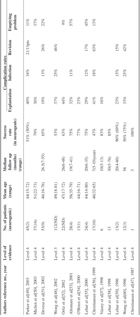
Table 2. Prevalence figures from literature of different causes of neurologic LUT dysfunction
Cerebro-vascular accidents poststroke 32 - 79% Brittain et al [1] On hospital discharge 25 – 28 % Borrie et al [2] after 12 weeks 29% Later 12 – 19 % Taub et al [3]
initial incontinence best single indicator of future disability
Cerebral tumor Case reports Maurice-Williams[4], Lang et al[5]
Normal pressure hydrocephalus Case reports Jonas –Brown[6], Black[7],
Mulrow et al[8]
Cerebral Palsy 36% Mc Neal et al[9], Decter et al[10]
Mental retardation depending
on grade disorder 12% - 65% Mitchell-Woodthorpe[11],
Reid et al[12]
Dementia 30-100% Campbell et al[13], Toba et al[14]
Parkinson’s disease 37,9 – 70 % Murnaghan[15] Campos-Sousa et al[16]
Multiple system atrophy 73% incontinence Chandiramani et al[17]
Multiple sclerosis 52-97 %
37-72%% incontinence Litwiller et al[18], Giannantoni at al[19]
Myelodysplasia 90% -97% Smith[20]
Spinal stenosis 61-62% Tammela et al[21], Kawaguchi[22]
Spine surgery 38%-60% Boulis et al[23], Brooks[24]
Disc disease 28%-87% (Bartolin et al[25], O’Flynn et al[26]
Spinal cord injury ?
Majority have neurologic lesions
Diabetes mellitus 25-48% Hampel et al[27]
43-87% insulin dependent Frimodt-Moller[28]
Rectal carcinoma resection <70% Hollabaugh et al[29]
20% or even 0% if nerve sparing Hojo et al[30]
Radical Hysterectomy 8 –10% Ketcham et al[31], Seski-Diokno[32]
57% Lin et al[33]
HIV 12 % advanced stage Gyrtrup et al[34] Shin et al[35] LOE2
Guillain Barré 25%- case studies Sakakibara et al[36]
Herpes 17 / 423 Chen et al[37]
gic bladder and investigation – 133 papers, and with keywords: neurologic bladder and diagnosis – 825 papers. Seventy one relevant papers have been selec-ted for this evaluation.
1. C
LINICAL EXAMINATIONWyndaele and De Sy [4] found that clinical neurolo-gical findings in 47 children with lumbosacral mye-lodysplasia did not correlate well with the existing dysfunction of the LUT as diagnosed by urodynamic studies. The level of intact skin sensation, and the presence or absence of bulbocavernosus and anal reflexes could not significantly predict the function of the detrusor muscle, proximal urethra and striated urethral sphincter. Therefore, one should not rely on a clinical neurological examination to outline the urological management in such patients. The same conclusion is given by Marshall and Boston [5] who examined the reliability of whether absence of sacrally-mediated anal reflexes in 76 children and adolescents (aged 3 - 18 years) with spina bifida gua-rantees a safe bladder and whether routine urodyna-mic studies can be, therefore, safely omitted in this group. They found the absence of anal reflexes to be a poor predictor of safe bladder pressures and thus no justification for depriving such a population of routi-ne urodynamic assessment on this basis. (LOE 3) In 92 patients with spinal cord lesion, out of spinal shock, Wyndaele [6] compared data from a clinical neurological examination of the lumbosacral area with the data from a full urodynamic investigation, including evaluation of sensation in the LUT. A significant correlation was found between different levels of spinal cord lesion, the function of bladder neck and sphincter and the anal/ bulbocavernosus reflexes. Higher lesions corresponded more with a reflexic LUT and somatic motor activity, lower lesions more with areflexia. In patients with lesions between thoracic 10 and lumbar 2 as many reflexic as areflexic LUT dysfunctions were found. The pre-sence or abpre-sence of perineal sensation of light touch corresponded significantly with the presence or absence of sensation in the LUT. Detrusor and stria-ted sphincter reflexia/areflexia corresponded signifi-cantly with the presence/absence of bulbocavernosus and anal reflexes.
Shenot et al [7] determined if intact perianal pin sen-sation (PPS) and bulbocavernosus reflex (BCR) shortly after spinal cord injury are predictive of blad-der function recovery in 28 patients, Frankel
Classi-fication A-D, spinal injury level C4-T12, admitted within 72 hours of injury. The presence of intact PPS and BCR were correlated with the patient’s voiding function and urodynamic evaluation results 1 year postinjury. Although PPS and BCR proved modera-tely sensitive in predicting the return of spontaneous voiding, they could not predict detrusor hyperre-flexia and sphincter dyssynergia.
Therefore, urodynamic study remains an essential component of initial urologic
evaluation after SCI. Also, Schurch et al [8] determi-ned whether early sensory examination, voluntary anal sphincter contraction, or bulbocavernosus reflex (BCR) might predict bladder function in patients with a spinal fracture at the thoracolumbar level. They found that in these patients neurologic voiding dysfunction cannot be predicted by the early sensory evaluation. Pinprick sensation in the perineal area is of negative predictive value: absence of pinprick sensation predicts poor bladder recovery. Most patients with a spinal fracture at T12-L1 did not improve in voiding function.
In men, who were at risk for obstructive uropathy, Nitti et al [9] evaluated whether, after a cerebrovas-cular accident, the cause of voiding dysfunction could be predicted by the type (obstructive or irrita-tive) or time of onset of symptoms. Presenting symp-toms did not predict the urodynamic findings of bladder outlet obstruction or DOA. The significant incidence of onset of symptoms after stroke suggests that the cerebrovascular accident induced voiding dysfunction in the face of preexisting bladder outlet obstruction may exacerbate the symptoms of the lat-ter condition or vice versa.
Conclusion
• These studies show that a combination of all data from a clinical neurological examination gives useful information which acceptably corresponds with the LUT function in patients with spinal cord injury but not in meningomyelocoele patients.
• In elderly male neurologic patients with pos-sible BPH related obstruction, symptoms and clinical examination are not sufficient to dif-ferentiate between outflow obstruction and neurologic DOA.
• To decide on a detailed individual diagnosis of LUT function in neurologic patients, histo-ry and clinical examination prove insuffi-cient.
2. U
RODYNAMIC TESTSClassic urodynamic techniques permit the acquisi-tion of multiple funcacquisi-tional parameters in patients with neurologic bladder. These techniques are dis-cussed in the chapter on dynamic testing.
The literature gives enough evidence of the value of different techniques in neurologic urinary inconti-nence: urodynamic investigations in patients with a traumatic SCI permit not only a urodynamic diagno-sis but also an objective follow up of treatment such as reeducation techniques, after sphincterotomy and after urethral overdilatation by indwelling catheters [10].
• CMG + EMG has been studied by several authors: Sundin and Petersen [11] used cystometry-electro-myography (EMG) investigation in patients with known or suspected neurologic disorders in whom a defect in bladder emptying, in spite of an active detrusor contraction, was found at cystometry. A voluntary control of the external urethral sphincter
relaxation—independent of the degree of bladder filling-was found in most of the healthy volunteers. The cystometry-EMG investigation gave reliable information as to whether a DSD exists. Perkash [12] found rhythmic detrusor contractions on cystomano-metry with associated marked increase in EMG acti-vity on attempted voiding to be relevant characteris-tics of patients with DSD. Rodriquez et al [13] used EMG-gas cystometrogram to select SCI patients for removal of the Foley catheter. Important factors governing success were the amplitude of the detrusor contraction, the presence of detrusor-sphincter synergy and the presence of a flaccid sphincter. Mayo and Kiviat [14] used multichannel urodynamic studies in patients with incomplete bladder emptying secondary to suprasacral SCL. They found that blad-der pressure and sphincter EMG measurement during voiding, combined with fluoroscopy, are ideal methods to identify the factors responsible for incomplete emptying in problem cases. Also Perlow and Diokno [15] and Koyanagi et al [16] found CMG-EMG very usefull in SCI patients.
Blaivas et al [17] described on the basis of CMG -EMG 3 types of dyssynergia: type 1 had a crescendo increase in electromyographic activity that reached a maximum at the peak of the detrusor contraction, type 2 had clonic sphincter contractions interspersed throughout the detrusor contraction and type 3 was characterized by a sustained sphincter contraction that coincided with the detrusor contraction. There was no correlation between the clinical neurologic level and the type of dyssynergia.
Simultaneous recording of intravesical pressure, sphincter electromyography and uroflowmetry (CMG.UFM.EMG study) was compared by Aoki et al [18] with cystometry + EMG. They found some influence of the catheter in the urethra. Micturition pressure and opening pressure were larger with CMG+ EMG, incidence of detrusor-sphincter dyssy-nergia was greater. The authors also found that the Credé maneuver exaggerated the DSD. Urodynamics with EMG permitted Kirby [19] to differentiate bet-ween patients with pelvic nerve injury, distal autonomic neuropathy, progressive autonoautonomic failure -multiple system atrophy, and idiopathic Parkinson’s disease. This influenced the selection of patients for transurethral surgery. Pavlakis et al [20] studied CMG concomitant with perineal floor and rectus abdominis EMG and concluded that the addition of rectus EMG can improve the recognition of intrave-sical pressure elevation owing to voluntary contrac-tion of the abdominal musculature.
• Perkash and Friedland (1987) found simultaneous transrectal ultrasonography helpful.
• They also recommended not to irritate the bladder when introducing the urodynamics catheter and to examine the entire curve of the CMG and not sim-ply the initial rise.
• Pressure-flow study can demonstrate an obstructi-ve pattern (high pressure voiding) also in neurolo-gic patients due to urethral relaxation failure [22].
• Video urodynamics permit a clear image of blad-der neck and urethral sphincter activity during filling and voiding [23].
• Zerin et al [24] found that the urographic position of the bladder neck in relation to the pubic sym-physis was correlated with lower motor neuron (LMN) denervation of the urethral sphincter as detected with electromyography in infants and children with myelodysplasia. They concluded that, although not as precise as urodynamic tes-ting, significant descent of the bladder neck is a reliable urographic finding of complete LMN denervation of the external urethral sphincter in infants and children with myelodysplasia.
• CMG filling rate seems to be very important espe-cially in neurologic patients:
De Gennaro et al [25] performed continuous urody-namic monitoring over 6 hours (CUM) in children and compared this with standard urodynamics. They found CUM feasible and permitting a better diagno-sis than standard cystometry in some. Zermann et al [26] investigated the diagnostic value of natural fill
cystometry (NFC) in children with neurologic blad-der in comparison to conventional videocystometry (CVC). In 45%, NFC detected new findings compa-red with CVC diagnoses. CVC findings were confir-med in another 45%.
Hess et al [27] studied how closely the intravesical pressures obtained before filling cystometry resem-bled those obtained during the filling phase of the cys-tometrogram. Filling pressures during cystometry were significantly higher than the pressures measured at rest. This study also suggests a strong correlation between both. Ko et al [28] determined in spinal cord injured patients with neurologic bladder whether cys-tometry performed by filling using diuretics (FCMG) reveals different findings compared with conventional cystometry (CMG). Significant differences were found between CMG and FCMG in hyperreflexic neurologic bladders with respect to a decrease in MPdet and increase in compliance with FCMG. However, there were no significant differences in MPdet and compliance in hyporeflexic or areflexic neurologic bladders between the two techniques.
• The determination of CMG filling sensation is important [29]. In 52 SCI patients, 26 % of those with a supposed complete lesion had sensation of bladder filling during cystometry [30]. Also in 41 patients with myelodysplasia the perception of bladder filling proved, rather unexpectedly, to be present in a majority of patients [31].
In a large cohort study it was clearly shown that impaired perception of bladder filling during CMG is a sign of neuropathy [32]. Ersoz and Akyuz [33] investigated bladder-filling sensation in 73 SCI patients with complete lesions above T11 and below T10 and with incomplete lesions Bladder-filling sen-sation was present to some degree in all incomplete SCI patients, in 82.4% of the patients with complete lesions below T10, and in 38.9% of the patients with complete lesions above T11. Bladder-filling sensa-tion investigasensa-tions were reproducible in terms of bladder filling sensation category in 36 SCI patients who had second cystometric examination. The authors concluded that presence of bladder-filling sensation in many SCI patients reveal the potential for sensation-dependent bladder emptying, especial-ly in the ones with complete lesions below T10 and the ones with incomplete lesions.
Madersbacher [34] introduced a specially designed, plastic, radiolucent micturition chair especially sui-table for the urodynamic evaluation in paraplegics, in myelomeningoceles and other handicapped persons.
3. S
PECIAL TESTSa) Ice water test
The Ice water test was first described by Bors and Blinn [35] for spinal cord injuried patients. It is based on the principle that stimulation of mucosal temperature receptors by rapid introduction of 100 ml water at 4°C into the bladder can elicit a spinal reflex contraction of the detrusor, a reflex that is nor-mally inhibited by supraspinal centers. A lesion above the detrusor motor cells interrupts these inhi-bitory pathways, resulting in manifestation of the reflex, whereas a lesion of the motor cells does not result in reflex contraction. A positive test should therefore theoretically occur in patients with supra-sacral lesions, whereas those with spinal supra-sacral lesions and neurologically normal patients should have a negative test. Sphincter spasiticity can pre-vent outflow of the filling fluid. Simultaneous mea-surement of intravesical pressure permits ruling out false negative tests.
In the more recent literature Geirsson et al [36] sho-wed in a large cohort study that 97% of patients with complete and 91% of those with incomplete neurolo-gic DOA had a positive or a false negative IWT. About 75% of the patients with multiple sclerosis, Parkinson’s disease or previous cerebrovascular accident had a positive IWT. All patients with sacral motor lesions or pure stress incontinence had a nega-tive IWT. There was a significant correlation bet-ween a positive IWT and an abnormal sensation of bladder filling and inability to inhibit micturition voluntarily, as well as between a negative IWT and the occurrence of phasic detrusor contractions during cystometry. The study shows that the IWT is a sensi-tive test for differentiating lesions from sacral motor nerves and suprasacral lesions with intact sacral motor nerves. It is also a useful parameter for func-tional subdivision of overactive bladders.
Conclusions
• Urodynamic tests are very useful in patients with neurologic urinary incontinence • A combination with EMG and /or imaging
adds to the diagnostic possibilities
• Filling rate can influence the outcome of several urodynamic parameters
• Evaluating sensation of filling during CMG is important for the neurological diagnosis and probably for treatment options.
In patients with voiding dysfunction in the absence of LUT inflammation, a positive test is an indicator of a silent or overt neurological disorder.
Geirsson and Fall [3] used the ice-water test (IWT), in patients suspected of DSD (cystometry and need-le EMG). A positive test with a high detrusor pressu-re indicates detrusor-external sphincter dyssynergia whereas the contrary applies to the negative test. All patients who responded to cold stimulation with detrusor contraction but without fluid leakage, called positive non-leakage IWT, presented DSD according to EMG. The authors conclude that in this situation, the cheap, non-invasive and simple IWT can replace a needle EMG study.
Ishigooka et al [37] evaluated urinary bladder sensa-tion to ice water instillasensa-tion in patients with diabetes mellitus. There was no apparent relationship bet-ween prevalence of peripheral neuropathy and that of negative sensation in the ice water test. Impairment of ice water perception was less frequent than that of mechanoreceptor sensation in patients with diabetic cystopathy.
Ronzoni et al [38] studied ice-water test (IWT) in 148 patients with neurologic bladder dysfunction resulting from a traumatic lesion and in 130 patients with neurologic bladder dysfunction and multiple pathogenic disorders. IWT was positive in 95% of patients affected by complete and in 86% of patients with incomplete medullary lesions. The IWT in patients with lower motor neuron medullary lesions was always negative. The test was used diagnostical-ly in patients with lower motor neuron lesions. In those with upper motor lesions it was used as a reha-bilitation method during the spinal shock phase to accelerate the appearance of the micturition reflex. In 9% of patients it was used to induce micturition during cystography. The authors consider IWT as a useful complement to urodynamic examinations in patients with neurological bladder disease.
Chancellor et al [39] determined the clinical utility of IWT during urodynamic evaluation in spinal cord injured (SCI) patients and found that it did not contribute to their management because of the insen-sitivity and nonspecificity. Autonomic hyperreflexia can occur during evaluation. The IWT did not influence clinical management in this group of SCI patients.
b) Bethanechol supersensitivity test
The Bethanechol test was developed by Lapides et al [40] to try to distinguish between a neurologic and a myogenic etiology in the presence of an acontractile bladder. It is based on the observation that after an organ is deprived of its nerve supply, it develops hypersensitivity to the normal excitatory neurotrans-mitters for that organ.A neurologically intact bladder should have a pressure increase of less than 15 cm H2O above the control value 10-20 minutes (or when sweating shows reaction on the drug has started) after subcutaneous injection of 5 mg bethanechol, whereas a denervated or decentralized bladder shows a response greater than 15 cm H 2O. The filling rate should be equal in both tests before and after betane-chol to permit comparison. A positive test suggests an interruption in the afferent or efferent peripheral or distal spinal innervation of the bladder. However, the test has been considered not very reliable by Blaivas et al [41].
Penders [42] considered the test reliable when the indications are good (large capacity, hypotonic blad-der, clinical suspicion of lower neuron lesion) and when the interpretation is based on a right unders-tanding of its mechanism. Pavlakis et al [20] suggest that the bethanechol chloride supersensitivity test is more sensitive and more specific than perineal floor electromyography in corroborating bladder neuropa-thy.
Sidi et al [2] studied patients with neurologic or non-neurologic detrusor areflexia with the bethanechol supersensitivity test, EMG of the urethral rhabdos-phincter and bulbocavernosus reflex latency and found the sensitivity of these tests in detecting
neu-Recommendation
• The ice water test should be interpreted in the light of all data from the diagnostic eva-luation. Its use is optional.
Conclusion
• The literature results from IWT show some value in the diagnosis of neurologic bladder and in the differentiation between reflex and areflex neurologic bladders. However stu-dies are contradictory and further stustu-dies will have to be done to position this test more clearly in the diagnosis of neurologic urina-ry incontinence.
rologic areflexia to be 90, 87.5 and 78.1 per cent, res-pectively, and the specificity 95.6, 76 and 80 per cent, respectively. When all 3 tests were performed together the combined accuracy approached 100 %. They conclude that these combined tests are useful in the diagnosis of patients with equivocal bladder neu-rologic conditions and in those with subtle neurolo-gical lesions.
Wheeler et al [43] found the positive BST not dia-gnostic of neurologic detrusor areflexia because of the many variables that can influence the test. In a study of 1990 Wheeler et al [44] suggest that flow rate, surface electromyography, and bethane-chol supersensitivity test can not help differentiate neurologic from non-neurologic detrusor failure. Although not one test can accurately differentiate neurologic from nonneurologic female urinary reten-tion, careful neurourologic evaluation will help guide to more appropriate management.
c) Electrodiagnostic tests 1. EMG OFSPHINCTER
Nordling and Meyhoff [45] used cystometry in com-bination with urethral and anal sphincter EMG in patients with suspected neurologic bladder dysfunc-tion and found anal sphincter EMG to be highly unreliable in the diagnosis of urethral sphincter dys-function. Koyanagi et al [16] also found in male patients with SCI, discordant activities between the anal and the external urethral sphincters in 39 per
cent. The degree of bladder dysfunction was related more to the degree of dyssynergia of the urethral than the anal sphincter.
Bauer et al [46] found EMG of the external urethral sphincter helpful in predicting which children with myelodysplasis and ileal conduit would be continent after undiversion and in managing the neurologic bladder postoperatively.
Fowler et al [47] introduced a technique of recording the EMG activity of striated muscle in the urethral sphincter by using a concentric needle electrode and an oscilloscope with a delay line and trigger. Indivi-dual motor units were isolated and measured. Also Vodusek [48] studied individual motor units. Both conclude that quantitative EMG may be a helpful technique in the investigation of patients with disor-ders of micturition.
Light et al [49] investigated patients with detrusor areflexia and a high spinal cord lesion with EMG of the pelvic floor muscles, lumbosacral-evoked poten-tial to tibial nerve stimulation, the bulbocavernosus reflex and water cystometry. Of those patients with initial detrusor areflexia evidence was found for a subclinical second lesion involving the lumbosacral arc, which accounted for the acontractile bladder. In the remaining patients who had an intact sacral reflex arc, a detrusor contraction developed after a mean of 16.6 months from the date of injury They found that the most predictive neurophysiological test was EMG of the pelvic floor.
Ziemann and Reimers [50] found the sphincter EMG the most sensitive technique in the diagnosis of chro-nic pudendal lesions. However, pure afferent lesions cannot be detected by the sphincter EMG. In this case, the BCR, using unilateral stimulation of the dorsal nerves of the penis, provides the opportunity to distinguish between afferent and efferent lesions of the sacral innervation.
Fowler [51] concluded that sphincter electromyogra-phy (EMG) has proved to be particularly valuable in identifying patients with parkinsonism who have multiple system atrophy. Tests which examine aspects of nerve conduction velocity have proved to be of lesser value both because such investigations test conduction of nerve fibres rather than levels of innervation, and furthermore examine large myelina-ted fibre conduction rather than that of the unmyeli-nated fibres which comprise the autonomic innerva-tion.
Recommendation
• The bethanechol supersensitivity test is an optional test for differentiation between neu-rologic and non neuneu-rologic detrusor are-flexia but the test has its limitations. Its result should be interpreted in the total of diagnostic results.
Conclusion
• The literature on the value of the bethane-chol test for the diagnosis of neurologic pathology is contradictory. Several authors state that a positive bethanechol supersensi-tivity test (BST) usually indicates neurologic detrusor areflexia. Others are more cautious and position the test as one of many in the global evaluation of neurologic LUT dys-function.
d) EMG of Detrusor muscle
Has been very little studied in neurologic patients. La Joie et al [52] recorded simultaneous electromyo-graphic (EMG) recordings from the bladder detrusor muscle and the inferior rectus abdominis muscle in 6 normal subjects, in 4 patients with LMN bladder disease and in 2 patients with an UMN type of blad-der lesion. Results of the study demonstrated that the bladder electrodes did not record remote muscle acti-vity from the abdominal muscles, so that any increa-sed detrusor electrical activity with abdominal contraction must have some other explanation such as a possible abdominal-detrusor reflex or the pro-duction of increased intra-abdominal pressure from abdominal contraction.Also Kaplan and Nanninga [53] analysed upper motor neuron type neurologic bladders by bladder EMG. We have to consider the technique as still experimental and not fit for clinical diagnostics at this time.
e) Dynamic Bulbocavernosus reflex (BCR)
Walter et al [1] studied a dynamic BCR (BDC) during micturition induced by using periodic dorsal penile nerve stimulation; the evoked reflex response was recorded with an anal sphincter pressure sensing balloon. Results indicate that an enhanced BC reflex is a major factor causing increased urethral resistan-ce during micturition.
Kaiho et al [54] recorded the evoked potential of the BCR (BCR-EP) by a concentric needle electrode at the periurethral striated muscle. They found BCR-EP suppressed during voluntary voiding in normal sub-jects, but insufficiently suppressed in the patients with neurologic bladder. It was suggested that the measurement of BCR-EP could distinguish
involun-tary voiding caused by pathological urethral sphinc-ter relaxation from voluntary voiding.
Kaiho et al [55] investigated the change of sacral reflex activity of the striated urethral sphincter in the urine storage phase using evoked potential reaction of the bulbocavernosus reflex (BCR-EP). In both normal male subjects and male patients with neuro-logic bladder due to suprasacral spinal cord injury, sacral reflex activity was accelerated by bladder filling; the acceleration in the SCI patients was more remarkable than that in the normal subjects. In addi-tion to the convenaddi-tional evaluaaddi-tion of the integrity of sacral reflex arc by BCR examination, the observa-tion of changes of BCR affected by bladder filling may provide information regarding the continuity of the sacral segment and supraspinal micturition cen-ter.
f) Nerve conduction study
In patients with diabetes mellitus, Andersen and Bradley [56] showed decreased conduction veloci-ties in those with a detrusor reflex as well as detrusor areflexia. The findings indicated that diabetic vesical dysfunction is principally the result of segmental demyelination in the peripheral nerve supply to the detrusor muscle and urethra.
Vereecken et al [57] found urethral and anal res-ponses produced by electrical stimulation of penis, bladder neck and anus delayed and the duration redu-ced in pathologic conditions.
Carbone et al [58] assessed the effect of urinary blad-der filling on the excitability of somatic spinal moto-neurones in patients affected by overactive bladder secondary to neurologic and non-neurologic causes with the H-reflex evoked by electrical stimuli applied to the tibial nerve at the popliteal fossa and recorded from the soleus muscle. In healthy subjects, a progressive reduction in the H-reflex amplitude during bladder filling was observed. In spinal cord-injured patients affected by a neurologic overactive bladder, bladder filling failed to inhibit the H-reflex amplitude; a decrease in the H-reflex amplitude simi-lar to that displayed by normal subjects was observed in patients with a non-neurologic overactive bladder.
Conclusion
• Very little data on bulbocavernosus –external sphincter conduction measurement exists in the literature, and this technique thus still has to be considered as experimental. Recommendation
• EMG of the urethral sphincter can be recommended as diagnostic method in patients with neurologic LUT dysfunction and neurologic urinary incontinence
Conclusion
• EMG can be valuable in the diagnosis of patients with neurologic bladder dysfunc-tion.
• EMG of the anal sphincter can be unreliable for the evaluation of LUT function.
By contrast, H-reflex behavior was unmodified in neurologic underactive bladder patients and was similar to normal subjects in psychogenic underacti-ve patients. H-reflex modulation may be considered a useful tool in the differential diagnosis of voiding dysfunctions.
g) Somatosensory evoked potentials (SSEP) Badr et al [59] described techniques of recording evoked potentials in humans in response to stimula-tion of the urinary bladder.
Galloway et al [60] described a simple method of sacral evoked response to measure the integrity and function of the lower sacral segments of the cord by stimulation at the urethral and anal sphincters. Mochida et al [61] studied evoked spinal cord poten-tials (ESCPs) in surgical patients with cervical mye-lopathy. The presence of neurologic bladder was clo-sely correlated with severe limb symptoms and rela-tively slow ESCP velocity. However, for 47% of the patients with urinary complaints, findings of urody-namic examinations were negative; these patients probably had pathologic or psychosomatic factors other than neurologic bladder due to cervical myelo-pathy.
Curt et al [62] studied the significance of SSEP recordings in predicting the recovery of bladder function in acute, traumatic spinal cord injury (SCI). They found a good correlation with the recovery of the external urethral sphincter function but not with the urodynamic impairment.
h) Electrosensitivity in the LUT
Measurement of the sensory threshold of the LUT towards electrical stimulation was performed by
Frankl-Hochwart and Zuckerkandl already in the 19th century [63].. After re-introduction of the tech-nique by Markland et al [64] several authors have studied its value in neurologic bladder dysfunction. Frimodt- Möller [65] described pathological electro-sensation in patients with Parkinson’s disease, with multiple sclerosis and meningomyelocoele. He also found abnormal electrosensation in half of patients with diabetes and generalized sensory neuropathy, but only in 10% of the diabetic patients with a neu-rologic bladder.
Kieswetter [66] and Powell and Feneley [67] demonstrated abnormal electrosensation in patients with neurologic LUT dysfunction.
Wyndaele [68] determined the threshold of sensitivi-ty to electrical stimulation in several parts of the LUT in 436 consecutive patients. In the groups with different patterns of disturbed sensation a higher incidence of neuropathy was found than in the group with a normal sensation. Further neurological inves-tigation revealed abnormal innervation in 29% of patients who lacked electrosensitivity in one or more parts of the LUT but who had shown no previous evidence of neuropathy.
Electrosensation proved present in many meningo-myelocoele patients with absent skin sensation and absent reflexes and in many patients with suspected complete spinal cord injury on clinical evaluation [30-31].
Standardization is necessary for reproducible results [69]
Recommendations
• The determination of electrosensitivity in the LUT can be recommended for evaluation of the afferent innervation in patients with a known neurologic disease and in patients with idiopathic LUT dysfunction if neurolo-gic pathology is suspected.
Conclusion
• Determination of electrosensation in the LUT is valuable to evaluate the afferent innervation.
• Absent electrosensitivity may help to decide on further neurologic tests in patients with LUT dysfunction of unknown cause. Conclusion
• Somatosensory evoked potentials can be of use in the further diagnosis of nervous defi-cits related to LUT dysfunction (Grade C).
Conclusion / Recommendation • Few data are found in the literature on nerve
conduction studies for LUT neurologic pro-blems.
• There are some arguments that the tech-nique can be useful in the further differen-tiation of the nerve deficits in cases in dia-betes and spinal cord lesion.
4. S
YMPATHETIC SKIN RESPONSESchurch et al [70] assessed the degree of sparing of the descending sympathetic spinal tract and correla-ted these findings with bladder neck function in SCI patients. Evidence is presented that the integrity of the descending sympathetic spinal tract is necessary for a synergic function of the vesicourethral complex and that sympathetic skin responses are of value in the diagnosis of bladder neck dyssynergia. For lesions below the T12 level other investigative methods to exclude bladder neck dyssynergia are necessary.
Rodic et al [71] investigated whether recording the perineal sympathetic skin response, which reflects the sympathetic function of the thoracolumbar spinal cord, represents a reliable and accurate diagnostic tool for assessing bladder neck competence and incompetence. They found that recording the per-ineal SSR in addition to that of the hand and foot represents a sensitive diagnostic tool for assessing sympathetic nerve function within the thoracolumbar spinal cord. It is of diagnostic value for evaluating neurologic bladder neck incompetence in spinal cord injured patients.
• Therapeutic Principles in different pattern of LUT dysfunction
Neurologic urinary incontinence may be due to 1 dysfunction of the detrusor
2 dysfunction of the sphincterand 3 a combination of both.
Neurologic detrusor overactivity leads to
reflex-incontinence, detrusor areflexia to incontinence with retention (overflow incontinence). An are-flexic (incompetent) sphincter causes neurologic stress-incontinence, a hyperreflexic (spastic) sphincter overflow-incontinence. Quite often
detrusor and sphincter are affected simultaneous-ly by the neurologic lesions with basically four com-binations.
In most patients the storage problem, leading to incontinence, is associated with an emptying pro-blem; therefore both aspects have to be considered at the same time.
Therapy of neurologic incontinence is primarily a conservative one. Timed bladder emptying, by wha-tever means, controlled fluid-intake and avoidance of urinary tract infections are the prerequisites for successful treatment.
In (a) SUPRASPINAL LESIONS neurologic detru-sor overactivity is mostly combined with normal sphincter function, reflex incontinence is the main symptom and anticholinergic therapy together with behavioural treatment, especially in patients with cognitive impairment, is the method of choice. (b) SPINAL LESIONS mostly cause simultaneous dysfunction of the detrusor and the sphincter. In suprasacral lesions the combination of a overac-tive detrusor with a hyperreflexic sphincteris cha-racteristic for the spinal reflex bladder.
Basically spontaneous reflex voiding is possible; however, it is uncontrolled, causing reflex-inconti-nence and is mostly unbalanced and basically unphy-siologic. Detrusor contractions are mostly inadequa-te, and detrusor striated sphincter dyssynergia is pre-sent, both leading to unbalanced voiding.
Triggered reflex voiding is recommended only if it is urodynamically safe and reflex incontinence is manageable. The method of choice nowadays to empty an unbalanced reflex bladder and to manage reflex-incontinence is intermittent (self-) catheteri-sation. However, to achieve the aims of therapy, - a low pressure LUT situation and continence between catheterisations - additional pharmacotherapy may be necessary.
If bladder relaxing agents fail or are not tolerable,
electrotherapy is an alternative in incomplete lesions : ano-genital electrostimulation (penile, clito-ral, vaginal and anal) can inhibit neurologic detrusor overactivity by stimulating pudendal nerve afferents. If none of the above mentioned treatment modalities
C3. CONSERVATIVE TREATMENT
Recommendation• From the publications sympathetic skin res-ponses seem promising and the further study of them are recommended for the eva-luation of the LUT sympathetic innervation.
Conclusion
• These publications indicate that sympathetic skin responses seem of value to evaluate the integrity of the LUT related sympathetic function and especially for bladder neck competence, incompetence and dyssynergia
is effective to control reflex incontinence and if ope-rative procedures are not indicated or possible, appliances, pads or condom-catheters, are the first choice in males and pads in females. To improve out-flow, treatment to lower tone and spasticity of the urethral sphincter can be used.
The indwelling catheter – a suprapubic catheter is preferable to transurethral – remains the last resort for conservative therapy.
For complete conus lesions, also named lower motor neuron lesions, areflexia of the detrusor with are-flexia of the sphincter is characteristic. Sphincter incompetence causes neurologic urinary stress incontinence and may be combined with overflow-incontinence if adequate emptying is not achieved. Basically, regular bladder emptying achieved by bladder expression, according to the individual blad-der capacity, in combination with controlled fluid intake may decrease neurologic urinary stress incon-tinence. However, continence is hard to achieve.
Bladder expression is potentially dangerous. Phar-macotherapy is not helpful in this situation, appliances and condom catheters are therefore often necessary. Continence can often be achieved only by operative therapy.
Areflexia of the detrusor combined with hyperre-flexia of the sphincter may occur in epiconal lesions; however, this pattern may be also due to a decompensation of a neurogenic overactive bladder after chronic urinary retention. With this combina-tion overflow incontinence can be controlled by intermittent catheterisation mostly without adjuncti-ve additional pharmacotherapy. If intermittent cathe-terisation is not possible, an indwelling catheter, pre-ferable suprapubic, may be needed.
If overactivity of the detrusor is combined with areflexia of the sphincter, a pattern sometimes found in epiconal lesions, especially in myelomenin-goceles, reflex incontinence is combined with neuro-logic stress incontinence. Bladder relaxant agents may abolish or diminish neurologic detrusor overac-tivity. In incomplete lesions electrical stimulation of the pelvic floor musculature may improve sphincter function. Thus the combination of pharmacotherapy to treat reflex incontinence with electrotherapy of the pelvic floor muscle may improve continence. Howe-ver, with this type of neurologic LUT dysfunction conservative treatment alone is generally unable to restore continence; therefore either appliances or operative treatment must be considered.
(c) SUBSACRAL (CAUDA EQUINA AND
PER-IPHERAL NERVES) LESION are often incomple-te lesions. Overactivity or areflexia of the detrusor may be combined with a normally functioning exter-nal striated sphincter, a combination which can be seen after intrapelvic surgery, when the pudendal nerves remain intact. On the other hand if the puden-dal nerve is lesioned and the pelvic plexus remains more or less intact, a combination of a normally functioning detrusor with a hypo- or areflexic exter-nal sphincter may be present. For the neurogenic overactive detrusor, again, pharmacotherapy is the first choice. In the hyporeflexic detrusor cholinergics may increase the tone. If the lesions were incomple-te, intravesical electrotherapy was reported to increa-se detrusor contractility. The chances for pharmaco-therapy to improve external sphincter weakness as well as to decrease external sphincter spasticity are poor. Injection of botulinum toxin in the striated sphincter has created a new tretament option. If conservative treatment fails several surgical options are available. They will be discussed in the corresponding part of this chapter.
Since only some conclusions and recommendations are changed and some new references are added, when comparing with the 2002 chapter, this chap-ter presents only the new lichap-terature, justifying the changes and actual conclusions and recommenda-tions. For previous references as well as for gene-ral outlines please refer to the ICI 2002 chapter. The following text will not deal specifically with the period of spinal shock or cerebral shock in acute neu-rological lesions when the urologic treatment consists of proper bladder drainage. For the post shock period or for slowly developing dysfunctions several conservative treatments exist:
I. BEHAVIORAL THERAPY
1 Triggered reflex voiding
2 Bladder expression (Crede and Valsalva maneuver)
3 Toileting assistance
II. CATHETERS
1 Intermittent catheterization 2 Indwelling catheterization
3 Condom catheter and external appliances
III PHARMACOTHERAPY IV. ELECTRO STIMULATION
1 Electrical Neuromodulation
2. Electrical stimulation of the pelvic floor musculature
1. T
RIGGERED REFLEX VOIDING(
REFERENCESSEE
ICI 2
REPORT)
• Background
The true automatic or reflex bladder occurs follo-wing recovery from spinal shock in spinal cord lesions not involving the conus or cauda equina. If the latters or the efferent branches of the pelvic nerve are involved, the reflex emptying is much less com-plete, and considerable voluntary straining is requi-red to empty the bladder to a satisfactory degree. The stimulation of the sacral and lumbar dermatomes should be used to elicit reflex contractions of the detrusor in cases with upper motor neuron bladders. The aims of regular triggered reflex voiding are to achieve balanced voiding, to decrease incontinence and/or to achieve continence. Prerequisites for this type of bladder emptying are: the possibility of col-lecting the urine in a socially acceptable way and an adequate time needed for bladder emptying.
Bladder reflex triggering comprises various manoeuvres performed by patients in order to elicit reflex detrusor contractions by exteroceptive stimuli. The most commonly used manoeuvres are: suprapu-bic tapping, thigh scratching and anal/rectal manipu-lation.
Frequency of use, intervals and duration has to be specified for each patient. Integrity of sacral reflex is requested for such voiding manoeuvres.
Today, learning triggered voiding should not be done without considering bladder outlet obstruction mana-gement, continence, appliances, gender, and level and type (complete or incomplete lesions, para- vs quadriplegic patients) of lesion.
Assessment of management by triggered reflex voi-ding is difficult because of the mostly retrospective nature of the reports and because the management of concomitant bladder outlet onstruction is not speci-fied or incompletely described.
An additional indication could be a quadriplegic patient who is unable to perform self-catheterization but is able to do tapping or triggered voiding. They may choose this option because it gives more inde-pendence.
Before considering triggered reflex emptying, one must check if the bladder situation is urodynamical-ly safe (mainurodynamical-ly low pressure bladder) and if regular
follow-up is guaranteed. The frequency of check-up is not validated, depends on risk factors, but should be between 6 months and 2 years.
To improve emptying and, control autonomic dysre-flexia related to bladder filling and contraction as well as to avoid upper tract damage, alpha-blockers [1] (LOE 3) or botulinum toxin sphincteric injections (see related part of this chapter) should be tried befo-re sphincterotomy and/or bladder neck incision is performed.
Triggered voiding should not be recommended as first line management of bladder hyperreflexia and neurogenic LUT dysfunction. Intermittent catheteri-sation has become the gold standard to achieve conti-nence, upper urinary tract protection and improve-ment of quality of life (see recommendations in the section of intermittent catheterisation).
Recommendations
• Triggered voiding could be recommended for patients whose situation has proven to be urodynamically safe and stable, and who can manage reflex incontinence. Moreover it is recommended for patients after sphincte-rotomy and/or bladder neck incision and/or alpha-blockers and or intrasphincteric botu-linum toxin injections, in order to improve spontaneous reflex voiding (Grade C). • Reflex voiding can be recommended only if
an adequate follow-up is guaranteed (Grade C).
Conclusions
• Reflex voiding is based on an unphysiological sacral reflex. It is potentially dangerous and has a limited role in managing the reflex bladder (LOE3).
• The long-term complication rate is not as high as with indwelling catheter, but enough to suggest a trend to avoid this triggered reflex voiding in detrusor overactivity (LOE2).
• Costs of appliances and of adjuvant thera-pies (pharmacotherapy, surgery, urethral prosthesis etc) have to be evaluated (LOE 2). • Treatment of co-existing sphincteric spastici-ty (botulinum toxin, αα-adrenolytics) and co-morbidity should be taken into consideration (LOE 3).
2. B
LADDER EXPRESSION(C
REDÉ ANDV
AL-SALVA
) (R
EFERENCES SEEICI 2
REPORT)
•Background
Bladder expression has been recommended for a long time for patients with a combination of an are-flexic detrusor with an areare-flexic sphincter or with an incompetent urethral closure mechanism of other origin (e.g. after sphincterotomy).Difficulties in emptying the bladder by expression may be due to an inability to open the bladder neck. However, espe-cially in men, these techniques induce a functional obstruction at the level of the striated external sphincter despite complete paralysis of the muscula-ture of the pelvic floor.
Bladder expression comprises various techniques aimed at increasing intravesical pressure in order to facilitate bladder emptying. The most commonly used are the Valsalva (abdominal straining) and the Credé (manual compression of the lower abdomen). With increasing time, using Valsalva and Credé tech-niques, more than 50 % of patients could show demonstrable reflux into the prostate and the seminal vesicles and other complications, e.g. epididymo-orchitis. Moreover, the high pressures could cause reflux into the upper urinary tract with all known complications. The stress to the pelvic floor with these techniques several times a day also has a nega-tive influence on the existing minimal storage func-tion of these structures and therefore makes inconti-nence worse, causes additional genital-rectal prolap-se and haemorrhoids.
Adjunctive therapy to decrease outflow assistance includes alpha-blockers, sphincterotomy or botuli-num toxin injections. If effective, they usually cause or increase neurologic urinary stress incontinence. Expression of the bladder for voiding by Credé and Valsalva can be effective. To empty the bladder, the pressures measured may be high and potentially dan-gerous for the upper urinary tract.Bladder expres-sion is often not safe. Sphincter-hyperreflexia and detrusor-sphincter dyssynergia are contra-indications for bladder expression.
3 T
OILETING ASSISTANCE:
TIMED VOIDING,
HABIT RETRAINING,
PROMPTED VOIDING(
REFERENCES SEEICI 2
REPORT)
For a more complete overview consult the chapters “adult conservative treatment” and “frail elderly”. a) Background
Adaptation of the drinking and voiding regimen is determined by education and can be implemented by the patient and/or caregivers.
Recommendations
• Before recommending bladder expression by Valsalva or Credé, it must be proven that the situation in the LUT is urodynamically safe. Basically the method is dangerous.Grade B • Exclude contraindications, such as a
vesico-uretero-renal reflux, vasal reflux, genito-rec-tal prolapse, hernias, urethra pathology and symptomatic UTIs before recommending this type of bladder emptying. Grade B • In general, bladder expression should be
replaced by CIC in most patients with neu-rologic bladder-sphincter dysfunction. Grade B
• Adjunctive therapy of outflow obstruction can be considered. Grade B.
• Valsalva and Credé guarantee a good quali-ty of life and are cost-effective in the long term only when the indication is proper and when the situation remains stable throu-ghout the years. Grade B
vesico-uretero-renal reflux are present. In addition, hernias, recto-genital prolapse and hemorrhoids as well as urethral pathology (stricture formation) and recurrent sympto-matic UTIs are further contraindications (LOE 3).
• It may have a negative influence on an exis-ting minimal outflow resistance of a flaccid pelvic floor and therefore incontinence may become worse (LOE 3).
• Alpha-blockers, sphincterotomy or botuli-num toxin may reduce the outflow resistan-ce, but may also induce or increase urinary stress incontinence (LOE 3).
Conclusions
• Bladder expression by Valsalva or Credé is potentially hazardous for the urinary tract due to functional obstruction at the level of the pelvic floor (LOE 3).
• It is contraindicated if it creates a high intra-vesical pressure, or/and if vasal reflux or/and
In patients with neurologic incontinence related to brain diseases, when independent continence cannot be achieved, social and/or dependent continence is sometimes achievable.
The aim of the behavioural process in adults is to re-establish the control of urinary continence. The goals include correcting faulty habit patterns of frequent urination, improving ability to control bladder urgen-cy, prolonging voiding intervals, increasing bladder capacity, reducing incontinent episodes, and building a patient’s confidence in controlling his/her bladder. Behavioural measures would seem to be beneficial for most neurologic patients in one way or another. Good indications are most common in brain diseases as cerebro vascular disease, Parkinson’s disease, multiple system atrophy, dementia, and cerebral palsy. Other diseases can also be good indications such as multiple sclerosis, incomplete spinal cord injury, transverse myelitis, diabetes mellitus and others. Frail elderly neurologic patients who need assistance can also benefit from these techniques independent of the disease they suffer from.
In dependent patients all these techniques can be pro-posed and tried, provided that caregivers (physiothe-rapist, nurse, member of the family…) are aware of them and are motivated to use them.
The following toileting assistance techniques require caregivers/ assistance in many of the patients:
• Timed voiding
• Prompted voiding
• Habit retraining
• Bladder retraining
• Patterned urge response toiletting 1. TIMED VOIDING
Timed voiding is characterized by a fixed interval between toileting. It is a passive toileting assistance program. It is initiated and maintained by caregivers. This technique is considered appropriate for patients who cannot participate in independent toileting. It has been used in patients whose incontinence may be associated with cognitive and/or motor deficits. Its aim is more to avoid incontinence than to restore a normal bladder function.
For neurologic patients it has also been considered as
an adjunct therapy to tapping and/or crede manoeu-ver and/or intermittent catheterisation. Timed voi-ding is one of the first steps of treating too high blad-der volumes, as in diabetes patients with loss of bladder filling sensation.
2. HABIT RETRAINING AND PROMPTED VOIDING
Both of these techniques have to be inititated and maintained by caregivers. They are more adapted to patients with brain diseases than to spinal cord diseases and for patients with cognitive and/or motor deficits.
The aim of habit retraining is to help patient to avoid incontinence and/or involuntary bladder contractions by decreasing voiding intervals. Such program has to be adapted to each patient and needs a specific ana-lysis of voiding patterns to select a good individual schedule for voiding. Such a program is very useful for instutionalised patients.
Prompted voiding is used to teach people to initiate their own toileting through requests for help and positive reinforcement from caregivers when they do this. This technique needs an outside individual’s participation in the process. There are no specific evaluations on neurologic patients in literature, though the technique may be useful in patients with incomplete neurologic lesions, and in patients with high dependence and good cognitive function.
Conclusions
• Behavioural techniques have to be used in conjunction and/or in addition with other therapies (pharmacological treatment, cathe-terisation) (LOE 2)
• There is no consensus, either on the definition of each technique nor on the population that can benefit from it. When available, toileting assistance should be used to improve conti-nence of neurologic impaired patients. (LOE 3)
• There is still some evidence that prompted voiding is able to decrease incontinence epi-sodes. Long-term effect of this therapy is not validated. Moreover there is evidence that patients who should have more benefit of this technique are those with less cognitive impairment and higher dependency (LOE 2/3)

![Table 2. Prevalence figures from literature of different causes of neurologic LUT dysfunction Cerebro-vascular accidents poststroke 32 - 79% Brittain et al [1]](https://thumb-us.123doks.com/thumbv2/123dok_us/8751203.2373128/7.892.106.799.126.883/prevalence-literature-different-neurologic-dysfunction-accidents-poststroke-brittain.webp)