PURDUE UNIVERSITY COOPERATIVE EXTENSION SERVICE • WEST LAFAYETTE, INDIANA
HERD HEALTH PIH-44
Internal Parasites in Swine
AuthorsT. Bonner Stewart, Louisiana State University Bert E. Stromberg, University of Minnesota
Reviewers
James and Annette Bellm, Carlinville, Illinois LeRoy Biehl, University of Illinois
Steve and Sharon Oetting, Concordia, Missouri
Internal parasitism in swine has been estimated to cause an annual loss of $538 million ($ value of 1984). This estimate includes losses caused from death, condemnation, reduction in performance, and cost of dewormers. However, this does not include losses to pneumonia, neonatal diarrhea, and other conditions in which parasites may be involved.
Internal parasites in the stomach, intestines, and other organs compete directly for food, thereby reducing the pig’s growth rate and feed efficiency. Also, tissue injury caused by migrating worms helps the establishment of other disease-producing organisms such as Brachyspira (Serpulina) hyodysenteriae and Mycoplasma hyopneumaniae.
The purpose of this fact sheet is to describe the internal parasites in swine, their life cycles, and the management practices necessary to control them.
Roundworms
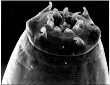
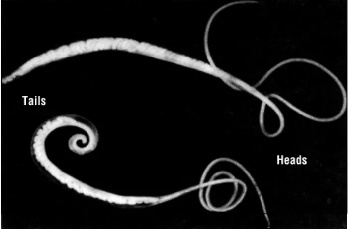
All dewormers placed on the market in the past 38 years have been very effective against the large roundworm (Ascaris suum ). Even so, it is still the most common parasite of swine. The large roundworm is the largest worm infecting pigs in the United States. It is about 12 inches (30.5 cm) in length and the diameter of a pencil (Figures 1 and 1a). The adults are found primarily in the small intestine but also may be found in the stomach or bile duct. The female ascarid produces large numbers of eggs (as many as one million/day) that are passed in the feces. The eggs have a thick, rough shell that provides protection from adverse environmental conditions (Figure 5c). These features also enable easy identification. Eggs require about four weeks to develop to the infective stage under ideal conditions. Eggs in fresh manure are not infective.
Figure 1a. Electron micrograph of the mouth and lips of the large roundworm (Ascaris suum). Note the rows of teeth on the lips edges and the two small pits on each of the three lips containing sensory organs. These worms graze along the small intestinal mucosa. Figure 1. Roundworm (Ascaris suum). The large one is the female; the small one with curled tail is the male.
Female Head
Female Tail Male Tail Male Head
2 The pig is infected by ingestion of the infective eggs, which hatch in the large intestine releasing larvae that penetrate the intestinal wall. These larvae utilize the circulatory system to migrate to the liver. The larvae migrate through the liver and again use the circulatory system to continue their migration to the lungs. They break out of the alveolar capillaries into the airspace where they travel up the bronchial tree and the trachea; and then they are coughed up and swallowed. Once in the small intestine, they continue their development to adult worms. Egg laying begins about six to eight weeks after infection.
When large numbers of larvae migrate through the liver, they cause localized hemorrhages and scarring. These are later replaced with connective tissue that appears as white spots on the surface and throughout the liver (Figure 2). These white spots are called milk spots and are responsible for liver condem-nation. Milk spots develop more rapidly on subsequent infections and are immune mediated. These lesions regress and the liver appears normal in about seven weeks.
Larvae that escape entrapment in the liver migrate to the lungs where they cause petechial (very small or minute) hemorrhages when they break out into the airspace. This may lead to verminous pneumonia and result in a cough. Pigs are more susceptible to secondary bacterial or viral infections during the parasite’s lung migration. Immunity develops subse-quent to infection, and the level of immunity increases according to the number of larvae trapped in the liver and lungs.
Nodular Worms
Nodular worms (Oesophagostomum spp.) are very com-mon in adult pigs throughout the United States. Adults, about .5 inch (1.3 cm) in length, are found in the large intestine (Figures 3 and 3a). Typical strongyle-type eggs (Figures 5d and 5e) are passed in the feces; they hatch and develop into infective larvae in about one week, and can survive for 12 months in protected areas.
Infected larvae are ingested, penetrate, and develop in the mucosal lining of the large intestine, causing small nodules. Most return to the lumen or cavity of the intestine in about one week, develop to adults, and begin laying eggs in about 3 to 5 weeks. The adults cause little harm; however, the pea-sized nodules caused by the larvae are thought to interfere with intestinal absorption. Severe infections can result in excessive weight loss and contribute to the “thin sow syndrome” during lactation. Nodules generally disappear within five weeks.
Whipworm
The swine whipworm (Trichuris suis) is present through-out the United States. The adult worms may be up to two inches (5 cm) in length, with the anterior two-thirds of the body thin and whip-like and the posterior one-third like the whip handle (Figures 4 and 4a). The adults are found in the large intestine; and the typical, bipolar plugged, football-shaped eggs (Figure 5) are passed in the feces. These eggs require about three weeks to reach the infective stage in the environment and must be ingested to continue the infection. The eggs are ingested and hatch, and the ensuing larvae penetrate into the intestinal wall. They remain in the mucosa (membrane lining the intestine) for two weeks, emerge from the mucosa in the third week, and develop into adults while attached to the cecal and large intestinal mucosa.
Figure 2. A pig liver showing “milk spots” resulting from the migration of large roundworm (Ascaris suum) larvae on their way from the large intestine through the liver, lungs, and mouth to the small intestine where they become adults.
Figure 3a. Electron micrograph of the head of a nodular worm (Oesophagostomum dentatum) showing food in the mouth opening surrounded by nine leaf-like lip elements. Note the four large papillae and the two bulges on the side with slits, all of which have sensory functions. These worms graze along the large intestinal mucosa.
Figure 3. Nodular worm (Oesophagostomum dentatum). The large one is the female; the small one with enlarged tail is the male.
Large numbers of worms may cause inflammation and result in diarrhea, dehydration, anorexia, and even death. Young animals are primarily affected; the diarrhea may resemble bloody dysentery. The thick-shelled eggs remain viable in the environ-ment for extended periods of time (up to 6 years) and are most frequently available to animals that have access to pasture. Recent experimental evidence suggests that the harmful effects of whipworm infection are caused by the interaction of the worms and resident intestinal bacteria.
where they become dormant. Those migrating to the intestine develop to adults (parthenogenetic females capable of reproduction without males), begin to lay eggs about one week after infection, and generally cause little tissue damage.
The dormant larvae become activated in the lactating sow and infect the nursing piglets via the colostrum. These larvae mature in four days and may cause severe damage when there are large numbers of worms. This may result in severe diarrhea in piglets during the first 14 days that can be confused with bacterial scours. Mortality may be as high as 75%. Those that survive may be stunted and feed conversion may be poor.
Stomach Worms
The red stomach worm (Hyostrongylus rubidus) is up to 3/8 inch (9.5 mm) in length, red in color (due to ingested blood), and found in the stomach. They are found throughout the United States, primarily in swine on pasture. Eggs passed in the feces develop to infective larvae in about one week. After ingestion, the larvae enter the stomach mucosa or lining, emerge in about two weeks, and develop into egg laying adults in about three weeks post-infection.
The presence of a large number of larvae and adults can produce anemia and stomach inflammation. Incidence of stom-ach worm has decreased due primarily to life cycle rearing in modern facilities. Eggs are typical strongyle type and difficult to distinguish from Oesophagostomum spp. (Figures 5d and 5e).
Lungworms
Lungworms (Metastrongylus spp.) are most prevalent in the Midwest and Southeast and require an intermediate host to complete the life cycle. The adult worms are almost 2 inches (5 cm) in length and lie in the bronchi and bronchioles. The eggs are coughed up, swallowed, and passed in the feces (Figure 5b). An earthworm must ingest these eggs for development to progress.
The pig ingests the earthworm containing the infective larvae. Larvae are freed on digestion, enter the lymphatics, and migrate to the right part of the heart and lungs, where they mature in the airspace. Clinical disease usually occurs with heavy infections and is caused by tiny hemorrhages caused by migrating larvae, or when adults may obstruct the airways.
Lungworm infection may predispose the animal to respira-tory infection due to influenza, mycoplasma, or other bacterial pathogens. Control is best accomplished by keeping pigs away from earthworms. The incidence of lungworm infection has decreased with confinement pig production.
Kidney Worms
The swine kidney worm (Stephanurus dentatus) is present in the Southeastern states and the lower Midwest. It has been introduced and survives in an isolated area of Canada. The worms are 1 to 1.5 inches (2.5 to 3.8 cm) in length and lie encysted in pairs along the ureters leading from the kidneys to the bladder. They also can be found in other locations in and around the kidneys.
Adult females produce eggs that are released through openings into the ureters and passed out in the urine. Eggs develop on the ground, hatch, and become infective larvae in about one week. Infection is by penetration of skin or by
Threadworms
Intestinal threadworms (Strongyloides ransomi) are less than 1/4 inch (6.35 mm) in length and are embedded in the surface of the small intestine. Threadworm infections are most frequently found in warmer climates, primarily in the southeast-ern United States and are rarely encountered in the north central states. Thin-shelled eggs (Figure 5a) pass in the feces, hatch, and develop to the infective larval stage on the ground. Infection is via penetration of the skin or oral mucosa.
The route of migration is thought to be via the circulatory system to the lungs, up the bronchial tree, and swallowed. A considerable number of larvae migrate to the mammary glands, Figure 4a. Electron micrograph of the anterior “whip” end of the whipworm (Trichuris suis). The double row of structures are gland openings located in the area which becomes attached to the large intestinal mucosa allowing the head to move freely in search of food. A small spear-like lancet just inside the mouth is protrusible and used to penetrate tissues. Whipworms feed on body fluids including blood.
Figure 4. Adult whipworms (Trichuris suis). The large one is the female; the small one with the curled tail is the male.
Heads Tails
4 ingestion. Ingested larvae, as well as those that penetrate the skin enter visceral lymph nodes, molt to 4th stage, and then
move to the liver in about 10 to 32 days where they grow and molt to young adults. Fetuses may become infected by errant, migrating larvae in gestating sows. The prolonged migration in the liver causes permanent damage that results in condem nation of a large number of swine livers, a condition most often occurring in the southeastern states.
Eventually the worms leave the liver as young adults, reach the kidney area, and become sexually mature. Worms frequently can be found in other organs, or in the spinal column where their presence has been shown to cause posterior paralysis in sows. Larvae can develop into egg-laying adults as early as six months after ingestion by a pig; however, it may take 9 to 16 months before eggs are found in urine.
Coccidia
Coccidiosis is a very important disease in nursing pigs raised inside. The disease is caused by the protozoan parasite Isospora suis and occurs in nursing pigs between 5 and 15 days of age. Infected pigs usually develop a yellowish diarrhea that can become very fluid. The pigs continue to nurse yet become dehydrated and debilitated. Affected pigs are unresponsive to antibacterial therapy. Mortality rate may be moderate but may increase with concurrent bacterial, viral, or parasitic infections.
See PIH-81, “Swine Coccidiosis” for more information.
Trichinella spiralis
Trichinosis is a disease of man and other animals caused by a tiny worm, Trichinella spiralis. Both domestic pigs and wild pigs can become infected with the parasite. For more information,
see PIH-103, “Trichinosis”.
Parasite Control Program
Worm infections with one or more species occur in 80% to 90% of U.S. swine herds, although most producers indicate that they deworm their hogs an average of 1.8 times during the
production cycle. Twenty-two million pounds (10 million kg) of liver were condemned in 1999 by USDA inspectors from 105.4 million hogs slaughtered. Most livers are condemned because of white spots caused by migrating immature worms (larvae). The percent of livers condemned has steadily decreased from 13% in 1976 to 6.3% in 1999. The higher rates of liver condemnation in the southeastern states are caused largely by kidney worms and in other states by ascarid larvae. A hidden loss from worms in pigs is a decrease in feed conversion efficiency. Results of controlled studies show the ascarid-infected pigs require 5.6% more feed per pound of gain than uninfected pigs. The incurred loss in the U.S. pig crop of 1999 is estimated at $15 million in finishing pigs (40 lb to 250 lb) with feed at $.065/lb. To effectively reduce the worm population in a swine herd and prevent condemnation losses, several management practices must be used along with dewormers. A good parasite control program will incorporate sound feeding and sanitation practices aimed at prevention of worm infections. Adequate nutrition, especially in regard to protein and vitamin levels, is important in protecting pigs against the effects of parasitism. Manure removal and thorough cleaning practices during the production cycle are essential in preventing reinfection. An all-in/all-out system of production provides the opportunity for thorough cleaning.
Worm parasites have enormous reproductive potential. The eggs of ascarids and whipworms have thick shells that protect the infective larvae inside. Larvae can survive in the egg for up to six years as long as the eggs are not exposed to direct sunlight, high temperatures, or allowed to dry completely. The eggs actually survive better when exposed to some disinfectants used on the farm to kill bacteria and other infectious organisms because their use prevents the organisms from attacking the worm egg shells. One of the best methods of preventing transmission of ascarids and whipworms in contaminated quarters is to thoroughly clean equipment, floors, and walls to remove all debris. This should be followed with a power wash to reach into cracks, crevices, and pores of all surfaces. Exposed concrete and wood need special attention because of their porous nature.
Knowledge of the life cycles of the worm species present in a herd will help in planning an appropriate control program.
5a. Strongyloides ransomi, intestinal threadworm eggs are thin-shelled and are in the larval stage when passed.
5b. Metastrongylus spp. lungworm eggs have rough thick shells. They look very much like the ascarid eggs (c) but are smaller, lighter in color, and are in the larval stage when passed. 5c. Ascaris suum, large roundworm eggs have rough thick shells, are dark yellow or brown, and are in the one-cell stage when passed. 5d and 5e. Oesophagostomum spp., nodular worm and
Hyostrongylus rubidus, stomach worm eggs are multicellular. The eggs are ellipsoidal with relatively thin, smooth shells. They vary in size and shape, are in the 8-32 cell stage, and brown in color when passed.
5f. Trichuris suis, whipworm eggs are easy to recognize by their football-like shape and end plugs, thick smooth shell, and dark yellow color. They are in the one cell stage when passed. Figure 5. Eggs of common pig internal parasites found in fresh manure. All photographs were taken at the same magnification (X400).
Roundworm
Ascaris suum ✓ ✓ ✓✻✻ ✓ ✓ ✓❖❖❖ ✓
Nodular worms
Oesophagostomum spp. ✓ ✓ ✓ ✓ ✓ ✓ ✓
Whipworm
Trichuris suis ✓ ✓ ✓✻✻
Lung worms
Metastrongylus spp ✓ ✓ ✓ ✓
Red stomach worm
Hyostrongylus rubidus ✓ ✓ ✓
Threadworm
Strongyloides ransomi ✓ ✓ ✓
Kidney worm
Stephanurus dentatus ✓ ✓ ✓ ✓
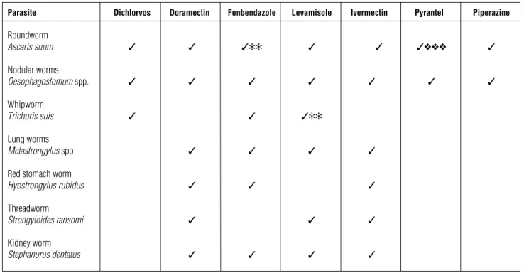
* Hygromycin is not included because it is a feed additive used as a prophylactic.
✻✻ Also effective against immature stages.
❖❖❖ Prevents ascarid larval migration by killing the larvae as they hatch from ingested embryonated eggs.
Table 1. Approved drugs for removal of internal parasites from pigs. A ✓ indicates the drug removes 90+% of adult worms*. Parasite Dichlorvos Doramectin Fenbendazole Levamisole Ivermectin Pyrantel Piperazine Effective programs will vary with the type of operation. Pigs
raised on pasture, dirt lots, or dirty concrete floors will need a more rigorous program because pigs will be constantly exposed to infective worm stages compared to pigs raised inside on slotted floors.
Before deciding on a worm treatment and control program, get an accurate diagnosis of the worm species present in the herd. Collect fresh fecal samples from several pigs in each age size group during each production cycle and have your veteri-narian or a laboratory examine a composite sample from each group for worm eggs. Carcass and viscera checks of slaughter pigs by your veterinarian can be very helpful in identifying parasite problems.
Dewormers
No single dewormer is effective against all species of worms (Table 1). The dewormers(s) chosen must be effective against the worms present and it must be given at the proper time for the maximum effect. The spectrum of efficiency, method of delivery, and cost should be considered for best economic results.
All dewormers are effective against the two most common worms of swine, the ascarid (Ascaris suum) and nodular worms (Oesophagostomum spp.). Dichlorvos is given in either a 1 or 2 day feeding. Fenbendazole can be given in feed for 3 to 12 days, piperazine and levamisole can be given in either feed or water for 1 day, and ivermectin and doramectin in a single injection. Ivermectin is also formulated as a feed additive to be fed for 7 days. Hygromycin is fed continuously and pyrantel tartrate can be fed continuously or in one feeding only. Continuous feeding of either one prevents infection with ascarids and nodular worms and in the case of hygromycin, whipworms (Trichuris suis).
Fenbendazole is effective against the immature stages of both ascarids and whipworms and can be used in preventing contamination of lots and pastures by deworming pigs every 6 weeks, before females of either parasite begin producing eggs. Ivermectin and doramectin are the only dewormers also effective against the ectoparasites. Injecting sows 10 to 14 days before farrowing prevents transmission of threadworm (Strongy-loides ransomi) larvae in the colostrum and clears the sow from mange mites that are normally acquired by the suckling pig while nursing.
REVISED 9/01
1-888-EXT-INFO
Reference to products in this publication is not intended to be an endorsement to the exclusion of others which may be similar. Persons using such products assume responsibility for their use in accordance with current directions of the manufacturer.