Original Article
Microleakage Evaluation of Bulk-fill Composites Used with
Different Adhesive Systems
Moosavi Ha; Moghaddas MJb; Kordnoshahri Fc; Zanjani Md*
aAssociate Professor, Dental Materials Research Center, Department of Operative Dentistry, Mashhad Dental School, Mashhad University of Medical Sciences, Mashhad, Iran
bAssociate Professor, Dental Materials Research Center, Department of Operative Dentistry, Mashhad Dental School, Mashhad University of Medical Sciences, Mashhad, Iran
cGeneral Dentist, Mashhad Dental School, Mashhad University of Medical Sciences, Mashhad, Iran
dPost graduate student, Department of Operative Dentistry, Mashhad Dental School, Mashhad University of Medical Sciences, Mashhad, Iran
ARTICLE INFO Abstract
Article History:
Received: 4 December 2017 Accepted: 21 January 2018
Statement of Problem: The integrity of adhesive bond at the tooth/resin interface performs an essential role in the clinical success and survival of restorations.
Objectives: The purpose of this study was to compare microleakage of Class
II cavities restored with total-etch or self-etch adhesives and different bulk-fill
composites.
Materials and methods: Conventional class II cavities were prepared on forty sound human premolar teeth with approximately same size and shape. Half of the cavities restored with a sonic-resin placement system (SonicFill, Kerr), and the other half of the cavities with Tetric N-Ceram (TNC) composite with total-etch; Optibond Solo Plus or self-etch; Optibond XTR, adhesives. The samples stored in distilled water at 37 ° C for 24 hours, and then specimens were under 1000 thermal cycles. The teeth were covered with two layers of nail varnish except for one mm around gingival margins, and then were dipped in glass vials for 48 h at 37°C in a neutral-buffered 2% methy¬lene-blue solution. After removal from the dye, the teeth were rinsed and the varnish was removed, and stored individually in a glass vial containing 65%
nitric acid. The vials were centrifuged, and 100 μl of the super¬natant from
each was then analysed in a UV-Visible spectro¬photometer after a kinetic assay at 550 nm wavelength using concentrat¬ed nitric acid as the blank. So dye extraction was conducted to investigate the leakage test. Data were analysed by ANOVA test (P<0.05).
Results: The highest and lowest microleakage was related to self-etch/ SonicFill and total-etch/Tetric N-Ceram groups respectively. The amount
of microleakage was not significantly different among experimental groups
(P>0.05).
Conclusions: Both of the bulk-fill composites with various adhesives had the
same microleakage. Key words:
Bulk fill
Adhesive resin Microleakage Dye extraction Corresponding Author: Maryam Zanjani Post graduate student, Department of Operative Dentistry, Mashhad Dental School, Mashhad University of Medical Sciences, Mashhad, Iran
Email: Zanjanim941@mums. ac.ir
Tel: +98-9155088028
Cite this article as: Moosavi H,Moghaddas MJ, Kordnoshahri F, Zanjani M.Microleakage Evaluation of Bulk-fill
Introduction
In recent years, the composite resins are the first choice of restorative materials due to ever growing esthetic demand from the patients. Though the substantial improvements in esthetic, wear, and physical properties are achieved, the polymerization shrinkage is persisting as a major limitation [1]. The clinical consequences of the marginal gap include marginal leakage, postoperative sensitivity, secondary caries, discoloration, and cuspal strain [2]. The prevalence of proximal and cervical caries is on the rise due to large elder population and improved survival of teeth [3]. Several researchers have suggested the various techniques to reduce the polymerization shrinkage and consequently the better marginal integrity. The proposed methods are incremental placement, three sided light curing, centripetal build up, pulse cure, precured composite inserts, and intermediate layer; glass ionomer cement, auto polymerizing composites, and flowable composites [4-9]. Scientists have presented a new generation of composites and adhesives with less contraction property and also various techniques to reduce polymerization shrinkage and thus, better marginal integrity [5,10]. Few authors also suggested the bulk placement and curing to reduce the marginal gap and stress at the cavosurface margins [11]. The investigators have reported the reduced polymerization shrinkage with bulk curing methods [12]. Obtaining optimum seal between composites and tooth structure is critical for the clinical longevity of restoration and will contribute significantly toward the improved public oral health. SonicFill is a single-step composite system that doesn’t require an additional capping layer. SonicFill system combines the advantages of a flowable composite with a universal composite. SonicFill system is comprised of a KaVo handpiece that enables sonic activation of a specially designed and conveniently delivered composite from Kerr. SonicFill’s activation significantly reduces the composite viscosity to rapidly fill the cavity. However, this technique does not have cost benefit [6]. To overcome this disadvantage, a group of new products has been introduced known as bulk-fill composites that could be inserted in 4 mm bulk [7,8]. Using bulk-fill composites, clinical steps will be reduced by filling the cavity in single increment,
leading to less porosity and a uniform consistency [7]. Dental adhesive systems can be categorized based on the clinical procedure in "etch-and-rinse adhesives" and "self-etch adhesives". Other essential considerations concern the different anatomical characteristics of enamel and dentine which are involved in the bonding procedures that have also implications for the technique used as well as for the quality of the bond [13]. The restorative procedure with composites are very time-consuming and technically demanding, particularly concerning the application of the adhesive system. Therefore, together bulk-fill composites and self-etch adhesives could reduce operator error and chair side time. The microleakage under composite restoration is the topic of intense research for a long time. Hence, the purpose of this in vitro study was to compare the microleakage of class II bulk-fill composite restorations either with total-etch or self-etch adhesives.
Materials and Methods
This in vitro experimental study was conducted on forty intact human premolars of approximately the same size extracted within the past six months. The teeth were sound and had no carious lesions, cracks or fracture. The teeth were stored in Chloramine T 0/5 % solution at room temperature for disinfection. Conventional Class II cavities were prepared on one of the proximal surfaces of the teeth. Cavities were prepared with diamond bur (835/012‡) in a high-speed hand piece with water spray and the same size as much as possible, by one operator. It was used periodontal probes for measurement the depth and width with pulpal floor depth of 2 ± 0.5 mm, gingival width of 1.5 ± 0.5 mm, axial height of 2± 0.5 mm, and buccolingual width to 1/3 the distance between the tip of cusps. Therefore, the cervical margin of the cavity was extended about to the cementoenamel junction. The cavity had 90 cavosurface margins. The burs (Mani, INC. 8-3 Kiyohara Industrial Park, Utsunomiya, Tochigi, 321-3231, JAPAN) were replaced every five cavity preparations. The teeth were randomly divided into two groups (n=20). A transparent matrix was then placed on the tooth. Half of the cavities were restored with viscose bulk-fill composite resin (SonicFill, Kerr) with
total-etch adhesives; Optibond Solo Plus (Kerr) or self-etch adhesive; Optibond XTR (Kerr) and the other half were restored with bulk-fill composite resin Tetric N-Ceram (Ivoclar Vivadent) with total-etch or self-etch technique, according to the manufacturer's instructions. For use Optibond Solo plus, the cavity was etched by 37% phosphoric acid on the enamel margins for five seconds and was then applied on dentin for 15 seconds, rinsed with air and water spray for 10 seconds and dried with cotton pellet. Optibond Solo Plus adhesive was then applied, air sprayed for 3-5 seconds from 1 cm distance and cured for 20 seconds. Using Optibond XTR adhesive, the initial Optibond XTR primer was applied on the enamel and dentin surfaces for 20 seconds and then was dried for 5 seconds with medium pressure air spray. To use Optibond XTR adhesive, it was primarily applied for 15 seconds on enamel and dentin and then dried out with air spray, light cured for 20 seconds using a Bluephase C8 (IvoclarVivadent, Schaan, Liechtenstein) light-curing unit with a light intensity of 800 mW ⁄ cm2. Cavities were then restored in one increment with the composite (SonicFill or Tetric N-Ceram) and cured with light for 20 seconds from occlusal, buccal and lingual sides. Restorative materials used in this study was shown in Table 1. After removing matrix, a scalpel and fine diamond burs were used
to eliminate any excess material, especially at the gingival margin. A series of paper disks (Sof-Lex) were used to finish margins. Samples were stored in 37°C distilled water for 24 hours and thermocycled for 1000 cycles between 5 and 55 °C with a six minutes dwell time. 40 teeth were covered with 2 layers of nail varnish (VepaKozmetik, Istanbul, Turkey), except for one mm around gingival margin. The teeth were dipped in glass vials for 48 hours at 37°C in a neutral-buffered 2% methy¬lene-blue solution, under normal atmospheric pressure. After removing the dye, the teeth were rinsed under tap water for 30 min and the varnish was removed using a sharp scal¬pel, and then stored individually in a glass vial containing 600 μl of concentrated (65%) nitric acid for 3 days. The vials were centrifuged (LABNT, Spectrafuge 16M, USA) at 14000 rpm for 5 min, and 100 μl of the super-natant from each was then analysed in a UV-Visible spectro¬photometer (UNIC visible with scanner S2150,2100PC,USA) after a kinetic assay at 550 nm wavelength using concentrat¬ed nitric acid as the blank. In this manner, we recorded wavelength of diffused dye at tooth-restorative material interface. To check the normality of data distribution, Kolmogorov-Smirnov test was used. ANOVA test was conducted for evaluating of microleakage of experimental groups. The
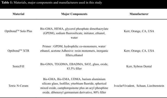
post-Table 1: Materials, major components and manufacturers used in this study
Material Major Components Manufacturer
OptibondTM Solo Plus Bis-GMA, HEMA, glycerol phosphate dimethacrylate
(GPDM), sodium fluorosilicate, initiator, ethanol, water
Kerr, Orange, CA, USA
OptibondTM XTR
Primer : GPDM, hydrophilic co-monomers, water/ ethanol, acetone Adhesive: resin monomers, inorganic
fillers,ethanol
Kerr, Orange, CA, USA
SonicFill Bis-GMA, TEGDMA, EBADMA, SiO2, glass, oxide, 83.5% filler Kerr, Sybron Dental
Tetric N-Ceram
Bis-GMA, Bis-EMA, UDMA, barium aluminium silicate glass, Isofiller, ytterbium fluoride, spherical mixed oxide, camphorquinone plus an acyl phosphine
oxide, dibenzoyl germanium derivative, 80% filler
hoc multiple comparisons Tukey’s test was used for pair wise comparisons. The statistical significance was set at p value less than 0.05. All statistical analyses were performed with Statistical Package for Social Sciences (SPSS) version 16 (SPSS Inc., Chicago, IL, USA).
Results
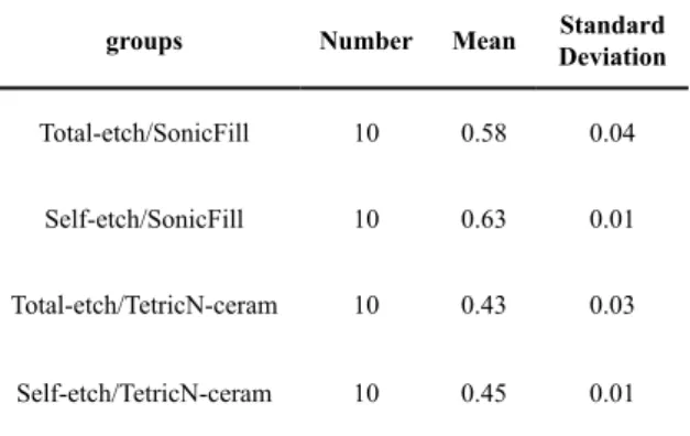
Table 2 illustrates the mean and standard deviation microleakage for all groups. ANOVA test was done to explore the difference in mean microleakage among studied groups. The analysis showed there was no difference among groups (P>0.05) and consequently, the type of adhesive and composite had no influence on the amount of microleakage (P= 0.13). All samples restored with SonicFill or Tetric N-Ceram composite and total-etch or self-etch adhesive showed the microleakage at the margins. The highest and lowest microleakage was related to self-etch/SonicFill (0.63) and total-etch/Tetric N-Ceram (0.43) groups respectively. However, this study showed that there was no difference between experimental groups in microleakage (P>0.05).
Discussion
The present study was an effort to find the desirable material, which had least microleakage. This trial evaluated and compared the microleakage of two relatively new composite resins SonicFill and Tetric N-Ceram along with total-etch and self-etch adhesives in conventional class II cavities. In order to simulate oral cavity environment and clinical states, we used 1000 thermocycling test at 5-55º C according to 11450 ISO protocol. In
Table 2: Mean microleakage values and standard deviations for the experimental groups
groups Number Mean DeviationStandard
Total-etch/SonicFill 10 0.58 0.04
Self-etch/SonicFill 10 0.63 0.01
Total-etch/TetricN-ceram 10 0.43 0.03
Self-etch/TetricN-ceram 10 0.45 0.01
our investigation, none of the tested adhesive systems eliminated microleakage in dentin margin of the cavity. This is in agreement with previ¬ous studies that evaluated microleakage of tooth-colour restorations at dentin interface [14-16]. In this study, selection of human permanent premolars of approximately the same size was based on maximum standardization and elimination of the possible effect of tooth anatomy on the results. It has been stated that etch-and-rinse adhesive systems generally perform better on enamel than self-etching systems which may be more appropriate for bonding to dentine. In order to escape a possible loss of the restoration, secondary caries or pulp damage due to bacteria penetration or due to cytotoxicity effects of eluted adhesive components, careful attention of several factors is essential in selecting the suitable bonding procedure and adhesive system for the individual patient situation [13]. Poggio et al. [17] evaluated microleakage of class II conventional and bulk-fill composite restorations with their gingival margin below the cementoenamel junction. All composite restorations showed some degrees of microleakage in their study but SonicFill composite showed minimum microleakage. However, in our study, Tetric N-Ceram bulk-fill with total-etch adhesive in comparison to SonicFill composite showed the best results, although the difference with other groups was not statistical significance. Moreover, it showed greater microleakage in dentin margin of conventional and bulk fill restorations, which was in accordance with our results and may be due to the lower thickness of enamel at the gingival margin, greater distance of light curing unit from the gingival margin and weaker bond to dentin compared to enamel [18]. A systematic review concluded that bulk-fill composites provide a bond to dentin as strong as that of conventional composites without the problems related to polymerization shrinkage of conventional composites and can be very useful particularly for deep cavities [19]. The microleakage test was designed taking into consideration; dye extraction, is the most frequent choices in test variables, as reported by prior studies [20,21]. Just like previous studies, in the present study, it was found that there was no statistical difference between SonicFill and Tetric N-Ceram composites in microleakage, as well two examined
adhesives [22-24].Thus, our null hypothesis was verified. The highest average of microleakage was contributed to SonicFill group with self-etch adhesive, and the least average of microleakage was contributed to Tetric N-Ceram group with total-etch adhesive. This result is in agreement with earlier research [15,16]. It was found that the best marginal compatibility is contributed to Tetric N-Ceram composite which resembles our findings that indicate the least amount of microleakage is in this group [25]. Bulk-fill composites have been introduced to facilitate the placement of deep direct resin composite restorations. An ideal bulk-fill composite would be one that could be placed into a preparation having a high C-factor design and still exhibit very little polymerization shrinkage stress, while maintaining a high degree of cure throughout [26].
According to manufacturing claimed that Tetric N-Ceram bulk-fill enables posterior teeth to be restored with only one layer measuring up to four-millimetres in thickness, which considerably heightens efficiency. The patented light activator Ivocerin is responsible for ensuring the complete cure of the filling. Compared with conventional light initiators, the Ivocerin polymerisation booster is much more reactive. Therefore, polymerization is initiated even in very deep cavities and the material is fully cured. A specially conditioned shrinkage stress reliever keeps shrinkage and shrinkage stress during polymerization to a minimum. In addition, Tetric N-Ceram contains a mixture of bisphenol-A diglycidyldimethacrylate, urethane dimethacrylate, and ethoxylated bisphenol A dimethacrylate, all of which are high-molecular-weight monomers with high viscosity and low polymerization shrinkage [27]. Based on the most of studies done till now, an incremental layering technique has been the standard procedure in direct posterior composite restorations to diminish polymerization shrinkage stress and achieve sufficient curing [28,29]. SonicFill is a composite restoration where the viscosity of the composite is dramatically reduced up to 87%, due to the special rheological modifiers that react to sonic activation of the material delivered through the SonicFill hand piece during its placement, thus, increasing its flow and enabling rapid filling of the cavity. Precise adaptation to the cavity walls make the frequency and size of critical
voids located at the margin and along line angles of the cavity less pronounced compared to the conventional putty-like composites [30]. When the sonic energy is stopped, the composite returns to a more viscous, non-slumping state that is perfect for carving and contouring [31]. Generally, increasing the filler load in the resin matrix results in reduction of overall shrinkage of the composite due to the reduced availability of the monomer for the curing reaction. However, it may also result in increasing the viscosity of a material, thus posing difficulty in placement and more chances of gap formation and microleakage [29]. SonicFill consist of a special composite formulation that contains about 83.5% of fillers by weight, which is more higher when compared to the filler content of Tetric N-Ceram (80%wt) [30]. There was no significant difference in the microleakage between the two different bulk-fill materials used in this study, which might be attributed to the similarity in their filler loading. It has also been documented that an increase in filler volume content results in an increase in the stiffness of the material with high modulus of elasticity, thereby increasing the microleakage, which consents with the results of the current study [32]. Al-Harbi et al. [33] showed that SonicFill composite, with total-etch adhesive, had the best marginal integrity, though not statistically significant. Although the result of that research was different from our findings in composite type, it was in agreement with our study in adhesive type. This finding is similar to the study of Campos et al. that found no difference between bulk-fill and conventional composites [34]. Moreover, in our trial, there was no difference between total-etch adhesive and self-etch adhesive, similar to Delbons et al.[35]. The reason of this finding seems to be the presence of ethanol base solvent and the same adhesives that were employed. In another study, SonicFill composite with total-etch adhesives have shown the best marginal integrity [20]. Despite different method of analysis tests used, our study yielded the same results. Nevertheless, there were some unavoidable limitations in our study such as long-term storage of tooth hydration condition, absence of mastication forces, and patient dental habits, which might not necessarily be expected in actual practice.
for a fast and reliable filling technique that allows reduction of layers, effort and time; therefore, the time-consuming incremental layering technique can be substituted with the bulk-fill technique using SonicFill as a bulk-fill material. Therefore, further research evaluating the properties of the materials with various adhesives and its clinical implications are recommended in future.
Conclusions
There was no difference between self-etch and total-etch adhesives with two bulk- fill composites, Tetric N-ceram and SonicFill resin composites, regarding their various placement methods, in term of cervical marginal microleakage.
Acknowledgments
Authors would like to thank for vice chancellor of research of Mashhad University of Medical Sciences for approve the grant for doing the research. The results presented in this study have been taken from a student thesis no: 2799, proposal code 931008.
Conflict of Interest: None declared. References
1. Agrawal VS, Parekh VV, Shah NC. Comparative evaluation of microleakage of silorane-based composite and nanohybrid composite with or without polyethylene fiber inserts in class II restorations: An in vitro study. Oper Dent. 2012;37:E1-7.
2. Kaya S, Yigit Özer S, Adigüzel Ö, et al. Comparison of apical microleakage of dual-curing resin cements with fluid-filtration and dye extraction techniques. Med Sci Monit. 2015;21:937-44.
3. Somani R, Jaidka S, Arora S. Comparative evaluation of microleakage of newer generation dentin bonding agents: An in vitro study. Indian J Dent Res. 2016; 27:86-90. 4. Park J, Chang J, Ferracane J, et al. How should
composite be layered to reduce shrinkage stress: incremental or bulk filling? Dent Mater. 2008;24:1501-5.
5. Lutz F, Krejci I, Luescher B,et al. Improved proximal margin adaptation of Class II composite resin restorations by use of light-reflecting wedges. Quintessence Int. 1986;17:659-64.
6. Eakle WS, Ito RK. Effect of insertion technique on microleakage in mesio-occlusodistal composite resin restorations. Quintessence Int. 1990;21:369-74.
7. Kanca J, Suh BI. Pulse activation: reducing resin-based composite contraction stresses at the enamel cavosurface margins. Am J Dent. 1999;12:107-12.
8. Hagge MS, Lindemuth JS, Mason JF, et al. Effect of four intermediate layer treatments on microleakage of Class II composite restorations. Gen Dent. 2001;49:489-95.
9. Majety KK, Pujar M. In vitro evaluation of microleakage of class II packable composite resin restorations using flowable composite and resin modified glass ionomers as intermediate layers. J Conserv Dent. 2011;14:414-7.
10. Giachetti L, Scaminaci Russo D, Bambi C, et al. A review of polymerization shrinkage stress: current techniques for posterior direct resin restorations. J Contemp Dent Pract. 2006;7:79-88.
11. Abbas G, Fleming GJ, Harrington E, et al. Cuspal movement and microleakage in premolar teeth restored with a packable composite cured in bulk or in increments. J Dent. 2003;31:437-44.
12. Belvedere PC. Contemporary posterior direct composites using state-of-the-art techniques. Dent Clin North Am. 2001;45:49-70.
13. Milia E, Cumbo E, Cardoso RJ, et al. Current dental adhesives systems. A narrative review. Curr Pharm Des. 2012;18:5542-52.
14. Sadeghi M. Influence of flowable materials on microleakage of nanofilled and hybrid Class II composite restorations with LED and QTH LCUs. Indian J Dent Res. 2009;20:159-63. 15. Osorio R, Toledano M, de Leonardi G, et al.
Microleakage and interfacial morphology of self-etching adhesives in class V resin composite restorations. J Biomed Mater Res B Appl Biomater. 2003;66:399-409.
16. Koubi S, Raskin A, Dejou J, et al. Effect of dual cure composite as dentin substitute on the
marginal integrity of Class II open-sandwich restorations. Oper Dent. 2010; 34:150-6. 17. Poggio C, Chiesa M, Scribante A, et al.
Microleakage in Class II composite restorations with margins below the CEJ: in vitro evaluation of different restorative techniques. Med Oral Patol Oral Cir Bucal. 2013;18:e793-8.
18. Juloski J, Carrabba M, Aragoneses JM, et al. Microleakage of Class II restorations and microtensile bond strength to dentin of low-shrinkage composites. Am J Dent. 2013;26:271-7.
19. Akah MM, Daifalla LE, Yousry MM. Bonding of bulk fill versus contemporary resin composites: A systematic review and meta-analysis. Indian J Sci Technol. 2016;9:1-13. 20. Asselin ME, Fortin D, Sitbon Y, et al. Marginal
microleakage of a sealant applied to permanent enamel: evaluation of 3 application protocols. Pediatr Dent. 2008;30:29-33.
21. Cehreli ZC, Gungor HC. Quantitative microleakage evaluation of fissure sealants applied with or without a bonding agent: results after four-year water storage in vitro. J Adhes Dent. 2008;10:379-84.
22. Sivakumar JS, Prasad AS, Soundappan S, et al. A comparative evaluation of microleakage of restorations using silorane-based dental composite and methacrylate-based dental composites in Class II cavities: An in vitro study. J Pharm Bioallied Sci.2016;8:S81-S5. 23. Cantekin K, Gumus H. In vitro and clinical
outcome of sandwich restorations with a bulk-fill flowable composite liner for pulpotomized primary teeth. J Clin Pediatr Dent.2014; 38:349-54.
24. Schwendicke F, Kern M, Dorfer C, et al. Influence of using different bonding systems and composites on the margin integrity and the mechanical properties of selectively excavated teeth in vitro. J Dent.2015; 43:327-34.
25. Agarwal RS, Hiremath H, Agarwal J, et al. Evaluation of cervical marginal and internal adaptation using newer bulk fill composites:
An in vitro study. J Conserv Dent. 2015; 18:56-61.
26. Tantbirojn D, Pfeifer CS, Braga RR, et al. Do low-shrink composites reduce polymerization shrinkage effects? J Dent Res. 2011; 90:596-601.
27. Gonçalves F, Kawano Y, Pfeifer C, et al. Influence of BisGMA, TEGDMA, and BisEMA contents on viscosity, conversion, and flexural strength of experimental resins and composites. Eur J Oral Sci. 2009 ;117:442-6. 28. Feilzer AJ, Gee AJD, Davidson CL. Setting
stress in composite resin in relation to configuration of the restoration. J Dent Res. 1987;66:1636-39.
29. Jang JH, Park SH, Hwang IN. Polymerization shrinkage and depth of cure of bulk- fill resin composites and highly filled flowable resin. Oper Dent. 2015;40:172-80.
30. Hirata R, Pacheco RR, Caceres E, et al. Effect of Sonic Resin Composite Delivery on Void Formation Assessed by Micro-computed Tomography. Oper Dent. 2018;43:144-150. 31. Eunice C, Margarida A, João CL, et al. 99mTc
in the evaluation of microleakage of composite resin restorations with SonicFillTM. An in vitro experimental model. Open J Stomatol. 2012;2:340-47.
32. Senawongse P, Pongprueksa P, Tagami J. The effect of the elastic modulus of low-viscosity resins on the microleakage of Class V resincomposite restorations under occlusal loading. Dent Mater J. 2010;29:324-9.
33. Al-Harbi F, Kaisarly D, Bader D, et al. Marginal Integrity of Bulk Versus Incremental Fill Class II Composite Restorations. Oper Dent. 2016; 41:146-56.
34. Campos EA, Ardu S, Lefever D,et al. Marginal adaptation of class II cavities restored with bulk-fill composites. J Dent. 2014;42:575-81. 35. Delbons FB, Perdigao J, Araujo E, et al.
Randomized clinical trial of four adhesion strategies in posterior restorations-18-month results. J Esthet Restor Dent. 2015;27:107-17.