Address for correspondence
Maryam Zare Jahromi E-mail:[email protected]
Funding sources
None declared
Conflict of interest
None declared
Received on January 22, 2019 Reviewed on April 3, 2019 Accepted on May 8, 2019
Published online on September 30, 2019
Abstract
Background. Various materials are used for vital pulp capping and the bond strength of restorative mate-rials to these pulp-capping agents significantly affects the success rate of vital pulp therapy.
Objectives. The aim of this study was to determine the shear bond strength of a flowable composite resin and a single-component glass-ionomer to mineral trioxide aggregate (MTA), calcium-enriched mixture (CEM) cement and BiodentineTM as pulp-capping agents.
Material and methods. Seventy-two cylindrical acrylic resin blocks, with a central hole 4 mm × 2 mm, were prepared. Mineral trioxide aggregate, CEM cement and Biodentine were placed in the cavities (n = 24 in each group) and incubated for 24 h. The blocks were subdivided into the composite resin and glass-ionomer subgroups. Cylindrical plastic molds, measuring 3 mm in height and diameter, were used to place the restorative materials on the samples. The shear bond strength test was performed at a strain rate of 1 mm/min in a universal testing machine. The samples were evaluated under a stereomicroscope at ×25 magnification for fracture modes. The data was analyzed with the one-way analysis of variance (ANOVA) and Tukey tests.
Results. The maximum and minimum mean shear bond strength values were recorded in the Bioden-tine–composite resin (4.77 MPa) and MTA–glass-ionomer (2.20 MPa) groups, respectively. There were significant differences in the mean shear bond strength values of MTA, CEM cement and Biodentine to the composite resin and glass-ionomer (p < 0.001).
Conclusions. A composite material may be preferable for definitive filling after pulp capping with Biodentine. Key words: shear bond strength, mineral trioxide aggregate, composite resin, glass-ionomer cement, Biodentine
Słowa kluczowe: wytrzymałość wiązania na ścinanie, agregat trójtlenków mineralnych, żywica kompo-zytowa, cement szkło-jonomerowy, Biodentyna
Cite as
Zarean P, Roozbeh R, Zarean P, Zare Jahromi M, Mirzakoochaki Broujeni P. In vitro comparison of shear bond strength of a flowable composite resin and a single-component glass-ionomer to three different pulp-capping agents. Dent Med Probl. 2019;56(3):239–244. doi:10.17219/dmp/109233
DOI
10.17219/dmp/109233
Copyright
© 2019 by Wroclaw Medical University This is an article distributed under the terms of the Creative Commons Attribution 3.0 Unported License (CC BY 3.0)
(https://creativecommons.org/licenses/by/3.0/)
In vitro comparison of shear bond strength of a flowable composite resin
and a single-component glass-ionomer to three different pulp-capping agents
Porównanie in vitro wytrzymałości na ścinanie wiązania
płynnej żywicy kompozytowej i jednoskładnikowego szkło-jonomeru
z trzema materiałami do pokrycia miazgi
Paridokht Zarean
1,D–F, Reza Roozbeh
2,B–D, Parichehr Zarean
3,D–F, Maryam Zare Jahromi
4,A,B,D, Parvin Mirzakoochaki Broujeni
5,A1 Dental Implant Research Center, Dental Research Institute, Isfahan University of Medical Sciences, Iran 2 Private dental office, Yazd, Iran
3 Dental Research Center, Dental Research Institute, Isfahan University of Medical Sciences, Iran 4 Department of Endodontics, Dental School, Islamic Azad University, Isfahan (Khorasgan) Branch, Iran 5 Department of Esthetic Dentistry, Dental School, Islamic Azad University, Isfahan (Khorasgan) Branch, Iran
A – research concept and design; B – collection and/or assembly of data; C – data analysis and interpretation; D – writing the article; E – critical revision of the article; F – final approval of the article
Introduction
Vital pulp therapy consists in placing a biocompatible material on the exposed pulp of the teeth with an open apex.1 Various materials are used for pulp capping, in-cluding calcium hydroxide, mineral trioxide aggregate (MTA) and newer silicate-based cements, such as BioAg-gregate®, EndoSequence®, BiodentineTM, etc.2
Mineral trioxide aggregate is a hydrophilic cement com-posed of calcium oxide, silica and bismuth oxide. Several successful clinical applications have been reported for MTA.1 Long-term studies (over 3 years) have reported that the success rate of vital pulp therapy with MTA is higher than in the case of calcium hydroxide (78% vs 60%).2 These favorable outcomes for direct pulp therapy with MTA have been confirmed in recent systematic reviews.3 De-spite the various reported advantages, MTA has also some disadvantages, including potential for discoloration, dif-ficult handling, long setting time, high cost, unavailability of a solvent, and difficulty with its removal after setting.4
Biodentine is a new calcium silicate-based cement with high purity, which has drawn attention as a substitute for dentin in composite resin restorations, direct pulp capping and endodontic treatment.5,6 Biodentine consists of trical-cium silicate, calof trical-cium carbonate (as a filler), zirconium oxide (as an opacifier), and a water-based liquid containing cal-cium chloride. Calcal-cium chloride serves as a water-reducing agent and decreases the initial and final setting times. The incorporation of calcium chloride into the liquid not only re-sults in the acceleration of the setting time of Biodentine, but also improves the handling properties and strength of the ce-ment.5 In previous studies, Biodentine has exhibited better sealing ability, higher compressive strength, shorter setting time, lower microleakage, better antimicrobial properties, less toxic effects, and better biocompatibility, bioactivity and biomineralization compared to MTA.7
Calcium-enriched mixture (CEM) cement is a newly introduced material in endodontics. It is a water-based cement with clinical properties similar to those of MTA; however, its chemical properties are different.8 This new material is recommended for direct pulp capping and
pulpotomy in deciduous and permanent molars.9
One of the most important issues in vital pulp therapy is the ability to seal the pulp-capping agents, because keep-ing them intact affects the prognosis of the therapeutic procedure. Composite resins are common restorative materials, especially in the esthetic zones. A proper bond between the composite resin and the pulp-capping agent can distribute stresses beyond the bonded area on the tooth surface, decrease microleakage and increase the strength of the remaining tooth structure.10
In cases with an insufficient amount of enamel around the access cavity, resin-modified glass-ionomer (RMGI) is considered a suitable restorative material for the recon-struction of the crowns of the teeth that have undergone pulp therapy.11 Glass-ionomers bond to the tooth structure
chemically, exhibit proper bio-compatibility, no polymer-ization shrinkage and no free monomers, and have dimen-sional stability in the presence of moisture, as their advan-tages.12 At present, newer types of glass-ionomer cements have been introduced to overcome the disadvantages of old cements, including sensitivity to water and low translucen-cy, and preserve their advantages, such as fluoride release and adhesion, at the same time.13 A proper bond between the restorative material and the pulp-capping agent results in the distribution of stresses on all bonded surfaces, en-sures the vitality of the pulp and its sealing, and improves the prognosis of vital pulp therapy.7,14
Cantekin and Avci evaluated the shear bond strength of a methacrylate-based and a silorane-based composite resin and a glass-ionomer to MTA and Biodentine.7 They reported that the highest bond strength was related to the methacrylate-based composite resin bonded to Biodentine.7 Later on, Doozaneh et al. reported that the bond strength of a self-adhering flowable composite resin to CEM cement and MTA was higher than in the case of improved RMGI with an additional application of an adhesive.15 Also, in 2018, Elmi et al. concluded that irrespective of the type of adhesive system, the shear bond strength of a composite resin to CEM cement is higher than that of RMGI.16
The purpose of the present in vitro study was to deter-mine the shear bond strength of a composite resin and a single-component glass-ionomer to MTA, CEM cement and Biodentine as pulp-capping agents.
Material and methods
A total of 72 cylindrical acrylic resin blocks (Acropars®; Marlic Medical Ind. Co., Tehran, Iran) were prepared for the purpose of this in vitro study. A cavity, measuring 4 mm in diameter and 2 mm in depth, was prepared at the center of each cylinder. Mineral trioxide aggregate (ProRoot® MTA; Dentsply Sirona Inc., York, USA), CEM cement (Yektazist Dandan, Tehran, Iran) and Biodentine (Septodont, Saint-Maur-des-Fossés, France) were used according to the manu-facturers’ instructions. Mineral trioxide aggregate was mixed at a powder-to-liquid ratio of 3:1.4 The liquid and powder of CEM cement were mixed according to the manufacturer’s instructions to achieve a proper consistency. Biodentine was prepared in an amalgamator by adding 5 drops of Biodentine liquid to the capsule containing its powder in 30 s. Then, the prepared materials were placed in the cavities at the center of the acrylic blocks.
The blocks were divided into 3 groups according to the material used: MTA, CEM cement and Biodentine (n = 24 in each group). The acrylic blocks were incubated at 37°C and 100% relative humidity for 24 h for the complete set-ting of the materials.7 As the acid-etching procedure af-fected the compressive strength and surface microhard-ness, after 24 h, surface changes occurred, which enhanced bonding.17 Then, each group was divided into 2 subgroups
– Grandio Flow® composite resin and Ionoseal® glass-ionomer (VOCO GmbH, Cuxhaven, Germany).
The blocks receiving the composite resin were acid-etched with 35% phosphoric acid for 15 s to avoid over-etching, which decreases the shear bond strength.18 Af-terward, they were rinsed with water for 30 s, followed by drying with an oil-free air stream for 5 s. At the next stage,
the adhesive Solobond® M (VOCO GmbH) was applied on
the specimen surfaces. It was applied twice and dried with an air flow for 5 s in order to evaporate its solvent. The next step was light-curing (LED D; Guilin Woodpecker Medical Instrument Co. Ltd., Guilin, China) for 15 s.
Cylindrical plastic molds, measuring 3 mm in diame-ter and height, were used to place the flowable compo-site resin.19 The molds were filled with the Grandio Flow composite resin and placed on the prepared surfaces of the samples before setting, followed by light-curing for 20 s from the top, based on the manufacturer’s instruc-tions. Then, the plastic molds were gently detached from the composite resin molds, which was followed by light-curing for 20 s from the sides. Similar plastic molds were used for the Ionoseal glass-ionomer. The glass-ionomer was placed within the transparent molds put on the samples, followed by light-curing from the top for 20 s. Then, the plastic molds were gently separated from the glass-ionomer samples, which was followed by light cur-ing from the sides for 20 s. Next, the samples were stored at 37°C and 100% relative humidity for 24 h.
Subsequently, the samples were transferred to a universal testing machine (Walter+Bai AG, Löhningen, Switzerland), equipped with a chisel-shaped head measuring 5 mm in width. A perpendicular force was applied at the restorative material–pulp-capping agent interface at a crosshead speed of 1 mm/min to detach the composite resin and glass-iono-mer from the endodontic materials and to draw a graph. Before carrying out statistical analyses, the resultant data, recorded in N, was divided by the surface area of the samples (7.06 mm2) in order to determine the bond strength in MPa. Finally, all samples were evaluated under a stereomicroscope (trinocular zoom stereo microscope SMP-200; HP Inc., Palo Alto, USA) at ×25 magnification to evaluate the fracture modes (cohesive, adhesive or mixed).
The data was analyzed with the one-way analysis of variance (ANOVA), t-test and post hoc Tukey tests.
The significance level was assumed at p < 0.05. The tables were drawn using the software IBM SPSS Statistics for Windows, v. 20 (IBM Corp., Armonk, USA).
Results
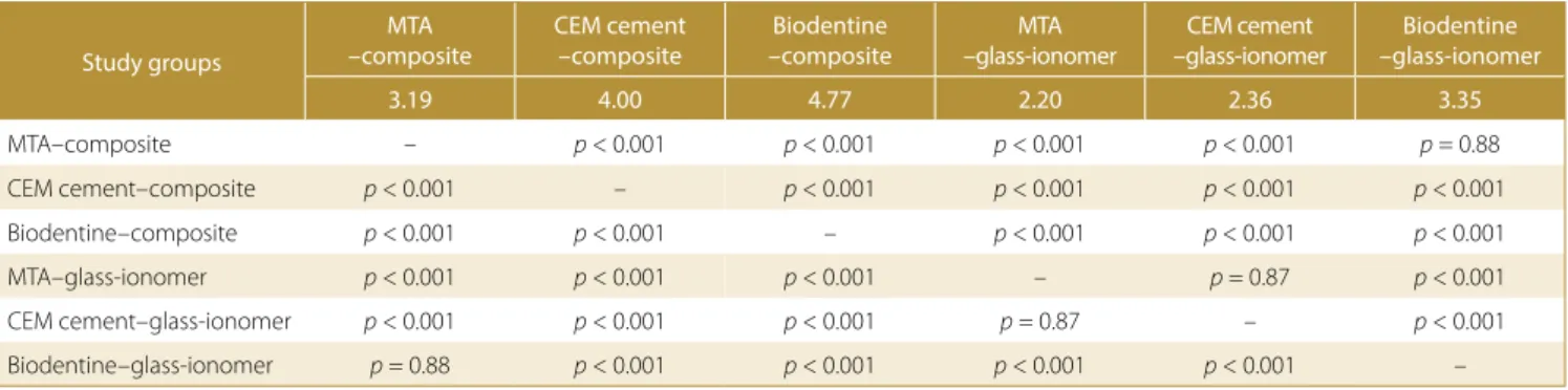
As shown in Table 1, the maximum and minimum mean shear bond strength values were recorded in the Bioden-tine–composite resin (4.77 MPa) and MTA–glass-ionomer (2.20 MPa) groups, respectively. There were significant dif-ferences in the mean shear bond strength values of MTA, CEM cement and Biodentine to the composite resin and glass-ionomer (p < 0.001). Since the differences in the mean shear bond strength value of MTA, CEM cement and Biodentine to the composite resin and glass-ionomer were significant, the post hoc Tukey tests were used. The mean shear bond strength value of MTA to the composite resin was 3.19 MPa, which was significantly higher than those in the MTA–glass-ionomer and CEM cement–glass-ionomer groups (p < 0.001). On the other hand, the mean shear bond strength value of the MTA–composite resin group was lower than those in the CEM cement–composite resin and Biodentine–composite resin groups, which was statistically significant (p < 0.001). However, despite the lower mean shear bond strength value in the MTA–composite resin group compared to the Biodentine–glass-ionomer group, the difference was not statistically significant (p = 0.88).
Based on the data in Table 2, the shear bond strength values for the CEM cement–composite resin group was
Table 2. Two-by-two comparisons of the shear bond strength value between the study groups
Study groups
MTA –composite
CEM cement –composite
Biodentine –composite
MTA –glass-ionomer
CEM cement –glass-ionomer
Biodentine –glass-ionomer
3.19 4.00 4.77 2.20 2.36 3.35
MTA–composite – p < 0.001 p < 0.001 p < 0.001 p < 0.001 p = 0.88
CEM cement–composite p < 0.001 – p < 0.001 p < 0.001 p < 0.001 p < 0.001
Biodentine–composite p < 0.001 p < 0.001 – p < 0.001 p < 0.001 p < 0.001
MTA–glass-ionomer p < 0.001 p < 0.001 p < 0.001 – p = 0.87 p < 0.001
CEM cement–glass-ionomer p < 0.001 p < 0.001 p < 0.001 p = 0.87 – p < 0.001
Biodentine–glass-ionomer p = 0.88 p < 0.001 p < 0.001 p < 0.001 p < 0.001 –
Table 1. Mean shear bond strength values of MTA, CEM and Biodentine to composite resin and glass-ionomer
Pulp-capping
agent Restorative material Number
Mean bond strength
[MPa] SD
MTA composite resin 12 3.19 0.23
glass-ionomer 12 2.20 0.27
CEM cement composite resin 12 4.00 0.47
glass-ionomer 12 2.36 0.39
Biodentine composite resin 12 4.77 0.30
glass-ionomer 12 3.35 0.38
SD – standard deviation; MTA – mineral trioxide aggregate; CEM – calcium-enriched mixture.
higher compared to all other groups except for the Bio-dentine–composite resin group and the differences, whether higher or lower, were significant (p < 0.001). The Biodentine–composite resin group had a higher shear bond strength value than all other 5 groups and the differ-ence was statistically significant (p < 0.001).
Also, the mean shear bond strength value in the MTA –glass-ionomer group was lower compared to other groups and this difference was not significant only with re-gard to the CEM cement–glass-ionomer group (p = 0.87); however, the difference was significant in the case of the other groups (p < 0.001).
The mean shear bond strength value in the CEM ce-ment–glass-ionomer group was not significantly different only from that in the MTA–glass-ionomer group (p < 0.87); however, the difference with regard to other groups was significant (p < 0.001). The comparison of the mean shear bond strength values between the Biodentine–glass-iono-mer and other groups showed no significant difference between this group and the MTA–composite resin group (p = 0.88); however, the differences between this group and other groups were significant (p < 0.001).
Discussion
After vital pulp therapy, the treated tooth requires a suit-able restoration. Recently, composite resins have been widely used for this purpose, but in some areas, where there is not enough enamel around the preparation site, RMGIs can be a good alternative.20 The bond strength of restor-ative materials to pulp-capping agents plays a crucial role in the coronal sealing, and consequently the success of vital pulp therapy.7 A proper bond between the restorative ma-terial and the pulp-capping agent also distributes stresses on the bonded surface area of dentin.14
Therefore, in the present study, the shear bond strength test was used to evaluate the adhesive properties of re-storative materials (composite resin and glass-ionomer) to 3 pulp-capping agents (MTA, CEM cement and Bio-dentine). The shear bond strength of different restorative materials to MTA has been evaluated in previous stu-dies.11,15 However, only a few studies on the bond strength of different restorative materials to CEM cementand Bio-dentine are available.11,15,16,21
The results of the present study showed that the highest shear bond strength between the pulp-capping agent and the restorative material was in the Biodentine–compo site resin group. Furthermore, Biodentine exhibited higher shear bond strength to the composite resin and glass-iono-mer compared to that of MTA and CEM cement. This result is in agreement with Cengiz and Ulusoy’s study.22 Such a difference might be explained by the fact that, un-like MTA, in which only distilled water is used for setting, the liquid of Biodentine contains a mixture of distilled water, calcium chloride and a water-soluble monomer.23
Calcium chloride accelerates the setting reaction, and the water-soluble monomer serves as a water-reducing agent and decreases the amount of water in the material, in-creasing the strength of the material.23,24
In the present study, the surfaces of the pulp-capping agents were not polished; therefore, they had some in-dentations, resulting in the greater penetration of the composite resin and glass-ionomer. The use of a bonding agent in the composite resin groups could result in higher bond strength. Composite resin is a hydrophobic material and forms a better bond with surfaces that have low wa-ter content. The bonding agent used in the present study was of the total-etch type (Solobond® M; VOCO GmbH), which creates a better bond with drier surfaces. Consider-ing the presence of a water-reducConsider-ing agent in Biodentine, the water content of the material decreases, resulting in better conditions for bonding with composite resin.
The exact mechanism of bonding restorative materials to Biodentine has not been elucidated. Since Biodentine and MTA have a similar chemical structure, it is prob-able that their water absorption is also similar. Etchants, during shorter than usual times, might cause the selec-tive elimination of the matrix around crystal structures, leading to successful bonding through micromechanical interlocking.17
The majority of studies on various adhesive systems ap-plied on MTA have shown favorable shear bond strength obtained with the use of total-etch adhesive systems; it has been demonstrated that phosphoric acid creates deeper and more retentive microscopic pores compared to self-etch adhesive systems.17 In the present study, the samples receiving composite resin were acid-etched with 35% phosphoric acid. Since no resin structure is present in MTA and CEM cement, it can be claimed that their bond-ing to restorative materials is completely mechanical.
Altunsoy et al. evaluated the shear bond strength of 2 different types of composite resin to MTA, CEM ce-ment and Biodentine.25 The results showed that the low-est bond strength was related to Biodentine–composite resin, contrary to the results of the present study. They used a self-adhesive flowable composite resin, without etching and dentin bonding. In addition, in their study, the specimen surfaces were polished with abrasive paper for 1 min, which resulted in a decrease in bond strength.25
Cantekin and Avci evaluated the shear bond strength of a methacrylate-based and a silorane-based compo site resin as well as a glass-ionomer to MTA and Biodentine.7 They reported that the highest bond strength was re-lated to methacrylate-based composite resin bonded to Biodentine. The results of their study are consistent with those of the present study. It should be pointed out that in the study by Cantekin and Avci, contrary to the present study, the composite resins were packable; in the present study, a flowable composite resin was used. In addition, the glass-ionomer used in the study by Cantekin and Avci was a type of conventional self-cured glass-ionomer.7
Ajami et al. compared the shear bond strengths of com-posite resin and glass-ionomer to MTA, CEM cement and
white MTA (WMTA) mixed with Na2HPO4 (NAMTA)
as pulp-capping agents.11 The highest bond strength
was recorded in the NAMTA–composite resin group. It should be pointed out that some properties of NAMTA are different from those of MTA. The study carried out by Ajami et al. was different from the present one with re-gard to the me thods and the materials used. In their study, all samples were sandblasted and a packable composite resin was used in association with a one-step self-etch ad-hesive system.11
Based on the results of the present study, the minimum shear bond strength value between the pulp-capping agent and the restorative material was recorded in the MTA–ionomer group. When a restorative glass-ionomer is placed on MTA, one of the following reactions might occur:
– the COO– group in polyacrylic acid might react with calcium in MTA to produce calcium salts;
– the MTA hydrated silicate gel might be compressed by the glass-ionomer hydrated silicate gel to create byproducts.26 Given the high percentage of metallic oxides in MTA and the porous surface topography of MTA, it is expect-ed that glass-ionomer will form a relatively strong bond with MTA.27 The glass-ionomer used in the present study was Ionoseal, which is a single-component material. Since no etching or bonding were used in the glass-iono-mer groups, possibly this material exhibited lower bond strength compared to the composite resin bonded to the pulp-capping agents. However, since in the present study, the surfaces of the pulp-capping agents were not polished and had some inherent porosity as well as considering the fact that the glass-ionomer used in the present study was flowable and exhibited proper adaptation to the surface, the restorative material formed a relatively strong bond to the pulp-capping agents.
In the study by Cantekin and Avci, in which the bond strength of Biodentine and MTA to a silorane-based composite resin, a methacrylate-based composite resin and a self-curing conventional glass-ionomer was evalu-ated, the lowest bond strength was recorded in the MTA –glass-ionomer group,7 which is consistent with the re-sults of the present study.
In the study by Ajami et al., in which the shear bond strength of composite resin and glass-ionomer to MTA, CEM cement and NAMTA was evaluated, the lowest bond strength values were recorded in the CEM cement –glass-ionomer group,11 which does not agree with the results of the present study. It should be pointed out that such a discrepancy in the results might be attributed to the differences in the methodologies, including the use of sandblasting and the use of polyacrylic acid as a condi-tioner before the application of RMGI.
In the present study, the fracture mode in all samples was cohesive within the pulp-capping agents. Recent
stu-dies have demonstrated that the fracture mode between MTA and dentin was cohesive within MTA. However, the number of cohesive fractures decreased and the number of adhesive fractures increased over time.28 In addition, the researchers pointed out that when CEM cement was used as a root-end filling material, the fracture mode was cohesive in the push-out test.29 In order to carry out a suc-cessful restorative procedure with 2 different materials, there should be a proper bond between the 2 materials. Generally, a bond is favorable when the fracture occurs within the material rather than at the bonded interface (i.e., a cohesive fracture is better than adhesive failure).19
Since in the present study, the failure mode in all samples was cohesive within the pulp-capping agents, the results indicate a favorable bond between the restorative mate-rial and the pulp-capping agent. In the evaluation of failure modes, especially in the shear bond strength tests, one im-portant issue should be taken into account. The tendency toward a cohesive fracture might be attributed to the un-even distribution of stresses within the bonded materials, resulting in early failure before the bonded surface is af-fected.30 This is an inherent problem with the shear bond strength test, in which a lot of tensile force is applied to the area below the force application point, with simultaneous compressive stresses at the point opposite the force appli-cation point.31
Conclusions
Within the limitations of an in vitro study, the composite resin exhibited stronger bonds to all the evaluated pulp-capping agents compared to the glass-ionomer. Among the pulp-capping materials, Biodentine exhibited a higher bond strength value to the flowable composite resin and the glass-ionomer. Therefore, to achieve a proper bond, the use of a composite resin on Biodentine is recommended. ORCID iDs
Paridokht Zarean https://orcid.org/0000-0002-9983-3066 Reza Roozbeh https://orcid.org/0000-0003-1831-8028 Parichehr Zarean https://orcid.org/0000-0002-6858-6146 Maryam Zare Jahromi https://orcid.org/0000-0002-4642-2810 Parvin Mirzakoochaki Broujeni https://orcid.org/0000-0001-9393-8222
References
1. Ford TR, Torabinejad M, Abedi HR, Bakland LK, Kariyawasam SP. Using mineral trioxide aggregate as a pulp-capping material. J Am Dent Assoc. 1996;127(10):1491–1494.
2. Mente J, Geletneky B, Ohle M, et al. Mineral trioxide aggregate or calcium hydroxide direct pulp capping: An analysis of the clinical treatment outcome. J Endod. 2010;36(5):806–813.
3. Didilescu AC, Cristache CM, Andrei M, Voicu G, Perlea P. The effect of dental pulp-capping materials on hard-tissue barrier forma-tion: A systematic review and meta-analysis. J Am Dent Assoc.
2018;149(10):903–917.e4.
4. Parirokh M, Torabinejad M. Mineral trioxide aggregate: A compre-hensive literature review. Part I: Chemical, physical, and antibacte-rial properties. J Endod. 2010;36(1):16–27.
5. Koubi G, Colon P, Franquin JC, et al. Clinical evaluation of the per-formance and safety of a new dentine substitute, Biodentine, in the restoration of posterior teeth – a prospective study. Clin Oral Investig. 2013;17(1):243–249.
6. Schmidt A, Schäfer E, Dammaschke T. Shear bond strength of lin-ing materials to calcium-silicate cements at different time intervals.
J Adhes Dent. 2017;19(2):129–135.
7. Cantekin K, Avci S. Evaluation of shear bond strength of two resin-based composites and glass ionomer cement to pure tricalcium sili-cate-based cement (Biodentine®). J Appl Oral Sci. 2014;22(4):302–306.
8. Asgary S, Shahabi S, Jafarzadeh T, Amini S, Kheirieh S. The proper-ties of a new endodontic material. J Endod. 2008;34(8):990–993. 9. Malekafzali B, Shekarchi F, Asgary S. Treatment outcomes
of pulpo-tomy in primary molars using two endodontic biomaterials: A 2-year randomised clinical trial. Eur J Paediatr Dent. 2011;12(3):189–193. 10. Heymann HO, Swift EJ Jr., Ritter AV, Sturdevant CM. Sturdevant’s
Art and Science of Operative Dentistry. 6th ed. St. Louis, MO: Elsevier/
Mosby; 2013:245.
11. Ajami AA, Jafari Navimipour E, Savadi Oskoee S, Abed Kahnamoui M, Lotfi M, Daneshpooy M. Comparison of shear bond strength of res-in-modified glass ionomer and composite resin to three pulp cap-ping agents. J Dent Res Dent Clin Dent Prospects. 2013;7(3):164–168. 12. Hilton TJ, Ferracane JL, Broome JC, eds. Summitt’s Fundamentals of
Operative Dentistry: A Contemporary Approach. 4th ed. Hanover Park,
IL: Quintessence Publishing Co. Inc.; 2013:516.
13. Sakaguchi RL, Powers JM, eds. Craig’s Restorative Dental Materials. 13th ed. Philadelphia, PA: Elsevier/Mosby; 2012:156–159.
14. Oskoee SS, Kimyai S, Bahari M, Motahari P, Eghbal MJ, Asgary S. Comparison of shear bond strength of calcium-enriched mixture cement and mineral trioxide aggregate to composite resin. J Con-temp Dent Pract. 2011;12(6):457–462.
15. Doozaneh M, Koohpeima F, Firouzmandi M, Abbassiyan F. Shear bond strength of self-adhering flowable composite and resin- modified glass ionomer to two pulp capping materials. Iran Endod J. 2017;12(1):103–107.
16. Elmi M, Ehsani M, Esmaeili B, Khafri S. Comparison of bond strength of a composite resin with two different adhesive systems and a resin modified glass ionomer to calcium enriched mixture. J Con-serv Dent. 2018;21(4):369–372.
17. Kayahan MB, Nekoofar MH, Kazandağ M, et al. Effect of acid-etch-ing procedure on selected physical properties of mineral trioxide aggregate. Int Endod J. 2009;42(11):1004–1014.
18. Al-Suleiman M, Baba F, Sawan MN, Suliman A. Mechanical evalua-tion of the effect of reducing phosphoric acid concentraevalua-tions and etching duration on the bond strength of orthodontic brackets.
J Dent Oral Disord Ther. 2014;2(2):1–5.
19. Tate WH, Friedl KH, Powers JM. Bond strength of composites to hybrid ionomers. Oper Dent. 1996;21(4):147–152.
20. Paterson RC. Bacterial contamination and the exposed pulp.
Br Dent J. 1976; 140(7):231–236.
21. Odabaş ME, Bani M, Tirali RE. Shear bond strengths of different adhe-sive systems to biodentine. ScientificWorldJournal. 2013;2013:626103. 22. Cengiz E, Ulusoy N. Microshear bond strength of tri-calcium
sili-cate-based cements to different restorative materials. J Adhes Dent.
2016;18(3):231–237.
23. Bortoluzzi EA, Broon NJ, Bramante CM, Felippe WT, Tanomaru Filho M, Esberard RM. The influence of calcium chloride on the setting time, solu- bility, disintegration, and pH of mineral trioxide aggregate and white Portland cement with a radiopacifier. J Endod. 2009;35(4):550–554. 24. Pradelle-Plasse N, Tran XV, Colon P. Physico-chemical properties.
In: ORE-FDI working group; Goldberg M, ed. Biocompatibility or Cytotoxic Effects of Dental Composites. Oxford, UK: Coxmoor Pub-lishing Company; 2009:184–194.
25. Altunsoy M, Tanriver M, Ok E, Kucukyilmaz E. Shear bond strength of a self-adhering flowable composite and a flowable base com-posite to mineral trioxide aggregate, calcium-enriched mixture cement, and Biodentine. J Endod. 2015;41(10):1691–1695.
26. Nandini S, Ballal S, Kandaswamy D. Influence of glass-ionomer cement on the interface and setting reaction of mineral trioxide aggregate when used as a furcal repair material using laser Raman spectroscopic analysis. J Endod. 2007;33(2):167–172.
27. Yesilyurt C, Yildirim T, Taşdemir T, Kusgoz A. Shear bond strength of conventional glass ionomer cements bound to mineral trioxide aggregate. J Endod. 2009;35(10):1381–1383.
28. Mozayeni MA, Milani AS, Marvasti LA, Asgary S. Cytotoxicity of cal-cium enriched mixture cement compared with mineral trioxide aggregate and intermediate restorative material. Aust Endod J.
2012;38(2):70–75.
29. Shokouhinejad N, Razmi H, Fekrazad R, et al. Push-out bond strength of two root-end filling materials in root-end cavities pre-pared by Er,Cr:YSGG laser or ultrasonic technique. Aust Endod J.
2012;38(3):113–117.
30. Asgary S, Eghbal MJ, Parirokh M, Ghoddusi J, Kheirieh S, Brink F. Comparison of mineral trioxide aggregate’s composition with Portland cements and a new endodontic cement. J Endod.
2009;35(2):243–250.
31. Armstrong S, Geraldeli S, Maia R, Raposo LH, Soares CJ, Yamaga-wa J. Adhesion to tooth structure: A critical review of “micro” bond strength test methods. Dent Mater. 2010;26(2):e50–e62.