Simple Factors Associated with Radiation-Induced Lung Toxicity
after Stereotactic Body Radiation Therapy of the Thorax: A
Pooled Analysis of 88 Studies
Jing Zhao, MD PhD1,2, Ellen D. Yorke, PhD3, Ling Li1,4, Brian D. Kavanagh, MD5, X. Allen Li, PhD6, Shiva Das, PhD7, Moyed Miften, PhD5, Andreas Rimner, MD8, Jeffrey Campbell1, Jinyu Xue, PhD9, Andrew Jackson, PhD3, Jimm Grimm, PhD10, Michael T. Milano, MD PhD11, and Feng-Ming (Spring) Kong, MD PhD1,*
1Department of Radiation Oncology, GRU Cancer Center/Medical College of Georgia, Georgia
Regents University, Augusta, GA, USA, 30912
2Department of Oncology, Tongji Hospital, Tongji Medical College, Huazhong University of
Science and Technology, Wuhan, China, 430030
3Department of Medical Physics, Memorial Sloan Kettering Cancer Center, New York City, NY
10065
4Department of Shanghai Cancer Hospital, Fudan University, Shanghai, China, 200032
5Department of Radiation Oncology, University of Colorado, Denver, CO 80045
6Department of Radiation Oncology, Medical College of Wisconsin, Milwaukie, WI 53226
7Department of Radiation Oncology, University of North Carolina, Chapel Hill, NC 27514
8Department of Radiation Oncology, Memorial Sloan Kettering Cancer Center, New York City, NY
10065
9Department of Radiation Oncology, MD Anderson Cancer Center at Cooper, Camden, New
Jersey 08103
10Bott Cancer Center, Holy Redeemer Hospital, Meadowbrook, PA 19046
11Department of Radiation Oncology, University of Rochester, Rochester, NY14642
Abstract
Purpose—To study the risk factors for radiation-induced lung toxicity (RILT) after stereotactic body radiotherapy (SBRT) of the thorax.
Mailing address, telephone, facsimile, and e-mail for corresponding author: 821 St Sebastian St, Augusta, GA 30912. Tel: (706) 723-0030; Fax: (706) 721-1937; [email protected].
Conflicts of interest: The remaining authors declare no conflict of interest.
This work was presented in part as an oral presentation at the 56th Annual Meeting of the American Society of Radiation Oncology held in San Francisco, California, September, 2014.
Publisher's Disclaimer: This is a PDF file of an unedited manuscript that has been accepted for publication. As a service to our
customers we are providing this early version of the manuscript. The manuscript will undergo copyediting, typesetting, and review of
HHS Public Access
Author manuscript
Int J Radiat Oncol Biol Phys
. Author manuscript; available in PMC 2017 August 03.Published in final edited form as:
Int J Radiat Oncol Biol Phys. 2016 August 01; 95(5): 1357–1366. doi:10.1016/j.ijrobp.2016.03.024.
A
uthor Man
uscr
ipt
A
uthor Man
uscr
ipt
A
uthor Man
uscr
ipt
A
uthor Man
uscr
Methods—Published studies on lung toxicity in patients with early stage non-small cell lung cancer (NSCLC) or metastatic lung tumors treated with SBRT were pooled and analyzed. The primary endpoint was RILT including pneumonitis and fibrosis. Data of RILT and risk factors were extracted from each study, and rates of grade 2-5 (G2+) and grade 3-5 (G3+) RILT were computed. Patient, tumor and dosimetric factors were analyzed for their correlation with RILT.
Results—Eighty-eight studies (7752 patients), that reported RILT incidence, were eligible. The pooled rates of G2+ and G3+ RILT from all 88 studies were 9.1% (95% CI: 7.15-11.4) and 1.8% (95% CI: 1.3-2.5), respectively. The median of median tumor sizes was 2.3 (range 1.4-4.1) cm. Among the factors analyzed, older patient age (P= 0.044) and larger tumor size (the greatest diameter) were significantly correlated with higher rates of G2+ (P= 0.049) and G3+ RILT (P= 0.001). Patients with stage IA vs. stage IB NSCLC had significantly lower risks of G2+ RILT (8.3% vs 17.1%, OR= 0.43, 95% CI: 0.29-0.64, P<0.0001). Among studies that provided detailed dosimetric data, the pooled analysis demonstrated a significantly higher mean lung dose (MLD) (P= 0.027) and V20 (P= 0.019) in patients with G2+ RILT comparing to that of grade 0-1 RILT.
Conclusions—The overall rate of RILT is relatively low after thoracic SBRT. Older age and larger tumor size are significant adverse risk factors for RILT. Lung dosimetry, specifically lung V20 and MLD also significantly affect RILT risk.
Summary—Risk factors for radiation-induced lung toxicity (RILT) after stereotactic body radiotherapy (SBRT) were analyzed from 88 published studies (7752 patients). The overall rate of RILT is relatively low after thoracic SBRT. Adverse risk factors for RILT after SBRT include older age, larger tumor size and greater lung dose-volume exposure as measured by mean lung dose and volume of lung receiving greater than 20 Gy.
Introduction
Stereotactic body radiotherapy (SBRT) is the standard of care for patients with medically inoperable stage I non-small cell lung cancer (NSCLC) (1). For operable early stage NSCLC patients, a meta-analysis (2) of 11921 patients in 63 studies demonstrated that overall survival or disease-free survival did not differ significantly between SBRT and surgery when patients' baseline condition was comparable. Recent studies (3-8) have demonstrated effectiveness of SBRT for oligometastasis(es) (1-5 pulmonary nodules).
Radiation induced lung toxicities (RILT), including radiation pneumonitis (RP) and clinical lung fibrosis, are important dose limiting toxicities. From published data to date, the rate of symptomatic RP ranged from 0% to 49% (9-12); the rate of radiation-induced lung fibrosis, which can continue to evolve beyond one year after SBRT, is reportedly up to 70-80% in high-dose regions (13), albeit most often asymptomatic. Fatal (grade 5) RILT after SBRT warrants serious consideration although is only sporadically reported (14).
Many factors, including patient-related, tumor-related and dosimetric may play important roles in the risk of RILT (15). SBRT dose and fractionation are often considered important, affecting clinical management decisions with respect to lung tolerance and tumor control, particularly in elderly patients and/or those with chronic lung interstitial diseases (12,16). Physical dosimetric measures such as mean lung dose (MLD) and lung V20 (percent or absolute volume receiving over 20 Gy), often used to guide clinical practice, were analyzed
A
uthor Man
uscr
ipt
A
uthor Man
uscr
ipt
A
uthor Man
uscr
ipt
A
uthor Man
uscr
in some studies, showing inconsistent relationship with RILT after SBRT (17-19), perhaps due to the limited number of toxicity events from each study.
Here we aimed to examine the risk of RILT after thoracic SBRT from the published studies to date and to study the significance of clinical and simple dosimetric risk factors in relation to RILT.
Materials and Methods
Selection criteria
Eligible studies included those that described treatment with SBRT either for primary NSCLC or metastatic lung tumors. Detailed reporting of RILT, including rate and grade, after SBRT was required. Studies of patients who received radiotherapy prior to SBRT, SBRT in conjunction with systemic therapy, and/or SBRT as a boost therapy after conventional radiotherapy were excluded. Publications were limited to those written in English.
Search methods
Studies published before Dec 2014 were sought by electronic databases (PubMed, Embase and Cochrane), using the search terms of lung tumor/cancer, SBRT/SABR and lung toxicity/ pneumonitis/lung fibrosis. When there was uncertainty about the eligibility of a study, the decision was made by the first and senior authors.
Data collection and calculation
The data collected for analysis included the number of patients and lesions, 14 clinical factors including age, gender, histology, stage, smoking status, tumor location, tumor size (maximum diameter of the tumor), gross tumor volume (GTV), planning target volume (PTV), total prescription dose, number of fractions, MLD and V20 for bilateral or ipsilateral lung, and prescribed biological effective dose with α/β= 10 (BED10). Male/female patients (M/F) ratio, centrally/peripherally located tumor ratio, the percentage of clinical stage I NSCLC, current smoking and percent of adenocarcinoma tumor were calculated. RILT data from grade 0 to grade 5 after treatment were also extracted, and data of RILT including grade 2-5 (G2+) and grade 3-5 (G3+) were summed from studies with such data provided.
Statistical analysis
To minimize potential bias due to heterogeneity, a pooled estimate for RILT rate was obtained using a Bayesian hierarchical model. The number of cases of RILT for each study were modeled with the binomial distribution with the logits of the binomial probabilities for each study following a normal distribution with mean and variance τ. These parameters had normal and inverse gamma priors, respectively. The overall rates of RILT and credible intervals were estimated as the posterior mean of μ. To compare the difference between asymptomatic (grade 0-1) and symptomatic (grade 2-5) RILT, odds ratio (OR) and 95% CI were estimated; P values were determined using the Chi-square test. χ2 tests and I2 were
used to study heterogeneity between trials which represented the percentage of total variation across studies that was attributable to heterogeneity rather than to chance. If the
A
uthor Man
uscr
ipt
A
uthor Man
uscr
ipt
A
uthor Man
uscr
ipt
A
uthor Man
uscr
test indicated heterogeneity across studies (p<0.10 or I2>50%, the random effects model (Der Simonian-Laird method) was selected. Otherwise, we used the fixed effects model (Mantel-Haenszel method) to analyze relations between toxicity groups (20). Analyses were carried out using Revman 5.2 software. All tests were two-sided, and a p-value <0.05 was considered statistically significant.
Results
Patients and study characteristics
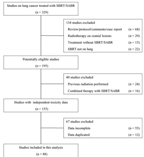
A total of 329 studies were identified at the initial search including all studies of SBRT for lung tumors; 241 studies were excluded due to 1) RILT data missing, 2) duplications of publication or publications with overlapping data, 3) prior thoracic radiation treatments with or without concurrent chemotherapy, or 4) non-original publications such as meta-analysis, review or case reports. Eighty-eight original studies including 7752 patients were eligible for this analysis (Fig 1 shows the detailed study selection). Pooled data of 14 clinical factors are shown in Tables 1 and 2. As not all the studies provided all elements of this study, numbers of study and patients varied such individual data provided for specific factors
Overall rates of RILT
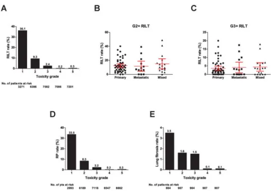
Of all 88 studies, the Bayesian hierarchical model estimates of RILT were 9.1% (95% CI: 7.15-11.4) and 1.8% (95% CI: 1.3-2.5), for G2+ and G3+ RILT, respectively. The crude rates and patient numbers with all RILT grades are shown in Fig 2A.
Fifty-four focused on primary lung cancers, 15 on lung metastases, and the remaining 19 on mixed patient populations. The average rates of G2+ RILT were 10.4% (95% CI: 9.8-15.2), 7.0% (95% CI: 4.3-18.9) and 12.8% (95% CI: 7.8-22.2), respectively. The average rates of G3+ RILT were 2.2% (95% CI: 2.4-4.9), 1.6% (95% CI: 1.0-7.2), and 3.0% (95% CI: 1.9-7.0), respectively. There was no significant difference in the RILT rates between patients with primary lung cancers and lung metastases from various primary sites (Fig 2B-C). This study thus analyzed the data without separating primary lung cancer and lung-metastatic patients.
Among these studies, 77 reported RP, 25 on lung fibrosis (of which 15 included detailed grading). From studies that provided detailed data, the average rates of G2+ / G3+ RP were 9.5% (95% CI:7.8-11.3) / 2.2% (95% CI:1.7-3.1) (Fig D), and the average rates of G2+ / G3+ lung fibrosis were 0.2% (95% CI: 0.008-4.7) / 0.2% (95% CI:0.005-1.3) (Fig E). Unless otherwise specified, all RILT refer to either RP or fibrosis, or both.
Patient and tumor factors and RILT
Older patient age was significantly associated with higher rate of G2+ RILT (P= 0.045). Histology, tumor location (central versus peripheral), GTV and PTV were not significant. However, the largest tumor linear dimension was significant: studies with larger median tumor sizes had higher rates of G2+ (P= 0.049) and G3+ (P= 0.001) RILT (Fig 3A-F).
Selected clinical factors were compared between asymptomatic (grade 0-1) and
symptomatic RILT (grade 2-5). In 6 studies of 1009 patients with stage IA (663 patients) or
A
uthor Man
uscr
ipt
A
uthor Man
uscr
ipt
A
uthor Man
uscr
ipt
A
uthor Man
uscr
IB (346 patients) NSCLC, 55 (8.3%, CI: 5.08-10.43) and 59 patients (17.1%, CI: 11.34-23.52) had G2+ RILT, respectively. The odds ratio was 0.40 (CI: 0.26-0.63,
P<0.0001). There was no significant heterogeneity with an I2 value of 0% (P= 0.66). There was no significant difference in gender for G2+ RILT, with rates of 10.5% (CI: 9.12-12.35) and 12.0% (CI: 7.7-17.42) in male and female patients, respectively (OR= 0.82, CI: 0.49-1.37, P= 0.44). Tumor location was not significant: 38 in 318 (11.9%, CI: 7.73-23.59) patients with centrally located tumors developed G2+ RILT after SBRT, comparing to 101/1005 (10.0%, CI: 10.64-18.30) patients with peripherally ones (OR= 1.14, CI: 0.65-1.99, P= 0.65) (Fig 3G-I). There were insufficient separated results for G3+RILT to perform a meaningful analysis.
Simple dosimetric factors and RILT
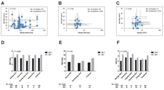
Simple dosimetric factors including prescription BED10, MLD and V20 were reported in 67 (6170 patients), 14 (1705 patients) and 19 (1928 patients) studies, resepctively. There were no significant correlations between the rates of G2+ and G3+ of RILT and median values of BED10, MLD, and V20. Data from each study were shown in Fig 4A-C.
A detailed breakdown of RILT by toxicity grade as a function of MLD was provided in only 6 studies (599 patients). In these reports, ‘lung’ was defined as the anatomical structure with GTV or CTV removed. Two of these studies (119 patients) (10,21) reported a significant correlation after the linear-quadratic (LQ) correction of the mean ipsilateral lung dose. Four studies (480 patients) (11,17,22,23) reported significant correlation between symptomatic RILT and the mean total lung dose, without LQ correction. After approximating the data of ipsilateral MLD to whole lung MLD (divide ipsilateral MLD by two), the MLD in patients with G2+ RP was significantly greater than those without (5.9 Gy vs 4.1 Gy, P= 0.027) (Fig 4D-E).
Five studies (595 patients) with detailed breakdown of RILT data were eligible for analysis of lung V20 between patients with G0-1 and G2+ RILT. In these studies, ‘lung’ was also defined as the anatomical structure of whole lung with GTV or PTV subtracted. Patients with G2+ RILT had significantly higher lung V20 than those with G0-1 RILT (6.2% vs 4.7%, P= 0.019) (Fig 4F).
Discussion
This is a pooled analysis of 88 published studies (7752 patients) for lung toxicity and risk assessment after lung SBRT. The overall estimated rate of 9.1% for G2+ RILT was acceptably low. Older age, larger tumor, stage IB (vs IA) NSCLC, MLD and V20 were significant factors for higher risk of symptomatic toxicity, while gender, prescription dose and dose-fractionation were not. To our knowledge, this is the first pooled analysis of lung toxicities after lung SBRT. Although the pooled rate of G2+ RILT was low, it is necessary to consider reported study population and complex patient-specific factors in designing treatments to avoid severe adverse events in clinical practice. Lack of significant correlation in this study does not ensure safe practice in the clinic. For instance, although central location was not significantly correlated with RILT risks in this pooled analysis, other toxicities such as those of bronchus and esophagus can limit the ability to escalate dose to
A
uthor Man
uscr
ipt
A
uthor Man
uscr
ipt
A
uthor Man
uscr
ipt
A
uthor Man
uscr
central tumors (24-26). Additionally, for central lung tumors, the number of patients treated and cumulative experience of radiation oncologists and physicists are relatively limited. In two studies, RILT rates were relatively high after moderate-dose SBRT to larger or centrally located lesions (27,28). For patients with severe COPD, the RP rate after a total dose of 40-50 Gy/5fx was relatively limited in Takeda's study (23.4% and 4.9% for G2+ and G3+ RP); however, the authors pointed out that this does not assure that SBRT is tolerable for all patients, and that pulmonary function, tumor location, interstitial lung diseases and other factors should be carefully considered before prescribing SBRT (29), consistent with recommendations from Yamashita et al (30,31).
It is expected that tumor size would be an important risk factor for RILT. Interestingly, our study showed significant correlation of RILT with the greatest tumor dimension, but not GTV or PTV. The pooled rate of G2+RILT was 17.1% in stage IB versus 8.3% in stage IA. Cuaron et al. reported 19.3% G2+ acute pulmonary toxicity in 63 patients with primary lung tumors >3 cm (28). Kanemoto et al. (32) reported significantly higher rate of G2+ RP for larger tumors vs. smaller ones (16.7% vs. 10.9%, P= 0.031) in 231 patients. Allibha et al. also reported (15) a significant difference in G2+ RP between larger and smaller tumors (17.9% vs 4.4%, P= 0.02) in a study of 185 patients. It is unclear to us why tumor volume (GTV and PTV) as continuous variables were not significantly associated with RILT while tumor size was a significant factor. This inconsistency may be partially explained by: 1) limited studies (22 studies for GTV and 36 studies for PTV) provided such data, and 2) the potential incremental effect of target volume (i.e. per cc) on RILT may be confounded by variation in the approach to delineating GTV i.e. use of breath-hold or free breathing CT scans, use of a single phase of 4DCT scan, or utilizing metabolic volume from PET. This suggests that SBRT with large-sized tumors (>3 cm) is most likely associated with higher risk of RILT, and should be done with careful consideration of lung dosimetry.
Prescription BED10, which accounts for both total dose and number of fractions of the tumor, is often used as a factor to evaluate safety for SBRT. This pooled analysis did not show a significant correlation of BED10 with either G2+ or G3+ RILT. This may be partially explained by the fact that here, BED10 specifically represents the dose to tumor instead of the normal lung, which has α/β of 3-4 Gy (33,34), which would generate different values of BED. Further, for highly conformal SBRT, the prescription dose does not necessarily correlate with the dose distribution in normal lung, which depends upon target volume, location and conformity. This is also supported by findings of a few individual reports for peripheral (35,36) and centrally located diseases (37).
MLD is a frequently used evaluation metric for radiotherapy treatment plans. Our findings that median MLD was not significant for RILT after SBRT at the pooled study level may be partially explained by several factors: 1) the wide variation of MLD within a study (i.e. a relatively low study median MLD will not account for individual patients with a high MLD); 2) many factors may have confounded the reported MLD, including but not limited to: a) the use of homogeneous unit density dose calculations or the use of different tissue
heterogeneity correction algorithms, b) inconsistent lung volume definitions (the whole lung volume minus GTV or PTV), lung from a free-breathing CT scan, a slow CT scan or minimum intensity projection, c) inconsistent target definition (GTV, CTV, ITV, PTV, and
A
uthor Man
uscr
ipt
A
uthor Man
uscr
ipt
A
uthor Man
uscr
ipt
A
uthor Man
uscr
type of CT scan used as described above), d) heterogeneous patient populations with various dose prescriptions (without LQ correlations), e) the biological effect of a given physical MLD can be expected to be very different when resulting from, e.g., prescriptions of 3×18 Gy vs 5×9 Gy and 3) most studies lack individual patient data to adjust for these potential confounding factors. In fact, there was a significant difference in the MLD reporting methods in the 6 studies (10,11,17,21-23) that provided breakdown data in patients with G0-1 and G2-5 RILT. Ricardi et al. (10) and Guckenberger et al. (21) both reported the correlation of symptomatic RILT with the LQ corrected mean EQD2 of ipsilateral ‘lung-CTV’. Guckenberger et al (20) estimated 0 to 11% symptomatic RILT at 8 Gy and 20% at approximately 14 Gy ipsilateral MLD, while Ricardi et al (9) reported a steeper dose-response, rising from 0 to 20% complication rate over a range from 8 -16 Gy. Chang et al. (18,38) found a physical ipsilateral MLD of 10 Gy to be a significant cutoff point for G2+ RP on univariate analysis, while for bilateral lungs (with GTV or PTV subtracted), MLD of 6 Gy was considered to be the significant cutoff. Takeda et al. (39,40) also reported that MLD was significant although no cutoff point was specified. While the current data do not provide enough evidence for us to set a safe limit for RILT, analogy to conventional fractionation leads us to expect that future studies will definitively identify MLD either of whole lung, or ipsilateral lung as a significant factor for RILT.
Similarly VDs, the percent or absolute volume of the lung or ipsilateral lung (minus targets
as defined in the study) receiving more than dose (or biologically equivalent dose) D, are frequently used plan evaluation metrics but again evidence is limited in literature. V20 was not a significant factor at the study level in this pooled analysis, as discussed above. Again, treatments with the same V20 but delivered in different numbers of fractions can be expected to have different biological effects, so pooling this data may obscure any real effects. In a study of 263 tumors, Baker et al. reported a significant correlation of RILT with V5 and V10, where 86% of the treatments were 5 fractions (41). In Chang's study (18), after treating 82 patients with SBRT in 4 fractions, the authors found the cutoff points of
V20<12% for bilateral lung-GTV and V30<15% for ipsilateral lung were significant predictors for G2+ RP on multivariate analysis. In an analysis of 133 tumors treated with SBRT in 5 fractions, Takeda et al. (42) reported that 15 Gy delivered to ≤6% of the bilateral lung-ITV resulted in a 5.4% rate of grade 2 RP vs. 32.2% for lung volumes > 6% (P= 0.002), and high V15 was a significant factor differentiating between grade 0-1 and grade 2+ RP (HR= 1.275, 95% CI: 1.017-1.599, P= 0.035). However, in Yamashita's study (9) of 25 cases treated with dose of 48 Gy in 4 fractions, RP was not significantly associated with MLD, V20, V30, V35, V40, V45, or V50, respectively.
The risk of symptomatic RILT after SBRT is apparently correlated with many factors, of which we analyzed fourteen frequently reported ones, yet almost none of these studies reported all these factors. Due to the limitations of the information provided in the published literature, this study is not sufficiently powerful to analyze all possible factors and provide concrete recommendation for safe clinical practice. We recommend future studies to provide more detailed data (e.g. DVHs, with patient specific clinical, outcome, and fraction number data), perhaps in electronic supplements, to allow more thorough analysis of factors involved in this important complication. Strengths of the current study include the large number of patients, allowing pooled analysis of RILT risks and relevant covariates. Limitations include
A
uthor Man
uscr
ipt
A
uthor Man
uscr
ipt
A
uthor Man
uscr
ipt
A
uthor Man
uscr
those inherent in retrospective analyses of data pooled from published studies, and are largely related to variability in treatment methodology and outcome reporting. Also, the relatively low rate of RILT could be confounded by publication bias in which a high rate of toxicity in any given patient cohort may be less likely to be reported.
Conclusions
This study demonstrates that SBRT for patients with lung tumors may be tolerated with overall modest rates of G2+ and G3+ RILT. Overall, age and tumor size as measured by the greatest dimension were significantly correlated with RILT. Higher MLD and V20 were significantly correlated with a higher rate of symptomatic RILT. Standardization of reporting for future studies should include risk factors such as patient, tumor, treatment and dosimetric details together with clearly defined RILT endpoints.
Supplementary Material
Refer to Web version on PubMed Central for supplementary material.
Acknowledgments
We are grateful to experts of AAPM SBRT Thoracic NTCP working group for their valuable discussions and comments.
source of funding: This work was funded in part by NIH/NCI R01 CA142840 (PI: FM Kong), preparation for a
research grant of SBRT from Varian Medical System.
References
1. Baumann P, Nyman J, Hoyer M, et al. Outcome in a prospective phase II trial of medically inoperable stage I non-small-cell lung cancer patients treated with stereotactic body radiotherapy. J Clin Oncol. 2009; 27:3290–3296. [PubMed: 19414667]
2. Zheng X, Schipper M, Kidwell K, et al. Survival outcome after stereotactic body radiation therapy and surgery for stage I non-small cell lung cancer: A meta-analysis. Int J Radiat Oncol Biol Phys. 2014; 90:603–611. [PubMed: 25052562]
3. Oh D, Ahn YC, Seo JM, et al. Potentially curative stereotactic body radiation therapy (SBRT) for single or oligometastasis to the lung. Acta Oncol. 2012; 51:596–602. [PubMed: 22548366] 4. Yu W, Tang L, Lin F, et al. Stereotactic radiosurgery, a potential alternative treatment for pulmonary
metastases from osteosarcoma. Int J Oncol. 2014; 44:1091–1098. [PubMed: 24535005] 5. Tree AC, Khoo VS, Eeles RA, et al. Stereotactic body radiotherapy for oligometastases. Lancet
Oncol. 2013; 14:e28–37. [PubMed: 23276369]
6. Rusthoven KE, Kavanagh BD, Burri SH, et al. Multi-institutional phase I/II trial of stereotactic body radiation therapy for lung metastases. J Clin Oncol. 2009; 27:1579–1584. [PubMed: 19255320] 7. Lo SS, Moffatt-Bruce SD, Dawson LA, et al. The role of local therapy in the management of lung
and liver oligometastases. Nat Rev Clin Oncol. 2011; 8:405–416. [PubMed: 21606970]
8. Chang JY, Senan S, Paul MA, et al. Stereotactic ablative radiotherapy versus lobectomy for operable stage I non-small-cell lung cancer: A pooled analysis of two randomised trials. Lancet Oncol. 2015; 16:630–637. [PubMed: 25981812]
9. Yamashita H, Nakagawa K, Nakamura N, et al. Exceptionally high incidence of symptomatic grade 2-5 radiation pneumonitis after stereotactic radiation therapy for lung tumors. Radiat Oncol. 2007; 2:21. [PubMed: 17553175]
10. Ricardi U, Filippi AR, Guarneri A, et al. Dosimetric predictors of radiation-induced lung injury in stereotactic body radiation therapy. Acta Oncol. 2009; 48:571–577. [PubMed: 19031164]
A
uthor Man
uscr
ipt
A
uthor Man
uscr
ipt
A
uthor Man
uscr
ipt
A
uthor Man
uscr
11. Barriger RB, Forquer JA, Brabham JG, et al. A dose-volume analysis of radiation pneumonitis in non-small cell lung cancer patients treated with stereotactic body radiation therapy. Int J Radiat Oncol Biol Phys. 2012; 82:457–462. [PubMed: 21035956]
12. Ueki N, Matsuo Y, Togashi Y, et al. Impact of pretreatment interstitial lung disease on radiation pneumonitis and survival after stereotactic body radiation therapy for lung cancer. J Thorac Oncol. 2015; 10:116–125. [PubMed: 25376512]
13. Takeda A, Kunieda E, Takeda T, et al. Possible misinterpretation of demarcated solid patterns of radiation fibrosis on ct scans as tumor recurrence in patients receiving hypofractionated stereotactic radiotherapy for lung cancer. Int J Radiat Oncol Biol Phys. 2008; 70:1057–1065. [PubMed: 17905527]
14. Nagata Y, Hiraoka M, Mizowaki T, et al. Survey of stereotactic body radiation therapy in japan by the japan 3-d conformal external beam radiotherapy group. Int J Radiat Oncol Biol Phys. 2009; 75:343–347. [PubMed: 19735861]
15. Allibhai Z, Taremi M, Bezjak A, et al. The impact of tumor size on outcomes after stereotactic body radiation therapy for medically inoperable early-stage non-small cell lung cancer. Int J Radiat Oncol Biol Phys. 2013; 87:1064–1070. [PubMed: 24210082]
16. Takeda A, Sanuki N, Enomoto T, et al. Subclinical interstitial lung disease: Is it a risk factor for fatal radiation pneumonitis following stereotactic body radiotherapy? Lung Cancer. 2014; 83:112. [PubMed: 24199683]
17. Inoue T, Katoh N, Onimaru R, et al. Stereotactic body radiotherapy using gated radiotherapy with real-time tumor-tracking for stage i non-small cell lung cancer. Radiat Oncol. 2013; 8:69. [PubMed: 23518013]
18. Chang JY, Li QQ, Xu QY, et al. Stereotactic ablative radiation therapy for centrally located early stage or isolated parenchymal recurrences of non-small cell lung cancer: How to fly in a “no fly zone”. Int J Radiat Oncol Biol Phys. 2014; 88:1120–1128. [PubMed: 24661665]
19. Yamaguchi S, Ohguri T, Ide S, et al. Stereotactic body radiotherapy for lung tumors in patients with subclinical interstitial lung disease: The potential risk of extensive radiation pneumonitis. Lung Cancer. 2013; 82:260–265. [PubMed: 24054547]
20. Higgins JP, Thompson SG, Deeks JJ, et al. Measuring inconsistency in meta-analyses. BMJ. 2003; 327:557–560. [PubMed: 12958120]
21. Guckenberger M, Baier K, Polat B, et al. Dose-response relationship for radiation-induced pneumonitis after pulmonary stereotactic body radiotherapy. Radiother Oncol. 2010; 97:65–70. [PubMed: 20605245]
22. Duncker-Rohr V, Nestle U, Momm F, et al. Stereotactic ablative radiotherapy for small lung tumors with a moderate dose. Favorable results and low toxicity. Strahlenther Onkol. 2013; 189:33–40. [PubMed: 23179248]
23. Hayashi S, Tanaka H, Kajiura Y, et al. Stereotactic body radiotherapy for very elderly patients (age, greater than or equal to 85 years) with stage I non-small cell lung cancer. Radiat Oncol. 2014; 9:138. [PubMed: 24935216]
24. Timmerman R, McGarry R, Yiannoutsos C, et al. Excessive toxicity when treating central tumors in a phase ii study of stereotactic body radiation therapy for medically inoperable early-stage lung cancer. J Clin Oncol. 2006; 24:4833–4839. [PubMed: 17050868]
25. Wu AJ, Williams E, Modh A, et al. Dosimetric predictors of esophageal toxicity after stereotactic body radiotherapy for central lung tumors. Radiother Oncol. 2014; 112:267–271. [PubMed: 25064471]
26. Modh A, Rimner A, Williams E, et al. Local control and toxicity in a large cohort of central lung tumors treated with stereotactic body radiation therapy. Int J Radiat Oncol Biol Phys. 2014; 90:1168–1176. [PubMed: 25303891]
27. Schanne DH, Nestle U, Allgauer M, et al. Stereotactic body radiotherapy for centrally located stage I NSCLC: A multicenter analysis. Strahlenther Onkol. 2015; 191:125–132. [PubMed: 25159135] 28. Cuaron JJ, Yorke ED, Foster A, et al. Stereotactic body radiation therapy for primary lung cancers
>3 centimeters. J Thorac Oncol. 2013; 8:1396–1401. [PubMed: 24077457]
29. Takeda A, Kunieda E, Ohashi T, et al. Severe copd is correlated with mild radiation pneumonitis following stereotactic body radiotherapy. Chest. 2012; 141:858–866. [PubMed: 21885726]
A
uthor Man
uscr
ipt
A
uthor Man
uscr
ipt
A
uthor Man
uscr
ipt
A
uthor Man
uscr
30. Yamashita H, Kobayashi-Shibata S, Terahara A, et al. Prescreening based on the presence of CT-scan abnormalities and biomarkers (KL-6 and SP-D) may reduce severe radiation pneumonitis after stereotactic radiotherapy. Radiat Oncol. 2010; 5:32. [PubMed: 20459699]
31. Yamashita H, Takahashi W, Haga A, et al. Radiation pneumonitis after stereotactic radiation therapy for lung cancer. World J Radiol. 2014; 6:708–715. [PubMed: 25276313]
32. Kanemoto A, Matsumoto Y, Sugita T. Timing and characteristics of radiation pneumonitis after stereotactic body radiotherapy for peripherally located stage I lung cancer. Int J Clin Oncol. 2014 33. Vogelius IS, Westerly DC, Cannon GM, et al. Hypofractionation does not increase radiation
pneumonitis risk with modern conformal radiation delivery techniques. Acta Oncol. 2010; 49:1052–1057. [PubMed: 20831495]
34. Mayyas E, Brown S, Liu J, et al. SU-E-T-289: Dose-volume-effect relationships for lung cancer patients treated with sbrt on a prospective protocol. Med Phys. 2015; 42:3399.
35. Ohashi T, Takeda A, Shigematsu N, et al. Differences in pulmonary function before vs. 1 year after hypofractionated stereotactic radiotherapy for small peripheral lung tumors. Int J Radiat Oncol Biol Phys. 2005; 62:1003–1008. [PubMed: 15990001]
36. Onishi H, Araki T, Shirato H, et al. Stereotactic hypofractionated high-dose irradiation for stage I non-small cell lung carcinoma: Clinical outcomes in 245 subjects in a japanese multiinstitutional study. Cancer. 2004; 101:1623–1631. [PubMed: 15378503]
37. Rowe BP, Boffa DJ, Wilson LD, et al. Stereotactic body radiotherapy for central lung tumors. J Thorac Oncol. 2012; 7:1394–1399. [PubMed: 22843088]
38. Chang JY, Liu H, Balter P, et al. Clinical outcome and predictors of survival and pneumonitis after stereotactic ablative radiotherapy for stage I non-small cell lung cancer. Radiat Oncol. 2012; 7:152. [PubMed: 22963661]
39. Takeda A, Oku Y, Sanuki N, et al. Feasibility study of stereotactic body radiotherapy for peripheral lung tumors with a maximum dose of 100 gy in five fractions and a heterogeneous dose
distribution in the planning target volume. J Radiat Res. 2014; 55:988–995. [PubMed: 24833770] 40. Oku Y, Takeda A, Kunieda E, et al. Analysis of suitable prescribed isodose line fitting to planning
target volume in stereotactic body radiotherapy using dynamic conformal multiple arc therapy. Pract Radiat Oncol. 2012; 2:46–53. [PubMed: 24674036]
41. Baker R, Han G, Sarangkasiri S, et al. Clinical and dosimetric predictors of radiation pneumonitis in a large series of patients treated with stereotactic body radiation therapy to the lung. Int J Radiat Oncol Biol Phys. 2013; 85:190–195. [PubMed: 22929858]
42. Takeda A, Ohashi T, Kunieda E, et al. Comparison of clinical, tumour-related and dosimetric factors in grade 0-1, grade 2 and grade 3 radiation pneumonitis after stereotactic body radiotherapy for lung tumours. Br J Radiol. 2012; 85:636–642. [PubMed: 22253343]
A
uthor Man
uscr
ipt
A
uthor Man
uscr
ipt
A
uthor Man
uscr
ipt
A
uthor Man
uscr
Fig. 1. Study selection schema
A
uthor Man
uscr
ipt
A
uthor Man
uscr
ipt
A
uthor Man
uscr
ipt
A
uthor Man
uscr
Fig. 2. RILT after SBRT
Plots show mean rates of RILT in all patients (A), primary and metastatic diseases separately (B-C), RP (D) and lung fibrosis (E). The number of patients differs from plot to plot, and point to point based on availability of such data. The comparison of RILT rates for primary and metastatic lung tumors are also shown and for G2+ (B) and G3+ RILT (C), when the horizontal bars show the Mean±95%CI values of RILT rate in each subgroup (B and C). SBRT= Stereotactic body radiation therapy, RILT = radiation induced lung toxicity, RP = radiation pneumonitis. RILT includes both RP and fibrosis.
A
uthor Man
uscr
ipt
A
uthor Man
uscr
ipt
A
uthor Man
uscr
ipt
A
uthor Man
uscr
Fig. 3. Patient/tumor factors and RILT
Linear regression analysis were performed for all continuous variables. Representative plots are shown. RILT= radiation induced lung toxicity. G2+= grade 2 and above, G3+= grade 3 and above. Plotted and analyzed data included only studies with relevant information provided. The error bars show the range of the data.
A: Age and G2+ RILT (P= 0.045).
B: Tumor size (the largest dimension) and G2+ RILT (P= 0.049). C: Tumor size (the largest dimension) and G3+ RILT (P= 0.001). D. Tumor location and G2+ RILT (P= 0.13).
E. Median GTV with G2+ RILT (P= 0.11). F: Median PTV with G2+ RILT (P= 0.21).
G: Forest plot for RILT between male and female (P= 0.44).
H: Forest plot for RILT between different tumor locations (centrally vs peripherally located) (P= 0.65).
I: Forest plot for RILT between stage IA and IB (P< 0.001).
A
uthor Man
uscr
ipt
A
uthor Man
uscr
ipt
A
uthor Man
uscr
ipt
A
uthor Man
uscr
Fig. 4. Dosimetric factors and RILT
This figure shows G2+ RILT rates versus BED10 (A), whole lung MLD (B) and V20 (C) of individual studies, and dosimetric comparisons between G2+RILT of limited studies provided detailed breakdown dosimetric data: MLD of the whole lung (D), MLD of ipsilateral lung (E), and V20 (F). BED10=biologically effective dose of using alpha/beta of 10. MLD= Mean Lung Dose; V20= volume at and above 20 Gy; RILT= radiation induced lung toxicity.
A
uthor Man
uscr
ipt
A
uthor Man
uscr
ipt
A
uthor Man
uscr
ipt
A
uthor Man
uscr
A
uthor Man
uscr
ipt
A
uthor Man
uscr
ipt
A
uthor Man
uscr
ipt
A
uthor Man
uscr
ipt
T ab le 1Data for analysis in e
xtracted from studies: continuous v
ariables.
V
ariable f
or analysis
No. of Studies
Min Max Median Mean SD Age (year) 85 51 82 74 72.6 5.77 T
umor size (greatest dimension) (cm)
34 1.5 4.1 2.3 2.4 0.61 GTV (cc) 22 2 34 8.1 9.6 7.07 PTV (cc) 36 2 165 42.6 50.1 31.76
Dose prescription (Gy)
84 26 60 48 48.4 7.98
No. of Fractions
76 1 10 4 4.3 1.72
Fraction dose (Gy)
75 5 30 12 13 4.94
Prescription BED10 (Gy)
67 72 180 105.6 112.1 25.23 MLD (Gy) * 14 3.0 6.4 4.17 4.4 1.00 V20 * 19 3.1 10.0 5.1 5.5 1.68
* calculated by total lung v
olume. Note median of each indi
vidual study w
as used to generate the numbers in this table; only studies pro
viding the data are counted.
GTV=gross tumor v
olume; PTV=planning tar
get v
olume; BED10=biologically ef
fecti
v
e dose of using alpha/beta of 10; MLD=mean lung dose; V20=v
olume at and abo
v
e 20 Gy; SD=standard de
A
uthor Man
uscr
ipt
A
uthor Man
uscr
ipt
A
uthor Man
uscr
ipt
A
uthor Man
uscr
ipt
T ab le 2Characteristic of discrete v
ariables of included studies.
Characteristic
No. of studies
Subgr oup V ariable f or analysis Median Mean SD Gender 73 Male Female Male/female ratio 1.8 2.2 1.95 38 Centrally Peripherally
Percentage of centrally located
25.7% 37.1% 32.85% T umor location 21
Upper lobe Middle lobe Lo
wer lobe
Percentage of lo
wer lobe located
31.7%
32.9%
13.79%
Stage
86
I II, III
Percentage of stage I
97.1% 71.6% 36.94% Smoking status 13 Metastasis/recurrence Ev
er/current smoking Ne
v
er smok
ers
Percentage of e
v er/current smok ers 93.3% 79.9% 27.11% P athology 53 Adenocarcinoma Non-adenocarcinoma
Percentage of adenocarcinoma
43.1%
42.6%