BIODEGRADATION OF N-HETEROCYCLIC AROMATIC HYDROCARBONS IN CONTAMINATED SOIL
Zhe Wang
A thesis submitted to the faculty of the University of North Carolina at Chapel Hill in partial fulfillment of the requirements for the degree of Master of Science in the Department of
Environmental Sciences and Engineering.
Chapel Hill
2017
ii © 2017 Zhe Wang
iii
ABSTRACT
Zhe Wang: Biodegradation of N-Heterocyclic Aromatic Hydrocarbons in Contaminated Soil
(Under the direction of David R. Singleton)
While progress has been made understanding the biodegradation of polycyclic aromatic hydrocarbons (PAHs) in contaminated soil, the fate of co-existing contaminants, such as nitrogen-containing heterocyclic aromatic hydrocarbons (azaarenes) is rarely
reported. Compared to their PAH analogues, azaarenes have higher toxicity and mobility in the environment, which might pose significant risk to public health. In this research, the fate of three azaarenes (carbazole, acridine, and phenanthridine) in contaminated soil was investigated. Carbazole was degraded in both untreated soil and soil treated in a lab-scale aerobic bioreactor. For phenanthridine, co-metabolism with phenanthrene was a major mechanism for its removal from untreated soil. Both acridine and phenanthridine were not significantly removed in untreated soil when these compounds were examined individually, but were removed in the bioreactor. Overall, this study of the fate and biodegradation of three azaarenes in contaminated soil provided insights and potential for future research on bioremediation of
iv
ACKNOWLEDGMENTS
I am extremely fortunate to be able to complete thesis under the support and guidance of Dr. David Singleton. He really set a model of scholarly research for me and the impacts from these valuable advice on me would beyond the boundary of science research. Surely, if it had not been for his valuable advice and stoic patience, nothing of this project can be achieved. In
v
TABLE OF CONTENTS
LIST OF TABLES ... vii
LIST OF FIGURES ... viii
CHAPTER 1: INTRODUCTION ... 1
CHAPTER 2: LITERATURE REVIEW ... 2
A. Soil Contamination and Sites of Manufactured Gas Plants ... 2
B. Polycyclic Aromatic Hydrocarbons ... 3
1. Introduction of Polycyclic Aromatic Hydrocarbons ... 3
2. Biodegradation of Phenanthrene and Anthracene ... 9
C. N-Heterocyclic Aromatic Hydrocarbons ... 12
1. Carbazole... 13
a. Introduction of Carbazole ... 13
b. Carbazole-Degrading Microorganisms ... 14
c. Metabolic Pathways of Carbazole Degradation ... 21
2. Acridine ... 26
a. Introduction of Acridine ... 26
b. Acridine-Degrading Microorganisms ... 27
c. Metabolic Pathways of Acridine Degradation ... 29
3. Phenanthridine ... 31
a. Introduction of Phenanthridine ... 31
b. Phenanthridine-Degrading Microorganisms ... 32
vi
CHAPTER 3: METHODS ... 36
A. Chemicals ... 36
B. Soil Preparation ... 36
C. Quantification of Azaarenes ... 37
D. Isolation of Putative Azaarene-Degrading Bacteria ... 38
E. Growth Tests ... 41
F. Tests of Azaarene Concentration on Soil Microbial Activity ... 43
G. Chemical Analysis of PAH and Azaarene Disappearance in Soils ... 44
H. Molecular Analysis ... 45
I. Pure Culture Tests for Co-metabolism of Azaarene ... 46
CHAPTER 4: RESULTS ... 48
A. HPLC Chromatogram for Identification of Three Azaarenes ... 48
B. Effects of Azaarene Concentration on Soil Microbial Activity ... 49
C. Co-Incubation of Azaarenes with Each Other and With PAHs ... 53
D. Transformation of Azaarenes by Isolates ... 57
E. Degradation Test of Azaarenes by Isolates ... 60
F. Community Analyses ... 62
G. Tests of Pure Cultures for Co-metabolism of Phenanthridine ... 67
1. Acidovorax sp. NA3 ... 67
2. Immundisolibacter cernigliae TR3.2T ... 68
3. Sphingobium sp. PAP1 ... 69
CHAPTER 5: DISCUSSION ... 70
APPENDIX ... 80
vii
LIST OF TABLES
Table 1. List of carbazole-degrading bacteria and fungi. ... 16 Table 2. Closest described relatives to 44 isolates based on partial 16S rRNA
genes and the azaarene-transforming ability of those isolates. ... 59 Table A1. Re-growth of select strains on a variety of (mostly) aromatic carbon
viii
LIST OF FIGURES
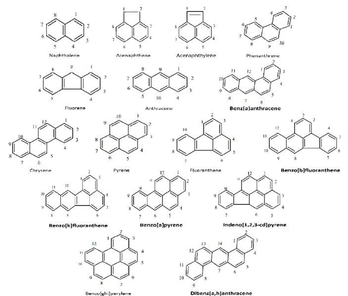
Figure 1. Numbered structures and nomenclatures of the 16 PAHs on the EPA
priority pollutant list. ... 5
Figure 2. Bay- and fjord-region of Dibenzo[a,l]pyrene. ... 7
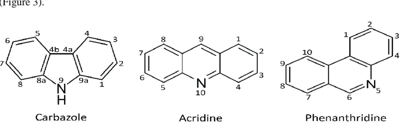
Figure 3. Numbered structure of 3 azaarenes: carbazole, acridine and phenanthridine. ... 12
Figure 4. A proposed metabolic pathway for the degradation of carbazole through Pseudomonas spp. Strain CA06, CA10 and Pseudomonas stutzeri OM1 ... 22
Figure 5. Other proposed metabolic pathways for the degradation of Carbazole. ... 25
Figure 6. Proposed metabolic pathways for the degradation of acridine. ... 30
Figure 7. Proposed metabolic pathways for the degradation of phenanthridine. ... 35
Figure 8. List of carbon sources and distribution in 96-well plates. ... 43
Figure 9. HPLC chromatograms for stock mixtures of three azaarenes: carbazole, acridine and phenanthridine. ... 48
Figure 10. Typical standard curve of three azaarenes from HPLC analysis. ... 49
Figure 11. Effects of two concentration of either carbazole (CAR), acridine (ACR), or phenanthridine (PHI) on the growth of microbes in feed soil (FS) or bioreactor-treated soil (BTS). ... 52
Figure 12. Concentration of azaarenes and PAHs in bioreactor-treated soil (BTS). ... 55
Figure 13. Concentrations of azaarenes and PAHs in feed soil samples (FS). ... 56
Figure 14. Negative image of the DGGE gel (1) of DNA extraction samples from incubations on testing co-metabolism of target azaarenes. ... 65
Figure 15. Negative image of the DGGE gel (2) of DNA extraction samples from incubations on testing co-metabolism of target azaarenes. ... 66
Figure 16. Removal of phenanthrene and phenanthridine from incubations of Acidovorax sp. NA3. ... 67
Figure 17. Removal of anthracene, phenanthrene and phenanthridine from incubations of Immundisolibactercernigliae TR3.2T. ... 68
1
CHAPTER 1: INTRODUCTION
2
CHAPTER 2: LITERATURE REVIEW
A. Soil Contamination and Sites of Manufactured Gas Plants
3
Conservation and Recovery Act of 1976) and Superfund sites (under the Comprehensive Environmental Response, Compensation, and Liability Act of 1980) [11].
Manufactured gas plants (MGPs) provided manufactured gas as an energy source to U.S. residents from the early 1800s through the mid-1900s [12]. The sites of former MGPs often become an environmental issue due to residual toxic contaminants leftover from the production process. Many toxic products, such as polycyclic aromatic hydrocarbons (PAHs), benzene, and cyanide, resulted from the production of manufactured gas, which included a coal carbonization process, carburetted water gas process, and oil gas process [13]. For example, an oil gas process would thermal-crack oil produced from a carburetted water gas process to obtain the raw
manufactured gas [14]. The raw gas was accepted by a vaporizer for the purpose of enrichment and then heated prior to distribution. Unavoidably, tars containing PAHs and other heterocyclic aromatic hydrocarbon compounds would also be produced at this stage.
B. Polycyclic Aromatic Hydrocarbons
1. Introduction of Polycyclic Aromatic Hydrocarbons
4
of total PAHs from contaminated sites is extremely difficult considering their toxicity and the low bioavailability of HMW PAHs. Besides the production of manufactured gas and the use of creosote to treat wood, PAHs come from the incomplete combustion of either other organic materials including plant biomass (phytogenic) and petroleum (petrogenic or pyrogenic) due to either anthropogenic or natural reasons [20]. Estimating the sources of PAH contamination is possible through plotting ratios of certain combination of PAHs (e.g. anthracene/anthracene+ phenanthrene) [21].
5
Figure 1. Numbered structures and nomenclatures of the 16 PAHs on the EPA priority pollutant list. The names of 6 PAHs classified as carcinogens by National Toxicity Program (NTP) are bolded [27].
6
naphthalene, acenaphthene and pyrene, have been reported as toxic compounds with mutagenic or cytotoxic characteristics [30]. PAHs are viewed as human carcinogens due to the formation of highly reactive metabolites which are able to combine with nuclear DNA [31]. The formation of these reactive metabolites is normally associated with the activity of cytochrome P450 (CYP) enzymes. This superfamily of heme-thiolate enzymes encoded by cytochrome P450 genes has the key function in living organisms of metabolizing xenobiotics by forming either detoxified hydrophilic metabolites or sometimes more toxic metabolites [32]. Members of CYP superfamily of enzymes, such as CYP1A1 and CYP1B1, were reported to play an essential role in
metabolizing PAHs in the body to form carcinogenic diol-epoxides under the co-function of epoxide hydrolase [33]. Diol-epoxides metabolites are able to bind to nuclear DNA to form adducts that cause further mutation and even cancer. Metabolites of PAHs with stereoisomeric bay or fjord regions are believed more reactive than others and have been called ultimate
7
Figure 2. Bay- and fjord-regions of dibenzo[a,l]pyrene.
The fate of PAHs in the soil can vary depending on the environmental conditions and the characteristics of the PAH. These include photooxidation, volatilization, chemical oxidation, adsorption to soil particles, leaching, bioaccumulation, and biodegradation [36]. Biodegradation is one of the most studied approaches for dealing with the challenges of PAH contamination. Especially in recent years, researchers have realized that a natural biological degradation process can be an effective remediation strategy to remove pollutants like PAHs and minimize the remedy carbon footprint.
Biodegradation of PAHs can happen either aerobically or anaerobically [37-39]. However, the rates of anaerobic biodegradation are typically slower than the biodegradation under aerobic conditions and the ecological function of anaerobic biodegradation was presumed to be minor historically [40, 41]. In spite of recent research progress revealing the unique role of anaerobic biodegradation under certain environmental conditions, such as when the oxygen demand exceeds its supply in natural environments, most attention has been focused on aerobic processes [42].
PAH-8
degrading microorganism provides information on the growth conditions and PAH growth substrates for microorganisms [43]. Contrary to metabolism of PAHs by fungi which is based on extracellular enzymes secreted outside cells to break down bonds of PAHs, metabolism of PAHs by bacteria is achieved by intracellular enzymes [43]. Therefore bioavailable PAHs, such as those dissolved in water or in the vapor phase, can be taken up by bacteria [44, 45]. Soil structure, other environmental factors (e.g. water, temperature, pH, and nutrients) and bioavailability of PAHs in the soil are all key factors affecting the biodegradation process. Bioavailability can be determined by the rate of substrate mass transfer into microbial cells with respect to the its intrinsic catabolic activity [43]. Bioavailability can be a limiting factor when the capacity of the cells consuming a PAH exceeds the capacity of acquiring this PAH from outside environment. As a result, substrate concentrations in the aqueous phase that can be consumed by microbes would be reduced significantly. Moreover, when PAHs sorb onto soil particles or enter non-aqueous phase liquids (NAPLs), the degradation of PAHs would be difficult as enzymes used by bacteria for the degradation of PAHs would not be able to encounter PAHs in these phases [26]. Solubility is one of the key factors affecting bioavailability. In addition,
bioavailability of a certain PAH compound is also reliant on the variability of desorption rates for different PAHs over different time periods [46]. Normally, PAHs are desorbed from solid materials quickly at the start, but the process of desorption would then become slow as concentration gradients between the sorbent and aqueous phases decrease [47].
Three biodegradation patterns are reported to be the major mechanisms associated with metabolism of multiple compounds: diauxie, simultaneous utilization, and competitive
9
determining which substrate would be preferentially used [48]. For example, the presence of ethylbenzene was reported to inhibit the metabolism of a Rhodococcus strain growing on other BTEX substrates [49]. Simultaneous utilization of substrates may lead to cell growth, but might also cause the degradation of compounds without cell growth. The pattern of simultaneous utilization of substrates without cell growth is considered as co-metabolism [50]. As a non-specific enzymatic mechanism, co-metabolism can be described as the reaction that occurs when a contaminant is fortuitously degraded by an enzyme or co-factor synthesized as a result of degradation process from another structurally similar primary substrate. Co-metabolic
remediation was found to be useful to degrade contaminants when their concentration was too low to be the growth substrate for certain bacteria [51]. It has also reportedly been involved in the degradation process of very recalcitrant contaminants, such as HMW PAHs,
trichloroethylene (TCE), tetrachloroethylene (PCE), and methyl tert-butyl ether (MTBE) [52]. Finally, competitive inhibition can be a process in which non-specific enzymes preferentially attack contaminants which are more easily degraded while more recalcitrant contaminants persist [53]. For instance, naphthalene, methylnaphthalene, and fluorene are frequently competitive substrates with phenanthrene for phenanthrene-degrading bacteria [54].
2. Biodegradation of Phenanthrene and Anthracene
The knowledge of biodegradation on phenanthrene and anthracene in contaminated soil are essential to this research due to their structural similarity to two azaarenes (phenanthridine and acridine, respectively) studied in this work. Therefore, a brief review of the known
10
As one of the more widely distributed PAHs, phenanthrene is viewed as a model compound for studies of biodegradation of LMW-PAHs. A variety of bacteria have been reported capable of metabolizing phenanthrene either directly or indirectly (e.g. through co-metabolism). Bacterial strains from the genera Mycobacterium [55, 56], Vibrio [57],
Pseudomonas [57, 58], Aeromonas [57, 59], Sphingomonas [60, 61], Sphingobium [62],
Arthrobacter [63], Acidovorax [61, 63], Brevibacterium [63], Burkholderia [64], Martelella [65],
Nocardia [66], Streptomyces [67] Staphylococcus [68] and Sinorhizobium sp. [69] are all reported to degrade phenanthrene. Fungal representatives, such as Cunninghamella elegans and
Pleurotus ostreatus,are also capable of metabolizing phenanthrene [70, 71].It is noteworthy that several members of the Sphingomonads (including Sphingomonas, Sphingobium, Sphingopyxis
and Novosphingobium spp.) are actively involved in the degradation of phenanthrene and are extremely important phenanthrene-degrading bacteria in soil [72].
11
pathways, Streptomyces flavovirens was reported to use a different pathway by attacking the 9,10- position (K-region) of phenanthrene under the modulation of cytochrome P-450
monooxygenase to produce phenanthrene-9,10-oxide [67]. 9-Phenanthrol was then formed after the conversion of phenanthrene-9,10-oxide to trans-9,10-dihydrodiolphenanthrene.
Anthracene is also widely detected at contaminated sites at high concentrations and is isomeric with phenanthrene [76]. Several bacteria have been reported to utilize anthracene as source of carbon and energy, including strains from the genera Pseudomonas [77, 78],
Mycobacterium [79, 80], Rhodococcus [81], Sphingomonas [82], Nocardia [66], Bacillus
[83], Microbacterium [84], Martelella [65], Brevibacterium [85], Burkholderia [86]. Fungi, such as Cunninghamella elegans, Aspergillus fumigatus, Pleurotus ostreatus, Fusarium solani,
and Penicillium simplicissimum, are also capable of metabolizing anthracene [70, 87-89]. One of the major biodegradation pathways of anthracene has been described with the initial dioxygenation at the 1,2 position to produce 1,2-dihydroxyanthracene [80]. This
12
C. N-Heterocyclic Aromatic Hydrocarbons
N-heterocyclic aromatic hydrocarbons (NHAs) - also known as nitrogen-containing polycyclic aromatic hydrocarbons, N-PAHs, PANHs, or azaarenes, are a class of organic molecules which can be toxic and are widely distributed in a broad range of environments including air, soil, and water systems [1, 91, 92]. Similar to PAHs, the major sources of azaarenes include incomplete combustion of fossil fuels, coal liquefaction, volcanic eruptions, forest fires, uncontrolled hazardous industries, and oily sludge [93]. Azaarenes have strong molecular bonds and π-electrons surrounding benzene rings, so they have the characteristics of being both highly recalcitrant and resistant to nucleophilic attack [43]. Microorganisms are involved in the removal of azaarenes at contaminated sites, and a variety of studies have been conducted on the biodegradation of various azaarenes such as carbazole, acridine, quinolone, pyridine, and phenanthridine. Accordingly, numerous microorganisms involved in the
degradation of azaarenes have been reported [94]. Despite the fact that azaarenes can be putative human carcinogens and usually have greater toxicity on microorganisms than their PAH
analogues, the fate, behavior and biodegradation of these compounds are still not fully
understood [95, 96]. This research is aimed to investigate the fate and behavior of three, three-ring azaarenes, including carbazole, acridine and phenanthridine, in the contaminated soil (Figure 3).
13
1. Carbazole
a. Introduction of Carbazole
Carbazole is an azaarene often present in soil, groundwater, and rivers contaminated by petroleum, wood-preserving wastes, or chemical pollution [97-99]. The sources of carbazole are mainly coal tars, creosote and shale oils, which contain many heterocyclic aromatic compounds [100]. Thousands of tons of carbazole are produced each year from coal tar and crude oil under high temperatures [101].
Carbazole and its derivatives are close to our daily life. It is commonly found in synthetic products such as dyes, pharmaceuticals, and plastics. It is also used to make photographic plates that are sensitive to ultraviolet light. The manufacturing process of some industrial products, including chemical reagents, pesticides, medicines and plastics require carbazole or its derivatives [101]. The structure of carbazole (Figure 3) is similar to dibenzofuran, which is mutagenic and toxic to living organisms [102]. The carcinogenic effect of carbazole tested in mice indicated that carbazole itself is a carcinogen [103]. Other previous reports have also shown that carbazole can be both mutagenic and carcinogenic [104, 105].
Given that the broad distribution of carbazole and its putative toxic and highly recalcitrant properties, there have been many studies on its degradation, including finding effective ways to remove it from the environment. Many studies concerning the transformation of carbazole have been conducted in the past 30 years - mostly on microbial degradation. Details concerning specific carbazole-degrading microorganisms and degradation pathways are
14
b. Carbazole-Degrading Microorganisms
A number of different microbes have been reported for their ability to degrade carbazole (Table 1), although their role in the bioremediation of carbazole from contaminated sites is typically not known. Among these various carbazole degraders, most are Gram-type negative bacteria, with only a few Gram-type positive.
In 1991, the first reported carbazole degrader was isolated from the soil of an abandoned coal gasification plant and was identified as a Xanthamonas sp. After growing the pure culture, the strain was reintroduced into contaminated soil [93]. The degradation of carbazole was enhanced to nearly 50% by these reintroduced bacteria after 7 days in comparison with little carbazole degradation observed for the indigenous bacteria. The process of degradation enhanced by additional bacteria, or bioaugmentation, is one approach to soil bioremediation [106].
Sphingomonads possess a wide variety of biodegradative capabilities and have the potential to be utilized for bioremediation. In 2001, four genera were proposed from the then-current Sphingomonas genus comprising the new genera Sphingomonas, Sphingobium,
Novosphingobium, and Sphingopyxis [107]. In addition to their ability to degrade phenanthrene and anthracene as mentioned above, many researchers have found strains of Sphingomonas that could also degrade other substrates, such as carbazole, dibenzofuran, dibenzothiophene, and benzothiophene through the process of co-metabolism [108]. Sphingomonas sp. strain CDH-7 was isolated during a screen of nearly 350 samples from soil, wastewater, and petroleum oil sludge collected by researchers. [109]. It used carbazole as a sole source of carbon and resting cells of Sphingomonas sp. CDH-7 were found to continuously degrade carbazole. The bacterium
15
Sphingomonas wittichii RW1 was viewed as an important species for its valuable ability to use dibenzo-p-dioxin and dibenzofuran as the sole sources of carbon and energy [110, 111].
Interestingly, while an initial study showed that purified dibenzofuran 4,4a-dioxygenase of strain RW1 was incapable of oxygenating carbazole, researchers re-tested Sphingomonas wittichii
RW1 and discovered that the color of the culture suspension in minimum salts medium (MSM) containing carbazole changed in a time-dependent manner - indicating that strain RW1 was actually capable of degrading carbazole [101]. Sphingomonas sp. strain XLDN2-5 (reclassified as Sphingobium yanoikuyae strain XLDN2-5) [108] is not only able to degrade carbazole, but also can co-metabolically catabolize dibenzofuran, dibenzothiophene, and benzothiophene [112].
Bacteria within the genus Pseudomonas are viewed as significant for health, agriculture, biotechnology, and environmental science due to their great adaptability [113]. Many researchers are interested in using Pseudomonas isolates for bioremediation given their ability to degrade various organic compounds, including carbazole [114]. Research on the carbazole-degrading
16 Table 1 List of carbazole-degrading bacteria and fungi.
Time of report Name of the strain Major metabolites
1986 Cunninghamella elegans ATCC 26269 [117] -
1992 Cunninghamella echinulata ATCC 9244[118] -
1993 Pseudomonas sp. CA06, CA10 [115] Anthranilic acid, Catechol
1993 Pseudomonas sp. NCIB 9816-4 [119] 3-hydroxycarbazole
1994 Pseudomonas stutzeri ATCC31258 [120] Anthranilic acid
1995 Bacillus sp. KUKK-4,5 [121]
-Pseudomonas sp. KUKK-1,2,3,8 [121] -
Escherichia coli KUKK-6 [121] -
Serratia sp. KUKK-7 [121] -
1995 Pseudomonas cepacia F297 [122] -
1995 Burkholderia cepacia CB1 [123] -
Xanthamonas sp. CB2 [123] -
Pseudomonas sp. CB3 [123] -
1996 Pseudomonas sp. LD2 [124] Anthranilic acid
1998 Pseudomonas stutzeri OM1 [125] Anthranilic acid, Catechol
1998 Sphingomonas sp. CB3 [126] -
1999 Sphingomonas sp. CDH-7 [109] Anthranilic acid
2000 Ralstonia sp. RJGII.123 [127] Anthranilic acid
2002 Pseudomonas rhodesiae KK1 [128] Anthranilic acid
2002 Sphingomonas sp. strain KA1 [129] -
2002 Sphingomonas sp. GTIN11 [130] Anthranilic acid
2003 Neptuniibacter sp. CAR-SF [131] -
2003 Sphingomonas sp. CP19 [132] -
2003 Pseudomonas sp. C3211 [133] -
2004 Janibacter sp. YY-1 [134] Catechol
2004 Pseudomonas sp. K23, K22, K15, J11 [135] -
Janthinobacterium sp. J3, J4 [135] -
Pantoea sp. J14 [135] -
Novosphingobium sp. J30 [135] -
Sphingomonas sp. J40, M2 [135] -
2004 Pseudomonas sp. XLDN4-9 [116] -
2004 Aspergillus flavus VKM F-1024 [136] 3-hydroxycarbazole
2005 Acinetobacter sp. IC001 [137] -
Pseudomonas sp. IC017 [137] -
Sphingomonas sp. IC033,IC075, IC081, IC097, IC145
[137] -
Burkholderia sp. IC049, IC129, IC138 [137] -
Achromobacter sp. IC074 [137] -
17
Table 1 (continued) List of carbazole-degrading bacteria and fungi.
Stenotrophomonas sp. IC193 [137] - Marinobacterium sp. IC961, IC977 [137] - Nocardioides aromaticivorans IC177 [137] Anthranilic acid
2006 Bacillus sp. T2.3-2.6, T3.1, T3.3, T4.1-4.3 T6.1-6.6, T7.0
[138] -
2006 Gordonia sp. F.5.25.8 [139] -
2006 Arthrobacter sp. P1-1 [140] Anthranilic acid
2006 Burkholderia sp. IMP5GC [141] -
2007 Sphingobium yanoikuyae XLDN2-5 [142] -
2008 Sphingomonas sp. VKM B-2434 [143] -
2008 Novosphingobium sp. NIY3 [144] Anthranilic acid
2008 Klebsiella sp. strain LSSE-H2 [145] -
2008 Chryseobacterium sp. NCY and Achromobacter sp. NCW
[146] -
2009 Kordiimonas sp. OC3, OC6S, OC9, OC11S [147] -
Erythrobacter sp. OC4, OC8S [147] -
Hyphomonas sp. OC5 [147] -
Sphingosinicella sp. OC5S [147] - Caulobacter sp. OC6, OC10 [147] -
Lysobacter sp. OC7 [147] -
2009 Sphingomonas sp. JS1 [148] -
2009 Ralstonia sp. strain SBUG 290 [149] 1-hydroxycarbazole
3-hydroxycarbazole
2011 Enterobacter sp. A8 [150] -
2011 Sphingomonas wittichii RW1 [151] Anthranilic acid, Catechol
2011 Acinetobacter spp. Alp6, Alp7 [91] -
2011 Pseudomonas BC039-046 [152] -
2011 Pseudoxanthomonas sp. DMVP2 [153] -
2012 Achromobacter sp. CAR1389 [154] Anthranilic acid
2013 Pseudomonas sp. GBS.5 [155] -
2014 Achromobacter sp. SL1 [113] Anthranilic acid, Catechol
Pseudomonas sp. SL4 [113] Anthranilic acid, Catechol Microbacterium esteraromaticum SL6 [113] Anthranilic acid, Catechol
Stenotrophomonas maltophilia BA [113] -
18
In crude oil, carbazole is often present with its alkylated derivatives [104]. These alkyl derivatives often have monomethyl, dimethyl, trimethyl, and tetramethyl side chains on different carbon positions of carbazole (called C1-, C2-, and C3-, C4-carbazoles, respectively). One study showed that mixed bacterial cultures were able to degrade alkyl carbazoles (from C1- to C4-carbazoles) in crude oil within eight days, and also showed that the alkyl-carbazoles were easier to degrade as the number of substituted alkyl groups became fewer [157]. However, pure
cultures rarely degrade alkylated derivatives in an oil phase with few exceptions; One example is that Sphingomonas sp. strain GTIN11 was demonstrated to degrade carbazole and C1-carbazoles in shale oil [130]. Pseudomonas sp. strain XLDN4-9 was also capable of degrading carbazole, C1-carbazoles, C2-carbazoles, and even benzocarbazoles, but it was shown that carbazole was harder to degrade than either C1-carbazoles and C2-carbazoles [158].
Ammonium can be produced by bacteria as a by-product during the degradation of
19
utilized NO3−–N and NO2−–N simultaneously, while others had a preference for utilizing NO3−– N. This study indicated that further understanding the process of nitrification and/or
denitrification process by carbazole-degrading bacteria can also contribute to efforts for controlling the release of ammonium to the environment.
Thus far, much of the work on carbazole degraders was performed with isolates identified as Sphingomonas and Pseudomonas species. However, isolates from other Gram-type negative genera can also degrade carbazole. Achromobacter species were demonstrated to degrade a wide range of xenobiotics and were specialized to degrade compounds such as hydrocarbons and PAHs [161-164]. Reports have shown that Achromobacter sp. strains IC074 [137], CAR1389 [154], and SL1 [113] can metabolize carbazole. Remarkably, strain CAR1389 was capable of growing on nominal 3.5 g·L-1 of carbazole in a minimal medium (MM) and degrading nearly half that amount [154]. Before this discovery, carbazole-degrading bacteria capable of growing at such high concentrations of carbazole were rarely reported [165]. This finding also suggested that carbazole might not have a strong inhibitory effect on the activity of strain CAR1389 even though the concentration of carbazole was extremely high.
Additional Gram-type negative carbazole degraders include Burkholderia cepacia strain CB1 [123], Escherichia coli strain KUKK-6, Serratia sp. strain KUKK-7 [121], Ralstonia sp. strain RJGII.123 (formerly Xanthomonas sp.) [93, 127], Janthinobacterium sp. strain J3 and strain J4, Pantoea sp. strain J14, Novosphingobium sp. strain J30 [135], Burkholderia sp. strain IMP5GC [141], Klebsiella sp. strain LSSE-H2 [145], Novosphingobium sp. strain NIY3 [166],
20
Several Gram-type positive bacteria have been reported as carbazole degraders. The carbazole degrader Nocardioides aromaticivorans strain IC177 was used to study the genes and metabolic pathway of carbazole degradation [159]. The test results indicated that the carbazole catabolic genes of strain IC177 were similar to those of Gram-type negative bacteria, such as
Pseudomonas spp. strain CA10 and Janthinobacterium sp. strain J3. Other Gram-type positive carbazole degraders include Bacillus sp. strain KUKK-4,5 [121], Janibacter sp. strain YY-1 [134], Arthrobacter sp. strain P1-1 [140], Bacillus sp. strains T2.3 to T2.6, T3.1 and T3.3, T4.1 to T4.3, T6.1 to T6.6, and T7.0 [138], Gordonia sp. strain F.5.25.8 [139], and Dietzia cinnamea
strain P4 [167]. The genome sequence of Dietzia cinnamea strain P4 has been reported and genes encoding enzymes for the degradation of aromatic hydrocarbons have been identified [168].
Not only pure cultures have been reported to degrade carbazole. A microbial consortium consisting of Chryseobacterium sp. strain NCY and Achromobacter sp. strain NCW was reported to utilize carbazole as the sole source of carbon, nitrogen, and energy [146]. However, neither of the pure strains NCY and NCW were able to degrade carbazole individually. The possible explanation reported for this phenomenon was neither strain NCY nor NCW possessed a complete pathway to transform carbazole.
Another kind of microorganism with the ability to metabolize carbazole are filamentous fungi, which often play an important role in the biodegradation process [169]. However,
21
c. Metabolic Pathways of Carbazole Degradation
Though the characterized carbazole degraders belong to multiple species, the research on metabolic pathway and enzymes linked to degradation was limited to only a few strains, such as
Pseudomonas resinovorans strain CA10 and Sphingomonas sp. strain CB3 [115, 123, 126]. In this section, the pathway and genes relative to carbazole degradation are described.
Pseudomonas spp. CA06 and CA10 were first used to determine a pathway for the biodegradation of carbazole (Figure 4) [115]. Two main metabolites, anthranilic acid (AN) and catechol (CAT), were found in the medium after analysis by high-performance liquid
chromatography (HPLC) and gas chromatography-mass spectrometry (GC-MS). Further experiments showed that CAT was detected when AN was used as the substrate. Additional analyses identified metabolites including 2'-aminobiphenyl-2,3-diol and 2-hydroxy-6-oxo-6-(2’-aminophenyl)-hexa-2,4-dienoic acid (HOADA). The proposed pathway begins with the
dioxygenation of carbazole at an angular position adjacent to the nitrogen atom. The product is a dihydroxylated intermediate which is converted to aminobiphenyl-2,3-diol spontaneously. 2'-aminobiphenyl-2,3-diol is converted to HOADA before hydrolysis to AN via a ortho-cleavage pathway. AN is a non-toxic substrate relatively easily degraded by microbes and can be used as a substrate in tryptophan biosynthesis [171, 172]. The final step of the carbazole pathway includes the conversion of AN to CAT through ortho-cleavage, to an intermediate of the tricarboxylic acid cycle (TCA) [173, 174]. Interestingly, Pseudomonas stutzeri OM1 had a meta-ring-cleavage pathway via conversion of CAT to 2-hydroxymuconic semialdehyde [125].
The pathway that uses an angular dioxygenase acting on the 9a and 1 carbons of
1,9a-22
dioxygenase (CarA or CARDO) is the first enzyme to participate in the reaction of carbazole oxidation. CarA can participate in several oxygenation reactions involving a large range of substrates and can also participate in monooxygenation reactions [174, 176]. CarA can not only oxidize carbazole, but also dibenzofuran (DBF), fluorene, naphthalene, and biphenyl [155]. The major product of CarA in carbazole dioxygenation is 2-aminobiphenyl-2,3-diol.
Figure 4. A proposed metabolic pathway for the degradation of carbazole through Pseudomonas
spp. strain CA06, CA10 and Pseudomonas stutzeri OM1 [115, 125]. 1. Carbazole (CAR); 2. 2'-aminobiphenyl-2,3-diol; 3. 2-hydroxy-oxo-(2'-aminophenyl)hexa-2,4-dienoic acid; 4. 6-dioxo-6-(2’-aminophenyl)hexa-4-enoic acid; 5. 2-hydroxy-4-pentenoate; 6. AN; 7. CAT; 8.
cis,cis-muconate; 9. 6-oxo-6-(2'-aminophenyl)hexa-4-enoic acid; 10.
5-oxo-5-(2'-aminophenyl)penta-3-enoic acid; 11. hydroxy-6-oxo-6-(2'-aminophenyl)hexanoic acid; 12. 2-hydroxymuconic semialdehyde. Compound within bracket was not detected.
A functional CarA enzyme depends on three gene products, including CarAa (a catalytic oxygenase containing a Rieske [2Fe–2S] cluster and a mononuclear iron domain), CarAc (a ferredoxin component, containing a Rieske [2Fe–2S] cluster), and CarAd (a reductase
2-aminobiphenyl-23
2,3-diol 1,2-dioxygenase (CarB) is the second major enzyme that participates in the
metabolization of carbazole. It is a multicomponent meta-cleavage dioxygenase encoded by both
carBa and carBb genes [178]. CarB can metabolize 2-aminobiphenyl-2,3-diol to form HOADA. CarC is the third enzyme encoded by the carC gene and transforms HOADA to produce
anthranilic acid via hydrolysis. CarC is also able to convert
2-hydroxy-6-oxo-6-(2,9-aminophenyl)-hexa-2,4-dienoic acid to a minor metabolite 2-hydroxypenta-2,4-dienoic acid [165]. CarD, which is a dienoate hydratase, converts 2-hydroxypenta-2,4-dienoic acid to 4-hydroxy-2-oxovaleric acid, CarE which is a 4-hydroxy-2-oxovalerate aldolase then converts 4-hydroxy-2-oxovaleric acid to acetaldehyde and pyruvic acid, and CarF which is an acetaldehyde dehydrogenase that can then transform acetaldehyde to acetyl coenzyme A are all major enzymes encoded by single genes that were involved in the degradation of carbazole [175].
Bacterial dioxygenases often play an important role in the first step of azaarene
metabolization [170]. Researchers found that naphthalene 1,2-dioxygenase (NDO) obtained from
Pseudomonas sp. strain NCIB 9816-4 strain and biphenyl dioxygenase (BPO) from Beijerinckia
sp. strain B8/36 can both oxidize carbazole [119]. The oxidation product 3-hydroxycarbazole was found after further analysis, while no further intermediates were detected, such as unstable
cis-carbazole-3,4-dihydrodiol. Products of dioxygenation may therefore be further metabolized via direct monooxygenation process.
24
study demonstrated that 3-hydroxycarbazole was the main metabolite of carbazole degradation while 1-hydroxycarbazole and 2-hydroxycarbazole could also be detected as minor products. Carbazole derivatives in dihydroxylated forms mainly comprised 2,6-hydroxycarbazole.
Aspergillus flavus could also transform N-benzoylcarbazole and N-acetylcarbazole. The metabolites of these two carbazole derivatives included carbazole, and small amounts of 1-hydroxycarbazole, 2-1-hydroxycarbazole, and 3-hydroxycarbazole. According to these results, the substitutent moiety influenced the position of hydroxylation. One report showed that
Cunnighamella elegans could also oxidize carbazole to produce 3-hydroxycarbazole [117]. As shown above, filamentous fungi can introduce hydroxyl groups in different positions of the carbazole ring. The monohydroxylation products can be viewed as a result of detoxification; the same process can happen at the mammalian metabolism of carbazole due to the structural similarity of fungal and mammalian liver microsomes [136].
25
Figure 5. Other proposed metabolic pathways for the degradation of Carbazole (A1). 1. Proposed pathway through Ralstonia sp. strain SBUG 290 [149]: A2. 1-hydroxycarbazole, A3.
3-hydroxycarbazole, A4. 3-hydroxy-1,2,3,9-tetrahydrocarbazol-4-one, A5. 9-3-hydroxycarbazole, A6. cis-carbazole-3,4-dihydrodiol. 2. Proposed pathway through Pseudomonas cepacia strain F297 [122]: B2. 4-(3'-Methoxy-2'-indolyl)-2-oxo-3-butenoic acid, B3. 4-(3’-Oxo-2’-indolinyl)-2-oxo-3-butenoic acid. 3. Proposed pathway through Pseudomonas sp. strain LD2 [124]: C2. indole-3-acetic acid. 4. Proposed pathway through Aspergillus flavus VKM F-1024 [136]: A2. 1-hydroxycarbazole, A3. 3-1-hydroxycarbazole, D2. 2-hydroxycarbazole. Compound within bracket was not detected.
Though there are many studies on carbazole biodegradation, less attention has been given to understanding the metabolic pathways of carbazole biodegradation compared to PAHs.
26
2. Acridine
a. Introduction of Acridine
Although a number of studies have analyzed the environmental fate and metabolic pathways regarding carbazole degradation by soil bacteria [124, 180, 181], the environmental fate and biodegradation pathways of other toxic multi-ring azaarenes, such as acridine and phenanthridine, have received far less attention [182-184].
The toxic tricyclic nitrogen-containing compound acridine was first isolated by Carl Gräbe and Heinrich Caro from high boiling fraction of coal tar in 1870 [185]. It is an environmental contaminant widely distributed in coal tar, petroleum wastes, motor vehicle exhaust, tobacco smoke, and the photolysis of carbamazepine when exposed to UV radiation [186-188]. Acridine has been used widely in electronic and electroluminescence devices, and light-emitting diodes [189]. In contrast to carbazole, its structure has a pyridine-ring instead of a pyrrole-ring (Figure 3). Acridine has been reported as a toxic compound; inhibiting the growth of many aquatic organisms and causing oxidative stress for crustaceans [190-193]. It is also
27
b. Acridine-Degrading Microorganisms
There is little prior research on the microbial degradation of acridine in soil. A few reports have indicated that bacterial, mammalian and fungal enzymes can transform acridine [188]. However, biodegradation of acridine by pure cultures of functional soil bacteria in a laboratory environment is rarely reported.
In one study investigating biodegradation of acridine under anaerobic conditions, a research group created lab-scale microcosms, including material from a mixed methanogenic culture from sewage sludge, a sulfate-reducing culture from alluvial sand aquifer contaminated by landfill leachate, and a methanogenic culture from alluvial sand aquifer [197]. At the first week, 1 mg·L-1 of acridine was added for enrichment of acridine-degrading microbial
communities. Two weeks later, 5 mg·L-1 of acridine was added to each microcosm with only 0.06 mg·L-1 of acridine detected one week later. Prior to this report, there was almost no information on the transformation of azaarenes containing three or more rings azaarenes.
28
The first acridine-degrading bacterium obtained in pure culture from contaminated soil was strain B1H1 that possessed 16S rRNA gene sequence similarity to sequences from the genus
Sphingomonas, isolated from a former shipyard site [194]. Strain B1H1 was isolated after a thirteen-week enrichment and used 10 mg·L-1 acridine as a sole source of carbon and energy in liquid medium. It removed >80% of amended acridine in liquid medium after 18 days. In addition to strain B1H1, six more isolates from that study with the potential ability to transform acridine were reported with similarity to sequences from the genera Rhodococcus, Pseudomonas,
Bradyrhizobium, Rhodococcus, and Afipia [194]. However, in contrast to strain B1H1, those six isolates could only transform acridine without growth.
Mycobacterium vanbaalenii strain PYR-1 was isolated from PAH-contaminated sediment of an oil tank farm and was reported as the first strain to be able to mineralize pyrene; it could additionally oxidize phenanthrene, fluoranthene, naphthalene, anthracene, and benz[a]anthracene [200]. Its capability to co-metabolize 47 mg·L-1 acridine in liquid medium for 7 days using phenanthrene to induce enzymes was investigated. Although a variety of both dioxygenases and monoxygenases are present in Mycobacterium vanbaalenii strain PYR-1 and four different metabolites associated with transformation of acridine were detected (see below), strain PYR-1 was not able to mineralize acridine [188, 201].
29
The ability of fungi to remove acridine in the soil is rarely reported even though many fungi can transform anthracene, the PAH analogue of acridine [70]. In one example,
Cunninghamella elegans ATCC 36112 transformed 10 mg·L-1 acridine in liquid Sabouraud medium over three days incubation [117, 205]. Interestingly, only 4% of acridine remained indicating that acridine was either efficiently transformed or alternatively, quantities of the hydrophobic compound may have been absorbed into the fungal mycelia.
c. Metabolic Pathways of Acridine Degradation
Currently, only fragmentary information exists regarding any microbial metabolic pathway for acridine degradation (Figure 6). Pseudomonas fluorescens TTC1 was reported to produce acridine cis-1,2-dihydrodiol [202]. An initial naphthalene dioxygenase was determined to be responsible for this transformation. Interestingly, the tolerance of this naphthalene
dioxygenase for high concentrations of acridine (4 g·L-1) indicate that it may be worth further investigation for in situ bioremediation purposes or during biotransformation of other azaarenes. Another metabolite of acridine transformation was produced by Sphingomonas sp. LH128 and identified as 9(10H)-acridinone [198]. It was also found as a major metabolite of acridine by zebra mussels, green algae, and bacteria in aquatic environments [206]. In comparison to acridine, 9(10H)-acridinone was reported as less acutely genotoxic for an aquatic invertebrate midge, however, it is still considered genotoxic according to the MutatoxTM test [206].
30
of a similar RHD was likely responsible for the presence of acridine cis-1,2-dihydrodiol by strain PYR-1. 4-Hydroxyacridine, however, was likely formed due to the activity of cytochrome P450 monooxygenase through a different pathway [208]. 9(10H)-Acridinone and 9,10-dihydroacridine may have been produced through either biological or abiotic processes [209, 210]. While
9(10H)-acridinone is considered a less toxic compound than acridine, the toxicity of the other three compounds still need to be investigated.
The transformation of acridine by the fungus Cunninghamella elegans ATCC 36112 produced acridine trans-1,2-dihydrodiol and 2-hydroxyacridine (Figure 6) [205]. These two metabolites of acridine were different than acridine metabolites produced by bacteria.
Figure 6. Proposed metabolic pathways for the degradation of acridine (A1). 1. Proposed pathway through Pseudomonas fluorescens TTC1 [202]: A2. acridine cis-1,2-dihydrodiol. 2. Proposed pathway through Sphingomonas sp. LH128 [211]: B2. 9(10H)-acridone. 3. Proposed pathway through Mycobacterium vanbaalenii PYR-1 [188]: A2. acridine cis-1,2-dihydrodiol, B2. 9(10H)-acridone, C2. acridine 3,4-oxide, C3. 4-hydroxyacridine, C4. 9,10-dihydroacridine. 4. Proposed pathway through Cunninghamella elegans ATCC 36112 [205]: D2. acridine trans-1,2-dihydrodiol, D3. 2-hydroxyacridine. 5. Proposed pathway under the anaerobic condition [197]: E2. 9-hydroxyacridine, E3. 3-hydroxyacridine or 4-hydroxyacridine, E4.
31
Researchers assumed that the first oxidation step of acridine involved a cytochrome P-450 monooxygenase to produce acridine 1,2-oxide [205, 212]. The trans-1,2-dihydrodiol could be formed after the oxide was metabolized by epoxide hydrolase [213]. This pathway is similar to one previously reported for the transformation of anthracene to anthracene
trans-1,2-dihydrodiol in Cunninghamella elegans [214]. The presence of 2-hydroxyacridine might be explained by a nonenzymatic rearrangement of acridine 1,2-oxide [215].
Under anaerobic conditions, a few metabolites of acridine were identified through a microcosms-based study and the possible pathways of acridine were presented (Figure 6) [197]. The metabolic route may begin with oxidation of either a benzene or pyridine ring to produce either 3-hydroxyacridine, 4-hydroxyacridine or 9-hydroxyacridine. All of these products can be converted to benzoic acid, a key intermediate for oxidized aromatic compounds [197, 216].
3. Phenanthridine
a. Introduction of Phenanthridine
Phenanthridine was first discovered in 1889 by Amé Pictet and H. J. Ankersmit through pyrolysis of the condensation product of benzaldehyde and aniline [217]. Similar to carbazole and acridine, phenanthridine is an azaarene widely distributed at contaminated sites due to its high water solubility and lower Kow values [95, 184]. Phenanthridine can be used in the
production of industrial solvents, dyes, explosives, and pharmaceuticals [216]. Although there is insufficient evidence to confirm the carcinogenic property of phenanthridine in humans, it was reported as acutely toxin to green algae, invertebrates, and fish [192, 206, 218-220].
32
b. Phenanthridine-Degrading Microorganisms
Streptomyces viridosporus strain T7A was reported to be able to co-metabolize five heterocyclic nitrogen-containing compounds, including phenanthridine, isoquinoline,
phthalazine, quinazoline, and quinoxaline, when growing in tryptone/yeast extract broth [182]. Strain TA7 was isolated for its ability to degrade substituted benzoic acids and it was the first isolate from soil reported to demonstrate microbial oxidation of phenanthridine [222]. In one test, 25% of phenanthridine was transformed by strain TA7 to 6(5H)-phenanthridinone during a 3-day transformation test (initial concentration 412 mg·L-1).
The phenanthrene-degrading bacterium Mycobacterium gilvum strain LB307T was also found to be able to transform phenanthridine [183]. While neither 2.5 mg·L-1 or 20 mg·L-1 phenanthridine supported the growth of strain LB307T, samples amended with 2.5 mg·L-1 phenanthridine demonstrated complete removal of phenanthridine over an 8-hour incubation. However, no degradation of phenanthridine was detected in samples containing 20 mg·L-1 phenanthridine, even after 215 hours of incubation, and no metabolites related to the transformation of phenanthridine were detected from incubations. This indicated a possible inhibitory effect of phenanthridine on this strain at high concentrations.
33
at concentrations >24.9 mg·L-1 for strain LH128, no inhibitory effect was observed even at the highest phenanthridine concentration (36.9 mg·L-1).
To the best of our knowledge, strain B1L4, which was isolated from a former shipyard site and possessed 16S rRNA gene sequence similarity to Rhodococcus sp., was the first reported isolate capable of using phenanthridine as a sole source of carbon and energy [194]. A liquid culture amended with 5 mg·L-1 phenanthridine demonstrated significant increase in turbidity relative to uninoculated controls. However, less than 20% phenanthridine was transformed by a strain, AB010907, over an 18-day incubation. Other genetically diverse
phenanthridine-transforming isolates from that same study possessed 16S rRNA gene sequence similarity to sequences from the genera Methylopila, Ralstonia, Xanthobacter, Afipia, Achromobacter,
Sphingomonas, and Pseudomonas. Isolates with low similarity to known genera were also recovered. These 13 isolates were able to transform either 5 mg·L-1 or 20 mg·L-1 of
phenanthridine in liquid medium but could not utilize phenanthridine as a source of carbon. Additionally, none of these isolates showed the ability to mineralize 14C-labeled phenanthrene, which has structural similarities to phenanthridine, indicating that the mechanism involved in the initial attack of phenanthridine may be different than the mechanism for phenanthrene.
Fungi have also been reported to be able to metabolize phenanthridine. The fungus
Umbelopsis ramanniana was reported to transform >70% of phenanthridine in liquid medium (41 mg·L-1 ) over 18 days [223].
c. Metabolic Pathways of Phenanthridine Degradation
34
transformed by other bacteria [183]. The bacterium Streptomyces viridosporus strain T7A is capable of transforming phenanthridine to 6(5H)-phenanthridinone, which was the only
metabolite detected [182]. 6(5H)-phenanthridinone was also detected after the transformation of phenanthridine by Sphingomonas sp. strain LH128 [211] and the larvae of the aquatic
invertebrate Chironomus riparius [224].
The fungus Umbelopsis ramanniana cantransformphenanthridine to phenanthridine N-oxide and 6(5H)-phenanthridinone (Figure 7) [223]. Phenanthridine N-N-oxide was also reported as one of the products of phenanthridine transformation by rat-liver homogenate [225]. Other major metabolites related to the transformation of phenanthridine by polychlorinated biphenyls (PCB)-induced rat-liver homogenate include 6(5H)-phenanthridinone,
9,10-dihydroxy-9,10-dihydrophenanthridine, 1,2-dihydroxy-1,2-9,10-dihydroxy-9,10-dihydrophenanthridine, and 2-hydroxyphenanthridine. Phenanthridine N-oxide can also be produced by CYP450 via oxidization of phenanthridine, and this compound was reported as non-mutagenic for Salmonellaenterica [220, 226]. However, 6(5H)-phenanthridinone was mutagenic but not tumorigenic for mice [227, 228]. The
35
Figure 7. Proposed metabolic pathways for the degradation of phenanthridine (A1). 1. Proposed pathway through Streptomyces viridosporus strain T7A [182]: A2. 6(5H)-phenanthridinone. 2. Proposed pathway through Umbelopsis ramanniana [223]: B2. phenanthridine N-oxide. 3. Proposed pathway through rat-liver homogenate [225]: A2. 6(5H)-phenanthridinone, B2. phenanthridine N-oxide, C2. 1,dihydroxy-1,dihydrophenanthridine, C3.
2-hydroxyphenanthridine, C4. 9,10-dihydroxy-9,10-dihydrophenanthridine. 4. Proposed pathway through Escherichia coli and Streptomyces lividans transformants [229]: C2. 1,2-dihydroxy-1,2- dihydrophenanthridine, C4. 9,dihydroxy-9,dihydrophenanthridine, D2.
10-hydroxyphenanthridine.
36
CHAPTER 3: METHODS
A. Chemicals
Carbazole (GC grade, ≥95% purity), acridine (97%), and phenanthridine (98%) were obtained from Sigma-Aldrich (St. Louis, MO, USA). Anthracene (scintillation grade) was purchased from Eastman Kodak (Rochester, NY, USA). Phenanthrene (97%) was obtained from Acros Organics (Thermo Fisher Scientific, New Jersey, USA). Other chemicals used for this research project were the highest available purity.
B. Soil Preparation
Contaminated soil was obtained from the site of a former manufactured gas plant (MGP) situated in Salisbury, North Carolina, USA. Soil properties were tested as previously described [230]. Soil samples were processed by air drying, sieving through 10 mm wire-mesh, mixing, and re-screening through 10 mm wire-mesh. After resuspension in a buffer containing
37
C. Quantification of Azaarenes
A high-performance liquid chromatography (HPLC) system method was developed to quantify the disappearance of azaarenes and select PAHs. The HPLC system included a Waters (Milford, MA, USA) 600E system controller, a Waters 717 Plus autosampler, an Alltech (Deerfield, Illinois, USA) 330 column heater, a Perkin Elmer (Beaconsfield, UK, USA) LS40 fluorescence detector, and a Kratos (Chestnut Ridge, NY, USA) Spectroflow 757 UV absorbance detector.
For the quantification of the PAHs phenanthrene and anthracene, analyte standards for calibration curves were acquired from dilutions of an EPA 610 polynuclear aromatic
hydrocarbons mixture stock (Sigma-Aldrich, MO, USA). Samples were injected through a 3 μm particle-size C18 Supelcosil™ LC-PAH column (Sigma-Aldrich, MO, USA). The analysis method had a run time of 26 minutes which included a 6 minute delay between samples to avoid retention time differences [232]. The initial conditions of the mobile phase 60% acetonitrile and 40% water with the flowrate of 1 mL·min-1. The proportion of acetonitrile was linearly increased from 60% to 100% within first 10 minutes, and the flowrate increased from 1 mL·min-1 to 2 mL·min-1 at 12.5 min. Fluorescence detection was used for the quantification of PAHs.
38
proportion of methanol was increased linearly from 30% to 70% over the first 14 minutes of each sample run, then increased to 95% at 16 minutes and kept constant for 6 minutes. The mobile phase was returned to initial conditions at 22 minutes. UV detection at 255 nm was used for the quantification of azaarenes.
D. Isolation of Putative Azaarene-Degrading Bacteria
To isolate putative azaarene-degrading bacteria, enrichments with both feed soil and bioreactor-treated soil were created. Carbazole, acridine, and phenanthridine stock solutions (50 mg·mL-1) were created by dissolving chemicals individually in acetone. 1 mL of each azaarene stock solution was then added individually to sterile, 1 L glass, screw-top, media storage bottles (final concentration of 200 mg·L-1) and caps left loose overnight to allow the organic solvent to evaporate. Incubations containing 250 mL of sterilized bioreactor buffer, and either 5 g of feed soil or 5 g bioreactor-treated soil (dry weight) were added to the 1 L glass screw-top media storage bottles. Enrichment cultures were incubated for 16 days at room temperature (20 – 23°C) with shaking at 180 rpm.
39
4 mg p-aminobenzoic acid, 1 mg biotin, 10 mg nicotinic acid, 5 mg Ca-pantothenate, 15 mg pyridoxine-HCl, in 100 mL 10 mM phosphate buffer at pH 7.1), 1 mL·L-1 thiamine solution (10 mg thiamine dissolved in 100 mL 25 mM sodium phosphate buffer at pH 3.4), 1.5 mL·L-1 thiosulfate solution (24.8 g Na2S2O3·5 H2O dissolved in 100 mL deionized water, sparged with 100% N2 gas) and 1 mL·L-1 vitamin B12 solution (5 mg cyanocobalamin dissolved in 100 mL deionized water) were added. Carbazole, acridine, and phenanthridine (final concentration of 50 mg·L-1) were added individually to sRB-agar in solvent prior to pouring plates to create enriched sRB-agar.
After 7 and 14 days of incubations, 1 mL samples were removed from each azaarene enrichment. Serial dilutions (10-1 to 10-6; in 0.85% NaCl) from each enrichment were created and 100 μL of liquid from the 10-3 to 10-6 dilutions were spread onto the surface of sRB-agar plates containing azaarenes (final dilutions 10-4 to 10-7). Plates were incubated at room temperature (~20 – 23°C) for 14 to 42 days. Isolated colonies were transferred to R2A (MD Difco, MD, USA) plates.
40
To obtain cells for PCR analyses and strain storage, a 1 mL sample was taken from each tube with observed turbidity or color change, centrifuged at 15,000 rpm for 5 minutes, and resuspended in 1 mL sRB medium. This wash process was repeated a total of 3 times. A 100 μL aliquot of suspended cell solution was transferred to a 15 mL Falcon centrifuge tube (Corning Life Science, NY, USA) containing 5 mL R2A broth (Himedia Laboratories, India) and
incubated at 30°C and 225 rpm for up to several days. After growth, strains were stored in R2A broth and glycerol (final concentration 15% v/v) at -80 °C.
Isolation of bacteria responsible for co-metabolizing phenanthridine. An additional enrichment incubation was set up to isolate bacteria responsible for co-metabolizing
phenanthridine and acridine. Phenanthrene, acridine, and phenanthridine stock solutions (7.5 mg·mL-1 each) were prepared by dissolving chemicals in methanol. 200 µL of each chemical (final concentration 50 mg·mL-1) was added individually to sterile, 125 mL glass screw-top flasks and the caps were left loosened overnight to allow the organic solvent to evaporate. 30 mL of sterilized bioreactor buffer, and 0.5 g bioreactor-treated soil (dry weight) were then added to the flask. The enrichment was incubated for 14 days at room temperature (20 – 23°C) with shaking at 180 rpm.
41
turbidity of the liquid medium compared to negative controls was recorded by visual observation.
PCR of 16S rRNA Genes. PCR was performed to obtain the partial 16S rRNA gene sequence of select putative azaarene-degrading isolates. Each reaction contained 1 μL fresh cell culture grown in R2A broth, 1.25 μL 10 μM forward primer 27F (5’→3’:
AGAGTTTGATCMTGGCTCAG), 1.25 μL 10 μM reverse primer 1492R (5’→3’:
TACGGYTACCTTGTTACGACTT), 8 μL 5 prime MasterMix (5 Prime GmbH, Germany), and 8.5 μL dH2O for a 20 μL final volume. The PCR program was 95°C for 10 min, 29 cycles of 95°C for 30 seconds, 55°C for 30 seconds, 72°C for 1.2 minutes, and a final dwell of 72°C for 10 minutes on an Eppendorf Mastercycler Gradient thermal cycler (Westbury, NY, USA).
PCR amplicons were analyzed by 1% agarose gel electrophoresis in 1X Tris-acetate-EDTA buffer (TAE). Each gel contained 7 μL Invitrogen SYBR Safe DNA Gel Stain (Thermo Fisher Scientific, NY, USA) added prior to casting. PCR products for sequencing were submitted to Eton Bioscience Inc. (Research Triangle Park, NC, USA) using 10 μM of forward primer 27F. The sequence was analyzed using BioEdit Sequence Alignment Editor 7.2.5 (Ibis Biosciences, CA, USA) and Sequencher 5.4.1 (Gene Codes, MI, USA). The closest 16S rRNA genes were determined using Basic Local Alignment Search Tool (BLAST) of the GenBank database [233] and the Sequence Match tool of the Ribosomal Database Project (RDP) [234].
E. Growth Tests
A novel designed test was performed to determine the ability of strains to grow on carbazole, acridine, or phenanthridine, as well as a variety of other PAHs and chemical
42
allowed to evaporate overnight (final chemical concentration of 320 mg/L; Figure 8). As this method relied on increases in protein mass to determine cellular growth, steps were first taken to determine the optimal concentration of inoculum. Fresh cell cultures grown in R2A broth were washed 3 times with sRB liquid medium. In a separate 96-well plate, 125 μL sRB liquid medium and varying volumes of cell suspension (20 μL, 10 μL, 5 μL, 2.5 μL, 1.25 μL, and 0 μL) washed in sRB liquid medium were added to wells in triplicate. 125 μL of Pierce BCA Protein Assay (50 parts of reagent A to 1 part of reagent B; Thermo Fisher Scientific, NY, USA) were added to each well and microplates were incubated for 45 minutes at 60°C. The optimum inoculum concentration was determined to be the wells with the lowest volume that also displayed a color change. This volume of cells was used to inoculate the 96-well plate containing carbon sources. A control plate containing carbon sources inoculated with 125 μL sRB liquid medium without cells was incubated identically. The microplates were placed in a temperature-controlled incubator for 30 days at 30°C.
43
Figure 8. List of carbon sources and distribution in 96-well plates.
F. Tests of Azaarene Concentration on Soil Microbial Activity
The potential inhibitory effect of high concentrations of either carbazole, acridine, or phenanthridine on microbial activity in feed soil and bioreactor-treated soil was investigated. Four different concentrations were tested for each azaarene: 50 mg·L-1, 25 mg·L-1, 10 mg·L-1, and 5 mg·L-1. Azaarene stock solutions (with acetone as solvent) were added to glass, sterile, screw-cap test tubes in triplicate and left open overnight in a chemical fume hood to evaporate solvent. After adding 5 mL sRB liquid medium into tubes, vortexing and an ultrasonic bath were used to mix azaarenes into the sRB liquid medium. A 0.5 grams (dry weight) aliquot of either feed soil or bioreactor-treated soil was then added to each tube. Controls included tubes with only sRB liquid medium, feed soil or bioreactor-treated soil without spiked azaarene, tubes with 0.2% sodium pyruvate solution instead of an azaarene, and tubes containing azaarenes without soil. All tubes were placed in a temperature-controlled shaking incubator at 30°C and 225 rpm. Initially, turbidity measured by spectrophotometric measurements was used to track microbial growth over the course of two weeks. However, this method was ineffective due to soil
constituents as well as undissolved azaarene crystals in the tubes. To overcome these effects, a new inoculum was created by mixing 1 mL of incubation samples from each of the four
concentrations (50 mg·L-1, 25 mg·L-1, 10 mg·L-1, 5 mg·L-1) into a sterile Falcon centrifuge tube
Fluorene Benzo(k)fluoranthene Benzo(a)anthrenequinone sRB
Phenanthrene Benzo(a)pyrene Carbazole sRB + Pyruvate
Anthracene Dibenzo(a,h)anthracene Phenanthridine R2A Fluoranthene Benzo(g,h,i)perylene Acridine
Pyrene 9-Fluorenone Salycilate
Benzo(a)anthracene 9,10-Phenanthrenequinone Phthalate
Chrysene 9-Anthrone Hexadecane
44
(Corning Life Science, NY, USA) to obtain 4 mL mixed inoculum. After vortexing, a 100 μL aliquot was used as inoculum into new tubes with the same carbon source and 4.9 mL of freshly prepared sRB liquid medium. For this experiment, azaarene concentrations of only 50 mg·L-1 and 10 mg·L-1 were tested but controls were otherwise identical to the prior setup. Each
concentration was prepared in triplicate. All tubes were placed in temperature-controlled shaking incubator at 30°C and 225 rpm. Spectrophotometric measurements of turbidity at OD600 were measured over two weeks.
G. Chemical Analysis of PAH and Azaarene Disappearance in Soils
45
After incubation, the pH of each vial was determined and adjusted to pH ≥ 7 if needed. Vials for time 0 incubations were extracted immediately after adjusting pH. Azaarenes and PAHs were extracted by adding 10 mL of ethyl acetate (15 mL ethyl acetate to acidic controls) directly and shaking vials on a wrist-action shaker for 12 hours at 160 rpm. The top organic layer of each vial was filtered through a 0.22 µm pore-size nylon filter (Millipore, MA, USA) and 1 mL filtered sample stored in 2 mL amber chromatography vials at -20°C until further analysis. Extraction samples were diluted with methanol to reach proper concentration prior to HPLC analyses as described earlier.
H. Molecular Analysis
The microbial communities of feed soil and bioreactor-treated soil communities in response to exposure to tested azaarenes, PAHs, and combinations of the chemicals were
analyzed. Incubations in sterile, screw-top, 40-mL amber EPA vials included 5 mL of soil slurry (0.2 g soil dry weight in bioreactor buffer) with tested chemicals at a final concentration of 50 mg/L (added in methanol and disrupted through ultrasonication). Aliquots of 1 mL were
removed at each time point (days 0, 7, 14, and 21), placed into sterile tubes, and centrifuged for 5 minutes at 15000 × g. The supernatant was discarded and the soil pellets stored at -20°C. DNA was recovered from select samples using the FastDNA Spin Kit for Soil (MP Biomedicals, OH, USA) according to manufacturer’s instructions.
Shifts in the microbial communities were analyzed by denaturing gradient gel
46
primer (341F-GC), 1.25 μL of 10 μM reverse primer (517 R), 10 μL 5 prime MasterMix (5 Prime GmbH, Germany), and 6.5 μL dH2O for a total volume of 20 μL. The PCR program consisted of 94°C for 5 min, 10 cycles of 94°C for 30 seconds, 58°C for 30 seconds (with a 0.5°C decrease each cycle), and 72°C for 1 minute. 25 additional cycles were 94°C for 30 seconds, 53°C for 30 seconds and 72°C for 1 minute using an Eppendorf Mastercycler Gradient thermal cycler. PCR amplicons were analyzed by gel electrophoresis using a 2% agarose gel. DGGE was performed with a DCodeTM Universal Mutation Detection system (Bio-Rad, CA, USA). A 10% polyacrylamide gel was made with a denaturant range from 40–60%. The gels were run in 1X TAE buffer at 60 V for 16 hours prior to staining with ethidium bromide.
Clone libraries for select samples were created using a TOPO TA Cloning® for Sequencing (Invitrogen, CA, USA). Plasmids of clones were purified using a QIAprep® Spin Miniprep Kit (Qiagen, CA, USA). Purified clonal products were analyzed by DGGE alongside fresh PCR products from extracted DNA samples. Clone products matching bands of interest were submitted to Eton Bioscience Inc. (Research Triangle Park, NC, USA) for sequencing. The sequence results were analyzed by BioEdit Sequence Alignment Editor 7.2.5 and Sequencher 5.4.1. The closest 16S rRNA gene was determined using BLAST searches of the GenBank database and the sequence match tool of the Ribosomal Database Project (RDP) [234].
I. Pure Culture Tests for Co-metabolism of Azaarene
Isolates capable of growth on PAHs were tested for their ability to co-metabolize acridine and phenanthridine. Incubations were set up for 3 strains: Acidovorax sp. NA3 [235],
47
amber-glass EPA vials with additional chemicals (individual azaarene or mixture of selected PAHs and azaarenes added in methanol prior to disruption through ultrasonication) and 5 mL sRB liquid medium were prepared in triplicate to obtain a final concentration of 50 mg·L-1. Acidified killed controls were created by adding 85% phosphoric acid to adjust the pH ≤ 2. 100 μL washed cells were added into both experimental and acidified vials and incubated for 7 days at room temperature (~20 – 23°C) with shaking at 180 rpm. Immundisolibactercernigliae
TR3.2T was grown to turbidity in sRB2 liquid medium (bioreactor buffer, 1 mM MgSO4·7 H2O, 1 mM CaCl2·2 H2O, 1 mL·L-1 trace element solution, 1 mL·L-1 vitamin B12 solution) with 0.2% sodium pyruvate [236]. The incubation for Immundisolibacter cernigliae TR3.2T was set up similarly to Acidovorax sp. NA3, while the incubation time for Immundisolibacter cernigliae
TR3.2T was 21 days given the slower growth of TR3.2T.The test of co-metabolism of
Sphingobium sp. PAP1 was set up with the same process of the incubation of Acidovorax sp. strain NA3. Strain PAP1 was tested in 125 mL sterile screw-top flasks incubated at room temperature with shaking at 180 rpm for a 4-day incubation.
After incubation, the pH of each vial was adjusted to pH ≥ 7 if required by adding NaOH. Chemicals were extracted, filtered, and stored using the same protocols as was described
48
CHAPTER 4: RESULTS
A. HPLC Chromatogram for Identification of Three Azaarenes
The HPLC chromatograms obtained from standard stocks of the mixture of three azaarenes dissolved in methanol (Figure 9). By comparing to peaks presented from standards containing single azaarene compounds, the three peaks identified based on lowest to highest retention time were acridine, phenanthridine, and carbazole, respectively. Standard curves representing values of 10 mg·L-1, 5 mg·L-1, 2.5 mg·L-1, 1.25 mg·L-1 and were regularly
performed (Figure 10). The lowest concentration tested was 0.625 mg·L-1 and the analytes were reliably detected at that concentration.
Figure 9. HPLC chromatograms for stock mixtures of three azaarenes: carbazole, acridine and
49
Figure 10. Typical standard curve of three azaarenes from HPLC analysis. Concentrations, including 10 mg·L-1, 5 mg·L-1, 2.5 mg·L-1, 1.25 mg·L-1 were performed for three azaarenes: A. carbazole (CAR), B. acridine (ACR) and C. phenanthridine (PHI).
B. Effects of Azaarene Concentration on Soil Microbial Activity
The ability of each of the three azaarenes to serve as a growth substrate for microbes in either feed soil (FS) or bioreactor-treated soil (BTS) was tested. Two concentrations of
azaarenes, 50 mg·L-1 and 10 mg·L-1,were added to soil slurries and the turbidity of soil-free supernatant (absorbance measured at OD600) was measured as a surrogate for microbial growth was measured (Figure 11). For both feed soil and bioreactor-treated soil samples, the turbidity of carbazole incubations indicated bacterial growth. For feed soil incubations amended with
![Figure 2. Bay- and fjord-regions of dibenzo[a,l]pyrene.](https://thumb-us.123doks.com/thumbv2/123dok_us/8325576.2207522/15.918.271.622.108.328/figure-bay-and-fjord-regions-of-dibenzo-pyrene.webp)


![Figure 6. Proposed metabolic pathways for the degradation of acridine (A1). 1. Proposed pathway through Pseudomonas fluorescens TTC1 [202]: A2](https://thumb-us.123doks.com/thumbv2/123dok_us/8325576.2207522/38.918.109.799.481.828/proposed-metabolic-pathways-degradation-acridine-proposed-pseudomonas-fluorescens.webp)
![Figure 7. Proposed metabolic pathways for the degradation of phenanthridine (A1). 1. Proposed pathway through Streptomyces viridosporus strain T7A [182]: A2](https://thumb-us.123doks.com/thumbv2/123dok_us/8325576.2207522/43.918.110.795.118.563/proposed-metabolic-pathways-degradation-phenanthridine-proposed-streptomyces-viridosporus.webp)


