i
ETHANOL ALTERS THE GABAERGIC NEUROACTIVE STEROID (3α,5α)-3-HYDROXYPREGNAN-20-ONE (3α,5α-THP or ALLOPREGNANOLONE) AT LOCAL BRAIN SITES: SIGNIFICANCE OF LOCAL 3α,5α-THP INCREASES IN
THE VENTRAL TEGMENTAL AREA
Jason B. Cook
A dissertation submitted to the faculty of the University of North Carolina at Chapel Hill in partial fulfillment of the requirements for the degree of Doctor of Philosophy in the Curriculum of Neurobiology
Chapel Hill 2013
Approved by:
iii ABSTRACT
JASON B. COOK: Ethanol Alters the GABAergic Neuroactive Steroid (3α,5α)-3-hydroxypregnan-20-one (3α,5α-THP or Allopregnanolone) at Local Brain Sites:
Significance of Local 3α,5α-THP Increases in the Ventral Tegmental Area (Under the direction of Dr. A. Leslie Morrow)
v
ACKNOWLEDGEMENTS
I would like to start by acknowledging Dr. A. Leslie Morrow for her outstanding guidance through the duration of my graduate career. Her support, understanding, patience, and wisdom have made a lasting impact on me as a scientist and a person. The training I have received from her will undoubtedly help and guide me throughout my career. All of my dissertation committee members, Dr. Hodge, Dr. Besheer, Dr. McCown, and Dr. Robinson have been amazing throughout this process by offering their time, support, and invaluable knowledge to guide me in my work. These studies would not have been possible without the help, support, and hard work of present and past members of the Morrow lab, especially Dr. David Werner, Dr. Antoniette Maldonado-Devincci, Todd O’Buckley, Ana Maria Dumitru, Maggie Leonard, and Stephanie Nelli. I must acknowledge my undergraduate mentor Dr. Harry June who sparked my interest in scientific research and shaped my early stages as a scientist. I would also like to thank the Bowles Center for Alcohol Studies and Dr. A. Leslie Morrow for providing me funding.
vii
TABLE OF CONTENTS
TABLE OF CONTENTS ... vii
LIST OF FIGURES ... ix
LIST OF ABBREVIATIONS ... xi
GENERAL INTRODUCTION ... 1
CHAPTER 1. ALCOHOLISM: SIGNIFICANCE OF THE PROBLEM... 2
EFFECTS OF ALCOHOLISM ON THE HUMAN BRAIN ... 4
EFFECTS OF ALCOHOL ON CNS FUNCTION ... 4
GABAA RECEPTORS... 6
ETHANOL EFFECTS ON GABAA RECEPTORS ... 6
NEUROACTIVE STEROIDS ... 8
NEUROACTIVE STEROID SYNTHESIS... 9
EFFECTS OF ETHANOL ON GABAERGIC NEUROACTIVE STEROID SYNTHESIS ... 13
GABAERGIC NEUROACTIVE STEROIDS CONTRIBUTE TO THE BEHAVIORAL AND NEUROPHYSIOLOGICAL EFFECTS OF ETHANOL ... 15
NEUROACTIVE STEROIDS AND ETHANOL SELF-ADMINISTRATION ... 17
RATIONALE FOR AIMS OF STUDIES ... 19
AIM I RATIONALE ... 20
EFFECTS OF ETHANOL ON 3,5-THP CHAPTER 2.
IMMUNOHISTOCHEMISTRY IN THE RAT BRAIN ... 22
INTRODUCTION ... 22
MATERIALS AND METHODS ... 26
RESULTS ... 30
DISCUSSION ... 32
FIGURES AND TABLES ... 40
CHAPTER 3. EFFECTS OF ADRENALECTOMY ON ETHANOL-3,5-THP INDUCED CHANGES IN IMMUNOHISTOCHEMISTRY... 47
INTRODUCTION ... 47
MATERIALS AND METHODS ... 50
RESULTS ... 54
DISCUSSION ... 57
FIGURES AND TABLES ... 65
CHAPTER 4. EFFECTS OF VIRAL VECTOR MEDIATED P450SCC OVEREXPRESSION IN THE NAC OR VTA ON ... 72
OPERANT ETHANOL SELF-ADMINISTRATION INTRODUCTION ... 72
MATERIALS AND METHODS ... 74
RESULTS ... 84
DISCUSSION ... 88
FIGURES AND TABLES ... 94
GENERAL DISCUSSION ... 105 CHAPTER 5.
ix
LIST OF FIGURES
Figure 1.1: Steroid biosynthetic pathway ... 10 Figure 2.1: Effect of acute ethanol administration on 3α,5α-THP
immunoreactivity in the mPFC and pyramidal cell layer of the CA1
hippocampus. ... 40 Figure 2.2: Effect of acute ethanol administration on 3α,5α-THP
immunoreactivity in the polymorph and granule cell layers of the DG ... 41 Figure 2.3: Effect of acute ethanol administration on 3α,5α-THP
immunoreactivity in the PVN of the hypothalamus and BNST ... 42 Figure 2.4: Effect of acute ethanol administration on 3α,5α-THP
immunoreactivity in NAc “shore” (core/shell border), DMS, and
VTA ... 43 Figure 2.5: Effect of acute ethanol administration on 3α,5α-THP
immunoreactivity in CeA, lateral amygdala, and basolateral
amygdala ... 45 Figure 3.1: Effect of ethanol on 3α,5α-THP levels in the whole cerebral
cortex or hippocampus following perfusion or pentobarbital
administration ... 65 Figure 3.2: Effect of ethanol on 3α,5α-THP immunoreactivity in the
mPFC of rats subjected to sham surgery or ADX. ... 66 Figure 3.3: Effect of ethanol on 3α,5α-THP immunoreactivity in the CA1
hippocampus and polymorphic cell layer of the DG following sham
surgery or ADX. ... 67 Figure 3.4: Effect of ethanol on 3α,5α-THP immunoreactivity in the
BNST and PVN of the hypothalamus following sham surgery or
ADX ... 69 Figure 3.5: Effect of ethanol on 3α,5α-THP immunoreactivity in the NAc
shore (core/shell border) after sham surgery or ADX... 70 Figure 3.6: Effect of ethanol on 3α,5α-THP immunoreactivity in the CeA
following sham surgery or ADX ... 71 Figure 4.1: The P450scc construct increases functional P450scc
Figure 4.2: rAAV2-P450scc transduction in the NAc shell increases
P450scc mRNA and protein expression ... 95 Figure 4.3: rAAV2-P450scc transduction in the NAc increases P450scc
mRNA but does not alter operant ethanol self-administration or
cellular 3α,5α-THP... 97 Figure 4.4: rAAV2-P450scc transduction in the VTA produces long-term
reductions in operant ethanol self-administration and increases
3α,5α-THP positive cells ... 99 Figure 4.5: Localization of viral vector infusions was determined using
GFAP immunofluorescence ... 101 Figure 4.6: Confocal scanning microscopy revealed that 3α,5α-THP
co-localizes with NeuN positive neurons, TH positive neurons, but not in
GFAP positive astrocytes in the VTA of P rats ... 102 Figure 4.7: Simplified schematic of potential mechanisms of
rAAV2-P450scc transduction-induced effects on VTA neurons believed to regulate ethanol reinforcement and consumption or in which optical
xi
LIST OF ABBREVIATIONS
3α,5α-THDOC (3,5)-3,21-dihydroxypregnan-20-one
3α,5α-THP (3,5)-3-hydroxypregnan-20-one (allopregnanolone) 3α-HSD 3α-hydroxysteroid dehydrogenase
5α-DHP 5α-dihydroprogesterone
ADX adrenalectomy
BNST bed nucleus of the stria terminalis CeA central nucleus of the amygdala
DG dentate gyrus
DMS dorsomedial striatum
GABA -aminobutyric acid
GFAP glial fibrillary acidic protein GFP green fluorescent protein
HPA hypothalamic-pituitary-adrenal axis
IHC immunohistochemistry
LHb lateral habenula
LTP long-term potentiation mPFC medial prefrontal cortex
NAc nucleus accumbens
NeuN Neuronal Nuclei
NMDA N-Methyl-D-aspartic acid
P450scc cytochrome P450 side chain cleavage PVN paraventricular nucleus of the hypothalamus
RIA radioimmunoassay
rAAV2 recombinant adeno-associated virus serotype 2 StAR steroidogenic acute regulatory protein
SEM standard error of the mean
TH tyrosine hydroxylase
1
GENERAL INTRODUCTION CHAPTER 1.
ALCOHOLISM: SIGNIFICANCE OF THE PROBLEM
1
approximately 18 million people suffer from alcohol use disorders in the United States. Excessive alcohol consumption is the third leading preventable cause of death in the United States (Mokdad et al., 2004), and the economic impact of alcohol abuse has been estimated to be approximately $235 billion per year (Rehm et al., 2009).
The causes of alcohol use disorders are not known but likely are attributable to many genetic and environmental influences that increase an individual’s susceptibility to develop these disorders. Genetic factors have been associated with an increased susceptibility for developing alcohol use disorders. For example, having a positive family history of alcoholism or low sensitivity to alcohol’s effects are associated with an increased incidence of alcohol use disorders (Schuckit, 2000). However, in some Asian populations the presence of a polymorphism in the alcohol metabolizing enzymes, alcohol or aldehyde dehydrogenase, can be protective against developing alcohol use disorders (Thomasson et al., 1991). An early age of onset of drinking is also a predictor of future development of alcohol use disorders (Dawson et al., 2008). Finally, as with other drugs of abuse, the incidence of men with alcohol use disorders is higher than that of women (Grant et al., 2004).
alcoholics do not respond to any of these drugs. Therefore, it is crucial to develop many different treatments for alcohol use disorders, since treatments will most likely only be effective in some individuals. In the future, an individualized medicine approach appears promising since genetic differences can determine the effectiveness of a particular pharmacological treatment (Johnson et al., 2013; Thorsell, 2013).
EFFECTS OF ALCOHOLISM ON THE HUMAN BRAIN
3 EFFECTS OF ALCOHOL ON CNS FUNCTION
Alcohol (ethyl alcohol or ethanol) is a very promiscuous drug that produces a plethora of effects in the body. Animal models and in vitro cell and slice cultures have been used to investigate the actions of ethanol in the brain. Multiple lines of biomedical research have determined that ethanol alters many neurotransmitter systems including dopamine, serotonin, γ-aminobutyric acid (GABA), glycine, and glutamate. Determining sites where ethanol produces its pharmacological effects is difficult since it is such a small molecule and physiological effects require a large dose. Nonetheless, electrophysiological studies have shown that ethanol alters conductance at multiple cell membrane bound ion channels. At physiologically relevant doses, ethanol enhances GABA type A (GABAA) receptor action (Suzdak et al., 1986) and inhibits excitatory glutamateric N-Methyl-D-aspartic acid (NMDA) receptors (Lovinger et al., 1989). Effects of ethanol at GABAA receptors are thought to mediate the anxiolytic and sedative effects of ethanol, while antagonism of NMDA receptors may contribute to ethanol’s impairing effects on cognition and memory. Ethanol also produces actions at acetylcholine nicotinic (Yu et al., 1996), glycine (Ye et al., 2001), and serotonin receptors (Lovinger and White, 1991) as well as G-protein inwardly rectifying (GIRK) and big potassium (BK) potassium channels (Kobayashi et al., 1999; Chiou et al., 2002).
reinforcement, also have been shown to co-release glutamate in the NAc shell during optical stimulation (Stuber et al., 2010). Most drugs of abuse, including alcohol, increase dopamine release in the NAc when administered acutely. This increase in dopamine release is thought to contribute to drug reward and influence the addiction process through associative learning processes (Wise, 2004). Ethanol also regulates levels of neuroactive steroids (Morrow et al., 2001) that contribute to behavioral and physiological effects of ethanol. Considering the many sites of ethanol action and complex effects on neurophysiology it is challenging from a basic research perspective to unravel fundamental actions of ethanol and identify promising therapeutic targets for treating alcoholism.
GABAA RECEPTORS
5
GABAA receptor. GABAA receptors remain an attractive target for the development of new compounds for treating psychiatric disorders.
ETHANOL EFFECTS ON GABAA RECEPTORS
Some of the effects of ethanol appear to be mediated via actions at the GABAA receptor. GABAA receptor agonists enhance and antagonists reduce ethanol effects (Lister and Linnoila, 1991). Electrophysiological studies have shown ethanol can affect neurotransmission through actions at synaptic GABAA receptors that produce phasic inhibition and consist of two , two β, and one , or at extrasynaptic GABAA receptors that produce tonic inhibition and consist of 4 or 6 and a instead of subunit. Extrasynaptic receptors respond to lower concentrations of ethanol, suggesting GABAergic effects at low doses of ethanol may be mediated via this population of receptors (Olsen et al., 2007). Ethanol’s effect on GABAA receptors is mainly thought to be due to enhancement of chloride current through the receptor, but there are a few reports of ethanol inhibition of GABAA receptors (Marszalec et al., 1994; Aguayo et al., 2002). Evidence from transgenic animals suggests ethanol produces sedative/hypnotic effects largely via 1 subunit containing receptors (Kralic et al., 2003; June et al., 2007), but specific ethanol effects mediated by other subunits remain unclear.
function (Kumar et al., 2005; Choi et al., 2008). Ethanol can increase presynaptic GABA release (Roberto et al., 2003; Ariwodola and Weiner, 2004) in many but not all brain regions (Kelm et al., 2011). The GABA agonist taurine also can be increased by ethanol (De Witte et al., 1994), which at low levels can increase tonic inhibition (Jia et al., 2008). Ethanol also increases levels of potent GABAergic neuroactive steroids (Barbaccia et al., 1999; Morrow et al., 1999; Khisti et al., 2003; O'Dell et al., 2004; Sanna et al., 2004; Boyd et al., 2010; Tokuda et al., 2011), which have similar pharmacologic effects as ethanol and are thought to contribute to some neurophysiological and behavioral effects of ethanol.
NEUROACTIVE STEROIDS
7
anxiety disorders (Strohle et al., 2002), post-traumatic stress disorder (Rasmusson et al., 2006), and schizophrenia (Marx et al., 2006).
effects that are similar to ethanol including anxiolytic, sedative, anticonvulsant, and cognitive impairing effects (for review see, Morrow et al., 2001).
NEUROACTIVE STEROID SYNTHESIS
9
The adrenal cortex receives neuroendocrine hormones from the pituitary gland, which regulates function and steroid synthesis. Structurally, the adrenal cortex is divided into three layers (i.e. zones). The zones of the cortex are functionally distinct based on the steroids they produce, which is due to the biosynthetic enzymes present in each zone. The zona glomerulosa is the outermost layer and is responsible for production of the mineralocorticoid aldosterone, which regulates sodium concentrations and blood volume. The zona fasciculata is the middle zone and the site of glucocorticoid production, which are involved in glucose metabolism, stress responses, and regulation of other hormones. The innermost layer of the adrenal cortex is the zona reticularis where androgens are produced, which produce many sex-specific effects. The enzymes for neuroactive steroid synthesis are located in the zona fasciculate and zona reticularis (Compagnone et al., 1995).
11
3α,5α-THDOC can also initiate the HPA stress response in C57BL/6J mice (Sarkar et al., 2011).
EFFECTS OF ETHANOL ON GABAERGIC NEUROACTIVE STEROID SYNTHESIS
Ethanol activates the HPA axis (Boyd et al., 2010) causing the adrenal glands to increase steroids and steroid precursors into the peripheral bloodstream. In rats, ethanol doses of ~1.3g/kg and higher produce physiologically relevant increases of the GABAergic neuroactive steroids 3α,5α-THP and 3α,5α-THDOC in the blood plasma (Porcu et al., 2010). Ethanol also increases levels of 3α,5α-THP and 3α,5α-THDOC in the whole cerebral cortex (VanDoren et al., 2000), frontal cortex (O'Dell et al., 2004) and hippocampus (Barbaccia et al., 1999). In humans, 0.8g/kg ethanol administered orally fails to elevate 3α,5α-THP or 3α,5α-THDOC levels in the blood plasma of healthy men (Porcu et al., 2010). Due to ethical concerns, higher doses of ethanol cannot be administered to humans in the laboratory. However, previous studies have shown that adolescents requiring emergency services for acute alcohol intoxication displayed elevated levels of plasma 3α,5α-THP (Torres and Ortega, 2003, 2004). Therefore, similar to what is seen in rats, doses of ethanol higher than 0.8g/kg may increase levels of 3α,5α-THP in humans.
13
cerebral cortex are dependent on adrenal gland activation to provide 3α,5α-THP precursor. Ethanol-induced increases of 3α,5α-THP in the cerebral cortex are also dependent on de novo adrenal synthesis of the cholesterol transporter StAR (Boyd et al., 2010). StAR transports cholesterol to the inner mitochondrial membrane where it is converted to pregnenolone by P450scc. Taken together, StAR synthesis in the adrenals may be necessary to provide precursor to the cerebral cortex where 3α,5α-THP is increased following ethanol. In vitro studies have shown that ethanol can increase levels of 3α,5α-THP in hippocampal slices (Sanna et al., 2004; Tokuda et al., 2011) in the absence of adrenal glands. It is not known in vivo, however, if ethanol can alter brain synthesis of 3α,5α-THP independent of the adrenal glands.
Therefore, it appears that ethanol dynamically regulates cellular levels of 3α,5α-THP in the rat brain.
GABAERGIC NEUROACTIVE STEROIDS CONTRIBUTE TO THE BEHAVIORAL AND NEUROPHYSIOLOGICAL EFFECTS OF ETHANOL
As mentioned above, ethanol administration in rats increases plasma, cerebral cortical, and hippocampal levels of the GABAergic neuroactive steroids 3α,5α-THP and 3α,5α-THDOC. The ethanol-induced increases in GABAergic steroids are dose as well as time dependent and levels of steroids reach concentrations that are pharmacologically significant (VanDoren et al., 2000). A large body of evidence suggests the ethanol-induced elevations of GABAergic neuroactive steroids contribute to many of the behavioral effects of ethanol in rats. For example, preventing GABAergic neuroactive steroid synthesis with the 5α-reductase inhibitor finasteride or performing adrenalectomy reduces the hypnotic (Khisti et al., 2003), anxiolytic-like, (Hirani et al., 2005), antidepressant-like (Hirani et al., 2002), and anticonvulsant (VanDoren et al., 2000) effects of ethanol. Importantly, in humans finasteride prevents some of the subjective effects of alcohol (Pierucci-Lagha et al., 2005).
15
NEUROACTIVE STEROIDS AND ETHANOL SELF-ADMINISTRATION
Systemic administration of endogenous and synthetic neuroactive steroids has been shown to reduce ethanol self-administration in rodents. The first steroid produced from cholesterol, pregnenolone, reduces operant ethanol self-administration in alcohol-preferring (P) rats (Besheer et al., 2010a). In this same study, the authors showed that one of the effective doses of pregnenolone (50mg/kg) increased 3α,5α-THP levels in the cerebral cortex of P rats that had been trained to self-administer ethanol, but not in ethanol naive P rats. These data suggest that increased brain levels of 3α,5α-THP contribute to pregnenolone’s ability to reduce ethanol self-administration, and that a history of ethanol self-administration may produce adaptations in neuroactive steroid biosynthesis.
17
The therapeutic value of exogenous 3α,5α-THP may be limited by rapid metabolism (Purdy et al., 1990a). Therefore, longer acting synthetic GABAergic neuroactive steroids such as ganaxolone may be more practical from a treatment standpoint. Ganaxolone has been shown to produce biphasic effects on operant ethanol self-administration in P rats, but the dose of ganaxolone that reduced ethanol responding also produced sedation (Besheer et al., 2010a). In mice, low (5 mg/kg) to mid (10mg/kg) dose ganaxolone has been shown to alter homecage ethanol consumption during a 24 hour exposure similar to low dose 3α,5α-THP, but over an extended duration (Ramaker et al., 2011). Furthermore, a 10 mg/kg dose of ganaxolone decreased consumption using a 2 hour limited access paradigm, but 5 and 10 mg/kg doses of ganaxolone did not significantly change operant ethanol self-administration (Ramaker et al., 2012). Higher doses of ganaxolone that may more consistently reduce ethanol drinking in mice hasn’t been tested, but there are concerns that 3α,5α-THP and ganaxolone may produce sedation at effective doses, thus, limiting therapeutic value. However, the synthetic GABAergic neuroactive steroid 3α,5β-20-oxo-pregnane-3-carboxylic acid (PCA) has been shown to dose dependently reduce operant ethanol self-administration in Wistar rats at doses that do not produce sedation (O'Dell et al., 2005).
discriminative stimulus (i.e. 3α,5α-THP feels like ethanol) properties (Grant et al., 1996; Bowen et al., 1999; Hodge et al., 2001). In DBA/2J mice, systemic administration of 3α,5α-THP has been shown to produce conditioned place preference (Finn et al., 1997). In rats, however, ICV administration of 3α,5α-THP has been shown to produce a conditioned place aversion (Beauchamp et al., 2000). Interestingly, a similar high dose of ICV 3α,5α-THP was shown to reduce dopamine release in the NAc and mPFC (Motzo et al., 1996). Furthermore, low dose ICV 3α,5α-THP administration increases dopamine release in the NAc (Rouge-Pont et al., 2002). Therefore, it is possible that 3α,5α-THP-mediated biphasic changes in dopamine release may underlie the observed biphasic changes in ethanol reinforcement and consumption.
Increasing levels of GABAergic neuroactive steroids may have therapeutic value for treating individuals suffering from alcohol use disorders. The preclinical studies mentioned here have shown 3α,5α-THP reduces ethanol reinforcement and consumption. Furthermore, human alcoholics have reduced blood plasma levels of 3α,5α-THP during alcohol withdrawal (Romeo et al., 1996). Therefore, restoring levels of 3α,5α-THP during abstinence and withdrawal may be therapeutic. GABAergic neuroactive steroids also may alleviate withdrawal symptoms by reducing anxiety and CNS excitability, which are characteristic of alcohol withdrawal.
RATIONALE FOR AIMS OF STUDIES
19
behavioral and neurophysiological effects of ethanol in rats. Importantly, 3α,5α-THP contributes to the subjective effects of alcohol in humans (Pierucci-Lagha et al., 2005) and levels of 3α,5α-THP are reduced in the blood of human alcoholics during withdrawal. Administration of 3α,5α-THP also can reduce ethanol reinforcement and consumption in rodent models of excessive alcohol consumption. Therefore, understanding how ethanol regulates 3α,5α-THP levels in the brain is important for understanding ethanol action and developing new treatments for alcohol use disorders. Although these studies are focused on ethanol-related effects, results and approaches from these experiments may be applied to other neuropsychiatric disorders where neuroactive steroids may be altered and/or changing levels of neuroactive steroids may be therapeutic.
AIM I RATIONALE
in the brain. Previous in vivo results have shown that ethanol-induced increases of 3α,5α-THP in the cerebral cortex are dependent on adrenal gland activation since adrenalectomy prevents 3α,5α-THP elevations. However, there is in vitro evidence for local brain synthesis of 3α,5α-THP in hippocampal slices, in the absence of adrenal influence. Therefore, aim I focused on examining the effects of ethanol on cellular 3α,5α-THP expression in several brain regions implicated in alcohol use disorders, and to determine the role of the adrenal glands in these effects.
AIM II RATIONALE
21
22
EFFECTS OF ETHANOL ON 3,5-THP CHAPTER 2.
IMMUNOHISTOCHEMISTRY IN THE RAT BRAIN
INTRODUCTION
Neuroactive steroids are endogenous neuromodulators capable of altering neuronal activity. Synthesis of neuroactive steroids occurs in the adrenal glands, gonads, and de novo in thebrain. The 5α-reduced pregnane steroids, (3,5)-3-hydroxypregnan-20-one (3α,5α-THP or allopregnanolone) and (3,5)-3,21-dihydroxypregnan-(3,5)-3-hydroxypregnan-20-one (3α,5α-THDOC), are positive allosteric modulators of γ-aminobutyric acid type A (GABAA) receptors. GABAA receptors are the primary inhibitory receptor family in the brain and mediate many of the behavioral effects of ethanol. Both THP and 3α,5THDOC enhance neuronal inhibition at a known binding site on GABAA receptor α-subunits (Hosie et al., 2006), and have corresponding behavioral effects similar to ethanol. These GABAergic neuroactive steroids are very potent positive modulators of GABAA receptors, which produce pharmacologically relevant effects at nanomolar concentrations (Morrow et al., 1987).
23
increases of 3α,5α-THP and 3α,5α-THDOC in the blood plasma, cerebral cortex, and hippocampus (Barbaccia et al., 1999; VanDoren et al., 2000; Porcu et al., 2009). Adrenalectomy or inhibition of 5α-reduced steroid synthesis with the 5α-reductase (5α-R) inhibitor finasteride reduces some of the behavioral effects of ethanol, including the hypnotic (Khisti et al., 2003), anxiolytic-like (Hirani et al., 2005), anticonvulsant (VanDoren et al., 2000), and anti-depressant-like (Hirani et al., 2002) effects in rats. Furthermore, finasteride reduces some of the subjective effects of alcohol in healthy men (Pierucci-Lagha et al., 2005). Finasteride also blocks ethanol inhibition of neuron firing in the medial septum (VanDoren et al., 2000), hippocampus (Tokunaga et al., 2003b), hippocampal slice (Sanna et al., 2004), and long-term potentiation (LTP) in the hippocampal slice preparation (Tokuda et al., 2011). Taken together, these findings suggest ethanol-induced elevations of 3α,5α-THP and 3α,5α-THDOC contribute to many of the physiological and behavioral effects of ethanol. However, outside of the cerebral cortex and hippocampus, it is not known if ethanol increases levels of 3α,5α-THP in other brain regions that contribute to ethanol’s myriad of pharmacological effects.
steroids are involved in initiation of the stress response (Sarkar et al., 2011). In the current study, we investigated ethanol-induced changes of 3α,5α-THP in the paraventricular nucleus (PVN) of the hypothalamus, the bed nucleus of the stria terminalis (BNST), and amygdala due to the involvement of these regions in stress, emotion, and ethanol responses (Armario, 2010; Cui et al., 2012; Koob, 2013).
Ethanol and 3α,5α-THP both impair learning and memory performance in a similar manner (Matthews et al., 2002), which may be due to modulation of activity of specific cellular populations in the hippocampus (Matthews et al., 2002; Tokunaga et al., 2003b; Tokuda et al., 2011). In vitro, ethanol increases 3α,5α-THP immunoreactivity in hippocampal pyramidal cells (Tokuda et al., 2011), and finasteride prevents ethanol’s inhibitory effect on these cells in anesthetized rats (Tokunaga et al., 2003b). Therefore, we examined the cellular layer specificity of ethanol effects on 3α,5α-THP levels in the hippocampal formation.
25
the ventral tegmental area (VTA), nucleus accumbens (NAc), striatum, and medial prefrontal cortex (mPFC).
MATERIALS AND METHODS
Subjects
Adult male Wistar rats (~250 g/7-8 per group) were purchased from Harlan Laboratories (Indianapolis, IN, USA). The animals were housed in Plexiglass cages (2 to 4 per cage) with food and water available ad libitum. The colony room was maintained on a normal 12 hr light-dark cycle (light onset at 0700 hr) and at a constant temperature of 22 ± 2 C and relative humitity of 65%. The animals were allowed 1 week to acclimate to the colony room. Following acclimation, the animals were habituated to handling and intraperitoneal (i.p.) saline injections for 5 days. Experiments were conducted between 0800 and 1300 hr to minimize potential circadian fluctuation in neuroactive steroid levels.
Ethanol (2g/kg, 20% v/v in saline) or saline was administered by i.p. injection 60 minutes before transcardial perfusion. Animal care and handling procedures followed National Institutes of Health Guidelines under University of North Carolina at Chapel Hill Institutional Animal Care and Use Committee approved protocols.
Antibody specificity tests
27
compounds were mixed with 10,000 CPM of [3H]-3α,5α-THP and a 1:500 dilution of the affinity purified 3α,5α-THP antiserum. Unbound [3H]- 3α,5α-THP was removed by centrifugation after adding dextran-coated charcoal. The supernatant was mixed with Ecoscint H (National Diagnostics) and [3H]-3α,5α-THP was measured in a scintillation counter. The resulting curves were analyzed using a one-site competition model (Prism, GraphPad Software, La Jolla, CA, USA) for EC50 values. We observed cross reactivity with 3α, hydroxy-4-pregnen-20-one (3α-HP; 41±0.14%), (3α,5β)-3-hydroxypregnan-20-one (3α,5β-THP; 22±0.43%), progester(3α,5β)-3-hydroxypregnan-20-one (14±1.95%), 3α,5α-THDOC (11±0.29%), and pregnenolone (9±1.61%) as expected from previous reports using a different antibody preparation (Janis et al., 1998; VanDoren et al., 2000; Khisti et al., 2003; Boyd et al., 2010).
Immunohistochemistry
Fifty minutes after 2g/kg ethanol or saline injection the animals were anesthetized with pentobarbital (100mg/kg, i.p.; Professional Compounding Centers of America, Houston, TX, USA) and transcardially perfused approximately 1hr following ethanol or saline injection with phosphate buffered saline (PBS) followed by 4% paraformaldehyde. Tissue was post-fixed in 4% paraformaldehyde for 24hr at 4°C, sectioned coronally on a vibrating microtome at 40μm, and stored at -30°C until further processing.
rabbit serum in PBS. Next, the tissue was incubated in sheep affinity purified anti-3α,5α-THP antiserum (targeted against 3α,5α-anti-3α,5α-THP carboxymethyl ether coupled to bovine serum albumin; purchased from Dr. R.H. Purdy) at a 1:2500 dilution for 48 hr at 4°C. Following rinsing in PBS, tissue was incubated in a rabbit anti-sheep biotinylated secondary antibody (1:200; Vector laboratories, Burlingame, CA, USA) for 1 hr. After rinsing in PBS, avidin biotin amplification was performed with a Vectastain Elite ABC kit (Vector Laboratories, Burlingame, CA, USA) and immunoreactivity was visualized with 3,3’-diaminobenzidine ( DAB; Polysciences, Inc., Warrington, PA, USA and Sigma-Aldrich, St. Louis, MO, USA) using the manufacturers’ recommended procedures.
Immunohistochemical analysis
29
experimental conditions. The polymorph dentate gyrus (DG), NAc, and mPFC were chosen randomly from 6 brain regions for analysis and values of r = 0.85 (p < 0.001), r = 0.78 (p < 0.01), and r = 0.77 (p < 0.005) were obtained, respectively. Intraclass-correlation coefficient was calculated using MATLAB (MathWorks, Natick, MA, USA). Immunoreactivity was measured separately for each brain region and statistically analyzed using Student’s t-test (Prism, GraphPad Software, La Jolla, CA, USA) to compare the ethanol versus saline group within each brain region.
RESULTS
Ethanol-induced increases of cellular 3α,5α-THP
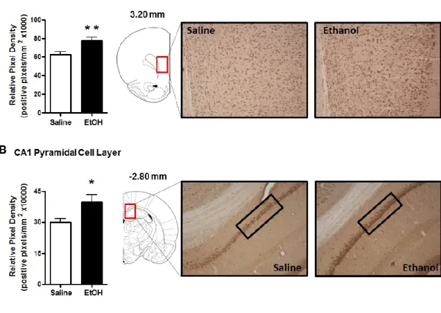
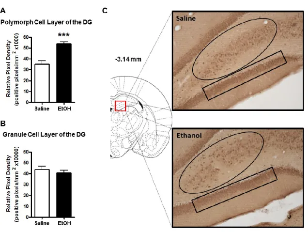
Previous studies using RIAs have shown ethanol-induced increases in 3α,5α-THP levels in the rat cortex and hippocampus (Barbaccia et al., 1999; VanDoren et al., 2000). Therefore, we first examined these regions to determine if the elevations displayed sub-region or cellular layer specificity. Ethanol administration (2g/kg, i.p.) increased 3α,5α-THP immunoreactivity in the mPFC [24±6%; t(13)=2.996, p < 0.01]. The effects of ethanol appear uniform across the cortical cell layers (Fig. 2.1A). In hippocampus, ethanol increased 3α,5α-THP immunoreactivity in the pyramidal cells of the CA1 region [32±12%; t(14)=2.401, p < 0.05] (Fig. 2.1B), and the polymorph cell layer of the DG [52±5%; t(14)=5.288, p < 0.001] (Fig. 2.2A), but had no effect on cellular 3α,5α-THP in the granule cell layer of the DG (Fig. 2.2B), indicating cellular layer specificity in the response to ethanol.
31
Ethanol-induced decreases of cellular 3α,5α-THP in regions associated with ethanol
reinforcement and consumption
DISCUSSION
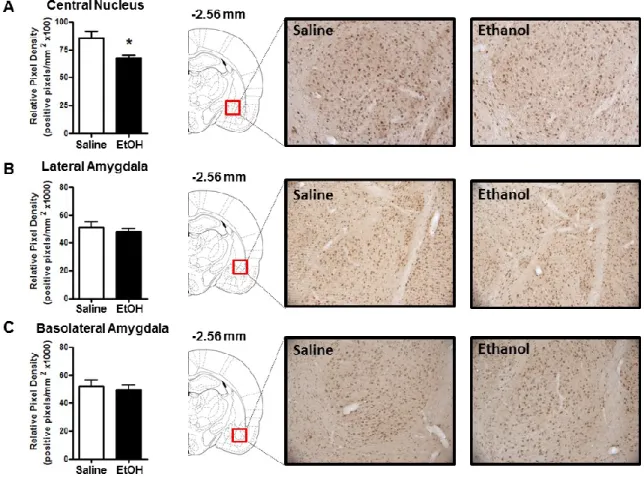
The goal of the present study was to use IHC to examine ethanol-induced changes in cellular 3α,5α-THP expression in multiple brain regions implicated in alcohol use disorders. Our findings support previous evidence that ethanol increases 3α,5α-THP concentrations in the rat cerebral cortex and hippocampus using RIA or GC-MS. We extend previous findings by showing that ethanol increases cellular 3α,5α-THP in the mPFC, CA1 pyramidal cell layer, and polymorph cell layer of the DG but not in granule cells of the DG. Therefore, ethanol-induced increases of cellular 3α,5α-THP in the hippocampus are isolated to specific cellular populations. We also show that ethanol increases cellular 3α,5α-THP levels in the BNST and the PVN of the hypothalamus. Interestingly, acute ethanol administration reduced cellular 3α,5α-THP levels in the NAc “shore” (core-shell border) and the CeA. To our knowledge, this is the first example of acute ethanol reducing 3α,5α-THP levels in the rat brain or periphery. We also determined that ethanol does not alter 3α,5α-THP levels in the VTA, DMS, or the lateral or basolateral amygdala. Therefore, ethanol produces divergent brain region and cellular layer specific changes in 3α,5α-THP concentrations.
33
3α,5α-THP using both fluorescent and DAB visualization, the magnitude of 3α,5α-THP changes may differ due to differences in immunohistochemical methodology.
The 3α,5α-THP antisera used in the present study was found to cross-react with 3α-HP (41±0.14%) and 3α,5β-THP (22±0.43%). This is not a major limitation since both of these steroids have similar GABAergic activities as 3α,5α-THP. Furthermore, there is no evidence that 3α-HP (Griffin and Mellon, 2001) or 3α,5β-THP are present in the brain in appreciable concentrations. In addition, ethanol does not elevate 3α,5β-THP levels in rat plasma (Porcu et al., 2010). Therefore, 3α,5α-THP is the most likely endogenous antigen immunolabelled using this approach. A potential limitation of the study is that only one dose of ethanol was examined. In the cerebral cortex, a threshold dose of ethanol (1.3-1.5 g/kg) is required to produce an increase of 3α,5α-THP and increasingly higher doses produce less of an increase of 3α,5α-THP (VanDoren et al., 2000; Boyd et al., 2010). However, since only one dose was used it is not known if a similar threshold dose is needed to produce increases or decreases of 3α,5α-THP in the cortical and subcortical brain regions examined in the present study. Similarly, it is not clear if ethanol produces a biphasic effect on 3α,5α-THP levels in these brain regions.
35
3α,5α-THP (Saalmann et al., 2007) as well as the biosynthetic enzymes 5α-R and 3α-hydroxysteroid dehydrogenase (3α-HSD) needed for 3α,5α-THP synthesis (Agis-Balboa et al., 2006). Furthermore, there was a reduction of cellular 3α,5α-THP in the NAc and the CeA. Based on these findings our data suggest there are unknown mechanisms contributing to ethanol’s effects on 3α,5α-THP concentrations. In the cerebral cortex, ethanol-induced elevations of 3α,5α-THP are dependent on the pituitary-adrenal axis (Khisti et al., 2003; O'Dell et al., 2004; Boyd et al., 2010). In vitro, however, there is evidence for ethanol-induced brain synthesis of 3α,5α-THP in hippocampal slices (Sanna et al., 2004; Tokuda et al., 2011). Thus, it is not known if ethanol alters brain synthesis of 3α,5α-THP in vivo independent of circulating steroids. Therefore, it will be important to determine the role of circulating steroids in the current observations, which may provide insight into this important question.
al., 2003). The synthesis of 3α,5α-THP from progesterone is accomplished by the sequential actions of 5α-R and 3α-HSD. Acute ethanol administration increases 5α-R type 1 (5α-RI) and 3α-HSD mRNA in the frontal cortex and 3α-HSD mRNA in the hypothalamus, but no change in transcript expression of either enzyme were detected in the hippocampus (Kim et al., 2003). Unfortunately, there are no data examining the effects of acute ethanol on expression of these enzymes in the NAc or CeA where we observed reductions in 3α,5α-THP. However, chronic intermittent ethanol administration reduces 3α,5α-THP levels in the hippocampus, which is associated with concurrent decreases in 5α-RI and 3α-HSD mRNA expression (Cagetti et al., 2004). Taken together, ethanol-induced changes in steroidogenic enzyme expression may underlie the divergent changes in 3α,5α-THP levels observed in the rat brain. Additionally, ethanol may directly or indirectly change the activity of steroidogenic enzymes, which could alter steroid concentrations. Ultimately, ethanol may alter the expression and/or activity of enzymes involved in steroid synthesis and metabolism, resulting in local changes in neurosteroid levels that are dependent on which enzymes are expressed in a particular cell.
37
Significance of ethanol-induced changes in cellular expression of 3α,5α-THP
role the hippocampus and DG play in learning and memory, 3α,5α-THP induction by ethanol may contribute to ethanol’s effects on memory and cognition.
Ethanol-induced changes in brain concentrations of 3α,5α-THP may also modulate neuronal activity in other brain regions examined in the present study. In vivo
evidence suggests ethanol-induced increases of 3α,5α-THP modulate neuronal activity outside of the hippocampus as well. For example, finasteride prevents ethanol (1.5g/kg) inhibition of spontaneous firing of medial septal/diagonal band of Broca neurons (VanDoren et al., 2000). In addition to hippocampal pyramidal cells, we observed increases of cellular 3α,5α-THP in the polymorph DG, mPFC, BNST, and PVN of the hypothalamus. The physiological consequences of 3α,5α-THP increases may alter neuronal activity and synaptic plasticity in these regions and related circuitry. Clearly, studies are needed to examine how increases of 3α,5α-THP in these brain regions may contribute to the physiological and behavioral effects of ethanol.
39
(Howard et al., 2009), but in the core or shell only ethanol associated cues increase dopamine release following operant training. It is important to note, however, that acute systemic ethanol administration increases dopamine release in the NAc core and shell (Imperato and Di Chiara, 1986; Yim et al., 2000) where ethanol did not alter 3α,5α-THP levels. Administration of 3α,5α-THP produces biphasic effects on ethanol consumption (Janak et al., 1998; Ford et al., 2005; Ford et al., 2007; Finn et al., 2010) and dopamine release in the NAc (Motzo et al., 1996; Rouge-Pont et al., 2002). Furthermore, 3α,5α-THP modulates ethanol’s effects on mPFC dopamine content (Dazzi et al., 2002). Therefore, investigating possible interactions of 3α,5α-THP and dopaminergic activity in the mesocorticolimbic system may be valuable in the effort to reduce ethanol consumption via neuroactive steroid modulation.
FIGURES AND TABLES
Figure 2.1: Effect of acute ethanol administration (2g/kg, i.p.) on 3α,5α-THP
immunoreactivity in the mPFC and pyramidal cell layer of the CA1 hippocampus. (A) Ethanol administration increased 3α,5α-THP immunoreactivity in the mPFC compared to saline controls. Representative photomicrographs (10x) of 3α,5α-THP immunoreactivity in the mPFC (3.20 mm relative to bregma) following saline (n=8) or ethanol (n=7) administration. (B) Ethanol administration increased 3α,5α-THP immunoreactivity in the pyramidal cell layer of the CA1 hippocampus compared to saline controls. Representative photomicrographs (10x) of 3α,5α-THP immunoreactivity in CA1 pyramidal cells
(highlighted in rectangle, -2.80 mm relative to bregma) following saline (n=8) or ethanol (n=8) administration. Ethanol (2g/kg, i.p.) or saline were administered 60 min prior to tissue fixation and collection. Data are expressed as mean positive pixels/mm2 SEM. * p < 0.05, ** p < 0.01 compared to saline administration. Medial prefrontal cortex
41
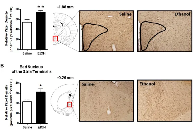
Figure 2.3: Effect of acute ethanol administration (2g/kg, i.p.) on 3α,5α-THP immunoreactivity in the PVN of the hypothalamus and BNST. (A) Ethanol
administration increased 3α,5α-THP immunoreactivity in the PVN compared to saline controls. Representative photomicrographs (10x) of 3α,5α-THP immunoreactivity in the PVN (-1.88 mm relative to bregma) following saline (n=8) or ethanol (n=8)
43
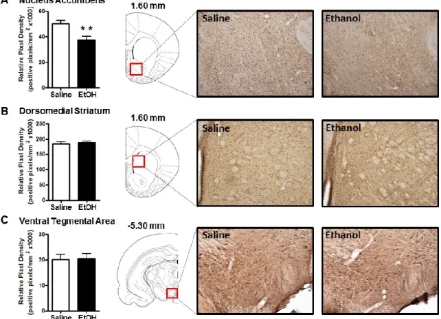
Figure 2.4: Effect of acute ethanol administration (2g/kg, i.p.) on 3α,5α-THP immunoreactivity in NAc “shore” (core/shell border), DMS, and VTA. (A) Ethanol administration reduced 3α,5α-THP immunoreactivity in the NAc “shore” compared to saline controls. Representative photomicrographs (10x) of 3α,5α-THP immunoreactivity in NAc “shore” (1.60 mm relative to bregma) following saline (n=7) or ethanol (n=7) administration. (B) Ethanol administration did not alter 3α,5α-THP immunoreactivity in the DMS compared to saline controls. Representative photomicrographs (10x) of 3α,5α-THP immunoreactivity in DMS (1.60 mm relative to bregma) following saline (n=8) or ethanol (n=7) administration. (C) Ethanol administration did not alter 3α,5α-THP immunoreactivity in the VTA compared to saline controls. Representative
45
47
EFFECTS OF ADRENALECTOMY ON ETHANOL-CHAPTER 3.
INDUCED CHANGES IN 3,5-THP IMMUNOHISTOCHEMISTRY
INTRODUCTION
Neuroactive steroids are endogenous neuromodulators capable of altering neuronal activity. Synthesis of neuroactive steroids occurs centrally in the brain and peripherally in the adrenal glands and gonads. The 5α-reduced pregnane steroids, (3,5)-3-hydroxypregnan-20-one (3α,5α-THP or allopregnanolone) and (3,5)-3,21-dihydroxypregnan-20-one (3α,5α-THDOC), are positive allosteric modulators of γ-aminobutyric acid type A (GABAA) receptors. GABAA receptors are the primary inhibitory receptor family in the brain, which mediate many of the behavioral effects of ethanol. Both 3α,5α-THP and 3α,5α-THDOC enhance neuronal inhibition at known binding sites on GABAA receptor α-subunits (Hosie et al., 2006), and produce behavioral effects similar to those produced by ethanol. These GABAergic neuroactive steroids are very potent positive modulators of GABAA receptors, producing pharmacological effects in nanomolar concentrations (Morrow et al., 1987), and modulating GABAergic steroid levels may have therapeutic value for treating multiple psychiatric disorders (Uzunova et al., 1998; Strohle et al., 2002; Marx et al., 2006; Rasmusson et al., 2006; Morrow, 2007).
48
Ethanol activates the hypothalamic-pituitary-adrenal (HPA) axis (Boyd et al., 2010) leading to physiologically significant increases of 3α,5α-THP and 3α,5α-THDOC in the blood plasma, cerebral cortex, and hippocampus (Barbaccia et al., 1999; VanDoren et al., 2000; Porcu et al., 2009). Adrenalectomy or inhibition of 5α-reduced steroid synthesis with the 5α-reductase (5α-R) inhibitor finasteride reduces some of the behavioral effects of ethanol, including the anticonvulsant (VanDoren et al., 2000), anti-depressant-like (Hirani et al., 2002), hypnotic (Khisti et al., 2003), and anxiolytic-like (Hirani et al., 2005) effects in rats. Finasteride also blocks ethanol inhibition of neuron firing in the medial septum (VanDoren et al., 2000), the hippocampus both in vivo (Tokunaga et al., 2003b) and in vitro (Sanna et al., 2004), and long-term potentiation (LTP) in hippocampal slice preparations (Tokuda et al., 2011). Importantly, finasteride also reduces some of the subjective effects of alcohol in healthy men (Pierucci-Lagha et al., 2005). Taken together, these findings suggest ethanol-induced elevations of 3α,5α-THP and 3α,5α-THDOC contribute to many of the physiological and behavioral effects of ethanol.
49
3α,5α-THP have used radioimmunoassay (RIA) or gas chromatography—mass spectroscopy (GC-MS), which lack the sensitivity to measure 3α,5α-THP in many brain regions.
previously observed ethanol-induced changes in 3α,5α-THP. We also investigated the effects of ethanol on 3,5-THP levels in perfused and non-perfused rat brain in order to determine the contribution of circulating steroids in brain measurements of 3α,5α-THP using RIA. Finally, we evaluated the effect of the pentobarbital anesthetic on 3α,5α-THP levels in the cerebral cortex and hippocampus.
MATERIALS AND METHODS
Subjects
For the ADX experiments adult male Wistar rats (~275 g/8-11 per group) that had undergone ADX or sham surgery were purchased from Harlan Laboratories (Indianapolis, IN, USA). The animals were single housed in Plexiglass cages with food and water (animals that underwent ADX received 0.9% saline instead of water) available
ad libitum. Animals were sacrificed 7 days following surgery and approximately 24hr after arriving in the colony room. For the RIA studies male Wistar rats (5-14 per group) were purchased from Harlan Laboratories (Indianapolis, IN, USA) and stayed in the colony room for 6 weeks to reach adulthood. The animals habituated to handling and saline i.p. saline injections for 1 week prior to the experiment. The colony room was maintained on a normal 12 hr light-dark cycle (light onset at 0700 hr) and at a constant temperature of 22 ± 2 C and relative humitity of 65%. Experiments were conducted between 0800 and 1300 hr to minimize potential circadian fluctuation in neuroactive steroid levels.
51
National Institutes of Health Guidelines under University of North Carolina at Chapel Hill Institutional Animal Care and Use Committee approved protocols.
Radioimmunoassay (RIA)
Prior to RIA, 3α,5α-THP was extracted from freshly frozen cerebral cortex or hippocampus. Recovery was monitored by incorporation of 4000 dpm of [3 H]-3α,5α-THP. Brain samples were homogenized in 2.5ml 0.3 N NaOH with a sonic dismembrator and extracted three times in 3ml aliquots of 10% (v/v) ethyl acetate in heptane. Aliquots were combined and 5ml of heptane was added to each sample. The extracts were run through solid phase silica columns, washed in pentane, and steroids of similar polarity to 3α,5α-THP were eluted off the column by 25% acetone in pentane. Eluant was dried, and steroids reconstituted in 50µl isopropanol and 150µl 50% sodium phosphate/bovine serum albumin assay buffer. Extraction efficiency was determined in 50µl the reconstituted extract by liquid scintillation spectroscopy. The remaining 150µl was used in RIA determination of 3α,5α-THP.
Immunohistochemistry
Fifty-three minutes after ethanol (2g/kg) or saline injection the animals were anesthetized with pentobarbital (100mg/kg, i.p.; Professional Compounding Centers of America, Houston, TX, USA) and transcardially perfused approximately 1hr following ethanol or saline injection with phosphate buffered saline (PBS) followed by 4% paraformaldehyde. Tissue was post-fixed in 4% paraformaldehyde for 24hr at 4°C, sectioned coronally at 40μm on a vibrating microtome, and stored at -30°C. Pentobarbital does not alter 3α,5α-THP levels using this procedure, and blood perfusion does not alter brain levels of 3α,5α-THP in intact animals using RIA (unpublished data).
Immunohistochemical assays were performed using a procedure modified from (Saalmann et al., 2007) and previously described in detail (Cook et al., 2013). Briefly, no detergents or organic solvents were used to prevent the possibility of neuroactive steroid leaching. Free floating brain sections (3-4 sections/animal/brain region) were rinsed, blocked in 10% rabbit serum, and incubated in sheep affinity purified anti-3α,5α-THP antiserum (purchased from Dr. R.H. Purdy) at a 1:2500 dilution for 48 hr at 4°C. A rabbit anti-sheep biotinylated secondary antibody (1:200; Vector laboratories, Burlingame, CA, USA) was used in collaboration with the Vectastain Elite ABC kit (Vector Laboratories, Burlingame, CA, USA) and immunoreactivity was visualized with 3,3’-diaminobenzidine (Sigma-Aldrich, St. Louis, MO, USA) using the manufacturers’ recommended procedures.
Immunohistochemical analysis
53
AP). All analyses were based on coordinates relative to bregma in the Rat Brain Atlas (Paxinos and Watson, 1998).
RESULTS
Effects of transcardial perfusion or pentobarbital administration on 3α,5α-THP
measurements in the whole cerebral cortex or hippocampus using RIA
Previous IHC results had found a discrepancy in the magnitude of ethanol-induced increases of 3α,5α-THP between IHC and RIA measurement. Using IHC we observed a 24% and 32% increase of 3α,5α-THP in the mPFC and CA1 pyramidal cells, respectively. RIA studies have shown 2-17 fold increases in the whole cortex (Barbaccia et al., 1999; VanDoren et al., 2000; Khisti et al., 2004; Boyd et al., 2010) and 4-11 fold increase in whole hippocampus (Barbaccia et al., 1999; Khisti et al., 2004). Therefore, since IHC brains are perfused, we tested whether the blood present in RIA tissue samples contribute to the discrepancy in 3α,5α-THP magnitude. As expected, there was a main effect of ethanol in the cerebral cortex [F (1,38) = 76.78, p < 0.0001] (Fig. 3.1A) and hippocampus [F (1,33) = 107.5, p < 0.0001] (Fig. 3.1B), but there was no effect of perfusion on 3α,5α-THP levels in the cortex [F (1,38) = 0.19, p = 0.6644] (Fig. 3.1A) or hippocampus [F (1,33) = 0.92, p = 0.3433] (Fig. 3.1B).
55
Ethanol-induced increases of cellular 3α,5α-THP
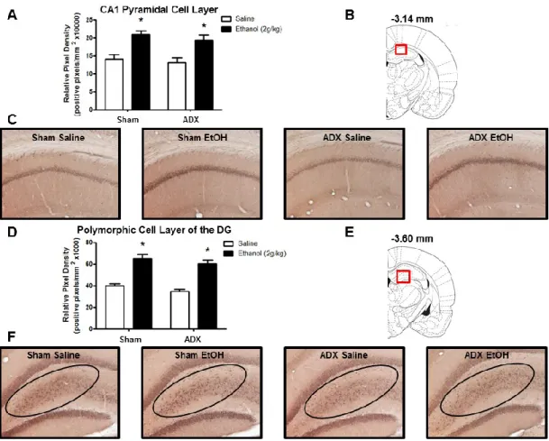
Recent work in our lab has shown that ethanol increases 3α,5α-THP immunoreactivity in the mPFC, CA1 pyramidal cell layer of the hippocampus, polymorphic cell layer of the DG, BNST, and PVN of the hypothalamus (Cook et al., 2013). Therefore, we tested whether ethanol-induced elevations of 3α,5α-THP in these regions are dependent upon adrenal gland activation. Ethanol administration increased 3α,5α-THP immunoreactivity in the mPFC, but this effect was moderated by adrenalectomy (Fig. 3.2A). The two-way ANOVA indicated a main effect of ethanol treatment [F (1,34) = 8.18, p < 0.01] and surgery condition [F (1,34) = 9.41, p < 0.005], but no significant interaction. Therefore, we performed an a priori planned comparison between the sham saline versus sham ethanol and ADX saline versus ADX ethanol groups. mPFC 3α,5α-THP immunoreactivity was significantly increased in the sham ethanol group compared to the sham saline group [25±11%; t(15)=3.583, p < 0.01], similar to previous immunohistochemical results (Cook et al., 2013). However, ethanol did not increase 3α,5α-THP in animals that received ADX [2±7%; t(19)=0.937, p > 0.05], suggesting that the adrenals contribute to the effects of ethanol in mPFC. The effects of ethanol appear uniform across the cortical cell layers (Fig. 3.2C).
immunoreactivity was increased by 63±10% in the sham ethanol versus sham saline group and by 74±11% in the ADX ethanol versus ADX saline group (main effect of ethanol treatment [F (1,34) = 61.16, p < 0.0001]) (Fig. 3.3D).
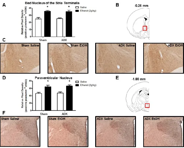
In the BNST and PVN of the hypothalamus, ethanol increased 3α,5α-THP immunoreactivity independent of the adrenal glands. In the BNST, 3α,5α-THP immunoreactivity was increased by 43±5% in the sham ethanol versus sham saline group and by 40±8% in the ADX ethanol versus ADX saline group (main effect of ethanol treatment [F (1,34) = 35.68, p < 0.0001]) (Fig. 3.4A). In the PVN, 3α,5α-THP immunoreactivity was increased by 38±10% in the sham ethanol versus sham saline group and by 39±7% in the ADX ethanol versus ADX saline group (main effect of ethanol treatment [F (1,34) = 27.70, p < 0.0001]) (Fig. 3.4D).
Ethanol-induced decreases of cellular 3α,5α-THP
57 DISCUSSION
assay showed that the pentobarbital anesthetic does not alter 3α,5α-THP levels in whole cerebral cortex or hippocampus.
The current IHC results and previous reports using RIA or GC-MS all suggest that ethanol-induced elevations of 3α,5α-THP in the cerebral cortex are dependent on the adrenal glands or the pituitary (Boyd et al., 2010). It is not clear why there is a dependence on HPA activation to observe cerebral cortical elevations of 3α,5α-THP, but one possibility is that there is a lack of precursor in the cortex. Indeed, it has previously been shown that administration of 5α-dihydroprogesterone (5α-DHP), the immediate precursor of 3α,5α-THP, restores ethanol-induced increases of 3α,5α-THP in the cerebral cortex and corresponding effects of ethanol on loss of righting reflex following ADX (Khisti et al., 2003). It has also been shown that de novo adrenal synthesis of the cholesterol transporter, steroidogenic acute regulatory protein (StAR), is necessary for ethanol-induced increases of 3α,5α-THP in the cerebral cortex (Boyd et al., 2010). StAR transports cholesterol to the inner mitochondrial membrane where it is converted to pregnenolone by cytochrome P450 side chain cleavage (P450scc). Therefore, de novo
59
increases of 3α,5α-THP in the cerebral cortex are dependent on adrenal derived precursor, which is converted to 3α,5α-THP by locally synthesized steroidogenic enzymes.
The current results in the hippocampal formation agree with the previous in vitro
studies that have shown ethanol-induced local brain synthesis of 3α,5α-THP. For example, it was first shown using RIA that incubation with ethanol (50 or 100 mM) can increase 3α,5α-THP in hippocampal minces from intact rats (Sanna et al., 2004) and rats that had undergone ADX/gonadectomy (Follesa et al., 2006). More recently, it was shown that ethanol increases cellular 3α,5α-THP in CA1 pyramidal cells using IHC with fluorescent detection of 3α,5α-THP (Tokuda et al., 2011). In each of these in vitro
studies, ethanol induction of 3α,5α-THP was shown to alter neuronal function using electrophysiological measures. In the current study, we show that ethanol increases 3α,5α-THP immunoreactivity in the CA1 pyramidal cell layer as well as the polymorphic cell layer of the DG, independent of adrenal gland activation. Furthermore, ethanol’s ability to stimulate brain synthesis of 3α,5α-THP in the hippocampal formation is isolated to specific cellular populations, since we did not previously observe ethanol-induced changes of 3α,5α-THP in the granule cell layer of the DG (Cook et al., 2013). The CA1 pyramidal cells, polymorphic DG, and granule cell layer of the DG all exhibit dense 3α,5α-THP staining. Therefore, the presence of this very specific effect of ethanol on cellular 3α,5α-THP in the hippocampus is intriguing and may underlie neuron specific responses to ethanol in the hippocampal formation.
are involved in stress, emotion, and ethanol responses (Armario, 2010; Cui et al., 2012; Koob, 2013), and we have previously shown that ethanol increases cellular 3α,5α-THP in these regions (Cook et al., 2013). It has also been shown that GABAergic neuroactive steroids contribute to negative feedback on the HPA axis at the level of the hypothalamus in rats (Owens et al., 1992; Patchev et al., 1994; Patchev et al., 1996), and activate the stress response in the hypothalamus of C57BL/6J mice (Sarkar et al., 2011). The present data suggest that ethanol-induced changes of cellular 3α,5α-THP in the PVN and BNST are independent of adrenal activation. Other studies are clearly needed to understand the physiological and behavioral impact these local elevations of 3α,5α-THP produce in the PVN and BNST.
61
Previous studies have shown that 3α,5α-THP administration produces biphasic effects on dopamine release in the NAc (Motzo et al., 1996; Rouge-Pont et al., 2002), and modulates ethanol’s effects on dopamine content in the mPFC (Dazzi et al., 2002). Therefore, investigating interactions between 3α,5α-THP levels and dopaminergic activity in the mesocorticolimbic system may aid in the effort to reduce ethanol consumption via neuroactive steroid modulation.
Significance on Neuronal Activity and Potential Mechanisms of Ethanol-Induced
Changes in Local 3α,5α-THP Levels
increases in amplitude of spontaneous and evoked IPSCs in CA1 pyramidal cells (Sanna et al., 2004), the latter of which is prevented by finasteride. Furthermore, in hippocampal slices, ethanol (60mM) increases 3α,5α-THP immunofluorescence and inhibits LTP in CA1 pyramidal cells (Tokuda et al., 2011). Importantly, pretreatment with finasteride prevents the increase in 3α,5α-THP immunofluorescence and ethanol’s ability to inhibit LTP. Thus, 3α,5α-THP appears to mediate ethanol’s effects on synaptic plasticity in CA1 pyramidal cells. In the present study, ethanol increased 3α,5α-THP in the CA1 pyramidal cell layer and the polymorphic cell layer of the DG. The most abundant cells in the polymorphic DG are mossy cells, which project to the molecular and granule cell layers of the DG (Amaral et al., 2007), with the granule cells being the only projection from the DG to the hippocampus. Ethanol-induced increases in cellular 3α,5α-THP within hippocampal pyramidal cells and the polymorphic DG may contribute to ethanol’s effects on memory and cognition by altering neuronal activity and synaptic plasticity.
63
most likely decrease GABAA receptor mediated inhibition. More studies are clearly needed to determine the extent to which these ethanol-induced changes of 3α,5α-THP contribute to the neurophysiological and behavioral effects of ethanol.
The presence of divergent local changes in cellular 3α,5α-THP suggest that ethanol may alter local synthesis and/or metabolism of 3α,5α-THP. One possibility is that ethanol alters the expression and/or activity of the cholesterol transporters and/or steroidogenic enzymes. Acute ethanol administration increases StAR mRNA in the hippocampus (Kim et al., 2003). In the hypothalamus, acute ethanol increases mRNA levels of StAR, P450scc, and 3α-HSD (Kim et al., 2003). Currently, there are no data examining ethanol’s effects on steroidogenic enzymes in the BNST. Similarly, there are no data examining ethanol’s effects on steroidogenic enzymes in the NAc or CeA where we observed ethanol-induced reductions in 3α,5α-THP. Chronic intermittent ethanol, however, has been shown to reduce 3α,5α-THP in the hippocampus along with concomitant decreases in 5α-R type I and 3α-HSD mRNA (Cagetti et al., 2004). Therefore, ethanol may alter levels and/or activity of steroid biosynthetic enzymes to produce divergent brain region specific changes in 3α,5α-THP. Ultimately, the changes in neuroactive steroid levels may depend on the combination of cholesterol transporter and/or synthetic enzymes expressed in a particular cell type.
there is no direct evidence for active release of 3α,5α-THP, we cannot rule out the possibility that ethanol causes a release or redistribution of 3α,5α-THP into the extracellular space.
65 FIGURES AND TABLES
Figure 3.2: Effect of ethanol (2g/kg, i.p.) on 3α,5α-THP immunoreactivity in the mPFC of rats subjected to sham surgery or ADX. (A) Ethanol increases 3α,5α-THP
immunoreactivity in the mPFC following sham surgery, but not ADX, compared to saline controls. (B) The red box indicates the location (3.20 mm relative to bregma) of
67
Figure 3.3: Effect of ethanol (2g/kg, i.p.) on 3α,5α-THP immunoreactivity in the CA1
hippocampus and polymorphic cell layer of the DG following sham surgery or ADX. (A) Ethanol increases 3α,5α-THP immunoreactivity in the CA1 pyramidal cell layer after sham surgery and ADX, compared to saline controls. (B) The red box indicates the location (-3.14 mm relative to bregma) of representative photomicrographs within the hippocampus. (C) Representative photomicrographs (10x) of 3α,5α-THP
69
71
72
EFFECTS OF VIRAL VECTOR MEDIATED P450SCC CHAPTER 4.
OVEREXPRESSION IN THE NAC OR VTA ON OPERANT ETHANOL SELF-ADMINISTRATION
INTRODUCTION
Neuroactive steroids are neuromodulators synthesized in the brain that modulate neuronal activity and influence motivation and emotional behaviors. The GABAergic neuroactive steroid (3,5)-3-hydroxypregnan-20-one (3,5-THP or allopregnanolone) is a potent positive allosteric modulator of γ-aminobutyric acid type A (GABAA) receptors. GABAA receptors are the primary inhibitory receptors in the brain and mediate many of the behavioral effects of ethanol. Ethanol-induced increases of 3,5-THP contribute to many of the neurophysiological and behavioral effects of ethanol in rats (VanDoren et al., 2000; Hirani et al., 2002; Khisti et al., 2003; Hirani et al., 2005) as well as subjective effects of alcohol in humans (Pierucci-Lagha et al., 2005). Furthermore, human alcoholics have reduced blood plasma levels of 3,5-THP during alcohol withdrawal (Romeo et al., 1996). Moreover, increasing evidence suggests that modulating GABAergic neuroactive steroid levels may have therapeutic value for treating multiple neurologic and psychiatric disorders (Marx et al., 2006; Rasmusson et al., 2006; Morrow, 2007; Rupprecht et al., 2010; Brinton, 2013).
73
neuroactive steroid 3α,5β-20-oxo-pregnane-3-carboxylic acid (PCA) (O'Dell et al., 2005) dose dependently reduce ethanol self-administration without producing sedation. Administration of 3,5-THP or the longer acting synthetic analog of 3,5-THP, ganaxolone, produce biphasic effects on ethanol self-administration (Janak et al., 1998; Ford et al., 2005; Besheer et al., 2010a). However, there are concerns that 3,5-THP and ganaxolone produce sedation (Belelli et al., 1989; Besheer et al., 2010a), limiting therapeutic effectiveness. The therapeutic potential of exogenously administered 3,5-THP may also be limited by rapid metabolism (Purdy et al., 1990a).
vector manipulation result in chronically elevated P450scc expression, pregnenolone synthesis, and levels of 3,5-THP, but regionally specific P450scc overexpression significantly influenced long-term operant ethanol self-administration. We also used confocal imaging to determine cell-type specific localization of 3,5-THP in the VTA. MATERIALS AND METHODS
Animals
Adult male Wistar rats (~275 g) were obtained from Harlan Laboratories (Indianapolis, IN, USA) and used for characterizing viral vectors (n=16 for in vivo
75 Apparatus
Self-administration chambers
Operant ethanol self-administration was performed in conditioning chambers measuring 30.5 x 24.1 cm (Med Associates, Georgia, VT, USA) located inside sound-attenuating cubicles. Cubicles were equipped with an exhaust fan for ventilation, which also masks external sounds. Both the left and right walls of the chambers contain a lever and liquid receptacle (i.e. 2 per chamber). The appropriate number of lever press responses simultaneously activates a stimulus light over the lever and a syringe pump (Med Associates) that delivers 0.1ml of liquid solution into a receptacle over 1.66 sec duration. Lever responses during reinforcer delivery were counted, but did not result in programmed consequences. The operant chambers were connected to a computer programmed to control sessions and record the resulting data.
Locomotor chambers
Operant self-administration
Training
The day prior to initial training, animals were water derived for approximately 24 hr. Immediately following water deprivation, animals were placed in the operant chambers for a 16 hr lever press training session where 0.1ml of sucrose (10%, w/v) or water were concurrently available contingent on lever press responses. Lever press responses were initially maintained on a concurrent fixed-ratio 1 (CONC FR1 FR1) schedule of reinforcement and were gradually increased to CONC FR2 FR2 following delivery of 4 reinforcers, followed by a CONC FR4 FR4 schedule after delivery of 10 reinforcers. Reinforcer presentations were paired with a light cue located above each lever. Following the 16 hr training session animals were returned to their homecage for a period of 24 hr with ad libitum water access thereafter.
Sucrose fading, operant baseline sessions, operant self-administration testing