Research Paper
Complement Activation in Arterial and Venous Thrombosis is Mediated
by Plasmin
Jonathan H. Foley
a,b,c, Bethany L. Walton
d, Maria M. Aleman
d, Alice M. O'Byrne
a, Victor Lei
a,
Micaela Harrasser
b, Kimberley A. Foley
e, Alisa S. Wolberg
d, Edward M. Conway
a,⁎
a
Centre for Blood Research, Department of Medicine, Life Sciences Institute, University of British Columbia, 2350 Health Sciences Mall, LSC4306, Vancouver V6T 1Z3, Canada b
Department of Haematology, UCL Cancer Institute, University College London, London, United Kingdom c
Katharine Dormandy Haemophilia Centre and Thrombosis Unit, Royal Free NHS Trust, London, United Kingdom d
Department of Pathology and Laboratory Medicine, University of North Carolina at Chapel Hill, 819 Brinkhous-Bullitt Building, CB# 7525, Chapel Hill, NC 27599-7525, USA eCancer Care and Epidemiology, Queen's Cancer Research Institute, Queen's University, Kingston, Canada
a b s t r a c t
a r t i c l e i n f o
Article history:
Received 3 November 2015
Received in revised form 4 February 2016 Accepted 5 February 2016
Available online 6 February 2016
Thrombus formation leading to vaso-occlusive events is a major cause of death, and involves complex interac-tions between coagulation,fibrinolytic and innate immune systems. Leukocyte recruitment is a key step, mediated partly by chemotactic complement activation factors C3a and C5a. However, mechanisms mediating C3a/C5a generation during thrombosis have not been studied. In a murine venous thrombosis model, levels of thrombin–antithrombin complexes poorly correlated with C3a and C5a, excluding a central role for thrombin in C3a/C5a production. However, clot weight strongly correlated with C5a, suggesting processes triggered during thrombosis promote C5a generation. Since thrombosis elicitsfibrinolysis, we hypothesized that plasmin activates C5 during thrombosis.In vitro, the catalytic efficiency of plasmin-mediated C5a generation greatly exceeded that of thrombin or factor Xa, but was similar to the recognized complement C5 convertases. Plasmin-activated C5 yielded a functional membrane attack complex (MAC). In an arterial thrombosis model, plasminogen activator administration increased C5a levels. Overall, thesefindings suggest plasmin bridges thrombosis and the immune response by liberating C5a and inducing MAC assembly. These new insights may lead to the development of strategies to limit thrombus formation and/or enhance resolution.
© 2016 The Authors. Published by Elsevier B.V. This is an open access article under the CC BY-NC-ND license (http://creativecommons.org/licenses/by-nc-nd/4.0/). Keywords:
Thrombosis Complement Leukocytes Thrombin Fibrinolysis
1. Introduction
The coagulation system and innate immunity are coordinately activated and highly integrated during venous and arterial thrombus formation and progression (von Bruhl et al., 2012; Engelmann and Massberg, 2013; Fuchs et al., 2012). Vascular endothelial activation or damage causes release of ultralarge von Willebrand factor (VWF) and P-selectin from Weibel-Palade bodies, and local activation of comple-ment with liberation of anaphylatoxic and chemotactic factors C3a and C5a. These pathways cooperate to trigger platelet, neutrophil, and monocyte recruitment and activation (von Bruhl et al., 2012). The locally accumulated cells release proteases, reactive oxygen species,
and nucleosomes, which provide a scaffold for aggregating platelets and red blood cells and further promote coagulation andfibrin formation (Fuchs et al., 2012). Several complement factors, including C3, C4, C3a, C5a and factor H are incorporated into the thrombus, where they modu-late thrombus stability and the inflammatory process (Distelmaier et al., 2009; Howes et al., 2012). Thefibrinolytic system and plasmin-mediated proteolysis are also intimately coupled to the axis of thrombus develop-ment and inflammation by controllingfibrin degradation, activation of matrix metalloproteinases, infiltration of monocytes/macrophages and other immune mediators, vessel wall remodeling, and ultimately throm-bus resolution (Wakefield et al., 2008).
The mechanisms by which leukocytes are recruited early in throm-bus formation and later during thromthrom-bus extension or resolution, are poorly understood. However, C5a, the most potent chemotactic comple-ment activation fragcomple-ment, is released following proteolytic cleavage of C5 and is considered a critical determinant of neutrophil recruitment and activation in thrombosis (Distelmaier et al., 2009; Salmon et al., 2002; Pierangeli et al., 2005). Moreover, terminal complement pathway complexes formed as C5 is activated, have multiple procoagulant prop-erties (Langer et al., 2013; Hamilton et al., 1990). Thus, there is interest in understanding how C5a and the other major complement-derived Abbreviations:MAC, membrane attack complex; VWF, von Willebrand factor; R751,
arginine 751; TAT, thrombin antithrombin; IVC, inferior vena cava; VFKck, Val-Phe-Lys-chloromethylketone; PPACK, Phe-Pro-Arg-Val-Phe-Lys-chloromethylketone; FeCl3, ferric chloride; tPA, tissue-type plasminogen activator; NETs, neutrophil extracellular traps; PAR1, protease activated receptor 1; MCP1-1, monocyte chemoattracant protein-1; IL-8, interleukin-8; FDP,fibrin degradation product.
⁎ Corresponding author at: Centre for Blood Research, 4306-2350 Health Sciences Mall, University of British Columbia, Vancouver, BC V6T 1Z3, Canada.
E-mail address:[email protected](E.M. Conway).
http://dx.doi.org/10.1016/j.ebiom.2016.02.011
2352-3964/© 2016 The Authors. Published by Elsevier B.V. This is an open access article under the CC BY-NC-ND license (http://creativecommons.org/licenses/by-nc-nd/4.0/).
Contents lists available atScienceDirect
EBioMedicine
chemotactic factor, C3a, are generated, so that novel therapeutic strate-gies may be designed to prevent thrombosis.
Complement activation typically proceeds via three pathways— classical, lectin and alternative—which converge to form C3 convertases that proteolyse C3 into C3b with release of C3a (Ricklin et al., 2010). As complement activation exceeds a threshold, and the density of C3b increases, the specificity of the convertase shifts from C3 to C5. The resultant C5 convertases—C3bBbC3b for the alternative pathway and C4b2aC3b for the classical/lectin pathway—efficiently cleave C5 at argi-nine 751 (R751), liberating C5a and generating C5b, the initiating factor for assembly of the lytic C5b-9 membrane attack complex (MAC). Although the C3/C5 convertases are well-recognized for their capacity to cleave C3 and C5, other serine proteases reportedly also exhibit convertase activity (Huber-Lang et al., 2006; Amara et al., 2010; Wiggins et al., 1981). Notably, thrombin was implicated in providing a“new pathway”to activate complement by cleaving C5 in a C3-independent manner, thereby bypassing the bonafide C5 convertases (Huber-Lang et al., 2006). However, C5 is a relatively poor substrate for thrombin cleavage at R751 (Krisinger et al., 2012), raising questions as to its impor-tance in contributing to C5a generation during thrombus formation in vivo. We therefore explored the mechanisms by which C3a and C5a are generated using biochemical approaches andin vivomodels of venous and arterial thrombosis.
2. Materials and Methods
2.1. Materials
Human complement C3a and C5a were measured using Quidel MicroVue C3a Plus or C5a ELISA kits (Cedarlane Laboratories, Burlington, Ontario). Murine thrombin–antithrombin (TAT) levels were measured using Enzygnost TAT micro ELISA (Siemens, Munich, Germany). Human complement proteins C3 and C5 were obtained from Comple-ment Technology, Inc. (Tyler, TX) and human hemostatic enzymes (plasmin, factor Xa and thrombin) were from Haematologic Technolo-gies, Inc. (Essex Junction, VT).
2.2. ELISAs to Measure C3a and C5a
Murine complement C5a levels were determined using the mouse complement component C5a duoset and accompanying standard from R&D systems (catalog #DY2150; Minneapolis, MN). An ELISA for murine complement C3a was established using a murine C3a standard and antibodies from BD Biosciences (Mississauga, Canada). The capture rat monoclonal anti-mouse C3a antibody (catalog # 55820, clone: I87-1162) was coated overnight onto 96-well plates in 100μL of PBS at a concentration of 2μg/mL. Wells were washed × 3 with wash buffer (R&D catalog #WA126) followed by blocking for 2 h with 300μl of reagent diluent (R&D systems catalog #DY995). Plasma samples in duplicate were diluted 1/50 and 1/125 in sample diluent to afinal volume of 100μl and incubated for 2 h. After 3 washes, 100μl of the biotinylated detection monoclonal rat anti-mouse C3a antibody (clone I87-419, catalog #55821) 0.5μg/mL in reagent diluent was incubated for 1 h. Wells were washed × 4 and incubated for 20 min at room temperature with 100μl of streptavidin-biotinylated horseradish per-oxidase (1:3000), followed by 2 washes, and development with the substrate solution containing o-phenylenediamine using a plate reader set to 450 nm. A standard curve was generated with purified murine C3a (catalog # 558618). The sensitivity range of the assay was 0.1 nM to 2.5 nM. Intra-assay and inter-assay precision was 8–10%.
2.3. Animal Models
All experiments with animals were approved by the University of North Carolina at Chapel Hill Institutional Animal Care and Use Commit-tees. The mice (C57Bl/6) were male and between 6 and 8 weeks of age.
The number of animals for each model was determined based on previ-ous work that showed a broad range of TAT levels in the respective models (Machlus et al., 2011a, 2011b). On each day of the experiments, animals were randomly assigned to receive the stated treatment or to be used for baseline measurements. Quantification of biomarkers was performed in a blinded fashion wherein an experimenter, different from the one who performed the procedures on the animals, carried out the ELISAs on coded samples that were only de-coded after results had been generated.
2.4. Murine model of Venous Thrombosis
The inferior vena cava (IVC) stasis model was performed as previ-ously described (Aleman et al., 2013). Briefly, mice were anesthetized with 1·5–2% isoflurane in oxygen and human prothrombin (to 300%, final, mouse plus human prothrombin) or vehicle was infused via tail vein injection. Prothrombin was infused to give a broader range of thrombin generation and clot weight. Following sterile laparotomy, the intestines were exteriorized, the IVC was dissected bluntly, and side branches were ligated with 8–0 prolene suture and lumbar branches closed by cautery. The IVC was separated from the aorta by blunt dissection and completely ligated with 8–0 prolene suture. After replacing the intestines, the muscle layer was closed with 5–0 vicryl suture and skin closed with 8–0 prolene suture and skin glue. Mice re-covered with analgesia (buprenorphine, 0·05 mg/kg subcutaneous). After 12 h, blood was drawn from the IVC above the ligation site into 3.2% sodium citrate and processed to platelet-poor plasma by centrifu-gation at 5000 ×gfor 10 min. Thrombi were collected and weighed. Plasmas were stored at−80 °C for analysis of TAT, C3a and C5a levels by a person that was blinded to the treatment group. Two samples showing hemolysis were excluded.
2.5. In Vitro Generation of C3a or C5a by Hemostatic Enzymes
The relative efficiency, time course, and rate of complement cleavage by plasmin, factor Xa or thrombin were determined using a series of assays. Briefly, complement C3 (20μM) or C5 (2μM) was incubated with 100 nM plasmin or 250 nM factor Xa or thrombin. Reactions were quenched at various time points and C3a or C5a levels were quan-tified by ELISA. To determine the relative efficiency of cleavage, 2μM of C5 was incubated with 100 nM of plasmin, factor Xa or thrombin at 37 °C. After 10 min the reaction was quenched with the appropriate chloromethylketone (Val-Phe-Lys-chloromethylketone (VFKck) for plasmin and Phe-Pro-Arg-chloromethylketone (PPAck) for factor Xa and thrombin). In similar experiments, aliquots of the reaction mixture were sub-sampled into chloromethylketones at various time points to determine the time courses of C3 and C5 cleavages by plasmin, factor Xa and thrombin. The kinetics of C5a generation were determined by incubating 0–3μM of C5 with 100 nM of plasmin at 37 °C. Reactions were quenched after 1 min and C5a levels were quantified by ELISA. Similar experiments substituting factor Xa or thrombin for plasmin were conducted, but the amount of C5a generated in these assays over 30 min (for factor Xa) or 1 h (for thrombin) was below the limit of detection for the assay.
2.6. In Vitro Generation of C5b,6 by Hemostatic Enzymes
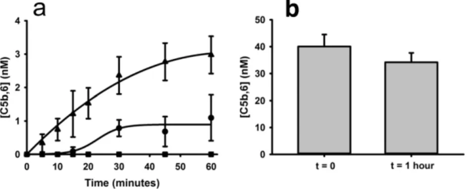
Complement factors C5 (400 nM) and C6 (500 nM) were incubated with 100 nM of plasmin or 250 nM of factor Xa or thrombin. Parallel reac-tions were quenched over time with an appropriate chloromethylketone. The amount of functional C5b,6 generated was subsequently quantified using a chicken erythrocyte hemolytic assay (Wat et al., 2014). Since plas-min readily cleaves C5, we also incubated 500 nM of C5b,6 with 100 nM of plasmin to determine if C5b,6 activity diminishes over time. In these ex-periments, plasmin was quenched with VFKck.
2.7. Ferric Chloride Model of Arterial Thrombosis
Mice were anesthetized with 1·5–2% isoflurane in oxygen. Ferric chloride injury to the carotid artery was performed as described (Aleman et al., 2013). Briefly, the right common carotid artery was ex-posed, dried, and treated with ferric chloride (7·5 or 10% on 0·5 × 1·0-mmfilter paper) for 2 min. The artery was washed with warm saline and bloodflow was continuously monitored by Doppler ul-trasonicflow probe (Indus Instruments). The time to vessel occlusion was defined as the time between FeCl3administration and lack offlow for 60 s. Blood was sampled into citrate from the IVC 5 min after stable vessel occlusion (defined as continuous occlusion for 1 min) or after 40 min if no occlusion occurred.
2.8. Thrombolysis Model
Thrombolysis was assessed in mice subjected to FeCl3carotid artery thrombosis. After 5 consecutive minutes of vessel occlusion, mice were infused with Tenecteplase (5 mg/kg, generous gift of Genentech, CA) through a saphenous vein intravenous catheter constructed of pulled PE-10 tubing (Braintree Scientific, Braintree, MA) with a 3·0-mil (0·076-mm diameter) cleaning wire (Hamilton Company, Reno NV) placed into the lumen as a stylet, as described (Machlus et al., 2011a), while continuously monitoring carotid bloodflow. Blood was sampled from the IVC 5 min after the return of bloodflow or 30 min after Tenecteplase infusion if the clot did not lyse.
2.9. Statistics
The relationships between prothrombotic and complement activa-tion markers were assessed using Spearman rank correlaactiva-tion. C3a and C5a levels were compared using t-tests or the Wilcoxon rank sum test (Tenecteplase-treatedversusuntreated mice). Pb0·05 was considered statistically significant.
3. Results
We studied the role of thrombin in complement activation using an in vivomurine model of ligation (stasis)-induced IVC thrombosis (von Bruhl et al., 2012; Aleman et al., 2013). Plasma levels of activation markers of coagulation and complement were measured by ELISA. Base-line TAT levels were 3·8 ± 4·0 ng/mL (n = 5), as previously reported (Aleman et al., 2013). 24 h after IVC ligation, plasma TAT levels rose to 21·2 ± 11 ng/mL (mean ± SD, n = 6). Prothrombin was infused in some mice to give a broader range of thrombin generation and clot weight (Aleman et al., 2013) (Fig. 1—open circles—mice infused with prothrombin; solid circles—mice infused with vehicle). When pro-thrombin was infused just prior to IVC ligation (Aleman et al., 2013), TAT levels measured at 24 h were significantly higher (47·9 ± 19 ng/mL, n = 8, pb0·009). As expected, clot weights directly correlated with TAT levels (r = 0·66, pb0·01). Plasma levels of C5a and C3a in ve-nous thrombosis were elevated as compared to baseline (unchallenged) levels (C5a = 0·43 ± 0·15 nM; C3ab0.1 nM; n = 6). Interestingly, cir-culating levels of complement activation markers C3a and C5a correlated poorly with TAT levels (Fig. 1a, b), suggesting that thrombin does not
directly activate complement in this experimental model of venous thrombosis. Notably, C5a also correlated poorly with C3a (Fig. 1c), sug-gesting that C5a was generated to a large extent via C3/C5 convertase-independent pathways. Furthermore, there was no relationship between clot weight and C3a levels (Fig. 1d). There was, however, a strong direct correlation between clot weight and C5a levels (Fig. 1e). Taken together, thesefindings show that processes triggered during venous thrombosis are associated with C5a generation, but suggest that thrombin is not the major activator of C5 under these conditions.
To determine the potential mechanisms of C5a generationin vivo, we usedin vitroassays in purified systems to compare C5a generation following cleavage of C5 by thrombin, factor Xa or plasmin. Plasmin was considered a likely candidate of complement activation in the set-ting of afibrin clot because, 1) plasmin is known to cleave C5 to yield chemotactically-active C5ain vitro(Amara et al., 2010), and 2)fibrin is an essential cofactor for tissue-type plasminogen activator (tPA)-mediated plasmin generation (Horrevoets et al., 1997).
Incubation of C3 or C5 with thrombin, factor Xa or plasmin revealed that plasmin is much more effective than thrombin or factor Xa in cleav-ing C3 and C5 to generate C3a and C5a, respectively (Fig. 2a, b). Plasmin more readily cleaved C5 than C3, with ~ 30% of C5 (~ 700 nM) being converted to C5a, and only ~2% of C3 (~450 nM) cleaved to form intact C3a. The low turnover of C3 by plasmin, thrombin and fXa precluded furtherin vitrointerrogation. The incomplete cleavage of C5 to C5a under the conditions employed may reflect competitive inhibition by an abundance of cleavage products (molecular weightN30–70 kDa) that were detected following SDS-PAGE (not shown and (Barthel et al., 2012)). We compared the efficiency of C5 cleavage by various con-centrations (0–100 nM) of each of the three enzymes (Fig. 2c). During a 10-minute incubation period, plasmin generated substantially more C5a than factor Xa or thrombin.
In kinetic assays, the rate of C5a generation by plasmin in-creased linearly as the concentration of C5 inin-creased (Fig. 2d). The catalytic efficiency, inferred from the slope of the plot, was 2·3 ± 0·6 × 104M−1s−1(Distelmaier et al., 2009). This is similar to the published rate of C5 cleavage by the bonafide alternative pathway C5 convertase and the soluble monomeric classical/lectin pathway C3/ C5 convertase (Rawal and Pangburn, 1998, 2001). This rate of C5 cleav-age by plasmin is therefore consistent with the premise that plasmin has a physiologically relevant role in generation of C5a.
Since plasmin is rarely, if ever, free in circulation, we next tested whether plasmin is capable of generating C5a in the presence of physi-ological concentrations of antiplasmin and fibrinogen, that when converted tofibrin, binds plasminogen and plasmin with high affinity. Wefirst confirmed that even at very high concentrations, thrombin generates almost undetectable amounts of C5a (Fig. 2e, conditions i, ii). In the absence of plasminogen, tPA was incapable of cleaving C5 to yield C5a (Fig. 2e, condition iii). When C5 was incubated with tPA and plasminogen in the presence offibrin(ogen) (Fig. 2e, condition v), readily detectable amounts of C5a were generated, and this occurred even in the presence of physiological concentrations of antiplasmin (condition iv). Absence offibrinogen, a cofactor for tPA-mediated conversion of plasminogen to plasmin, resulted in the generation of measurable, but less, C5a than withfibrinogen (condition vversusvi). Overall, the data confirm that in the presence of physiological concen-trations of hemostatic proteins, C5a can be generated in a plasmin-dependent manner.
concentration of factor X) generated only 1·1 ± 0·7 nM of C5b,6. Consistent with its inefficient cleavage of C5, thrombin (250 nM) did not generate any detectable C5b,6. Thus, although plasmin can cleave C5 at several sites (Barthel et al., 2012), exposure of C5b,6 to plasmin for 1 h did not appreciably decrease the functionality of the MAC (Fig. 3b).
In view of thesein vitrofindings, we tested the association between thrombin generation and complement activation in a second, indepen-dent thrombosis/thrombolysis model, and used this model to determine whether C5 is activated by thrombolytic pathwaysin vivo.Using wild-type mice with stable carotid artery thrombi induced by ferric chloride, wefirst showed that 5 min after occlusion, TAT levels correlated poorly with systemic levels of C3a and C5a (r =−0·36, p = 0·39 for C3a; r =−0·05, p = 0·91 for C5a; n = 8), consistent with our observations in the venous thrombosis model. To determine the impact of
generation of C5. We further excluded this possibility by showing in vitrothat exposure of C5 to high concentrations of tPA (up to 200 nM) for 10 and 30 min, did not yield measurable amounts of C5a (data not shown). Taken together, these data are consistent with a direct effect of plasmin on complement activation during thrombosis.
4. Discussion
Thrombus formation leading to pathological vaso-occlusive events (e.g. acute coronary syndrome, stroke, deep vein thrombosis and pul-monary embolus) is a major cause of death worldwide (Mozaffarian
Fig. 3.Hemostatic enzymes generate C5b,6. a. Cleavage of C5 by plasmin (▴), factor Xa (•), or thrombin (■) in the presence of excess C6, C7, C8 and C9, resulted in the generation of functional C5b,6, measured by a terminal pathway erythrocyte hemolytic assay. Thrombin did not generate any measurable C5b,6 in this experimental system. b. The functional integrity of C5b,6, measured by the terminal pathway hemolytic assay, did not decay appreciably over a 1-hour period when incubated with plasmin. The data presented represent means ± SD for 3 replicates.
Fig. 4.Effects of plasminogen activator-mediated thrombolysis on C3a and C5a levels. a, b. Stable carotid artery thrombosis was induced in wild-type mice using the ferric chloride model (seeMaterials and Methodssection), after which the plasminogen activator Tenecteplase was administered intravenously as noted. C3a (a) and C5a (b) levels were significantly increased by Tenecteplase infusion. Each dot represents a separate mouse (n = 9 controls, n = 8 infused with Tenecteplase).
activation through positive feedback loops (Oikonomopoulou et al., 2012). Given the currentfindings, we propose a model in which plas-min, via liberation of C5a, contributes to leukocyte trafficking during thrombus formation, propagation and/or resolution (Fig. 5). The precise local contribution of C5a (and C3a) to thrombus formation and resolu-tion, is difficult to ascertain, particularly since clearance of these pep-tides is short and likely dynamically changes in this setting. Nonetheless, with generation of C5a, terminal complement pathway complexes form which also regulate coagulation. C5b-7 induces tissue factor expression by monocytic cells (Langer et al., 2013), while the MAC induces VWF and P-selectin secretion, platelet microparticle re-lease, and endothelial cell and platelet membrane changes that favor prothrombinase assembly and thrombin generation (Hamilton et al., 1990; Wiedmer et al., 1986; Sims et al., 1988). Since C5 activation is as-sociated with many disease states, including acute lung injury, arthritis, sepsis (Huber-Lang et al., 2006; Kessel et al., 2014; Yan and Gao, 2012), and thrombosis (Distelmaier et al., 2009; Cheung et al., 1994), the pres-ent studies suggest that intervpres-entions at the level of plasmin may have broad clinical utility.
From studies with mouse models (Huber-Lang et al., 2006; Hoth et al., 2014; Khan et al., 2013; Auger et al., 2012; Zecher et al., 2014; Borkowska et al., 2014), several groups have concluded that thrombin is the major coagulation enzyme that generates C5a under pathologic conditions. This role for thrombin was supported by observations that thrombin generates C5in vitro(Huber-Lang et al., 2006), and inhibition of thrombin dampens severity of disease and reduces C5a levels in murine models of disease (Huber-Lang et al., 2006; Hoth et al., 2014; Khan et al., 2013; Auger et al., 2012; Zecher et al., 2014; Borkowska et al., 2014). How do we reconcile thesefindings with the fact that the residuesflanking the R751 C5 convertase cleavage site necessary to gen-erate C5a lack similarity to thrombin cleavage sites in all other classic thrombin substrates (e.g., protein C, PAR1,fibrinogen, factor V, factor VIII) (Krisinger et al., 2012), and that thrombin is an inefficient cutter of C5 at that site (Krisinger et al., 2012)?
That thrombin participates in C5a generation during coagulation is not challenged by the presentfindings. However, this reaction likely does not occur via direct C5 cleavage. Indeed, in our experiments, with thrombin concentrations that more closely approximate the dynamics of thrombin generation in plasma and blood (Brummel et al., 2002; Dielis et al., 2008), C5a could not be measured. Moreover, the C5T product that is generated by thrombin-mediated cleavage of C5 at the thrombin-sensitive R947 site (Krisinger et al., 2012), does not exhibit C5a-like chemotactic/migration propertiesin vitro(data not shown). The present studies are consistent with the concept that thrombin con-tributes to C5a generation, butindirectlyvia plasmin-mediated events. Thrombin is fundamentally important for the initiation offibrinolysis since it generatesfibrin, an important cofactor for tPA-mediated plas-min generation. Thrombin further amplifies plasmin generation by inducing endothelial secretion of tPA and expression of urokinase-type plasminogen activator (van den Eijnden-Schrauwen et al., 1995). Given the relative kinetics of thrombinversusplasmin in generating C5a, it is unlikely that thrombin, alone, generates C5a. Rather, our data support the premise that, in combination with C5 convertase, plasmin (and/or downstream proteolytic effectors of plasmin) is the major mediator, and that the amount of thrombin only affects C5a levels inso-far as thrombin affects the kinetics offibrinolysis/plasmin generation. The caveat to this premise is that very high thrombin concentrations that may transiently accumulate during thrombus formation ( Brummel-Ziedins et al., 2005) could cleave C5 at R751 to generate C5a (Krisinger et al., 2012). Interestingly, reports of a role for thrombin in generating C5a come from studies with mice lacking C3 (Huber-Lang et al., 2006; Khan et al., 2013; Auger et al., 2012; Borkowska et al., 2014). These mice, for unexplained reasons, have elevated levels of prothrombin (Huber-Lang et al., 2006) which can result in increased generation of thrombin following activation of coagulation (Aleman et al., 2013; Kyrle et al., 1998). It is reasonable to consider that the high thrombin levels
reached in C3-deficient mice might contribute to C5a generation. How-ever, with excess thrombin andfibrin deposition, plasmin generation would also be markedly increased, and plasmin would be significantly favored over thrombin in generating C5a from C5. Indeed, this apparent paradox of heightened plasmin generation with a larger thrombus is in line with our observation that IVC clot weights strongly correlated with C5a levels. By interfering with deposition offibrin, a critical cofactor for plasmin generation, thrombin inhibition would reduce C5a generation. Overall, thrombin is important in C5a generation, but indirectly, via enhanced production of plasmin. Thesefindings are consistent with, and indeed, extend previousin vitrostudies in which high concentrations of plasmin cleaved C5 (Amara et al., 2010). Attempts to measure murine levels of plasma plasmin–antiplasmin complexes were confounded by a lack of reliable assays. However,in vivogain-of-function studies using pharmacologic-intervention (Tenecteplase infusion), allow us to con-clude that plasmin plays a physiologically relevant role in the generation of C5a.
Although plasmin generation is increased acutely and to a lesser extent, chronically, in patients with thrombotic disorders (Wada et al., 1989), the role of plasmin in complement-mediated events during thrombus formation, propagation, and resolution is unknown. Our finding that plasmin drives C5a generation in mouse models of throm-bosis exposes plasmin and/or its downstream effectors as potential therapeutic targets for limiting production of procoagulant and pro-inflammatory effectors. These insights are likely to be applicable to thrombi in arteries and veins, in spite of the differences with regard to etiology of these presentations. Indeed, and most notably, both presen-tations involve activation of inflammatory pathways and liberation of fibrinolytic activity in response to the thrombus (Engelmann and Massberg, 2013; Wolberg et al., 2012). The impact of any intervention would likely be dependent on its timing in relation to thrombus forma-tion and resoluforma-tion (Fig. 5). Thus, early dampening of C5a and MAC assembly may thwart leukocyte accumulation and thrombus initiation and propagation. Later interventions may hinder resolution of the clot, but may also reduce long-term inflammatory sequelae of thrombosis, such as occurs in post-thrombotic syndrome. Intuitively, administering agents to suppressfibrinolysis during a thrombotic event at any stage may be unappealing, and thus an optimal intervention strategy/agent might preservefibrinolysis while restricting C5 activation. Direct blocking of C5 activation is highly efficacious in preventing complement-mediated thrombosis in atypical hemolytic uremic syndrome and paroxysmal nocturnal hemoglobinuria (Wong and Kavanagh, 2015). Similar suc-cesses were not, however, observed for acute myocardial infarction (APEX AMI Investigators et al., 2007), which might be due to the fact that therapy was initiated early at presentation. The variable responses in these reports underline the need for further study, using models that represent different vascular disorders.
Contributors
JHF, ASW and EMC designed experiments, analyzed data and wrote the manuscript and managed the project. JHF, MMA, BLW, AMO, VL and MH performed experiments. KAF analyzed data and edited the manuscript.
Declaration of Interests
We declare no competing interests.
Ethical Research Conduct
Role of the Funding Sources
None of the funders had any input into the design of the experi-ments, the collection, analysis or interpretation of the data, the writing of the manuscript, or the decision to submit it for publication.
Acknowledgments
JHF was supported by a Banting Fellowship. EMC is supported by operating grants from the Canadian Institutes of Health Research (CIHR), the Natural Sciences and Engineering Research Council of Canada (NSERC), and the Canada Foundations for Innovation (CFI). He holds a CSL Behring Research Chair and a Tier 1 Canada Research Chair in Endothelial Cell Biology and is an Adjunct Scientist with the Canadian Blood Services. ASW was supported by funding from the National Insti-tutes of Health (R01HL094740 and R56HL094740). BLW was supported by a training grant to the University of North Carolina (T32HL069768) and MMA was supported by NIH grant F31HL112608. KAF was support-ed by the Queen's University Terry Fox Foundation Training Program in Transdisciplinary Cancer Research in partnership with the CIHR. Partial funding was provided by Biogen. We thank Drs. Amit Nathwani and Jay L. Degen for critically reviewing the manuscript.
References
Aleman, M.M., Walton, B.L., Byrnes, J.R., et al., 2013.Elevated prothrombin promotes venous, but not arterial, thrombosis in mice. Arterioscler. Thromb. Vasc. Biol. 33 (8), 1829–1836.
Amara, U., Flierl, M.A., Rittirsch, D., et al., 2010.Molecular intercommunication between the complement and coagulation systems. J. Immunol. 185 (9), 5628–5636. APEX AMI Investigators, Armstrong, P.W., Granger, C.B., et al., 2007.Pexelizumab for acute
ST-elevation myocardial infarction in patients undergoing primary percutaneous cor-onary intervention: a randomized controlled trial. JAMA 297 (1), 43–51.
Auger, J.L., Haasken, S., Binstadt, B.A., 2012.Autoantibody-mediated arthritis in the absence of C3 and activating Fcgamma receptors: C5 is activated by the coagulation cascade. Arthritis Res. Ther. 14 (6), R269.
Barthel, D., Schindler, S., Zipfel, P.F., 2012.Plasminogen is a complement inhibitor. J. Biol. Chem. 287 (22), 18831–18842.
Bennett, W.R., Yawn, D.H., Migliore, P.J., et al., 1987.Activation of the complement system by recombinant tissue plasminogen activator. J. Am. Coll. Cardiol. 10 (3), 627–632. Borkowska, S., Suszynska, M., Mierzejewska, K., et al., 2014.Novel evidence that crosstalk
between the complement, coagulation, andfibrinolysis proteolytic cascades is involved in mobilization of hematopoietic stem/progenitor cells (HSPCs). Leukemia. Brummel, K.E., Paradis, S.G., Butenas, S., Mann, K.G., 2002.Thrombin functions during
tissue factor-induced blood coagulation. Blood 100 (1), 148–152.
Brummel-Ziedins, K.E., Vossen, C.Y., Butenas, S., Mann, K.G., Rosendaal, F.R., 2005. Throm-bin generation profiles in deep venous thrombosis. J. Thromb. Haemost. 3 (11), 2497–2505.
Cheung, A.K., Faezi-Jenkin, B., Leypoldt, J.K., 1994.Effect of thrombosis on complement activation and neutrophil degranulation during in vitro hemodialysis. J. Am. Soc. Nephrol. 5 (1), 110–115.
Dielis, A.W., Castoldi, E., Spronk, H.M., et al., 2008.Coagulation factors and the protein C system as determinants of thrombin generation in a normal population. J. Thromb. Haemost. 6 (1), 125–131.
Distelmaier, K., Adlbrecht, C., Jakowitsch, J., et al., 2009.Local complement activation triggers neutrophil recruitment to the site of thrombus formation in acute myocardial infarction. Thromb. Haemost. 102 (3), 564–572.
Engelmann, B., Massberg, S., 2013.Thrombosis as an intravascular effector of innate immunity. Nat. Rev. Immunol. 13 (1), 34–45.
Fuchs, T.A., Brill, A., Wagner, D.D., 2012.Neutrophil extracellular trap (NET) impact on deep vein thrombosis. Arterioscler. Thromb. Vasc. Biol. 32 (8), 1777–1783. Hamilton, K.K., Hattori, R., Esmon, C.T., Sims, P.J., 1990.Complement proteins C5b-9
induce vesiculation of the endothelial plasma membrane and expose catalytic surface for assembly of the prothrombinase enzyme complex. J. Biol. Chem. 265 (7), 3809–3814.
Horrevoets, A.J., Pannekoek, H., Nesheim, M.E., 1997.A steady-state template model that describes the kinetics offibrin-stimulated [Glu1]- and [Lys78]plasminogen activation by native tissue-type plasminogen activator and variants that lack either thefinger or kringle-2 domain. J. Biol. Chem. 272 (4), 2183–2191.
Hoth, J.J., Wells, J.D., Jones, S.E., Yoza, B.K., McCall, C.E., 2014.Complement mediates a primed inflammatory response after traumatic lung injury. J. Trauma Acute Care Surg. 76 (3), 601–608 (discussion 8-9).
Howes, J.M., Richardson, V.R., Smith, K.A., et al., 2012.Complement C3 is a novel plasma clot component with anti-fibrinolytic properties. Diab. Vasc. Dis. Res. 9 (3), 216–225. Huber-Lang, M., Sarma, J.V., Zetoune, F.S., et al., 2006.Generation of C5a in the absence of
C3: a new complement activation pathway. Nat. Med. 12 (6), 682–687.
Kessel, C., Nandakumar, K.S., Peters, F.B., Gauba, V., Schultz, P.G., Holmdahl, R., 2014.A single functional group substitution in c5a breaks B cell and T cell tolerance and protects against experimental arthritis. Arthritis Rheum. 66 (3), 610–621. Khan, M.A., Maasch, C., Vater, A., et al., 2013.Targeting complement component 5a
pro-motes vascular integrity and limits airway remodeling. Proc. Natl. Acad. Sci. U. S. A. 110 (15), 6061–6066.
Krisinger, M.J., Goebeler, V., Lu, Z., et al., 2012.Thrombin generates previously
unidenti-fied C5 products that support the terminal complement activation pathway. Blood 120 (8), 1717–1725.
Kyrle, P.A., Mannhalter, C., Beguin, S., et al., 1998.Clinical studies and thrombin generation in patients homozygous or heterozygous for the G20210A mutation in the prothrom-bin gene. Arterioscler. Thromb. Vasc. Biol. 18 (8), 1287–1291.
Langer, F., Spath, B., Fischer, C., et al., 2013.Rapid activation of monocyte tissue factor by antithymocyte globulin is dependent on complement and protein disulfide isom-erase. Blood 121 (12), 2324–2335.
Machlus, K.R., Cardenas, J.C., Church, F.C., Wolberg, A.S., 2011a.Causal relationship between hyperfibrinogenemia, thrombosis, and resistance to thrombolysis in mice. Blood 117 (18), 4953–4963.
Machlus, K.R., Lin, F.C., Wolberg, A.S., 2011b.Procoagulant activity induced by vascular injury determines contribution of elevated factor VIII to thrombosis and thrombus stability in mice. Blood 118 (14), 3960–3968.
Mozaffarian, D., Benjamin, E.J., Go, A.S., et al., 2015.Heart disease and stroke statistics– 2015 update: a report from the American Heart Association. Circulation 131 (4), e29–322.
Oikonomopoulou, K., Ricklin, D., Ward, P.A., Lambris, J.D., 2012.Interactions between coagulation and complement–their role in inflammation. Semin. Immunopathol. 34 (1), 151–165.
Pierangeli, S.S., Girardi, G., Vega-Ostertag, M., Liu, X., Espinola, R.G., Salmon, J., 2005. Requirement of activation of complement C3 and C5 for antiphospholipid antibody-mediated thrombophilia. Arthritis Rheum. 52 (7), 2120–2124.
Raskob, G.E., Angchaisuksiri, P., Blanco, A.N., et al., 2014.Thrombosis: a major contributor to global disease burden. Arterioscler. Thromb. Vasc. Biol. 34 (11), 2363–2371. Rawal, N., Pangburn, M.K., 1998.C5 convertase of the alternative pathway of complement.
Kinetic analysis of the free and surface-bound forms of the enzyme. J. Biol. Chem. 273 (27), 16828–16835.
Rawal, N., Pangburn, M., 2001.Formation of high-affinity C5 convertases of the alternative pathway of complement. J. Immunol. 166 (4), 2635–2642.
Ricklin, D., Hajishengallis, G., Yang, K., Lambris, J.D., 2010.Complement: a key system for immune surveillance and homeostasis. Nat. Immunol. 11 (9), 785–797.
Salmon, J.E., Girardi, G., Holers, V.M., 2002.Complement activation as a mediator of antiphospholipid antibody induced pregnancy loss and thrombosis. Ann. Rheum. Dis. 61 (Suppl. 2), ii46–ii50.
Sims, P.J., Faioni, E.M., Wiedmer, T., Shattil, S.J., 1988.Complement proteins C5b-9 cause release of membrane vesicles from the platelet surface that are enriched in the mem-brane receptor for coagulation factor Va and express prothrombinase activity. J. Biol. Chem. 263 (34), 18205–18212.
van den Eijnden-Schrauwen, Y., Kooistra, T., de Vries, R.E., Emeis, J.J., 1995.Studies on the acute release of tissue-type plasminogen activator from human endothelial cells in vitro and in rats in vivo: evidence for a dynamic storage pool. Blood 85 (12), 3510–3517.
von Bruhl, M.L., Stark, K., Steinhart, A., et al., 2012.Monocytes, neutrophils, and platelets cooperate to initiate and propagate venous thrombosis in mice in vivo. J. Exp. Med. 209 (4), 819–835.
Wada, K., Takahashi, H., Tatewaki, W., Takizawa, S., Shibata, A., 1989.Plasmin-a2-plasmin inhibitor complex in plasma of patients with thromboembolic diseases. Thromb. Res. 56 (6), 661–665.
Wakefield, T.W., Myers, D.D., Henke, P.K., 2008.Mechanisms of venous thrombosis and resolution. Arterioscler. Thromb. Vasc. Biol. 28 (3), 387–391.
Wat, J., Foley, J.H., Krisinger, M.J., et al., 2014.Polyphosphate suppresses complement via the terminal pathway. Blood 123 (5), 768–776.
Wiedmer, T., Esmon, C.T., Sims, P.J., 1986.Complement proteins C5b-9 stimulate procoagulant activity through platelet prothrombinase. Blood 68 (4), 875–880. Wiggins, R.C., Giclas, P.C., Henson, P.M., 1981.Chemotactic activity generated from the
fifth component of complement by plasma kallikrein of the rabbit. J. Exp. Med. 153 (6), 1391–1404.
Wolberg, A.S., Aleman, M.M., Leiderman, K., Machlus, K.R., 2012.Procoagulant activity in hemostasis and thrombosis: Virchow's triad revisited. Anesth. Analg. 114 (2), 275–285.
Wong, E.K., Kavanagh, D., 2015.Anticomplement C5 therapy with eculizumab for the treatment of paroxysmal nocturnal hemoglobinuria and atypical hemolytic uremic syndrome. Transl. Res. 165 (2), 306–320.
Yan, C., Gao, H., 2012.New insights for C5a and C5a receptors in sepsis. Front. Immunol. 3, 368.