1 POTENTIAL ROLE OF NEUTROPHIL GELATINASE-ASSOCIATED LIPOCALIN (NGAL) AS A MEDIATOR OF LIVER INJURY AND FIBROGENESIS IN ALCOHOLIC HEPATITIS
Palak K. Patel
Department of Nutrition
University of North Carolina at Chapel Hill 2014
2 ACKNOWLEDGEMENTS
3 TABLE OF CONTENTS
CHAPTER
4 I. ABSTRACT
Author: Palak K. Patel1
Advisors: Gemma Odena1,2, Ramon Bataller1,2
Affiliations: University of North Carolina at Chapel Hill Department of Nutrition,1 University of North Carolina at Chapel Hill Department of Medicine2
Background: Alcoholic hepatitis (AH) is a severe form of alcoholic liver disease (ALD)
associated with significant short-term mortality. Existing treatment therapies are not completely effective, highlighting the need to identify molecular drivers in AH. NGAL has recently been identified as one of the most overexpressed genes in patients with AH.
Aims: To investigate the role of NGAL as a potential target for therapy in AH using a
translational approach including human samples and animal models.
Methods: NGAL expression was analyzed in patients with AH, animal models of liver
inflammation and fibrogenesis, and cell lines of human hepatocytes. Fibrosis- and inflammation-marker genes were analyzed for expression in hepatic stellate cell (HSC) cell lines treated with NGAL. RNA was extracted from all samples and gene expression of cDNA was measured using real-time qPCR analysis.
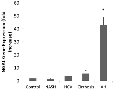
Results: Gene expression of NGAL in patients with AH had a 44-fold increase in comparison to
5 Conclusion: NGAL is overexpressed in patients with AH and in animal models of liver
6 II. INTRODUCTION
Alcohol-related illness and death has become increasingly prevalent worldwide. ALD progression includes steatosis, AH, fibrosis, and cirrhosis.1 Steatosis, or fatty liver, develops in over 95% of heavy drinkers1 and is marked by small- and large-droplet fat beginning in
perivenular regions.2 Persistent alcohol consumption leads to fibrosis in 20-40% of patients with steatosis, characterized by excessive accumulation of collagen and other extracellular matrix proteins.1 In 8-20% of patients who have developed fibrosis, further scar tissue formation through cirrhosis can occur as a result of continued alcohol consumption.1 AH is an acute inflammatory syndrome3 whose features include hepatocyte ballooning and neutrophil-rich parenchymal inflammation.2 AH ranges in severity from mild forms that resolve with
conservative management to severe presentations that are associated with mortality as high as 65%.3 Although there are several therapeutic measures currently available to improve survival in patients with AH, the overall quality of treatments is lacking.4 The use of glucocorticoids, the most commonly administered treatment for severe AH, is still controversial and there is a
significant group of patients who have glucocorticoid-resistant disease.1 The development of new therapeutic approaches in AH has been difficult due to the fact that available animal models do not reproduce all of the key histological features found in humans.5 Thus, it is imperative that molecular drivers of AH be identified as new targets for therapy.
A preliminary transcriptome analysis performed by our lab group indicated that NGAL expression had a 44-fold increase in patients with AH when compared to other genes. NGAL is an inflammatory protein belonging to the lipocalin superfamily released by activated
7 most promising biomarker of acute kidney injury (AKI) through its release by damaged
parenchymal cells,8 but its role in AH is largely unknown.
Alcohol intake affects inflammation in the liver through induction by LPS. LPS is a key cell wall component of Gram-negative bacteria and it mimics bacterial infection by causing an acute inflammatory response.9 Kupffer cells are macrophages in the liver that, when exposed to LPS, activate and produce pro-inflammatory cytokines.10 This study analyzed the association between LPS induction and NGAL expression in several animal models and human cell lines.
8 III. METHODS
AH Patient Samples
Patients admitted to the Liver Unit, Hospital Clínic of Barcelona from 2007 to 2010 with
clinical, analytical and histological features of AH were prospectively included in our
sample.13,14,15 Inclusion criteria have been defined in a previous study.16 All patients had a
histological diagnosis of AH (n = 34) and patients with hepatocellular carcinoma or any other
potential liver disease were excluded from the study. Liver biopsies were obtained using a
transjugular approach. For controls, fragments of normal liver tissue (n = 6) were obtained from
optimal cadaveric liver donors (n = 3) or by resection of liver metastases (n = 3) as described in
detail in a previous study.16 Liver specimens were analyzed by a pathologist. Part of the biopsy
was submerged into a RNA stabilization solution (RNAlater, Ambion, Austin, Texas, USA). All
patients gave informed consent and the protocol was approved by the Ethics Committee of the
Hospital Clínic of Barcelona.
Mouse Models
In the fibrosis model, hepatic fibrosis was induced in male BALB/c mice (Bar Harbor, ME,
USA) following administration of carbon tetrachloride (CCL4) (Sigma-Aldrich, St Louis, MO,
USA) injected intraperitoneally at a dose of 0.5 mL/kg, 12.5% diluted in corn oil, once a week
for four weeks. Control mice were given corn oil at the same dose. Mice were sacrificed 48
hours after administration of the final injection.
In the fibrosis-plus-alcohol model, hepatic fibrosis was induced in male C57BL/6J mice (Bar
Harbor, ME, USA) following the administration of CCL4 (Sigma-Aldrich) injected
intraperitoneally at a dose of 0.5 mL/kg, 12.5% diluted in olive oil, once a week for six weeks.
9
a daily gavage of ethanol in the amount of 4g/kg was administered for six days to the
fibrosis-plus-ethanol group and the ethanol-only control group. The other control group did not receive
the ethanol gavage. Mice were sacrificed following completion of the six day gavage treatment.
In the endotoxin-induced inflammation model, LPS (Sigma-Aldrich) was administered to male
BALB/c mice via an intraperitoneal injection in the amount of 10 mg/kg four hours before
sacrifice. At the time of sacrifice, mice were ten weeks old. Control mice were not given the LPS
injection.
Liver and blood samples were collected. Each group included at least three mice. Mice were kept
on a 12-hour light/dark cycle and housed in temperature- and humidity-controlled rooms. All
experimental procedures were reviewed and approved by the University of North Carolina at
Chapel Hill Institutional Animal Care and Use Committee.
Rat Models
10 and were in accordance with National Institute of Health regulations for the care and use of animals in research.
Cell Cultures
Cells from HepG2 (Sigma-Aldrich), a human hepatocyte cell line,17 were serum-starved for 24 hours before incubation with LPS 1µg/mL (Sigma-Aldrich) for 20 hours.
Cells from LX-2 (Scott L. Friedman, Mount Sinai Hospital, New York, NY), a human HSC cell line,18 were serum-starved for 24 hours before incubation with NGAL 100 ng/mL (Novus Biologicals, Littleton, CO) for 20 hours.
FBS 20%/mL (Sigma-Aldrich) was used as a control for HepG2 and LX-2 cells. RNA Isolation and qPCR Analysis
RNA was isolated from liver tissues, animal models, and human cells using Trizol (Invitrogen, Carlsbad, CA, USA), following the manufacturer’s protocol. A high-capacity complementary DNA (cDNA) reverse-transcription kit (Applied Biosystems, Foster City, CA, USA) was used to retro-transcribe 2,000 ng of RNA. cDNA was then amplified using Taqman Technology
(Applied Biosystems), with a final PCR volume of 10 µL using a StepOnePlusTM Real-Time PCR System (Applied Biosystems). Various assay-on-demand probes and primers (Applied
Biosystems) were used. For all samples, primer-probe pairs for NGAL were utilized.
Additionally, MMP2, ACTA2, ICAM1, and MCP1 were used for LX-2 samples. Results were
normalized to 18s rRNA expression for mouse and human samples, and 45s rRNA expression
for rat samples. Gene expression was calculated based on the ΔΔCt method. Results are
11 Statistical Analysis
Results of quantitative variables are expressed as the mean plus standard error unless otherwise
specified. Differences between groups were analyzed using non-parametric tests (Mann-Whitney
U test) for continuous variables. Statistical analysis was performed using SPSS version 22 for
Mac (SPSS Inc., Chicago, IL, USA).
IV. RESULTS
NGAL Overexpression in Patients with AH
To confirm preliminary analyses done by our lab group that indicated NGAL was one of the most up-regulated genes in patients with AH, we analyzed hepatic NGAL expression in a cohort of patients with AH using real-time qPCR analysis. Samples were obtained using a transjugular biopsy. Our results confirmed significant NGAL overexpression in patients with AH compared to controls and patients with several other liver diseases. Patients with AH had a 44-fold increase in expression when compared to these groups (Figure 1).
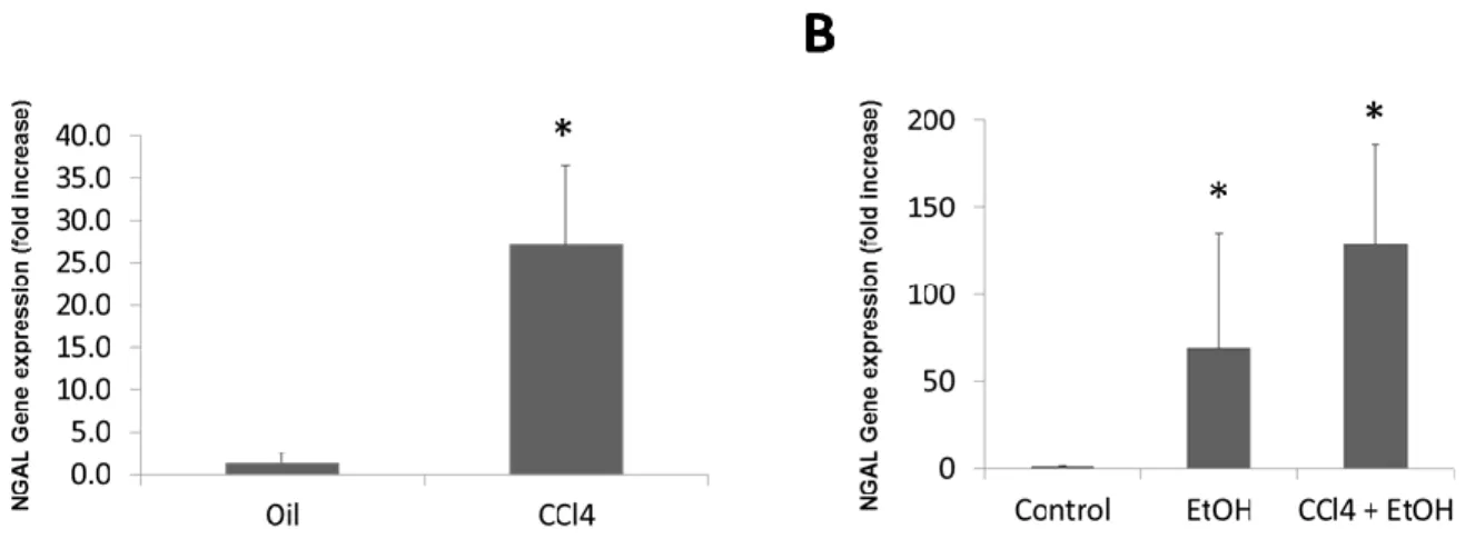
12 NGAL Overexpression in Animal Models of Fibrosis
Although there are no fully-developed animal models that precisely characterize the pathology seen in patients with AH,5 we used CCL4-induced hepatic fibrosis in mice along with an alcohol model to assess NGAL expression. NGAL expression was significantly up-regulated with CCL4 induction in the fibrosis model when compared to controls that were only given corn oil (Figure 2A). Similarly, expression of NGAL was significantly greater in fibrosis-plus-alcohol and alcohol-only mice, which both received a daily gavage of ethanol for six days, in comparison to controls (Figure 2B).
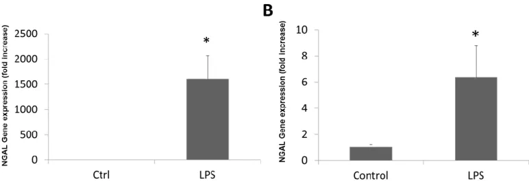
13 NGAL Overexpression in Animal Models of Inflammation
Liver inflammation is also a hallmark of AH.19 LPS induction was analyzed for NGAL expression in mouse and rat samples. NGAL expression was significantly greater in
LPS-induced mice than in controls (Figure 3A). These results were confirmed in the liver slices of rats induced with LPS (Figure 3B).
Figure 3. NGAL gene expression in animal models of inflammation. (A). Hepatic NGAL expression in mice treated with LPS. (B) Hepatic NGAL expression in liver slices of rats treated with LPS. (*p<0.05 compared to controls)
The Role of Hepatocytes in NGAL Expression and Inflammation
Because hepatocytes are primarily responsible for regeneration of the liver following inflammation-induced injury,20 the hepatocyte cell line HepG2 was analyzed for NGAL
14 Figure 4. NGAL gene expression in HepG2 cells. (A) NGAL expression in HepG2 and LX-2 cells (*p<0.05 compared to LX-2). (B) NGAL expression in LPS-induced HepG2 cells.
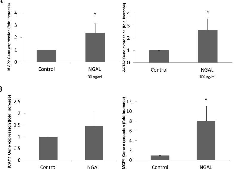
Expression of Fibrosis and Inflammation Markers in LX-2 Cells
As major responders to liver injury, HSCs play a central role in mediating fibrosis and inflammation.11 Accordingly, we investigated the expression of several fibrotic and pro-inflammatory markers in LX-2 cells treated with NGAL to assess NGAL’s role in the regulation of these genes. MMP2 and ACTA2, genes that are markers of liver fibrosis, had significantly greater expression in LX-2 cells treated with NGAL (Figure 5A). For the markers of
15
16 V. DISCUSSION
AH is a severe form of ALD characterized by hepatic fibrosis and inflammation, along with overall damage to the liver.3 Because existing treatments have not significantly improved mortality rates in patients with AH over the past few decades,1 it is essential that new, effective targets for therapy be identified. In the present study, we investigated the role of NGAL in patients with AH, animal models of liver fibrosis and inflammation, and human liver cell lines in order to determine whether NGAL may be a novel target for therapy. NGAL overexpression was confirmed in patients with AH. Furthermore, NGAL expression was up-regulated in animal models of both fibrosis and inflammation, indicating that NGAL may play a role in regulating the development of these injuries with respect to the liver. Increased expression of several markers of fibrosis and inflammation in LX-2 cells treated with NGAL suggests that HSCs may play a role in NGAL’s regulation. Because there was no observed association between induction of HepG2 cells with LPS and NGAL expression, it is possible that hepatocytes are not involved in the regulation of NGAL during the post-injury phase.
Several limitations were present in our study. Becausealcohol metabolism in rodents differs from humans and rodents are averse to the consumption of alcohol, accurate animal models reflecting the disease seen in human patients have not been fully developed.5 As a result, the models used in our study (consisting of CCL4, ethanol, and LPS) likely do not represent AH exclusively, but rather ALD in general. Furthermore, due to time and resource constraints, we were not able to extend our analyses to human primary cells which would have provided a more precise overview of NGAL’s role in AH.
17 Primary HSCs treated with NGAL should also be analyzed for other markers of fibrosis and inflammation in order to expand our results on NGAL’s role in the regulation of these genes during liver injury. Several other targets for treatment of AH have been studied as well. Namely, many studies have identified T cells as playing an important future role in AH treatment due to their function in the recruitment of neutrophils and the perpetuation of inflammation,21
suggesting that NGAL’s role in association with T cells may also be of forthcoming interest.
VI. CONCLUSION
18 VII. REFERENCES
1. Gao, B., & Bataller, R. (2011). Alcoholic Liver Disease: Pathogenesis and New
Therapeutic Targets. Gastroenterology,141(5), 1572-1585.
2. Theise, N. (2013). Histopathology of Alcoholic Liver Disease. Clinical Liver
Disease,2(2), 64-67.
3. Drinane, M., & Shah, V. (2013). Alcoholic Hepatitis: Diagnosis and Prognosis. Clinical
Liver Disease,2(2), 80-83.
4. Kim, W., & Kim, D. (2014). Severe alcoholic hepatitis-current concepts, diagnosis and
treatment options. World J Hepatol,6(10), 688–695-688–695.
5. Affo, S., Morales-Ibanez, O., Rodrigo-Torres, D, et al. (2014). CCL20 mediates
lipopolysaccharide induced liver injury and is a potential driver of inflammation and
fibrosis in alcoholic hepatitis. Gut,63(11), 1782-1792.
6. Bolignano, D., Donato, V., Coppolino, G, et al. (2008). Neutrophil Gelatinase–
Associated Lipocalin (NGAL) as a Marker of Kidney Damage. American Journal of
Kidney Diseases,52(3), 595-605.
7. Soler-Rodriguez, A., Zhang, H., Lichenstein, H, et al. (2000). Neutrophil Activation by
Bacterial Lipoprotein Versus Lipopolysaccharide: Differential Requirements for Serum
and CD14. The Journal of Immunology,164(5), 2674-2683.
8. Martensson, J., & Bellomo, R. (2014). The Rise and Fall of NGAL in Acute Kidney
Injury. Blood Purification,37(4), 304-310.
9. Wang, H., Zakhari, S., & Jung, M. (2010). Alcohol, inflammation, and gut-liver-brain
interactions in tissue damage and disease development. World J Gastroenterol,16(11),
1304-1313.
10. Su, G., Goyert, S., & Fan, M. (2002). Activation of human and mouse Kupffer cells by
lipopolysaccharide is mediated by CD14. American Journal of Physiology -
Gastrointestinal and Liver Physiology,283(3), G640-G645.
11. Yin, C., Evason, K., & Asahina, K. (2013). Hepatic stellate cells in liver development,
regeneration, and cancer. J Clin Invest,123(5), 1902-1910.
12. Rocha, S., De Franca, M., Rodrigues, G, et al. (2014). Diethylcarbamazine Reduces
Chronic Inflammation and Fibrosis in Carbon Tetrachloride- (CCl4-) Induced Liver
19
13. Dominguez, M., Rincon, D., Abraldes, J, et al. (2008). A new scoring system for
prognostic stratification of patients with alcoholic hepatitis. Am J Gastroenterol,103(11),
2747-2756.
14. Colmenero, J., Bataller, R., Sancho-Bru, P, et al. (2007). Hepatic Expression of
Candidate Genes in Patients With Alcoholic Hepatitis: Correlation With Disease
Severity. Gastroenterology,132(2), 687–697.
15. Dominguez, M., Miguel, R., Colmenero, J, et al. (2009). Hepatic Expression of CXC
Chemokines Predicts Portal Hypertension and Survival in Patients With Alcoholic
Hepatitis.Gastroenterology,136(5), 1639-1650.
16. Affo, S., Dominguez, M., Lozano, J, et al. (2012). Transcriptome analysis identifies TNF
superfamily receptors as potential therapeutic targets in alcoholic hepatitis. Gut,62(3),
452-460.
17. Bokhari, M., Carnachan, R., Cameron, N, et al. (2007). Culture of HepG2 liver cells on
three dimensional polystyrene scaffolds enhances cell structure and function during
toxicological challenge. J Anat,211(4), 567-576.
18. Xu, L., Hui, A., Arthur, M, et al. (2005). Human hepatic stellate cell lines, 1 and
LX-2: New tools for analysis of hepatic fibrosis. Gut,54(1), 142-151.
19. Sandahl, T., Gronbaek, H., Moller, H, et al. (2014). Hepatic macrophage activation and
the LPS pathway in patients with alcoholic hepatitis: A prospective cohort study. Am J
Gastroenterol,109(11), 1749-1756.
20. Fausto, N., & Campbell, J. (2003). The role of hepatocytes and oval cells in liver
regeneration and repopulation. Mechanisms of Development,120(1), 117-130.
21. Dhanda, A., Lee, R., Collins, P., & McCune, C. (2012). Molecular targets in the