Copyright © 1999, American Society for Microbiology. All Rights Reserved.
Characterization of Antimicrobial Resistance in
Streptococcus pyogenes
Isolates from the San Francisco Bay Area of Northern California
MARY K. YORK,1* LAUREL GIBBS,1FRANCOISE PERDREAU-REMINGTON,2
ANDG. F. BROOKS1
Department of Laboratory Medicine1and Department of Medicine, Infectious Diseases Division,2
University of California, San Francisco, California
Received 21 August 1998/Returned for modification 17 October 1998/Accepted 23 February 1999
During 1994 and 1995, 157 isolates of Streptococcus pyogenes from patients with invasive disease were consecutively collected in the San Francisco Bay area to determine the frequency of antimicrobial resistance. Susceptibility testing was performed according to the guidelines of the National Committee for Clinical Laboratory Standards by the disk method and by broth microdilution. For comparison of susceptibility patterns, an additional 149 strains were randomly collected from patients with pharyngitis. For San Francisco County, 32% of the isolates from invasive-disease-related specimens but only 9% of the isolates from throat cultures from the same period were resistant to erythromycin (P50.0007). Alameda County and Contra Costa County had rates of resistance of<10% from isolates from all cultures. When the data were analyzed by
hospital, the San Francisco County Hospital had a statistically higher rate of erythromycin resistance (39%) among the strains from serious infections compared to those from other counties (P 5 <0.0003). For tetracycline, high rates of resistance were observed in San Francisco County for both isolates from patients with invasive disease (34%) and pharyngitis (21%) in the same period. Using pulsed-field gel electrophoresis, two clones, one at the San Francisco County Hospital and a second in the entire area, were identified. The latter clone exhibited resistance to bacitracin. Of 146 strains that were tested by microdilution, all were susceptible to penicillin. Clindamycin resistance was not seen among the erythromycin-susceptible strains, but two of the 39 erythromycin-resistant strains were also resistant to clindamycin. An additional 34 strains showed resis-tance to clindamycin when exposed to an erythromycin disk in the double-disk diffusion test, suggesting that the mechanism of erythromycin resistance is due to anerm gene. This study demonstrates a high rate of resistance to macrolides and tetracycline amongS. pyogenesisolates in San Francisco County and shows that macrolide resistance is more common in strains from patients with invasive disease than in strains from those with pharyngitis.
Antimicrobial resistance among group A beta-hemolytic streptococci (GABHS) is an emerging concern. Although pen-icillin is the first choice for treatment of pharyngeal and most other infections with this organism, erythromycin or one of the newer macrolides is the second-line drug of choice and is used in penicillin-hypersensitive patients (8, 9). Addition of clinda-mycin to the therapy of serious streptococcal infections is rec-ommended to inhibit protein synthesis and, therefore, toxin production by the organism. Clindamycin is also the drug of choice for chronic, recurrent pharyngitis.
Erythromycin resistance ranges from as low as 1.3 to 5% at endemic levels (11) to.45% during outbreaks in Finland (24), Sweden (9), and Japan (7). High rates of resistance have also been reported sporadically in Australia (17%) (28), the United Kingdom (22.8%) (20), Taiwan (rate not specified) (10), and Italy (40%) (5). Documentation shows that changes in the prescribing patterns of physicians to reduce macrolide antibi-otic use have often resulted in a decrease in resistance (5, 23). Outbreaks of resistant GABHS above 5% have almost never been reported in the United States (8, 11, 21), and those reports may have been flawed if disk testing methodology was used (3). In 1993, Rathore and Jenkins in Jacksonville, Fla., reported that 2mg of erythromycin/ml was needed to inhibit
growth of 99% of isolates by agar dilution (21). They con-cluded that while most isolates in their area remain susceptible to erythromycin, many no longer were fully susceptible, with MICs of#0.5mg/ml.
Resistance to tetracycline has been reported to be high, making it virtually unusable as an alternative for the treatment of GABHS infections (8). All studies agree that there is a need for surveillance of antimicrobial susceptibility to detect emerg-ing local patterns, since resistance varies widely in different parts of the world, as well as in different areas of the same country.
Because the level of erythromycin resistance in San Fran-cisco has been persistently elevated, this study was undertaken to systematically survey resistance to erythromycin, tetracy-cline, and clindamycin among GABHS in the San Francisco Bay area of Northern California. To our knowledge, this work is the first to document high-level macrolide resistance in the United States. The results were confirmed by the current Na-tional Committee for Clinical Laboratory Standards (NCCLS) microdilution test method with lysed horse blood supplemen-tation (14, 19).
(A preliminary report of this work was presented previously [30].)
MATERIALS AND METHODS
Strains.A total of 306 GABHS isolates were studied. The 157 isolates from patients with invasive infections were received sequentially between May 1994 and November 1995 from 32 institutions representing San Francisco Bay area counties (San Francisco, 50%; Alameda, 33%; Contra Costa, 15%; other or
* Corresponding author. Mailing address: Department of Labora-tory Medicine, L 515, Box 0100, University of California, San Fran-cisco, CA 94143. Phone: (415) 476-3233. Fax: (415) 502-0929. E-mail: york@labmed.ucsf.edu.
unknown counties 1%) as part of the Centers for Disease Control and Preven-tion Active Surveillance Program (1). San Francisco County is separated from Alameda and Contra Costa counties by San Francisco Bay. Of the strains from patients with invasive disease, 118 were isolated from blood, 20 from joint specimens, 1 each from peritoneal fluid and pericardial fluid, and 2 each from lymph node and pleural fluid. The remaining 13 isolates were from wounds and tissues associated with necrotizing fasciitis or toxic shock.
Seven hospitals plus an Alameda County central laboratory that receives specimens from several counties in the area randomly collected 149 respiratory site isolates (San Francisco, 78%; Alameda, 7%; Contra Costa, 5%; and central laboratory, 10%) from November 1994 to November 1995 for comparison of the strains with those from the surveillance program. Statistical analysis was per-formed by the one-sided test on the difference between proportions.
Identification to species level.The isolates were identified as group A strep-tococci by the submitting institution. All identifications were confirmed upon receipt asStreptococcus pyogenesby colony morphology, catalase reaction, and beta-hemolysis. All isolates were pyrrolidonyl arylamidase (PYR) positive. The first 200 strains were also tested on the BactiCard Strep (Remel, Lenexa, Kans.) and were leucine aminopeptidase positive and esculin negative.
Erythromycin and tetracycline disk susceptibility testing.Initially, disk sus-ceptibility testing was performed according to the guidelines in NCCLS docu-ment M2-A5 (15). Mueller-Hinton sheep blood agar (PML, Tualatin, Oreg.) was inoculated with a suspension of each organism equivalent to a 0.5 MacFarland turbidity standard. Disks with 15mg of erythromycin or 30mg of tetracycline (BBL, Cockeysville, Md.) were dispensed on the agar, and zone diameters were measured after 20 to 24 h of incubation in ambient air at 35°C. Breakpoints were determined according to NCCLS standard M100-S5 (17), using 14 to 22 mm for the “intermediate” range.Staphylococcus aureusATCC 25923 was used as a control. For strains isolated in 1995, the tests were performed according to the NCCLS standards found in M100-S6 (18), which are identical to those of the previous standard, except that incubation is in 5% CO2, breakpoints are revised,
with a range of 16 to 20 mm for the intermediate category, andStreptococcus pneumoniaeATCC 49619 is used as a control. Bacitracin susceptibility was measured by the same method, with bacteria producing no zone being consid-ered resistant. The disks contained 0.04 U of bacitracin (6).
Erythromycin and clindamycin double-disk susceptibility testing.To test the effect of erythromycin on the expression of clindamycin resistance, a disk con-taining 15 mg of erythromycin was placed 20 mm from the center of a disk containing 2 mg of clindamycin. Inhibition of the circular zone around the clindamycin disk was considered positive for inducible resistance (25).
Dilution susceptibility test.MICs were determined by the NCCLS microdilu-tion method (14, 18, 19). Antimicrobial agents were diluted in camicrodilu-tion-adjusted Mueller-Hinton broth containing 2% lysed horse blood, 100ml of each solution was dispensed into each well of microdilution plates, and the plates were kept frozen at270°C until use. Inoculations were performed with a Dynatech 2000 inoculator to a final concentration of 33105to 73105CFU/ml, and the
inoculated trays were incubated in ambient air at 35°C for 20 to 24 h and read visually by transmitted light with a reflective viewing device.S. pneumoniae ATCC 49619 was used as a control organism. A control plate was inoculated from each growth control well with a calibrated loop to confirm the purity of the culture and the initial inoculum density. MICs were interpreted by using the NCCLS breakpoints for streptococci (19).
PFGE.Preparation of chromosomal DNA for pulsed-field gel electrophoresis (PFGE) was performed as outlined by Bert et al. (2). PFGE was performed as described by Maslow et al. (12). Restriction fragments of chromosomal DNA after digestion with the enzymesSmaI andApaI (Boehringer Mannheim,
Indi-anapolis, Ind.) were analyzed as described by Tenover et al. (29). Isolates were considered identical if they had exactly the same electrophoretic pattern, and they were considered to be clonally related if they showed differences of three or fewer bands between strains (29).
RESULTS
Of the 157 strains from patients with invasive disease, 25 were resistant to erythromycin. The frequency was 27% for San Francisco County, 6% for Alameda County, and 4% for Con-tra Costa County (Table 1). MICs for erythromycin-resistant isolates ranged from 2 to.8mg/ml. No MIC results with an interpretation in the intermediate category (0.5 mg/ml) were detected. An unusually high rate of resistance (39%) was seen at one hospital, the San Francisco County Hospital, compared to those of the counties (P5 ,0.0003).
Because of the high rate of erythromycin resistance in iso-lates from patients with invasive disease, 149 strains of GABHS were randomly collected from patients with pharyn-gitis from several hospitals in the three counties between No-vember 1994 and NoNo-vember 1995. Results of erythromycin testing of these specimens were compared to results for all the strains from patients with invasive disease submitted during the same period (Table 1). For San Francisco County, 32% of the invasive-disease-related specimens, but only 9% of the isolates from patients with pharyngitis from the same period, were resistant to erythromycin (P50.0007). For the San Fran-cisco County Hospital, 50% of 26 strains from patients with invasive disease were erythromycin resistant, compared to 13% of 39 strains from patients with pharyngitis. The San Francisco County Hospital had statistically more resistant strains from pharyngitis compared to those from Alameda County (P 5 0.0004).
Tetracycline resistance was found in 31 of the 39 erythro-mycin-resistant strains. In addition, resistance was found in 24 erythromycin-susceptible strains. The rate of tetracycline resis-tance was 25% in San Francisco County, with no significant difference between strains from patients with invasive infec-tions and those from throat cultures. Tetracycline resistance was uncommon (,10%) in isolates from the other counties.
Broth microdilution susceptibility testing was performed on all of the strains that were classified as resistant or intermedi-ate to erythromycin by disk test plus approximintermedi-ately 40% of the strains that were classified as susceptible to erythromycin. All 146 strains tested were susceptible to penicillin, with MICs of
[image:2.612.49.553.83.234.2]#0.015mg/ml. There was no resistance to clindamycin among
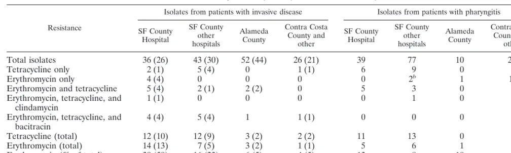
TABLE 1. Resistance patterns by county for San Francisco Bay areaa
Resistance
Isolates from patients with invasive disease Isolates from patients with pharyngitis
SF County Hospital
SF County other hospitals
Alameda County
Contra Costa County and
other
SF County Hospital
SF County other hospitals
Alameda County
Contra Costa County and
other
Total isolates 36 (26) 43 (30) 52 (44) 26 (21) 39 77 10 23
Tetracycline only 2 (1) 5 (4) 0 1 (1) 6 9 0 1
Erythromycin only 4 (4) 0 0 0 0 2b 1 1b
Erythromycin and tetracycline 5 (4) 2 (1) 2 (2) 0 5 3 0 1
Erythromycin, tetracycline, and
clindamycin 1 (1) 0 0 0 0 1 0 0
Erythromycin, tetracycline, and
bacitracin 4 (4) 5 (4) 1 1 (1) 0 0 0 0
Tetracycline (total) 12 (10) 12 (9) 3 (2) 2 (2) 11 13 0 2
Erythromycin (total) 14 (13) 7 (5) 3 (2) 1 (1) 5 6 1 2
Erythromycin (% of total) 39 (50) 16 (23) 6 (5) 4 (5) 13 8 10 9
the erythromycin-susceptible strains. Of the erythromycin-re-sistant strains, only two strains (one from a blood culture and one from a throat culture) were resistant to clindamycin by both disk and microdilution methods, with MICs of.1mg/ml. Of the 37 remaining erythromycin-resistant strains, all except three strains isolated from throat cultures demonstrated inhi-bition of the zone of susceptibility to clindamycin in the pres-ence of an erythromycin disk. Eleven strains isolated from invasive specimens were resistant to erythromycin, tetracy-cline, and bacitracin. Bacitracin resistance was not seen among the throat isolates.
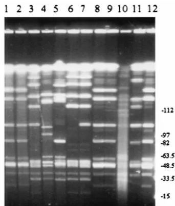
In an effort to determine if there was a clonal strain resistant to erythromycin within San Francisco County, PFGE was per-formed on the 12 strains collected between May 1994 and March 1995. The results identified two clones (five strains with pattern A and three strains with pattern B) by restriction with eitherApaI orSmaI (Fig. 1 and Table 2). All strains of clone
A had identical restriction patterns with both enzymes. One isolate of clone A was from the San Francisco County Hospi-tal; three strains of clone A were collected from patients at three different hospitals in San Francisco within a 5-week pe-riod. All five strains of clone A were resistant to bacitracin. Clone B was isolated only at the San Francisco County Hos-pital. Strain SF1612 differed from the other strains of clone B by one restriction fragment.
DISCUSSION
No resistance to penicillin was seen in 146 GABHS isolates that were studied, reinforcing the fact that penicillin resistance has not developed in this organism, despite the presence of resistance in other species of streptococci. High levels of eryth-romycin and tetracycline resistance were found by surveillance of GABHS in the San Francisco Bay Area. San Francisco County contributed 82% of the erythromycin-resistant strains and 87% of the tetracycline-resistant strains. Resistance has been prevalent at the San Francisco County Hospital for at least 20 years and continues to be problematic, with a docu-mented rate of resistance in 1998 of 25% (personal communi-cation). The cause of this resistance has not been determined. Macrolide resistance usually results from overuse of these drugs for treatment of pharyngitis (5, 23), but such usage has not been identified in San Francisco. The higher number of macrolide-resistant strains among the strains from patients with serious infections suggests that erythromycin resistance has an association with strains that are more invasive. This association could be a result of the unsuccessful treatment of infections with macrolide antibiotics, resulting in more invasive infections. Bacitracin resistance was also associated with the more invasive strains. Possibly resistance to these agents is merely a marker for a clone with pathogenic factors.
By PFGE, two distinct clones were identified among the erythromycin-resistant strains from patients with invasive dis-ease. Initially, an attempt was made to separate clones by M and T typing (1); however, these methods do not have the discriminatory power of PFGE (27). The PFGE results were reinforced by the observation that all 5 strains of clone A, and none of the other erythromycin-resistant strains, were resistant to bacitracin. Historically, susceptibility to bacitracin has been used to identify GABHS and to separate them from group B streptococci. Because the test lacks specificity, most laborato-FIG. 1. PFGE of 12 erythromycin-resistant strains digested withSmaI. Lanes
[image:3.612.79.250.73.275.2]1 to 12 contain strains SF1818, SF3413, SF1608, SF1544, SF1611, SF1612, SF1613, SF1810, SF1615, SF1618, SF1617, and SF1923, respectively; a molecular weight ladder was in well 13. The PFGE patterns (from left to right) are desig-nated A, A, B, C, D, B2, B, A, A, E, F, and A, respectively.
TABLE 2. Characterization of 12 erythromycin-resistant strains ofS. pyogenesfrom patients with invasive infections
Strain Hospital of Originc Collection date
(mo/day/yr) Bacitracind
PFGE pattern
ApaI SmaI
SF1818 Hospital A (SF) 5/31/94 R A A
SF3413 Hospital B (Alameda) 8/11/94 R A A
SF1923 Hospital C (SF) 3/31/95 R A A
SF1810 Hospital A (SF) 2/21/95 R A A
SF1615 SF County Hospital 2/21/95 R A A
SF1608 SF County Hospital 9/27/94 S B B
SF1612 SF County Hospital 11/22/94 S B2 B2
SF1613 SF County Hospital 1/7/95 S B B
SF1544 Hospital D (SF) 10/20/94 S C C
SF1611a SF County Hospital 11/17/94 S D D
SF1618 SF County Hospital 3/22/95 S E E
SF1617b SF County Hospital 3/10/95 S F F
aStrain is constitutively resistant to clindamycin; all other strains were inducibly resistant to clindamycin. bOnly strain susceptible to tetracycline.
[image:3.612.52.552.551.695.2]ries currently use the presence of PYR as a better test to identify GABHS (6). Our results confirm the importance of using an alternative to bacitracin susceptibility testing.
Erythromycin resistance in GABHS has been reported to be of three types. The most common is a target site modification, which involves dimethylation of adenine in 23S rRNA. This leads to reduced binding of macrolide, lincosamide, and strep-togramin B antibiotics to their shared 50S rRNA target site (called the MLS phenotype). At least eight classes of theseerm
(erythromycin resistance methylase) genes have been identi-fied in various species, including staphylococci (22), strepto-cocci (4), andS. pneumoniae(26). The phenotype for this type of resistance is demonstrated by the double-disk diffusion test (25), in which resistance to clindamycin is demonstrated after induction with erythromycin. Most (34 of 39) of the erythro-mycin-resistant strains in this study demonstrated inducible clindamycin resistance, suggesting that the mechanism of re-sistance is anermgene.
Uncommonly, another type of macrolide resistance is seen when the erm gene mutates and is constitutively expressed. Then the isolate demonstrates in vitro resistance to clindamy-cin without induction. Constitutive resistance to clindamyclindamy-cin was seen in two strains in this study, one from a throat culture and one from a blood culture (SF1611).
Three strains from throat cultures did not show inducible or constitutive resistance to clindamycin and probably had a dif-ferent type of erythromycin resistance. Other known mecha-nisms of resistance involve an active efflux mechanism (msr
genes) in staphylococci, an erythromycin esterase (eregenes), an undefined mechanism inS. pneumoniae(26), and an ener-gy-dependent efflux pump inS. pyogenescalledmefA(4).msr
resistance confers inducible coresistance to macrolides and type B streptogramins but not to lincosamides, such as clinda-mycin.mefAconfers resistance only to the macrolides but not to 16-membered macrolides, lincosamides, or streptogramin B. It is possible that three of our strains carrymefA, since this gene has been found in GABHS. Alternatively, an uncharac-terized mechanism of resistance that does not confer resistance to clindamycin may be present in these strains.
Prior to 1994, the NCCLS did not separately address break-points in susceptibility to erythromycin for different species (13, 15). The documents did indicate that for streptococci testing should be modified to use Mueller-Hinton sheep blood agar for disk testing without increased CO2. Zone sizes in CO2
tend to be smaller than those in air. In our initial study, more than 50% of the GABHS isolates were classified as interme-diate in susceptibility to erythromycin compared to being clas-sified as susceptible by microdilution testing (30). Using the current NCCLS standards (16, 19), this problem has been alleviated. It is imperative that laboratories use the current NCCLS standards to avoid reporting results with intermediate interpretations. In addition, we found that it was often difficult to determine the zone size for erythromycin. Because the drug is bacteriostatic, it can produce an uneven demarcation of the zone of inhibition. Care must be taken to avoid measuring the zone of hemolysis or the slight haze of growth within the zone of inhibition. Based on our results with disk and microdilution testing, we recommend that disk test results with interpreta-tions in the intermediate category be confirmed by an MIC methodology.
Conclusions.This study indicates that, for GABHS isolates from patients with invasive disease, San Francisco County has a higher rate of erythromycin resistance (27%) than those found in two neighboring counties. Only two strains with eryth-romycin resistance also demonstrated constitutive resistance to clindamycin. Tetracycline resistance was also high in San
Fran-cisco County but was rarely found in the other counties. Eryth-romycin resistance, but not tetracycline resistance, was more common in isolates from patients with invasive disease than in isolates from patients with pharyngitis. No resistance to peni-cillin was found; however, a clone showing bacitracin resis-tance was identified among the erythromycin-resistant isolates from patients with invasive disease.
ACKNOWLEDGMENTS
We thank Stuart Beal for statistical analysis, R. Facklam for review of the manuscript, and Nhung Huynh for her technical assistance with the PFGE.
REFERENCES
1.Beall, B., R. Facklam, T. Hoenes, and B. Schwartz.1997. Survey ofemmgene sequences and T-antigen types from systemicStreptococcus pyogenes infec-tion isolates collected in San Francisco, California; Atlanta, Georgia, and Connecticut in 1994 and 1995. J. Clin. Microbiol.35:1231–1235.
2.Bert F., C. Branger, and N. Lambert-Zechovsky.1997. Pulsed-field gel elec-trophoresis is more discriminating than multilocus enzyme elecelec-trophoresis and random amplified polymorphic DNA analysis for typing pyogenic strep-tococci. Curr. Microbiol.34:226–229.
3.Brorson, J., and P. Larsson.1987. The regression line for erythromycin is not valid for beta-hemolytic streptococci group A. Scand. J. Infect. Dis.19:243– 246.
4.Clancy, J., J. Petitpas, F. Dib-Hajj, W. Yuan, M. Cronan, A. V. Kamath, J. Bergeron, and J. A. Retsema.1996. Molecular cloning and functional anal-ysis of a novel macrolide-resistance determinant,mefA, fromStreptococcus pyogenes. Mol. Microbiol.22:867–879.
5.Cornaglia, G., M. Ligozzi, A. Mazzariol, M. Valentini, G. Orefici, the Italian Surveillance Group for Antimicrobial Resistance, and R. Fontana.1996. Rapid increase of resistance to erythromycin and clindamycin in Streptococ-cus pyogenesin Italy, 1993–1995. Emerg. Infect. Dis.2:339–342.
6.Facklam, R. R., and J. A. Washington II.1991.Streptococcusand related catalase-negative gram-positive cocci, p. 238–257.InA. Balows, W. J. Hau-sler, Jr., K. L. Herrmann, H. D. Isenberg, and H. J. Shadomy (ed.), Manual of clinical microbiology, 5th ed. American Society for Microbiology, Wash-ington, D.C.
7.Fujita, K., K. Murono, M. Yoshikawa, and T. Murai.1994. Decline of erythromycin resistance of group A streptococci in Japan. Pediatr. Infect. Dis. J.13:1075–1078.
8.Gerber, M. A.Antibiotic resistance in group A streptococci. 1995. Pediatr. Clin. N. Am.42:539–551.
9.Ho¨lmstro¨m, L., B. Nyman, M. Rosengren, S. Wallander, and T. Ripa.1990. Outbreaks of infections with erythromycin-resistant group A streptococci in child day care centres. Scand. J. Infect. Dis.22:179–185.
10. Hsueh, P. R., H. M. Chen, A. H. Huang, and J. J. Wu.1995. Decreased activity of erythromycin againstStreptococcus pyogenesin Taiwan. Antimi-crob. Agents Chemother.39:2239–2242.
11. Kaplan, E. L.Recent evaluation of antimicrobial resistance inb-hemolytic streptococci. 1997. Clin. Infect. Dis.24(Suppl 1):S89–S92.
12. Maslow, J. N., A. M. Slutsky, and R. D. Arbeit.1993. Applications of pulsed-field gel electrophoresis to molecular epidemiology, p. 563–572.InD. H. Persing, T. F. Smith, F. C. Tenover, and T. J. White (ed.), Diagnostic molecular microbiology: principles and applications. American Society for Microbiology, Washington, D.C.
13. National Committee for Clinical Laboratory Standards.1993. Methods for dilution antimicrobial susceptibility tests for bacteria that grow aerobically, 3rd ed. Approved standard M7-A3. National Committee for Clinical Labo-ratory Standards, Villanova, Pa.
14. National Committee for Clinical Laboratory Standards.1997. Methods for dilution antimicrobial susceptibility tests for bacteria that grow aerobically, 4th ed. Approved standard M7-A4. National Committee for Clinical Labo-ratory Standards, Wayne, Pa.
15. National Committee for Clinical Laboratory Standards.1993. Performance standards for antimicrobial disk susceptibility tests, 5th ed. Approved stan-dard M2-A5. National Committee for Clinical Laboratory Stanstan-dards, Vil-lanova, Pa.
16. National Committee for Clinical Laboratory Standards.1997. Performance standards for antimicrobial disk susceptibility tests, 6th ed. Approved stan-dard M2-A6. National Committee for Clinical Laboratory Stanstan-dards, Wayne, Pa.
17. National Committee for Clinical Laboratory Standards.1994. Performance standards for antimicrobial susceptibility testing. Fifth information supple-ment M100-S5. National Committee for Clinical Laboratory Standards, Vil-lanova, Pa.
supple-ment M100-S6. National Committee for Clinical Laboratory Standards, Wayne, Pa.
19.National Committee for Clinical Laboratory Standards.1998. Performance standards for antimicrobial susceptibility testing. Eighth information supple-ment M100-S8. National Committee for Clinical Laboratory Standards, Wayne, Pa.
20.Phillips, G., D. Parratt, G. V. Orange, I. Harper, H. McEwan, and N. Young. 1990. Erythromycin-resistantStreptococcus pyogenes. J. Antimicrob. Che-mother.25:723–724.
21.Rathore, M. H., and S. G. Jenkins.1993. Group A beta-hemolytic Strepto-coccus: issue of resistance. Pediatr. Infect. Dis. J.12:354–355.
22.Sanchez, M. L., K. K. Flint, and R. N. Jones.1993. Occurrence of macrolide-lincosamide-streptogramin resistances among staphylococcal clinical isolates at a university medical center. Diagn. Microbiol. Infect. Dis.16:205–213. 23. Seppa¨la¨, H., T. Klaukka, J. Vuopio-Varkila, A. Muotiala, H. Helenius, K.
Lager, P. Huovinen, and the Finnish Study Group for Antimicrobial Resis-tance.1997. The effect of changes in the consumption of macrolide antibi-otics on erythromycin resistance in group A streptococci in Finland. N. Engl. J. Med.337:441–446.
24. Seppa¨la¨, H., A. Nissinen, H. Ja¨rvinen, S. Huovinen, T. Henriksson, E. Herva, S. E. Holm, M. Jahkola, M.-L. Katila, T. Klaukka, S. Kontiainen, O. Liima-tainen, S. Oinonen, L. Passi-Metsomaa, and P. Huovinen.1992. Resistance
to erythromycin in group A streptococci. N. Engl. J. Med.326:292–297. 25. Seppa¨la¨, H., A. Nissinen, Q. Yu, and P. Huovinen.1993. Three different
phenotypes of erythromycin-resistantStreptococcus pyogenesin Finland. J. Antimicrob. Chemother.32:885–891.
26. Shortridge, V. D., R. K. Flamm, N. Ramer, J. Beyer, and S. K. Tanaka.1996. Novel mechanism of macrolide resistance inStreptococcus pneumoniae. Di-agn. Microbiol. Infect. Dis.26:73–78.
27. Stanley, J., M. Desai, J. Xerry, A. Tanna, A. Efstratiou, and R. George.1996. High-resolution genotyping elucidates the epidemiology of group A strep-tococcus outbreaks. J. Infect. Dis.174:500–506.
28. Stingemore, N., G. R. Francis, M. Toohey, and D. B. McGechie.1989. The emergence of erythromycin resistance in Streptococcus pyogenes in Fremantle, Western Australia. Med. J. Aust.150:626–627, 630–631. 29. Tenover, F. C., R. D. Arbeit, R. V. Goering, P. A. Mickelsen, B. E. Murray,
D. H. Persing, and B. Swaminathan.1995. Interpreting chromosomal DNA restriction patterns produced by pulsed-field gel electrophoresis: criteria for bacterial strain typing. J. Clin. Microbiol.33:2233–2239.