0095-1137/06/$08.00⫹0 doi:10.1128/JCM.01043-06
Copyright © 2006, American Society for Microbiology. All Rights Reserved.
Design, Construction, and Evaluation of a Specific Chimeric Antigen
To Diagnose Chagasic Infection
Sebastia
´n Aguirre,
1Ariel M. Silber,
2Maria Edileuza F. Brito,
3Marı´a E. Ribone,
4Claudia M. Lagier,
4and Iva
´n S. Marcipar
1*
Instituto de Tecnologı´a Biolo´gica, INTEBIO, Universidad Nacional del Litoral, Santa Fe, Argentina1; Departamento de Parasitologia,
Instituto de Cieˆncias Biome´dicas, Universidade de Sa˜o Paulo, Sa˜o Paulo, Brasil2; Centro de Pesquisas Aggeu Magalha˜es,
Fundac¸a˜o Oswaldo Cruz, Recife, Brasil3; and Departamento de Quı´mica Analı´tica, Facultad de Ciencias Bioquı´micas y
Farmace´uticas, Universidad Nacional de Rosario, Rosario, Argentina4
Received 18 May 2006/Returned for modification 4 July 2006/Accepted 31 July 2006
Chagas’ disease is routinely diagnosed by detecting specific antibodies (Abs) using serological methods. The methodology has the drawback of potential cross-reactions with Abs raised during other infectious and autoimmune diseases (AID). Fusion of DNA sequences encoding antigenic proteins is a versatile tool to engineer proteins to be used as sensitizing elements in serological tests. A synthetic gene encoding a chimeric protein containing the C-terminal region of C29 and the N-terminal region of TcP2 was constructed. A 236-serum panel, composed of 104 reactive and 132 nonreactive sera to Chagas’ disease, was used to evaluate the performance of the chimera. Among the nonreactive sera, 65 were from patients with AID (systemic lupus erythematosus and rheumatoid arthritis) or patients infected with Leishmania brasiliensis, Brucella abortus,
Streptococcus pyogenes, orToxoplasma gondii. The diagnostic performances of the complete TcP2(TcP2FL)
and its N-terminal region (TcP2N) were evaluated. TcP2FLshowed unspecific recognition toward
leishman-iasis (40%) and AID Abs (58%), while TcP2Nshowed no unspecific recognition. The diagnostic utility of the
chimera was evaluated by analyzing reactivity and comparing the results with those obtained with TcP2N. The
chimera reactivity was higher than that of the peptide fractions (0.874 versus 0.564 optical density,Pⴝ0.0017). The detectability and specificity were both 100% for the whole serum panel tested. We conclude that the obtained chimera shows an improved selectivity and sensitivity compared with other ones previously reported, therefore displaying an optimized performance forTrypanosoma cruziinfection diagnosis.
Chagas’ disease is a parasitic illness affecting 16 to 18 million people, mainly in Latin America. Its etiological agent is the protozoan parasiteTrypanosoma cruzi(http://www.who.int/ctd /chagas/disease.htm). Although 80% of the disease is vectori-ally transmitted, interhuman transmission is also significant. This is explained by the prevalence of Chagas’ infected reser-voirs in blood banks, which ranges from 1.7 to 53%, depending on the geographical location (http://www.who.int/ctd/chagas /burdens.htm). Considering there are chagasic blood donors who are nonsymptomatic, an accurate screening for chagasic infection is the main means to prevent interhuman transfu-sional transmission.
Most of the conventional serological reactions use whole extracts of the noninfective insect-stage epimastigote, since this is the easiest, cheapest, and safest parasite stage to be cultured, which allow for detection of antibodies (Abs) against the mammalian infective stages. Indeed, epimastigote-derived antigens (Ags) are broadly accepted for serological methods, and they have shown to be sensitive enough to be used as a screening primary tool at blood banks (15). However, the methodology is very difficult to standardize, since these kinds of Ags are constituted by largely undefined complex mixtures, and frequently render false-positive or undetermined results that lead to an unnecessary disposal of whole-blood reservoirs
(33, 34). In addition, the cross-reactivity of several components of these Ag mixtures with sera from patients infected with phylogenetically related organisms, such asLeishmaniaspp. or Trypanosoma rangeli, leads frequently to wrong diagnosis (3, 8, 32). To overcome this problem, many authors have proposed to use purified Ags (1, 2, 9, 10, 18, 29, 30, 36, 39, 45) or recombinant Ags to enhance specificity and sensitivity (some of them reviewed in reference 12).
Several recombinant proteins have shown to be suitable tools for the specific diagnosis of chagasic infection. However, none of them were antigenic enough to be recognized by all of the chagasic human sera. To address this drawback, combina-tions of more than one recombinant protein were used to enhance their overall performance, thus improving assay sen-sitivity to values close to that of conventional serology (44).
Recombinant Ags may also fail in specificity when some fragments of their amino acid (aa) sequence (not necessarily the whole sequence) are shared with their orthologues in other organisms. In these cases, the DNA fragment encoding for the unspecific region may be removed, yielding an optimized re-combinant Ag in terms of specificity. This is the case for theT. cruzicalflagin, in which a fragment responsible for cross-reac-tivity with sera fromLeishmaniaspp. has been identified, and the Ag has been successfully optimized for diagnostic purposes by the excision of this low-specificity fragment (28).
Some anti-T. cruziAbs show cross-reactivity with epitopes of other orthologous proteins of phylogenetically related micro-organisms and certain host proteins. The latter have been involved in the autoimmune pathological process occurring in * Corresponding author. Mailing address: Ciudad Universitaria,
Paraje “El pozo,” INTEBIO, CC 242, 3000 Santa Fe, Argentina. Phone: 54 342 4575206, ext. 125. Fax: 54 342 4575206, ext. 118. E-mail: imarcipr@fbcb.unl.edu.ar.
3768
on May 16, 2020 by guest
http://jcm.asm.org/
genes encoding for chimeric proteins to be used in diagnostic tests. In the present work, we optimized the above-described Ag, TcP2, for the detection ofT. cruziinfection by excision of a fragment which diminished its specificity, and we fused its DNA coding sequence to that of a previously reported calfla-gin-derived protein. Finally, we evaluated the performance of this two-component chimeric protein forT. cruziinfection di-agnostic purposes.
MATERIALS AND METHODS
Reagents.All standard reagents were purchased from Sigma (St. Louis, Mo.), unless otherwise indicated.
Parasite cultures and homogenates. Epimastigotes ofT. cruzi (Tulahuen strain) were grown in infusion tryptose medium supplemented with 10% fetal calf serum (Cultilab, Sa˜o Paulo, Brazil) (7). Total homogenates of epimastigotes were obtained by resuspension of the washed cells in five volumes of 1 mM N-p-tosyl-L-lysine chloromethyl ketone and 1 mM phenylmethylsulfonyl fluoride
in distilled water, freeze and thaw (four cycles), and sonication (20 kHz, 30 W, 2 min).
Patients’ serum samples.Serum samples fromT. cruzi-infected patients (n⫽ 104) were obtained from a region of endemicity located in northeast Argentina. TheT. cruziinfection status of the patients was established by using two different conventional tests, namely, commercial enzyme-linked immunosorbent assay (ELISA) (Chagatest ELISA) and indirect hemagglutination (Chagatest IHA) from Wiener Lab (Argentina), both of them based on epimastigote total homog-enate Ags. The serological status was established by the WHO recommended criterion, i.e., any sample is considered to be positive or negative toT. cruzi infection when concordant results are obtained by using both conventional tests (11). All individuals were serologically negative for syphilis, human immunode-ficiency virus, and hepatitis B or C virus. Negative sera were obtained from healthy blood donors (n⫽117) from the same Argentinean region. Fifteen sera from individuals infected withLeishmania(Viannia)braziliensiswere obtained from patients recruited at the Centro de Pesquisas Aggeu Magalha˜es, Fundaca˜o Oswaldo Cruz, Recife PE, Brazil, with clinical manifestations of cutaneous leish-maniasis. All of these individuals live in Pernambuco State, Brazil, a region where the infection byL.(V.)braziliensisis endemic (6). The patients were defined as epidemiologically negative forT. cruziinfection, since there are no reports of the presence of the insect vector or cases of infection in this region. These individuals reported not having traveled to areas whereT. cruziis endemic or having received blood transfusion or organ grafting.
Serum samples were gathered into different groups as follows. Group Ch⫹, containing 32 Chagas-positive serum samples, without other reactivity, was used as a positive control in all trials. Group Ch⫺, containing 32 Chagas-negative serum samples, without other reactivity, was used as a negative control in all trials. Control groups Ch⫹and Ch⫺were enlarged with an additional 72 Cha-gas’ disease positive and 35 ChaCha-gas’ disease negative serum samples, respectively, to evaluate only the chimeric protein. Group AID consisted of 17 and 7 serum samples from patients with systemic lupus erythematosus (SLE) and rheumatoid arthritis (RA), respectively, all of them being negative for Chagas’ disease; group AID was used as an autoimmune disease serum control. Group ID contained 10, 9, and 7 serum samples from patients infected withToxoplasma gondii,Brucella abortus, andStreptococcus pyogenes, respectively, all of them being negative for chagasic infection; group ID was used as an infectious disease serum control.
brasiliensis, all of them being negative for Chagas’ disease; group L was used as a leishmaniasis disease serum control.
Polyclonal serum.The recombinant C-terminal region of the 29-kDa calfalgin protein (C29C) was expressed and purified as previously described (28). Serum against C29Cwas obtained from rabbits inoculated twice subsequently, as pre-viously described (37). Polyclonal sera were kept at⫺20°C.
Expression vector engineering.Molecular biology reagents were purchased from Promega (Madison, WI). TheT. cruzicDNA encoding the full-length TcP2protein (TcP2FL) was obtained by screening aT. cruzitripomastigote cDNA library, gently gifted by Mariano Levin (INGEBI—University of Buenos Aires, Buenos Aires, Argentina), as previously described (26). The cDNA en-coding TcP2FLwas subcloned in the EcoRI site of the vector pET-32a (named pET-32a/TcP2FL), digested with HindIII enzyme, and ligated without the ex-cised fragment (pET-32a/TcP2N) (Fig. 1 A). TheT. cruzicDNA encoding the full-length calflagin (C29FL) subcloned in the EcoRI site of the vector pMALC2 (pMALC2/C29FL) was obtained as previously described (28). The construction was digested with HindIII, and the 3⬘fragment of 400 bp was purified by GFX PCR (Amersham), ligated in the HindIII site of the pET-32a/TcP2Nvector, and used to transform BL21(DE3) competent cells. The clones expressing the chi-mera (named pET-32a/TcP2N-C29C) were selected by immunoscreening, using the immune serum against C29C. The obtained plasmids were tested for the presence of the expected DNA fragment by restriction endonuclease analysis and automatic sequencing by the dideoxy chain termination method (35).
Expression and purification of recombinant proteins.Escherichia colicells bearing the plasmids pET-32a, pET-32a/TcP2FL, pET-32a/TcP2N, and pET-32a/TcP2N-C29Cwere grown overnight in 10 ml of LB medium, supplemented with 0.1 mg/ml ampicillin at 37°C, with agitation. The respective TcP2FL, TcP2N, TcP2N-C29C, and tioredoxin reductase (TRX) peptides were purified according to the manufacturer’s specifications. Briefly, cells were induced for 3 h with isopropyl--D-thiogalactopyranoside, washed with phosphate-buffered
sa-line (PBS), centrifuged, and resuspended in 50 mM NaH2PO4(pH 8), 300 mM NaCl, 10 mM imidazole buffer. Once the supernatants were applied to the columns, they were washed with the same buffer and then eluted into different fractions using the mentioned buffer plus 50, 100, and 250 mM imidazole, respectively. Protein quantification was performed by using the Bradford assay (5). The purity of the recombinant protein was analyzed by 12% sodium dodecyl sulfate (SDS)–polyacrylamide gel electrophoresis (PAGE), followed by staining with Coomassie blue.
SDS-PAGE.The samples were resuspended in loading buffer, subjected to 12% SDS–PAGE, and stained with Coomassie brilliant blue, according to the method of Laemmli (20).
Western blotting.Protein extracts or the purified fraction (50g per lane) were subjected to 12% SDS–PAGE and electroblotted to a nitrocellulose mem-brane. Membranes were incubated with sera diluted 1:1,000 in PBS with 1% bovine serum albumin and, subsequently, with anti-rabbit immunoglobulin G peroxidase conjugate, diluted 1:1,000 in PBS with 1% bovine serum albumin. The reaction was developed with 0.05% diaminobenzidine and 0.04 volumes of H2O2 in PBS.
ELISA using recombinant proteins.The optimal concentrations of serum, Ag, and conjugate were determined by a chessboard titration. The optimal Ag con-centration (500 ng/well) was determined by using 50, 100, 500 and 1,000 ng per well of each protein and analyzing the best discrimination between two positive and two negative sera. Polystyrene microplates (Costar) were then sensitized with 500 ng/well of each protein. The plates were incubated overnight at 4°C, washed three times with 0.01% Tween in PBS, and blocked for 1 h at 37°C with 1% bovine serum albumin in PBS. Microplates were incubated with a 1:100
on May 16, 2020 by guest
dilution of human serum in 1% low-fat milk in PBS. After washing, microplates were incubated with anti-rabbit immunoglobulin G-peroxidase conjugate (Sigma, St. Louis, MO), diluted 1:1,000 in 1% milk in PBS. The reaction was developed using tetramethyl benzidine in H2O2. All incubations were performed at 37°C for 60 min. Absorbances were read at 450 nm.
Data analysis.The results, recorded as optical density at 450 nm (OD450), were distributed by using a scatter computer graphic software (GraphPad Prism, version 2.00). The cutoff values of ELISA were calculated as the mean OD450of the true negative sera plus 3 standard deviations. Results of ELISAs were compared with the serologic status obtained by the respective reference tech-nique, as described above. The GraphPad Prism software was used to perform Student’sttest to compare population distributions. Sensitivity was expressed as 100⫻detectability index of positives, i.e., the number of positive samples detected by using our Ags over the number of true-positive samples evaluated, confirmed by two alternative commercial kit assays (ELISA and indirect hem-agglutination [IHA]) (43). Specificity was expressed as 100⫻detectability index of negatives, defined as the number of negative samples detected by using our Ags over the number of true-negative samples evaluated, confirmed by two alternative commercial kit assays (ELISA and IHA) (43).
Nucleotide sequence accession numbers. Calflagin, ribosomal TcP2, and
TcP2N-C29Cwere deposited in GenBank under the accession numbers X75030, AF192980, and DQ648783, respectively.
RESULTS
Evaluation of TcP2lacking the C-terminal fraction.Since the ribosomal 13-aa TcP2C-terminal fragment shows cross-reactivity toward SLE (25, 38), we excised this C-terminal coding sequence from the full-length gene and expressed both TcP2FL and TcP2N. The protein TRX, which is fused to
proteins expressed in pET-32a, was also expressed alone as control. The expressed proteins were purified and run in a 12% SDS–PAGE (Fig. 2). The obtained products were successfully purified to apparent homogeneity; the molecular weights were those expected in each case.
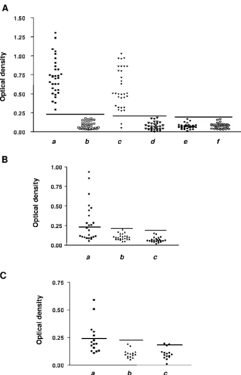
The three obtained proteins were then evaluated as capture Ags in ELISAs, using three different serum groups from the panel previously described. When using Ch⫹and Ch⫺groups, the sensitivities obtained for TcP2FLand TcP2Nwere 100%
and 94%, respectively (Fig. 3A). When testing TcP2FLwith
sera from the AID group (autoimmune patients), 58% of them showed cross-reactivity (Fig. 3B). The same holds true for samples from group L (cutaneous leishmaniasis patients), with the reactivity being 40% (Fig. 3C). However, cross-reactions
were avoided when using TcP2N. TRX protein was also
[image:3.585.301.539.66.436.2]eval-uated in each assay for all of the serum samples, showing no unspecific reactivity. All of these results are summarized in Table 1. The obtained results prompted us to explore the possibility of expressing this protein fused to C29C, which
pre-sents lower sensitivity than C29FLbut higher specificity toward
chagasic Abs (28).
The pET-32a vector containing the TcP2N-encoding DNA
was cut with the HindIII enzyme and then purified. The C29C
-encoding fragment, previously subcloned into a pMAL vector (28), was excised by digestion with the HindIII enzyme. The FIG. 2. Sodium dodecyl sulfate-polyacrylamide gel electrophoresis
of proteins, followed by staining with Coomassie blue. Lanes:a, mo-lecular marker;b, TcP2FL;c, TcP2N;d, TRX.
FIG. 3. Optical density values obtained by performing ELISA with several serum groups. Horizontal lines indicate the cutoff values cal-culated for each peptide. (A) Results obtained for each fragment when assaying Ch⫹and Ch⫺control serum groups. Points in columnsaand bshow results for TcP2FL, points in columnscanddshow results for
TcP2N, and points in columnseandfshow results for TRX. Points in
columnsa,c, andecorrespond to results for Chagas’ disease-positive sera; points in columnsb,d, andfcorrespond to results for Chagas’ disease-negative sera. (B) Results obtained for each fragment when assaying the AID serum group. Points in columnashow results for TcP2FL, points in columnbshow results for TcP2N, and points in
columncshow results for TRX. (C) Results obtained for each frag-ment when assaying the L serum group. Points in columna show results for TcP2FL, points in columnbshow results for TcP2N, and
points in columncshow results for TRX.
on May 16, 2020 by guest
http://jcm.asm.org/
[image:3.585.47.282.71.226.2]obtained fragment corresponded to a sequence encoding a 6-kDa peptide (Fig. 4B). The fragment was ligated to the HindIII site, which was present in the vector containing the TcP2N-encoding sequence (Fig. 4A). Then, a synthetic gene
encoding a chimera of 113-aa was obtained (Fig. 4B). The expression of this synthetic protein was induced and evaluated by 12% SDS–PAGE (Fig. 5A), followed by Western blotting, using a specific polyclonal anti-C29Cserum (Fig. 5B). It should
be noted that, even when the hyperimmune serum unspecifi-cally recognized some protein bands of the bacteria extract, it did not recognize TcP2N. Contrarily, the serum showed
re-activity towards C29FLand the fusion peptide TcP2N-C29C.
Evaluation of the TcP2N-C29Cchimera.To assess the
Cha-gas’ disease diagnostic utility of the protein obtained, we as-sayed the reactivity toward different sets of sera from the panel previously typified; the results were compared with those
ob-previously verified to be so by two other conventional serolog-ical tests, without other serologserolog-ical reactivity. We also studied cross-reactivity with the ID serum group, which includes sera from patients with the infectious diseases that have been re-ported in the literature as causative of cross-reactions when performing tests for diagnosis of T. cruzi infection. The ID serum group, consisted of Chagas-negative serum samples showing reactivity toward Toxoplasma gondii, Streptococcus pyogenes, or Brucella abortus (Fig. 6B). The sensitivity and specificity obtained with these additional serum panels was 100%, indicating that this synthetic protein behaves as an op-timized Ag for the diagnosis of the chagasic infection.
DISCUSSION
Chagasic infection is currently diagnosed serologically by detecting anti-T. cruziimmunoglobulin G Abs in the patient’s
FIG. 4. (A) Schematic representation of the method used to obtain the plasmid encoding the chimera protein. (B) C29Cand chimera amino
acid sequences. Letters in bold correspond to the C29Cfraction fused to TcP2N.
on May 16, 2020 by guest
[image:4.585.43.283.97.180.2] [image:4.585.136.452.442.697.2]serum. However, conventional serological methods often show cross-reactivity with related protozoan infections, particularly leishmaniasis. Recombinant Ags are specific, but a panel of them must be used to obtain appropriate sensitivity (12). Moreover, the utilization of several antigenic determinants gathered in the same molecule, which allows for an equili-brated adsorption of the Ag to the ELISA plaque, thus facil-itating standardization during reagent production has also been reported (12, 16).
Late in the 1980s, Levin et al. obtained a 34-aa TcP2 protein decoding a DNA sequence by using aT. cruzicDNA library (22). As mentioned above, this protein has a 13-aa, C-terminal fragment, R13, which shares 12 aa with a homo-logue human protein. Other works have described the pres-ence of Abs against the complete TcP2peptide, or against a fraction of it, in sera from patients suffering from several in-fections and autoimmune diseases (13, 38, 40, 42). These facts led researchers to propose that this peptide might be involved in the development of an autoimmune response in patients with Chagas’ disease (24, 31). A multicenter study evaluated the diagnostic performances of some chagasic recombinant Ags, among which TcP2peptide had been included (21). The
results obtained for TcP2(76% sensitivity) showed that the peptide did not enhance immunoassay sensitivity, compared with other Ags, and displayed high cross-reactivity with sera of patients with autoimmune or related parasitic diseases. This seems to be the reason why this peptide was not used in tests for diagnosis of chagasic infection. However, since anti-R13 peptide Abs were found in serum of chagasic patients with cardiopathies, quantification of these Abs was proposed to be useful to evaluate the patient’s status in terms of cardiopathy development (4). Additionally, the orthologous protein of Leishmaniaspp., whose C-terminal fraction showed cross-re-activity when assaying sera from chagasic patients, has been FIG. 5. (A) Coomassie blue staining of protein extracts after
elec-trophoresis in a 12% sodium dodecyl sulfate–polyacrylamide gel. Lane a,E. coliBL21(DE3)-(TcP2N-TRX) cell extract, used as a negative
control; laneb,E. coliBL21(DE3) cell extract of the isolated clone; lanec,E. coliDH5␣(C29-MBP) cell extract. (B) Western blot assay for TcP2N, the C29Cfraction fused to TcP2N, and C29Cin different
E. coliextracts, using a polyclonal anti-C29Cserum. Lanea,E. coli
DH5␣(C29-MBP) cell extract, used as a positive control; laneb,E. coliBL21(DE3) cell extract of the chimera clone; lanec,E. coli
BL21(DE3)-(TcP2NTRX) cell extract, used as a negative control. FIG. 6. Optical density values obtained by performing ELISA with
several serum groups. Horizontal lines correspond to calculated cutoff values. (A) Results obtained for the chimera by assaying the Ch⫹, Ch⫺, AID, and L serum groups. Columnsaandbshow results for Chagas’ disease-positive and Chagas’ disease-negative sera, respec-tively, columnscanddshow results for the AID serum group for SLE and RA sera, respectively, and columneshows results for the L serum group. (B) Results obtained for the chimera when assaying additional control serum samples and the ID serum group. Columnsaandbshow results for 72 Chagas’ disease-positive and 35 Chagas’ disease-negative control serum samples, respectively, columnsc,d, andeshow ID group results for sera of patients infected withToxoplasma gondii,Brucella abortus, andStreptococcus pyogenes, respectively.
on May 16, 2020 by guest
http://jcm.asm.org/
[image:5.585.301.544.68.448.2] [image:5.585.92.236.69.362.2]nia-truncated protein exhibits no cross-reactivity toward chagasic sera when used as the sensitizing element.
We have previously described that the use of TcP2FL
pro-tein as a sensitizing Ag detects all of the positive sera assayed (26). Considering that it has been described that the chagasic antigenic determinants are among the first 15 aa, whereas the final 20 aa are not immunogenic (23), we used this information as a criterion to eliminate more than 13 terminal amino acids, generating the Ag TcP2N. The peptide obtained showed a
higher specificity but lower sensitivity than TcP2FL. The
en-hanced specificity was displayed by the absence of reactivity when assaying sera from SLE and RA patients, together with the lack of cross-reactivity when assaying sera from L. (V.) brasiliensis-infected patients.
To increase the Ag sensitivity, we fused TcP2Nto another
studied protein region. Taking into account that previous works have shown thatT. cruzicalflagins are useful as sensi-tizing agents for diagnosis of chagasic infection (14, 19, 22) and considering that it has also been demonstrated that the C-terminal fraction of this protein shows high specificity when assaying leishmanial sera (28), C29C was chosen to assemble
the chimera. We therefore built up an artificial protein com-posed of C29Cand TcP2N. The usage of this fused peptide as
a sensitizing Ag rendered a sensitivity of 100% (Fig. 6 and Table 1). The results obtained for the serum samples of the Ch⫹and Ch⫺control groups (Table 2) additionally showed an increased reactivity for the positive sera with respect to that obtained when testing the same samples against TcP2N(P⫽
0.0017). In contrast, the negative serum samples showed no reactivity enhancement (P⫽0.45). These results show that this new Ag displays better discrimination efficiency between pos-itive and negative samples than that of TcP2N alone. The
evaluation of the fusion protein obtained also included testing those Chagas ’ disease-positive sera that had rendered positive results when using the complete TcP2peptide but had ren-dered negative results when using only TcP2N. Again, 100%
consistency with the sera typification was obtained, indicating that the sensitivity drop occurring when using TcP2Nalone
was fully prevented.
To complete the assessment of the new Ag, we analyzed Chagas ’ disease-negative sera of patients with SLE, AR, and leishmaniasis. Once more, the recognizing effectiveness of TcP2N-C29Cwas 100%, since all of the samples studied
ren-dered negative results. However, it should be noted that 4 of the 132 negative and 3 of the 108 positive serum samples evaluated presented OD values close to the cutoff which
be-development of a multicomponent protein for the diagnosis of chagasic infection (17), which was further improved later (16). This protein allowed a very important discrimination among positive and negative sera. However, the validation did not include sera from patients with leishmaniasis.
The optimization of the diagnosis of infection with micro-organisms is a multivariable task involving, among others, the search for suitable Ags, the analysis of their avidity (27), and in the case of recombinant Ags, the possibility of optimizing their antigenic profile by selecting an adequate expression system (26). Regions that are responsible for undesired cross-reactiv-ity can be removed (28), and regions showing high specificcross-reactiv-ity can be grafted one to another to raise sensitivity (16, 17). In line with this, the construction of the chimeric protein herein reported constitutes a promising land to obtain Ags that allows for standardization of diagnosis techniques and provides a novel platform to obtain new immunogens.
ACKNOWLEDGMENTS
This work was funded by ANPCyT, PICTR2002-00057 (C.M.L., A. Marcipar, and I. Malan Borel).
We are grateful to Alberto Marcipar for essential preliminary suggestions and discussion. We also dedicate this work to his lovely memory.
REFERENCES
1.Aguillon, J. C., R. Harris, M. C. Molina, A. Colombo, C. Cortes, T. Her-mosilla, P. Carreno, A. Orn, and A. Ferreira. 1997. Recognition of an immunogenetically selectedTrypanosoma cruziantigen by seropositive cha-gasic human sera. Acta Trop.63:159–166.
2.Almeida, I. C., D. T. Covas, L. M. Soussumi, and L. R. Travassos.1997. A highly sensitive and specific chemiluminescent enzyme-linked immunosor-bent assay for diagnosis of activeTrypanosoma cruziinfection. Transfusion
37:850–857.
3.Araujo, F. G.1986. Analysis ofTrypanosoma cruziantigens bound by specific antibodies and by antibodies to related trypanosomatids. Infect. Immun.
53:179–185.
4.Aznar, C., P. Lopez-Bergami, S. Brandariz, C. Mariette, P. Liegeard, M. D. Alves, E. L. Barreiro, R. Carrasco, S. Lafon, D. Kaplan, et al.1995. Preva-lence of anti-R-13 antibodies in humanTrypanosoma cruziinfection. FEMS Immunol. Med. Microbiol.12:231–238.
5.Bradford, M. M.1976. A rapid and sensitive method for the quantitation of microgram quantities of protein utilizing the principle of protein-dye bind-ing. Anal. Biochem.72:248–254.
6.Brito, M. E., M. G. Mendonca, Y. M. Gomes, M. L. Jardim, and F. G. Abath.
2000. Identification of potentially diagnosticLeishmania braziliensisantigens in human cutaneous leishmaniasis by immunoblot analysis. Clin. Diagn. Lab. Immunol.7:318–321.
7.Camargo, E. P.1964. Growth and differentiation inTrypanosoma cruzi.I. Origin of metacyclic trypanosomes in liquid media. Rev. Inst. Med. Trop. Sao Paulo12:93–100.
8.Camargo, M. E., E. L. Segura, I. G. Kagan, J. M. Souza, R. Carvalheiro Jda, J. F. Yanovsky, and M. C. Guimaraes.1986. Three years of collaboration on the standardization of Chagas’ disease serodiagnosis in the Americas: an appraisal. Bull. Pan. Am. Health Organ.20:233–244.
on May 16, 2020 by guest
[image:6.585.41.284.98.161.2]9.Carbonetto, C. H., E. L. Malchiodi, M. Chiaramonte, E. Durante de Isola, C. A. Fossati, and R. A. Margni.1990. Isolation of aTrypanosoma cruzi antigen by affinity chromatography with a monoclonal antibody. Preliminary evaluation of its possible applications in serological tests. Clin. Exp. Immu-nol.82:93–96.
10.Cuna, W. R., C. Rodriguez, F. Torrico, D. Afchain, M. Loyens, and P. Desjeux.1989. Evaluation of a competitive antibody enzyme immunoassay for specific diagnosis of Chagas’ disease. J. Parasitol.75:357–359. 11.Cura, E., S. Wendel, F. P. Pinheiro, and M. Weinserbacher.1996. Manual de
procedimientos de control de calidad para laboratorios de serologı`as de los bancos de sangre. PAHO, Washington, D.C.
12.da Silveira, J. F., E. S. Umezawa, and A. O. Luquetti.2001. Chagas disease: recombinantTrypanosoma cruziantigens for serological diagnosis. Trends Parasitol.17:286–291.
13.Elkon, K., S. Skelly, A. Parnassa, W. Moller, W. Danho, H. Weissbach, and N. Brot.1986. Identification and chemical synthesis of a ribosomal protein antigenic determinant in systemic lupus erythematosus. Proc. Natl. Acad. Sci. USA83:7419–7423.
14.Godsel, L. M., R. S. Tibbetts, C. L. Olson, B. M. Chaudoir, and D. M. Engman.1995. Utility of recombinant flagellar calcium-binding protein for serodiagnosis ofTrypanosoma cruziinfection. J. Clin. Microbiol.33:2082– 2085.
15.Guhl, F., C. Jaramillo, J. C. Carranza, and G. A. Vallejo.2002. Molecular characterization and diagnosis ofTrypanosoma cruziandT. rangeli. Arch. Med. Res.33:362–370.
16.Houghton, R. L., D. R. Benson, L. Reynolds, P. McNeill, P. Sleath, M. Lodes, Y. A. Skeiky, R. Badaro, A. U. Krettli, and S. G. Reed.2000. Multiepitope synthetic peptide and recombinant protein for the detection of antibodies to Trypanosoma cruziin patients with treated or untreated Chagas’ disease. J. Infect. Dis.181:325–330.
17.Houghton, R. L., D. R. Benson, L. D. Reynolds, P. D. McNeill, P. R. Sleath, M. J. Lodes, Y. A. Skeiky, D. A. Leiby, R. Badaro, and S. G. Reed.1999. A multi-epitope synthetic peptide and recombinant protein for the detection of antibodies to Trypanosoma cruzi in radioimmunoprecipitation-confirmed and consensus-positive sera. J. Infect. Dis.179:1226–1234.
18.Kirchhoff, L. V., A. A. Gam, R. A. Gusmao, R. S. Goldsmith, J. M. Rezende, and A. Rassi.1987. Increased specificity of serodiagnosis of Chagas’ disease by detection of antibody to the 72- and 90-kilodalton glycoproteins of Trypanosoma cruzi. J. Infect. Dis.155:561–564.
19.Krautz, G. M., L. M. Galvao, J. R. Cancado, A. Guevara-Espinoza, A. Ouaissi, and A. U. Krettli.1995. Use of a 24-kilodaltonTrypanosoma cruzi recombinant protein to monitor cure of human Chagas’ disease. J. Clin. Microbiol.33:2086–2090.
20.Laemmli, U. K.1970. Cleavage of structural proteins during the assembly of the head of bacteriophage T4. Nature227:680–685.
21.Levin, M. J., J. Franco da Silveira, A. C. Frasch, M. E. Camargo, S. Lafon, W. M. Degrave, and R. Rangel-Aldao.1991. RecombinantTrypanosoma cruzi antigens and Chagas’ disease diagnosis: analysis of a workshop. FEMS Mi-crobiol. Immunol.4:11–19.
22.Levin, M. J., E. Mesri, R. Benarous, G. Levitus, A. Schijman, P. Levy-Yeyati, P. A. Chiale, A. M. Ruiz, A. Kahn, M. B. Rosenbaum, et al.1989. Identifi-cation of majorTrypanosoma cruziantigenic determinants in chronic Chagas’ heart disease. Am. J. Trop. Med. Hyg.41:530–538.
23.Levin, M. J., R. Rossi, G. Levitus, E. Mesri, S. Bonnefoy, N. Kerner, and M. Hontebeyrie-Joskowicz.1990. The cloned C-terminal region of a Trypano-soma cruziP ribosomal protein harbors two antigenic determinants. Immu-nol. Lett.24:69–73.
24.Levin, M. J., M. Vazquez, D. Kaplan, and A. G. Schijman. 1993. The Trypanosoma cruziribosomal P protein family: classification and antigenicity. Parasitol. Today9:381–384.
25.Levitus, G., M. Hontebeyrie-Joskowicz, M. H. Van Regenmortel, and M. J. Levin.1991. Humoral autoimmune response to ribosomal P proteins in chronic Chagas heart disease. Clin. Exp. Immunol.85:413–417.
26.Marcipar, I. S., M. L. Olivares, L. Robles, A. Dekanty, A. Marcipar, and A. M. Silber.2004. The diagnostic performance of recombinant Trypano-soma cruziribosomal P2protein is influenced by its expression system. Protein Expr. Purif.34:1–7.
27.Marcipar, I. S., M. G. Risso, A. M. Silber, S. Revelli, and A. J. Marcipar.
2001. Antibody maturation inTrypanosoma cruzi-infected rats. Clin. Diagn. Lab. Immunol.8:802–805.
28.Marcipar, I. S., C. Roodveldt, G. Corradi, M. L. Cabeza, M. E. Brito, L. M. Winter, A. J. Marcipar, and A. M. Silber.2005. Use of full-length recombi-nant calflagin and its c fragment for improvement of diagnosis of Trypano-soma cruziinfection. J. Clin. Microbiol.43:5498–5503.
29.Marcipar, I. S., E. Welchen, C. Roodveldt, A. J. Marcipar, and A. M. Silber.
2003. Purification of the 67-kDa lectin-like glycoprotein ofTrypanosoma cruzi, LLGP-67, and its evaluation as a relevant antigen for the diagnosis of human infection. FEMS Microbiol. Lett.220:149–154.
30.Martinez, J., O. Campetella, A. C. Frasch, and J. J. Cazzulo.1991. The major cysteine proteinase (cruzipain) fromTrypanosoma cruziis antigenic in human infections. Infect. Immun.59:4275–4277.
31.Masuda, M. O., M. Levin, S. F. De Oliveira, P. C. Dos Santos Costa, P. L. Bergami, N. A. Dos Santos Almeida, R. C. Pedrosa, I. Ferrari, J. Hoebeke, and A. C. Campos de Carvalho.1998. Functionally active cardiac antibodies in chronic Chagas’ disease are specifically blocked byTrypanosoma cruzi antigens. FASEB J.12:1551–1558.
32.Saez-Alquezar, A., A. O. Luquetti, J. Borges-Pereira, E. F. Moreira, M. Gadahela, M. T. Garcia Zapata, and A. H. Strugo Arruda.1997. Estudo multice´ntrico: validac¸a˜o do desempenho de conjuntos diagno´sticos de hem-aglutinac¸a˜o indirecta disponı´veis no Brasil para o diagno´stico serolo´gico da infec¸a˜o peloTrypanosoma cruzi. Rev. Patol. Trop.26:343–374.
33.Saez-Alquezar, A., E. C. Sabino, N. Salles, D. F. Chamone, F. Hulstaert, H. Pottel, E. Stoops, and M. Zrein.2000. Serological confirmation of Chagas’ disease by a recombinant and peptide antigen line immunoassay: INNO-LIA chagas. J. Clin. Microbiol.38:851–854.
34.Salles, N. A., E. C. Sabino, M. G. Cliquet, J. Eluf-Neto, A. Mayer, C. Almeida-Neto, M. C. Mendonca, P. Dorliach-Llacer, D. F. Chamone, and A. Saez-Alquezar.1996. Risk of exposure to Chagas’ disease among seroreac-tive Brazilian blood donors. Transfusion36:969–973.
35.Sanger, F., S. Nicklen, and A. R. Coulson.1977. DNA sequencing with chain-terminating inhibitors. Proc. Natl. Acad. Sci. USA74:5463–5467. 36.Scharfstein, J., M. Schechter, M. Senna, J. M. Peralta, L.
Mendonca-Pre-viato, and M. A. Miles.1986.Trypanosoma cruzi: characterization and iso-lation of a 57/51,000 m.w. surface glycoprotein (GP57/51) expressed by epimastigotes and bloodstream trypomastigotes. J. Immunol.137:1336–1341. 37.Silber, A., I. Marcipar, C. Roodveldt, P. Cabeza Meckert, R. Laguens, and A. Marcipar.2002.Trypanosoma cruzi: identification of a galactose-binding protein that binds to cell surface of human erythrocytes and is involved in cell invasion by the parasite. Exp. Parasitol.100:217–225.
38.Skeiky, Y. A., D. R. Benson, J. A. Guderian, P. R. Sleath, M. Parsons, and S. G. Reed.1993.Trypanosoma cruziacidic ribosomal P protein gene family. Novel P proteins encoding unusual cross-reactive epitopes. J. Immunol.
151:5504–5515.
39.Solana, M. E., A. M. Katzin, E. S. Umezawa, and C. S. Miatello.1995. High specificity ofTrypanosoma cruziepimastigote ribonucleoprotein as antigen in serodiagnosis of Chagas’ disease. J. Clin. Microbiol.33:1456–1460. 40.Soto, M., J. M. Requena, M. Garcia, L. C. Gomez, I. Navarrete, and C.
Alonso.1993. Genomic organization and expression of two independent gene arrays coding for two antigenic acidic ribosomal proteins of Leishma-nia. J. Biol. Chem.268:21835–21843.
41.Soto, M., J. M. Requena, L. Quijada, and C. Alonso.1996. Specific serodi-agnosis of human leishmaniasis with recombinantLeishmaniaP2 acidic ri-bosomal proteins. Clin. Diagn. Lab. Immunol.3:387–391.
42.Soto, M., J. M. Requena, L. Quijada, S. O. Angel, L. C. Gomez, F. Guzman, M. E. Patarroyo, and C. Alonso.1995. During active viscerocutaneous leish-maniasis the anti-P2 humoral response is specifically triggered by the para-site P proteins. Clin. Exp. Immunol.100:246–252.
43.Tijsen, P.1985. Practice and theory of enzyme immunoassays, vol. 15. Elsevier Science Publishers, Amsterdam, The Netherlands.
44.Umezawa, E. S., S. F. Bastos, M. E. Camargo, L. M. Yamauchi, M. R. Santos, A. Gonzalez, B. Zingales, M. J. Levin, O. Sousa, R. Rangel-Aldao, and J. F. da Silveira.1999. Evaluation of recombinant antigens for serodiagnosis of Chagas’ disease in South and Central America. J. Clin. Microbiol.37:1554– 1560.
45.Umezawa, E. S., M. S. Nascimento, N. Kesper, Jr., J. R. Coura, J. Borges-Pereira, A. C. Junqueira, and M. E. Camargo.1996. Immunoblot assay using excreted-secreted antigens ofTrypanosoma cruziin serodiagnosis of congen-ital, acute, and chronic Chagas’ disease. J. Clin. Microbiol.34:2143–2147.