Copyright © 2003, American Society for Microbiology. All Rights Reserved.
Evaluation of the Etest and Disk Diffusion Methods for Determining
Susceptibilities of 235 Bloodstream Isolates of
Candida glabrata
to Fluconazole and Voriconazole
M. A. Pfaller,
1,2* D. J. Diekema,
1,3L. Boyken,
1S. A. Messer,
1S. Tendolkar,
1and R. J. Hollis
1Departments of Pathology1and Medicine,3Roy J. and Lucille A. Carver College of Medicine, and Department
of Epidemiology, College of Public Health,2University of Iowa, Iowa City, Iowa 52242
Received 21 November 2002/Returned for modification 12 January 2003/Accepted 27 January 2003
The performances of the Etest and the disk diffusion methods for testing of the susceptibilities of 235
Candida glabrataisolates to fluconazole and voriconazole were compared with that of the National Committee
for Clinical Laboratory Standards (NCCLS) approved standard broth microdilution (BMD) method. The NCCLS method used RPMI 1640 broth medium, and MICs were read after incubation for 48 h at 35°C. Etest MICs were determined with RPMI 1640 agar containing 2% glucose (RPG agar) and with Mueller-Hinton agar containing 2% glucose and 0.5g of methylene blue per ml (MBE agar) and were read after incubation for 48 h at 35°C. Disk diffusion testing was performed with MBE agar, 25-g fluconazole disks, and 1-g voriconazole disks and by incubation at 35°C for 24 h. Overall agreements between the Etest and the BMD MICs obtained with RPG and MBE agars were 91 and 96%, respectively, for fluconazole and 93 and 95%, respectively, for voriconazole. Categorical agreements between the agar-based methods and BMD were 52.3 to 64.7% with fluconazole and 94.8 to 97.4% with voriconazole. The vast majority of the discrepancies by the disk diffusion and Etest methods with fluconazole were minor errors. The agar-based methods performed well in identifying isolates with resistance to fluconazole and decreased susceptibility to voriconazole.
The agar-based methods for performing fluconazole and voriconazole susceptibility testing with Candida spp. include both the disk diffusion and the Etest stable-agar-gradient MIC methods (1, 3, 4, 6, 8, 9, 12, 16, 17). Barry et al. (3) demon-strated that both the disk diffusion and the Etest methods were accurate and precise when they were used to determine the fluconazole susceptibilities of 495 isolates ofCandidaspp. Al-though published data for the disk diffusion and Etest methods with voriconazole are limited (4, 9, 12), the results of both methods show good agreement with those of the reference broth microdilution (BMD) method.
The studies to date that have documented the efficacies of agar-based methods for the testing of susceptibilities to flucon-azole and voriconflucon-azole have generally included adequate num-bers ofCandida albicansisolates but relatively fewC. glabrata
isolates (1, 3, 8, 17). Among the four most common species of
Candida causing bloodstream infections (BSIs; C. albicans,
C. glabrata,C. parapsilosis, andC. tropicalis) (10),C. glabrata
alone tends to be less susceptible to fluconazole, with a signif-icant percentage of isolates classified as susceptible-dose de-pendent (S-DD; MIC, 16 to 32 g/ml) or resistant (R; MIC, ⱖ64g/ml) (11). This relative lack of susceptibility to flucon-azole means that BSIs due toC. glabratamust be treated with high doses of fluconazole (800 mg/day) or an alternative agent pending the results of antifungal susceptibility testing (14). Voriconazole may be useful as an alternative agent, given its excellent activity againstC. glabrataisolates that are suscepti-ble (S) or S-DD to fluconazole, but it is not reliably active in
vitro against fluconazole-R strains (11). For these reasons, antifungal susceptibility testing may play a very important role in optimizing the treatment of BSIs due toC. glabrata, and new testing methods (i.e., agar-based methods) should be rigor-ously examined with a large number of clinically important isolates of this species (15).
The purpose of this study was to expand the evaluation of agar-based methods for determining the in vitro susceptibili-ties ofC. glabratato fluconazole and voriconazole by testing an international collection of 235 BSI isolates obtained from more than 60 medical centers worldwide. The fluconazole and vori-conazole Etest MICs obtained with two different media and the disk diffusion zone diameters obtained with each agent were compared to the MICs determined by the National Com-mittee for Clinical Laboratory Standards (NCCLS) reference BMD method, the M27-A method (7).
MATERIALS AND METHODS
Organisms.A total of 235 clinical isolates ofC. glabratawere obtained from 61 medical centers worldwide in 2001. All were incident clinical isolates obtained from cultures of blood from 235 different patients with candidemia. Isolates were identified with the Vitek and API yeast identification systems (bioMerieux, Inc., Hazelwood, Mo.), and identification tests with these systems were supplemented by conventional methods as needed (18). Isolates were stored as water suspen-sions until use. Prior to testing, each isolate was passaged on potato dextrose agar (Remel, Lenexa, Kans.) and CHROMagar (Hardy Laboratories, Santa Monica, Calif.) to ensure purity and viability.
Susceptibility testing.Reference antifungal susceptibility testing ofC. glabrata
was performed by the BMD method described by NCCLS (7). Reference pow-ders of fluconazole and voriconazole were obtained from Pfizer Pharmaceuticals (Groton, Conn.).
Fluconazole and voriconazole Etest strips were provided by AB BIODISK (Solna, Sweden). MICs were determined by the Etest as described previously (8, 9) with RPMI 1640 agar with 2% glucose (RPG agar; Remel), an inoculum suspension adjusted to the turbidity of a 0.5 McFarland standard (⬃106cells/ml),
and incubation at 35°C for 48 h. In addition, a second medium prepared as * Corresponding author. Mailing address: Medical Microbiology
Di-vision, C606 GH, Department of Pathology, University of Iowa Col-lege of Medicine, Iowa City, IA 52242. Phone: (319) 384-9566. Fax: (319) 356-4916. E-mail: michael-pfaller@uiowa.edu.
1875
on May 15, 2020 by guest
http://jcm.asm.org/
described by Barry et al. (3) with Mueller-Hinton agar (Difco Laboratories) supplemented with 2% glucose and methylene blue (0.5g/ml) (MBE agar) was used for both Etest and disk diffusion testing (see below). The MICs of both fluconazole and voriconazole obtained with both RPG and MBE agars were read as the lowest concentration at which the border of the elliptical inhibition zone intercepted the scale on the strip. Any growth such as microcolonies throughout a discernible inhibition ellipse was ignored.
Disk diffusion testing of fluconazole and voriconazole was performed as de-scribed by Barry et al. (3) and Meis et al. (5). Fluconazole (25-g) and voricon-azole (1-g) disks were obtained from Becton Dickinson (Sparks, Md.). For disk diffusion testing, 90-mm-diameter plates containing MBE agar at a depth of 4.0 mm were used. The agar surface was inoculated by using a swab dipped in a cell suspension adjusted to the turbidity of a 0.5 McFarland standard. The inoculum was allowed to dry, and both the disks and the Etest strips were placed on the same plates. The plates were incubated in air at 35°C, and the zone diameters surrounding the fluconazole and voriconazole disks were read at 24 h. Zone diameter endpoints were read at 80% growth inhibition by using the BIOMIC image analysis plate reader system (version 5.9; Giles Scientific, Santa Barbara, Calif.) (5).
MIC interpretive criteria for fluconazole were those published by Rex et al. (13) and the NCCLS (17): S,ⱕ8g/ml; S-DD, 16 to 32g/ml; R,ⱖ64g/ml. The interpretive criteria for the fluconazole disk test were those published by Barry et al. (3): S,ⱖ19 mm; S-DD, 15 to 18 mm; R,ⱕ14 mm. Although inter-pretive breakpoints have not yet been established for voriconazole, we have elected to use the following criteria for purposes of comparison in this study (10–12): S,ⱕ1g/ml (zone diameter,ⱖ14 mm); R,ⱖ2g/ml (zone diameter, ⱕ13 mm).
QC.Quality control (QC) was performed for the BMD and Etest methods in accordance with NCCLS document M27-A by usingC. kruseiATCC 6258 and
C. parapsilosisATCC 22019 (2, 7). QC determinations made on each day of testing were within the control limits for fluconazole and voriconazole described by Barry et al. (2). QC for disk diffusion testing was performed by usingC. al-bicansATCC 90028 andC. parapsilosisATCC 22019 (3, 5).
Analysis of results.The Etest MICs of fluconazole and voriconazole on both RPG and MBE agars were read at 48 h and were compared to the reference BMD MICs read at 48 h. The Etest MICs were rounded up to the next even log2
concentration in order to simplify analysis (3, 8, 9). Discrepancies of no more than 2 dilutions were used to calculate the percent agreement.
The diameters of the zones of inhibition surrounding the fluconazole and voriconazole disks at 24 h of incubation were plotted against the respective BMD MICs read at 48 h (3). The method of least squares was used to calculate a regression line for each comparison.
The interpretive breakpoints described above were used to determine the categorical agreement between the results of the agar-based tests (the disk diffusion and Etest methods) and the results of the reference BMD method for fluconazole and voriconazole. Major errors were identified as a classification of R by the disk or Etest methods and a classification of S by BMD, very major errors were identified as a result of S by the disk diffusion or Etest method and a result of R by the BMD method, and minor errors were identified as a result of S or R by one of the tests and a result of S-DD by the other method.
RESULTS AND DISCUSSION
In vitro susceptibility testing by both the reference BMD method and the Etest with either RPG or MBE agar demon-strated the relatively high MICs of fluconazole forC. glabrata
(Table 1). The MICs obtained by the BMD method tended to cluster at the upper end of the S category (4 to 8g/ml) and in the S-DD category. A similar distribution was observed by the Etest, although on both media the Etest MICs tended to be slightly higher than the BMD MICs. The overall levels of agreement (within 2 dilutions) between the Etest and the BMD method were 91% with RPG agar and 96% with MBE agar, consistent with those reported previously (3, 8).
The MIC results obtained by both the BMD and the Etest methods demonstrated that voriconazole is very active against the vast majority ofC. glabrataBSI isolates (Table 1). Overall, 92 to 93% of isolates were inhibited byⱕ1g of voriconazole per ml, as determined by the Etest and the BMD methods. Similar to the results obtained with fluconazole, the level of agreement between the results of the BMD method and those of the Etest was good (93 to 95% agreement within 2 dilu-tions). Again, these results are similar to those reported pre-viously (9). Voriconazole MICs wereⱖ2g/ml for 14 of the
235C. glabrataisolates tested. Among these 14 isolates, 2 were
S-DD and 12 were R to fluconazole, confirming their lack of susceptibility to azole antifungal agents.
When susceptibility testing methods are compared, it is gen-erally useful to determine the more qualitative categorical agreement of the investigational methods and the established reference method. Despite the availability of interpretive breakpoints for fluconazole MIC and disk testing ofCandida
[image:2.603.42.283.98.196.2]spp., very few of the published evaluations of these methods provide data on overall categorical agreement and error rates (3, 6, 8). As seen in Table 2, despite reasonably good quanti-tative agreement between the Etest and the BMD MICs for fluconazole andC. glabrata, the overall categorical agreements TABLE 1. In vitro activities of fluconazole and voriconazole against
235 clinical isolates ofC. glabrataas determined by the reference BMD method and the Etest with two different media
Antifungal
agent methodTest a
MIC (g/ml)b %
Agree-mentc
Range 50% 90%
Fluconazole BMD 1–⬎128 8 32
ET-RPG 1–⬎256 16 64 91
ET-MBE 0.5–⬎256 16 64 96
Voriconazole BMD 0.03–8 0.25 1
ET-RPG 0.012–64 0.25 1 93
ET-MBE 0.012–64 0.25 1 95
aThe BMD method was performed according to the guidelines for the NCCLS M27-A method (7); ET-RPG, Etest with RPG agar; ET-MBE, Etest with MBE agar.
b50 and 90%, MICs at which 50 and 90% of isolates tested, respectively, are inhibited.
cPercentage of Etest MICs (read at 48 h) that are within 2 log
2dilutions of the
reference BMD MICs.
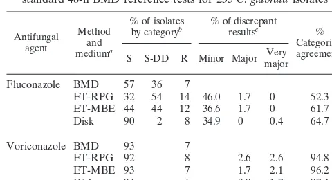
TABLE 2. Overall interpretive agreement between results of fluconazole and voriconazole agar-based susceptibility tests and of
standard 48-h BMD reference tests for 235C. glabrataisolates
Antifungal agent
Method and mediuma
% of isolates
by categoryb % of discrepantresultsc % Categorical agreementd S S-DD R Minor Major Verymajor
Fluconazole BMD 57 36 7
ET-RPG 32 54 14 46.0 1.7 0 52.3
ET-MBE 44 44 12 36.6 1.7 0 61.7
Disk 90 2 8 34.9 0 0.4 64.7
Voriconazole BMD 93 7
ET-RPG 92 8 2.6 2.6 94.8
ET-MBE 93 7 1.7 2.1 96.2
Disk 94 6 0.9 1.7 97.4
aSee footnoteaof Table 1 for definitions of BMD, ET-RPG, ET-MBE; disk, disk diffusion test with fluconazole (25-g disk) and voriconazole (1-g disk).
bPercentage of isolates classified in the different susceptibility categories. See Materials and Methods for definitions.
cPercentage of test results with minor, major, or very major discrepancies compared to the results of the reference BMD method at 48 h. See Materials and Methods for definitions.
dAgreement rates reflect the percentage of isolates classified in the same category by both the agar-based (Etest and disk) and the reference BMD meth-ods.
on May 15, 2020 by guest
http://jcm.asm.org/
[image:2.603.301.541.513.643.2]were rather poor: 52.3% for the Etest with RPG agar and 61.7% for the Etest with MBE agar. This is almost entirely due to minor errors consisting of shifts between the S and S-DD categories. Etest results tended to be slightly higher (usually 1 dilution) than the BMD results, with a higher percentage of isolates being in the S-DD category. Small numbers of major errors (false-positive resistance) were seen by the Etest, but no very major errors were observed.
By using the putative breakpoints for voriconazole of S being an MIC ⱕ1g/ml and R being an MIC ⱖ2g/ml, with no intermediate or S-DD category, the categorical agreements by the Etest were 94.8% with RPG agar and 96.2% with MBE agar. There were⬍3% major or very major errors, indicating that the Etest with either medium may be useful in determin-ing the in vitro susceptibilities of C. glabrataisolates to vori-conazole.
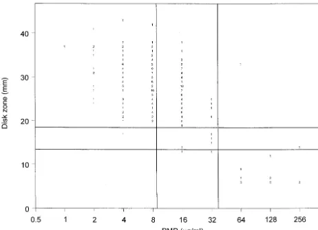
Although disk diffusion testing with MBE agar is relatively new, it is clear that this medium formulation supports the growth of C. glabrata and allows the measurement of zone diameters surrounding both fluconazole and voriconazole disks within 24 h (3, 6, 12). Figure 1 shows the correlation of the 25-g fluconazole disk zone diameters read at 24 h with the BMD MIC results. The clustering of the results for the isolates around the breakpoint values is evident from the scattergram,
with most of the isolates for which the fluconazole MICs were 16g/ml (S-DD) appearing to be susceptible by the disk meth-od, with zone diameters of⬎19 mm. This shift toward larger numbers of susceptible isolates by disk testing resulted in an overall categorical agreement of 64.7%, with 34.9% minor errors, no major errors, and only one very major error (Table 2). Thus, although the fluconazole disk test was unable to distinguish S versus S-DD isolates, it did reliably detect those strains with resistance (MICs,ⱖ64g/ml) to fluconazole.
The results of testing with the 1-g voriconazole disk are shown in Fig. 2 and Table 2. The regression statistics (y⫽55.8
⫺3.8x;R⫽0.7) show a good level of agreement between the two methods. Zone diameters of ⱕ13 mm identified those isolates with decreased susceptibility to voriconazole (MICs, ⱖ2g/ml), and the resulting categorical agreement was 97.4%, with⬍2% very major and major errors. Again, those isolates with smaller inhibition zone diameters and for which voricon-azole MICs were higher also exhibited decreased susceptibility to fluconazole.
[image:3.603.60.522.68.403.2]The results of this study complement the previous observa-tions of Barry et al. (3) and Morace et al. (6) regarding the use of Etest and disk diffusion testing to determine the suscepti-bilities ofC. glabrataisolates to azole antifungal agents. Barry et al. (3) found that both the Etest and the disk diffusion FIG. 1. Zones of inhibition around 25-g fluconazole disks on MBE agar plotted against the MICs determined at 48 h by the reference BMD method for 235C. glabrataisolates. The method of least squares was used to calculate a regression line (y⫽71.1⫺3.6x;R⫽0.7). The horizontal lines indicate the S (ⱖ19 mm) and R (ⱕ14 mm) zone diameter breakpoints for the fluconazole disk test. The vertical lines indicate the S (ⱕ8g/ml) and R (ⱖ64g/ml) MIC breakpoints for fluconazole. The numbers inside the graph indicate numbers of isolates.
on May 15, 2020 by guest
http://jcm.asm.org/
methods were accurate, precise, and reproducible for the test-ing of susceptibility to fluconazole, although they included only
37C. glabrataisolates among the 495 isolates tested and did
not analyze the data for this species separately. Those investi-gators did examine the performance of the Etest on MBE agar and found that its performance was comparable to but not superior to that on RPG agar.
More recently, Morace et al. (6) reported the results of a multicenter evaluation of the Etest and disk diffusion methods, as well as other commercially available methods, for the testing of the fluconazole susceptibilities of 793 clinicalCandida iso-lates, including 133C. glabrataisolates. Their conclusions were similar to those of Barry et al. (3), stating that both the Etest and the disk diffusion methods were accurate, reproducible, and potentially useful for determining susceptibility to flucon-azole in the clinical microbiology laboratory. However, when the data forC. glabratawere analyzed separately, those inves-tigators found categorical agreements of 68% for the Etest and 50% for disk diffusion testing. The vast majority of the discrep-ancies by both of these agar-based tests were minor errors involving changes from S to S-DD.
On the basis of the results of the previously published stud-ies described above plus the data presented herein, it appears that both the Etest and the disk diffusion test methods may be useful in identifying isolates ofC. glabrataexpressing resistance to fluconazole but are not reliable in differentiating isolates that are S versus those that are S-DD. Given the fact that the fluconazole dosing recommendations for the treatment ofC.
glabrataBSIs (800 mg/day) are intended to account for those
isolates in the S-DD category, the inability to differentiate S from S-DD isolates may not be a critical failing of these tests (14). It is far more important to identify those strains with resistance (15), and these tests appear to perform that function reasonably well.
Although there are very few data regarding agar-based tests for voriconazole, it does appear that the results of both the Etest and the disk diffusion test methods provide good agree-ment with those of the BMD method for the testing ofCandida
[image:4.603.46.538.68.436.2]spp., includingC. glabrata(4, 9, 12). Previously, we evaluated the voriconazole Etest and demonstrated overall levels of agreement with the BMD method of 98% forCandidaspp. and 91% forC. glabrata; however, we did not determine categorical FIG. 2. Zones of inhibition around 1-g voriconazole disks plotted against the MICs determined at 48 h by the reference BMD method for 235
C. glabrataisolates. The method of least squares was used to calculate a regression line (y⫽55.8⫺3.8x;R⫽0.7). The horizontal line indicates the putative S zone diameter breakpoint (ⱖ14 mm) for the voriconazole disk test. The vertical line indicates the putative S MIC breakpoint (ⱕ1 g/ml) for voriconazole. The numbers inside the graph indicate numbers of isolates.
on May 15, 2020 by guest
http://jcm.asm.org/
agreement in that study (9). More recently, we have demon-strated (12) a good overall correlation between BMD and disk diffusion testing for voriconazole and a categorical agreement of 99% when a collection of 1,586 isolates of Candida was tested by using the putative interpretive criteria described herein. The data presented in Fig. 2 and Table 2 demonstrate that both the Etest and the disk diffusion test with voriconazole perform comparably to the BMD method in identifying C.
glabratastrains with decreased susceptibility to this agent. The
fact that thoseC. glabrataisolates that appear to be less sus-ceptible to voriconazole by all three methods also exhibit de-creased susceptibility to fluconazole suggests that these ap-proaches may be useful clinically to guide therapeutic decision making for infections due to C. glabrata. Establishment of clinical correlates is essential; however, these results are very promising.
In summary, we have performed an extensive analysis of agar-based testing methods for determination of the in vitro susceptibilities ofC. glabrata isolates to fluconazole and vori-conazole. We have demonstrated the usefulness of MBE agar for the performance of both the Etest and the disk diffusion methods to determine the susceptibilities of this relatively fas-tidious species ofCandidato both of these antifungal agents. We have shown that both Etest and disk diffusion testing may reliably identify C. glabrataisolates that express resistance to fluconazole but that neither agar-based method can differen-tiate fluconazole-S from fluconazole-S-DD isolates. Likewise, both agar-based methods can identify those few C. glabrata
isolates with decreased susceptibility to voriconazole.C.
gla-brata isolates that are S-DD to fluconazole are virtually all
“susceptible” to voriconazole as determined by the BMD and Etest methods (MICs, ⱕ1 g/ml) and disk diffusion testing (zone diameters,ⱖ14 mm).
From the standpoint of antifungal resistance,C. glabratais clearly the Candidaspecies with the greatest potential to ac-quire resistance to fluconazole and other azoles (14, 15). The availability of test methods to reliably identify thoseC. glabrata
strains that express resistance to fluconazole, voriconazole, and other azoles will allow clinicians to optimize their therapeutic approaches to infections due to this important species (14, 15). We have demonstrated that the relatively simple agar-based methods for the testing of susceptibility to fluconazole and voriconazole may be used for this purpose. The commercial availability of broth- and agar-based antifungal susceptibility testing methods should bring this technology into the main-stream of clinical microbiology and infectious disease practice.
ACKNOWLEDGMENTS
This study was supported in part by unrestricted research grants from Pfizer Pharmaceuticals (ARTEMIS Program).
Linda Elliott provided excellent support in the preparation of the manuscript. We express our appreciation to all ARTEMIS Program participants. Participants contributing isolates to the study included Hershey Medical Center, Hershey, Pa. (P. Appelbaum); Stanford Hos-pitals and Clinics, Stanford, Calif. (E. J. Baron); University of Califor-nia, Los Angeles, Medical Center (D. Brucker); New York State De-partment of Health, Albany (V. Chaturvedi); Wishard Health Services, Indianapolis, Ind. (T. Davis); Summa Health System, Akron, Ohio (J. DiPersio); Case Western Reserve University Hospital, Cleveland, Ohio (M. Ghannoum); Cleveland Clinic Foundation, Cleveland, Ohio (G. Hall); University of Rochester Medical Center, Rochester, N.Y. (D. Hardy); University of Virginia Health System, Charlottesville (K.
Hazen); Veterans Affairs Medical Center, Ann Arbor, Mich. (C. Kauffman); New York Presbyterian Hospital, Cornell Medical Center, New York, N.Y. (D. Larone); Harper Hospital, Division of Infectious Diseases, Detroit, Mich. (J. Sobel); Christiana Care ID Lab, Wilming-ton, Del. (L. Steele Moore); Temple University School of Medicine, Philadelphia, Pa. (B. Suh); Good Samaritan Medical Regional Center, Phoenix, Ariz. (D. Sussland); Westchester Medical Center, Valhalla, N.Y. (K. Van Horn); Scaolabrini Ortiz, Buenos Aires, Argentina (J. Finquelievich); Arabe Seria, Buenos Aires, Argentina (I. N. Tirabos-chi); Rua Diogo de Faria, San Paulo, Brazil (A. Lopes Colombo); Laboratorio de Micologia, Medellin, Columbia (A. Restrepo); Hospi-tal Militar Central, Bogota, Columbia (R. Vega); HospiHospi-tal Militar, Quito, Ecuador (J. Ayabaca); Instituto de Patologia Infecciosa y Ex-perimental, Guadalajara, Mexico (E. Rodriquez Noriega); Hospital General O’Horan, Merida, Mexico (M. Zaidi); SAIMR, Johannes-burg, South Africa (H. H. Crewe-Brown); Department of Medical Microbiology, Medunsa, South Africa (A. Hoosen); Groote Schuur Hospital, Cape Town, South Africa (D. Roditi); Johannesburg Hospi-tal, Parktown, South Africa (T. Towindo); Pelanomi HospiHospi-tal, UOFS, Bloemfontein, South Africa (J. van Rensburg); Women’s and Chil-dren’s Hospital, North Adelaide, Australia (D. Ellis); Department of Medical Microbiology, Kuala Lumpur, Malaysia (N. K. Peng); Chiang Mai University, Chiang Mai, Thailand (P. Tharavichitkul); China Med-ical College Hospital, Taichung, Taiwan (J.-H. Wang); National Tai-wan University Hospital, Taipei, TaiTai-wan (K.-W. Yu); Vzhongshan Hospital, Shanghai, China (H. Bijie); Peking Union Medical College Hospital, Beijing, China (Y. Xu); Yonsei University College of Med-icine, Seoul, Korea (Y. Chong, K. Lee); St. Cyril and Metod Hospital, Bratislava, Slovakia (H. Hupkova); National Cancer Institute, Bratislava, Slovakia (J. Trupl); Derer University Hospital, Bratislava, Slovakia (A. Vaculikova); Nernocnice C. Budejovice, Ceske Budejov-ice, Czech Republic (N. Mallatova); Odd. Klinicke Myckkologie, Ostrava, Czech Republic (D. Stanislava); Children’s Memorial Health Institute, Warsaw, Poland (D. Dzierzanowska); Pescara Civil Hospital, Pescara, Italy (D. D’Antonio); Ospedale di Novara, Novara, Italy (G. Fortina); Departimento di Biotechnologie Cellulari ed Ematologia, Rome, Italy (P. Martino); Azienda Ospedaliera Umbertol, Torrett, Italy (G. Scalise); Ospedale di Genova, Genoa, Italy (G. C. Schito); University of Rome “Tor Vergata,” Rome, Italy (G. P. Testore); versita degli Studi di Torino, Torino, Italy (V. Tullio); Hopital Uni-versitario 12 de Octobre, Madrid, Spain (A. del Palacio); Canisius-Wilhelmina Ziekerhuis Megishe microbiologie C70, Nijmegen, The Netherlands (J. F. G. M. Meis); Friarage Hospital, Northallerton, United Kingdom (N. Weightman); Children’s Hospital, Ankara, Tur-key (D. Gur); Mamara Medical School Hospital, Istanbul, TurTur-key (V. Korten); University of Alberta Hospitals, Edmonton, Alberta, Canada (R. Rennie); Department of Microbiology and Immunology, Saska-toon, Saskatchewan, Canada (K. Mochoruk); NHS Trust, Aberdeen Royal Infirmary, Foresterhill, United Kingdom (I. Gould); and Ro-swell Park Cancer Institute, Buffalo, N.Y. (B. Segal).
REFERENCES
1. Arendrup, M., B. Lundgren, I. M. Jensen, B. S. Hansen, and N. Frimondt-Møfller.2001. Comparison of Etest and a tablet diffusion test with the NCCLS broth microdilution method for fluconazole and amphotericin B susceptibility testing ofCandidaisolates. J. Antimicrob. Chemother.47:521– 526.
2. Barry, A. L., M. A. Pfaller, S. D. Brown, A. Espinel-Ingroff, M. A. Ghan-noum, C. Knapp, R. P. Rennie, J. H. Rex, and M. G. Rinaldi.2000. Quality control limits for broth microdilution susceptibility tests of ten antifungal agents. J. Clin. Microbiol.38:3457–3459.
3. Barry, A. L., M. A. Pfaller, R. P. Rennie, P. C. Fuchs, and S. D. Brown.2002. Precision and accuracy of fluconazole susceptibility tests by broth microdi-lution, Etest, and disk diffusion methods. Antimicrob. Agents Chemother. 46:1781–1784.
4. Kronvall, G., and I. Karlsson.2001. Fluconazole and voriconazole multidisk testing ofCandidaspecies for disk calibration and MIC estimation. J. Clin. Microbiol.39:1422–1428.
5. Meis, J., M. Petrou, J. Bille, D. Ellis, D. Gibbs, and the Global Antifungal Surveillance Group.2000. A global evaluation of the susceptibility of Can-didaspecies to fluconazole by disk diffusion. Diagn. Microbiol. Infect. Dis. 36:215–223.
6. Morace, G., G. Amato, F. Bistoni, G. Fadda, P. Marone, M. T. Montagna, S. Oliveri, L. Polonelli, R. Rigoli, I. Mancuso, S. La Face, L. Masucci, L. Romano, C. Napoli, D. Tato, M. G. Buscema, C. M. C. Belli, M. M. Picirillo, S. Coni, S. Covan, F. Fanti, C. Cavanna, F. D’Alo, and L. Pitzurra.2002.
on May 15, 2020 by guest
http://jcm.asm.org/
Multicenter comparative evaluation of six commercial systems and the Na-tional Committee for Clinical Laboratory Standards M27-A broth microdi-lution method for fluconazole susceptibility testing of Candidaspecies. J. Clin. Microbiol.40:2953–2958.
7. National Committee for Clinical Laboratory Standards.1997. Reference method for broth dilution antifungal susceptibility testing of yeasts. Ap-proved standard M27-A. National Committee for Clinical Laboratory Stan-dards, Wayne, Pa.
8. Pfaller, M. A., S. A. Messer, A. Karlsson, and A. Bolmstro¨m.1998. Evalu-ation of the Etest method for determining fluconazole susceptibilities of 402 clinical yeast isolates by using three different agar media. J. Clin. Microbiol. 36:2586–2589.
9. Pfaller, M. A., S. A. Messer, A. Houston, K. Mills, A. Bolmstro¨m, and R. N. Jones.2000. Evaluation of the Etest method for determining voriconazole susceptibilities of 312 clinical isolates ofCandidaspecies by using three different media. J. Clin. Microbiol.38:3715–3717.
10. Pfaller, M. A., D. J. Diekema, R. N. Jones, H. S. Sader, A. C. Fluit, R. J. Hollis, S. A. Messer, and the SENTRY Participant Group.2001. Interna-tional surveillance of bloodstream infections due toCandidaspecies: fre-quency of occurrence and in vitro susceptibilities to fluconazole, ravucon-azole, and voriconazole of isolates collected from 1997 through 1999 in the SENTRY Antimicrobial Surveillance Program. J. Clin. Microbiol.39:3254– 3259.
11. Pfaller, M. A., S. A. Messer, R. J. Hollis, R. N. Jones, and D. J. Diekema. 2002. In vitro activities of ravuconazole and voriconazole compared with those of four approved systemic antifungal agents against 6,970 clinical isolates ofCandidaspp. Antimicrob. Agents Chemother.46:1723–1727. 12. Pfaller, M. A., D. J. Diekema, S. A. Messer, L. Boyken, and R. J. Hollis for
the ARTEMIS Global Antifungal Susceptibility Program Participants Group.Activity of fluconazole and voriconazole determined by broth mi-crodilution, disk diffusion, and Etest methods against 1,586 recent clinical isolates ofCandidaspecies: report from the ARTEMIS Global Antifungal Susceptibility Program, 2001. J. Clin. Microbiol., in press.
13. Rex, J. H., M. A. Pfaller, J. N. Galgiani, M. S. Bartlett, A. Espinel-Ingroff, M. A. Ghannoum, M. Lancaster, M. G. Rinaldi, T. J. Walsh, and A. L. Barry. 1997. Development of interpretive breakpoints for antifungal susceptibility testing: conceptual framework and analysis of in vitro-in vivo correlation data for fluconazole, itraconazole, andCandidainfections. Clin. Infect. Dis. 24:235–247.
14. Rex, J. H., T. J. Walsh, J. D. Sobel, S. G. Filler, P. G. Pappas, W. E. Dismukes, and J. E. Edwards.2000. Practice guidelines for the treatment of candidiasis. Clin. Infect. Dis.30:662–678.
15. Rex, J. H., and M. A. Pfaller.2002. Has antifungal susceptibility testing come of age? Clin. Infect. Dis.35:982–989.
16. Simor, A. E., G. Goswell, L. Louie, M. Lee, and M. Louie.1997. Antifungal susceptibility testing of yeast isolates from blood cultures by microbroth dilution and the Etest. Eur. J. Clin. Microbiol. Infect. Dis.16:693–697. 17. Warnock, D. W., E. M. Johnson, T. R. Rogers, et al.1998. Multi-centre
evaluation of the Etest method for antifungal drug susceptibility testing of
Candidaspp. andCryptococcus neoformans. J. Antimicrob. Chemother.42: 321–331.
18. Warren, N. G., and K. C. Hazen.1999.Candida, Cryptococcus,and other yeasts of medical importance, p. 1184–1199.InP. R. Murray, E. J. Baron, M. A. Pfaller, F. C. Tenover, and R. H. Yolken (ed.), Manual of clinical microbiology, 7th ed. ASM Press, Washington, D.C.