How to cite this paper: Asma, B., Abdellatif, B., Mohame, H., Mokhtar, H. and Hamza, R. (2014) Bone Healing by the Use of Intramedullary Pinning in Dogs. Open Access Library Journal, 1: e668. http://dx.doi.org/10.4236/oalib.1100668
Bone Healing by the Use of Intramedullary
Pinning in Dogs
Bouknine Asma
*, Boudra Abdellatif, Hamdi Mohame, Hamri Mokhtar, Rezki Hamza
Institute of Veterinary Science, University of Tiaret, Tiaret, AlgeriaEmail: *[email protected]
Received 15 April 2014; revised 20 May 2014; accepted 14 July 2014
Copyright © 2014 by authors and OALib.
This work is licensed under the Creative Commons Attribution International License (CC BY). http://creativecommons.org/licenses/by/4.0/
Abstract
Femur is the most commonly fractured bone in dogs. This work is interested in the technical as-pects of ostheosythesis with intramedullary pinning, and a quality of functional and bone healing results. Our experimentation involved the use of four healthy male dogs, by causing a transverse osteotomy in the mid shapt of in the left femur, with clinical and radiological follow established. Our results show that the intramedullary pinning is the most secure method and gives fewer com-plications with second bone consolidation. At the end of our study, we have concluded that using intramedullary pins is safe, economical and successful treatment method if basic principles of re-pair are used.
Keywords
Femur, Intramedullary Pinning, Consolidation
Subject Areas: Anatomy & Physiology, Orthopedics
1. Introduction
The femur fracture in canine was frequently recorded as compared to other long bone fractures. To treat frac-tures of long bones of small animals both conservative and operative methods are used, although numerous op-tions are available for fracture treatment, surgical internal fixation various intramedullary pin are used [1].
Stability in orthopedics is defined as the degree of displacement between the fragments involved in a fracture and stiffness as the capacity of the implant to oppose deformation [2].
Intramedullary pinning acts primarily as internal splint of medullary canal of long bone that shares loading with bones maintain axial alignment of the fracture and resists bending forces in all directions applied to the bone [3].
The objective of the present study was to evaluate the femur fractures and treatment options in dogs by intra-medullary pinning fixation.
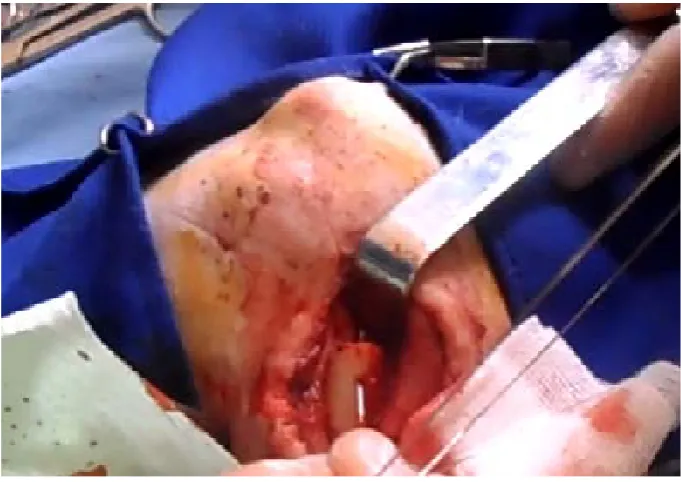
OALibJ | DOI:10.4236/oalib.1100668 2 July 2014 | Volume 1 | e668 lateralis was done to expose the femoral shaft. The left femur of all animals was experimentally fractured trans-versally by using a wire saw in the mid-diaphysis under constant cooling with a 0.9% NaCl solution with antibi-otic then the femur were fixed by intramedullary pinning by retrograde manner and renforced by hemicerclage wire (Figures 1-4).
Dogs were clinically observed daily during the study period for recording any postoperative complications No postoperative immobilisation was used, all dogs received antibiotic during 10 days.
[image:2.595.172.457.315.491.2]A radiograph examination is made room immediately after surgery to enjoy a good quality of osteotomy, the correct position of the implants and the absence of intraoperative fractures. To assess the degree of bone healing, radiographs were taken at 1, 2, 3, 4, 5, 6 weeks postoperatively until bone healing.
Figure 1. Separation between two muscles (vastus lateralis and biceps femoris).
[image:2.595.171.457.526.710.2]OALibJ |DOI:10.4236/oalib.1100668 3 July 2014 | Volume 1 | e668 Figure 3. Penetration of metallic wire.
Figure 4. Introduction of pin in medullary canal.
3. Results
OALibJ | DOI:10.4236/oalib.1100668 4 July 2014 | Volume 1 | e668 (a) (b)
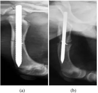
Figure 5. Immediately postoperative radiography of case N˚1 (a), showing presence of pin which happen until the distal epiphysis, and clear line (b), showing the callus formation.
[image:4.595.91.540.101.186.2](a) (b)
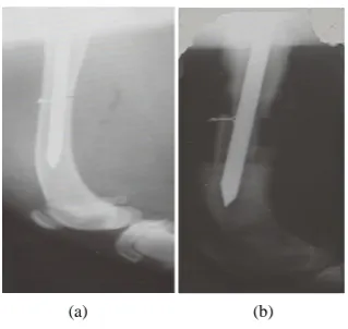
Figure 6. Immediately postoperative radiography of case N˚ 4˚ (a) showing presence of pin which happen until the distal epiphysis, with a good alignement of two abouts, and second- ary ossification at fourth week (b).
4. Discussion
There are three areas of osteognic potential in the healing of any diaphyseal Fracture: the periosteal reaction, the endosteal callus, and time fractures hematoma.
[image:4.595.237.393.209.367.2] [image:4.595.233.396.413.567.2]OALibJ |DOI:10.4236/oalib.1100668 5 July 2014 | Volume 1 | e668 (a) (b)
Figure 7. Radiography of case N˚3 showeing alignement of two abouts (a), (b) a beginning of callus formation.
it has been demonstrated that the marrow isable to regenerate vascularisation after one week and to irrigate the bone cortex, with the exception of areas where it is in direct contact with the nail [4]. Moreover, the insertion of intramedullary pin promote bone healing even by bringing in contact with the bone fragments, pluripotent cells derived from bone marrow [5].
A complication in the assembly is recorded osteomylitis, it results in a diffused light on the radiograph. The bone infection in case N˚2, caused the release and migration of pin, from the medullary canal of the bone to the outside. A modification of support within the same group were found, the support case N˚1, 3 and 4 was been early compared case N˚2, which shows two complication which are osteomyelitis, and migration of pin.
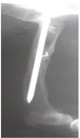
On radiological features of fractured femur in dogs at scheduled intervals following reduction, revealed callus formation on day 28 post treatment. The 35-day radiograph indicated the beginning of reabsorption of excessive callus, reduced periosteal reaction and process of remodeling of new bone. Radiographs on day 60 (Figure 8) revealed completion of the reparative process supported by reabsorption of excessive callus and disappearance of periosteal reaction with a good healing of bone.
This coincides with a good clinical recovery and full support of dogs was observed during the 4th week (Table 1). The Case N˚4 had delayed consolidation report, relative cases N˚1 and 3 whom are the same age, this is due to the turbulent behavior of the animal and its movement who were responsible for the delay of consolidation and secondary ossification; the migration of pin before the complete consolidation showed an angulation of the bone in the dog N˚2. This complication is the essential defect of intramedullary pinning recorded during our study. According to Fossu, 2007 [5], the micro movements of the pin caused by the inequality between a pin and the diameter of the medullary canal, so, are responsible for the migration of the pin. This has necessitated, that is why, we are used, a complementary fastener as hemi-wire to increase the rotational and axial strength. This dif-ference in diameter, according Duhautois, 1993 [6]; back to the fact that changes in diaphyseal sections and the curvature of the bone in the large dog, do not allow the pin to hold the entire medullary canal, by cons, the join-ing between the pin and the medullary canal is easier in small dogs. It has been reported that intramedullary pins are satisfactory for shaft fractures of the femur in small dogs and cats [7]. As Boyd has said, an intramedullary pinning is justified only in situations when its mechanical advantages from the stand point of the stress involved outweigh the biological disadvantages and when rotation can be controlled. From a biomechanical point of view, the callus decreases the lever arm which tends to stretch the fracture, which is why the callus will be even larger than the home is unstable [8]. Implants failure from fatigue fracture is not common with intramedullary pinning because the location of an intramedullary pin in the centre of the shaft tends to spare some of the bending forces responsible for fatigue failure and the effect of the law of wolf which is very favorable by the intramedullary pinning.
5. Conclusions
OALibJ | DOI:10.4236/oalib.1100668 6 July 2014 | Volume 1 | e668 Figure 8. A radiograph at two months showing a complete inte-
gration of bone with significant narrowing of callus volume.
• Application of other imaging means for assessment of fracture repair; • Histological study of fracture healing;
• Fracture repair by intramedullary nailing.
Acknowledgements
My thanks addressed to the radiology centers of Tiaret (Algeria) (Dr Efret and Dr Chikhaoui), for all authors of this article for their true help, mainly Dr Boudra Abdellatif for his ideas and suggestions in orthopedic surgery.
References
[1] Brinker, W.O., Piermattei, D.L. and Flo, G.L. (1994) Le Manuel d'orthopédie et de traitement des fractures des petits animaux. 2nd Edition, Point Vétérinaire, Maisons Alfort, 9-137.
[2] Wagner, M. and Frigg, R. (2006) Internal Fixators. Thieme Medical Publishers, New York.
[3] Beale, B. (2004) Orthopedic Clinical Techniques Femur Fracture Repair. Clinical Techniques in Small Animal Practice,
19, 134-150. http://dx.doi.org/10.1053/j.ctsap.2004.09.006
[4] Autefage, A. (1992) Consolidation des fractures, encyclopédie vétérinaire, Paris. Orthopédie, 3100, 1-8.
[5] Fossum, T.W. (2007) Small Animal Surgery. 3rd Edition, Elsevier, Mosby, 935-1005.
[6] Duhautois, B. (1993) L’enclouage verrouillé en chirurgie vétérinaire de la conception aux premiers cas cliniques. Practice Medicale et Chirurgiacl de l’animal de Compagnie, 28, 657-683.
[7] Phillips, I.R. (1979) A Survey of the Bone Fractures in the Dog and Cat. Journal of Small Animal Practice, 20, 661- 674. http://dx.doi.org/10.1111/j.1748-5827.1979.tb06679.x