An Autoimmune Disease-Associated Risk Variant in the
TNFAIP3
Gene Plays a Protective Role in Brucellosis That Is
Mediated by the NF-
B Signaling Pathway
Lixin Lou,
aWanguo Bao,
aXianjun Liu,
bHongxiao Song,
cYang Wang,
aKaiyu Zhang,
aWenjing Gao,
bHaijun Li,
cZhengkun Tu,
cShaofeng Wang
baDepartment of Infectious Diseases, The First Hospital of Jilin University, Jilin, China bThe Bethune Institute of Epigenetic Medicine, The First Hospital of Jilin University, Jilin, China cDepartment of Translational Medicine, The First Hospital, Jilin University, Jilin, China
ABSTRACT
Naturally occurring functional variants (rs148314165 and rs200820567,
collectively referred to as TT
⬎
A) reduce the expression of the tumor necrosis factor
alpha-induced protein 3 (TNFAIP3) gene, a negative regulator of NF-
B signaling, and
predispose individuals to autoimmune disease. In this analysis, we conducted a
ge-netic association study of the TT
⬎
A variants in 1,209 controls and 150 patients with
brucellosis, an infectious disease, and further assessed the role of the variants in
bru-cellosis. Our data demonstrated that the TT
⬎
A variants were correlated with cases
of brucellosis (P
⫽
0.002; odds ratio [OR]
⫽
0.34) and with individuals who had a
positive serum agglutination test (SAT) result (titer of
⬎
1/160) (P
⫽
4.2
⫻
10
⫺6;
OR
⫽
0.23). A functional study demonstrated that brucellosis patients carrying
the protective allele (A) showed significantly lower expression levels of the TNFAIP3
gene in their peripheral blood mononuclear cells and showed increased NF-
B
sig-naling. Monocytes from individuals carrying the A allele that were stimulated with
Brucella abortus
had lower mRNA levels of TNFAIP3 and produced more interleukin-10
(IL-10), IL-6, and IL-1

than those from TT allele carriers. These data showed that
au-toimmune disease-associated risk variants, TT
⬎
A, of the TNFAIP3 locus play a
pro-tective role in the pathogenesis of brucellosis. Our findings suggest that a disruption
of the normal function of the TNFAIP3 gene might serve as a therapeutic target for
the treatment of brucellosis.
KEYWORDS
brucellosis, inflammation, single nucleotide polymorphism, TNFAIP3,
NF-
B
B
rucellosis is a zoonosis caused by Gram-negative bacteria of the genus
Brucella
that infect many farm animals, including cattle, sheep, goats, and pigs. There are
approximately half a million human cases of
Brucella
infections per year, even though
humans are only incidental hosts (
http://data.stats.gov.cn/index.htm
). There is no
ap-proved human vaccine available. In humans, brucellosis typically presents with a high,
undulating fever. However, chronic brucellosis may affect multiple host organs, leading
to arthritis, orchitis, encephalomyelitis, and endocarditis. Osteoarticular disease
repre-sents the most common complication (1). The diverse manifestations of brucellosis
make the diagnosis of this disease even more complicated. Brucellosis in humans and
livestock is relatively uncommon in industrialized nations. However, brucellosis is
endemic in many developing regions, including China. As shown in Fig. 1, the annual
incidence rate of brucellosis in China has increased from 2002 to 2015 (
http://data.stats
.gov.cn/index.htm
).
The interaction of
Brucella
with the human immune system is critical for the
development of chronic parasitism and the clearance of infection (2–4). NF-
B signaling
Received25 August 2017Returned for modification2 October 2017Accepted5 January 2018
Accepted manuscript posted online17 January 2018
CitationLou L, Bao W, Liu X, Song H, Wang Y, Zhang K, Gao W, Li H, Tu Z, Wang S. 2018. An autoimmune disease-associated risk variant in theTNFAIP3gene plays a protective role in brucellosis that is mediated by the NF-κB signaling pathway. J Clin Microbiol 56:e01363-17.https://doi.org/10.1128/JCM .01363-17.
EditorYi-Wei Tang, Memorial Sloan Kettering Cancer Center
Copyright© 2018 Lou et al. This is an open-access article distributed under the terms of theCreative Commons Attribution 4.0 International license.
Address correspondence to Zhengkun Tu, tuzhengkun@hotmail.com, or Shaofeng Wang, shaofengwang@jlu.edu.cn.
L.L. and W.B. contributed equally to this work.
crossm
on May 16, 2020 by guest
http://jcm.asm.org/
plays a vital role in the immune system by regulating innate and adaptive immunity (5).
Bacterial and viral infections rapidly induce the activation of NF-
B signaling, producing
a potent inflammatory response. It is known that immune responses vary among
individuals. These differences between individuals can be partly explained by the
presence of genetic polymorphisms within immune-response-related genes that
reg-ulate the activities of inflammatory signaling pathways.
The tumor necrosis factor alpha-induced protein 3 (TNFAIP3) gene encodes the
ubiquitin-editing enzyme A20, a key negative regulator of NF-
B activity. Genetic
studies have suggested a role for
TNFAIP3
in susceptibility to complex genetic
auto-immune disorders (6–12), including systemic lupus erythematosus (SLE) (13–24). In
our previous studies, we described a pair of tandem polymorphic dinucleotides
(rs148314165 and rs200820567, collectively referred to as TT
⬎
A) that decrease the
gene expression level of
TNFAIP3
(25–27). Further characterization of the molecular
mechanisms involved revealed that the TT
⬎
A variants reside in an enhancer element
that binds the transcription factors NF-
B and SATB1, enabling the interaction of the
TT
⬎
A enhancer with the
TNFAIP3
promoter through long-range DNA looping.
Impair-ment of NF-
B binding to the TT
⬎
A risk alleles inhibits the looping interaction,
resulting in reduced A20 expression. Additionally, transcription activation-like effector
nuclease (TALEN)-mediated knockout of the TT
⬎
A enhancer in HEK293T cells enhanced
the activity of NF-
B signaling.
Cell-mediated immunity serves a crucial role in host protection against
Brucella.
Among the cells responsible for this, macrophages are key elements in the cellular
immune response against intracellular
Brucella
(28, 29). Activated macrophages use a
broad range of phagocytic and inducible bactericidal functions to kill
Brucella.
More-over, infected macrophages produce proinflammatory cytokines and chemokines.
A previous study showed that A20 knockdown in macrophages promoted
Brucella
abortus-induced NF-
B activation and macrophage cell death, which suppressed
B.
abortus
intracellular replication (30). Additionally, several regulatory molecules secreted
by monocytes may contribute to the intracellular survival of
Brucella
(4, 31).
FIG 1The annual incidence rate of brucellosis in China has increased from 2002 to 2015. These national data were collected from the official website of the National Bureau of Statistics of China. (A) Total number of brucellosis patients in China each year from 2002 to 2015. (B) Annual total incidence of brucellosis in China from 2002 to 2015 (incidence range, 0.41 to 4.22 per 106individuals).
on May 16, 2020 by guest
http://jcm.asm.org/
[image:2.585.41.376.69.324.2]The TT
⬎
A variants at the
TNFAIP3
gene are important modifiers of the NF-
B
signaling pathway, and we hypothesized that the TT
⬎
A variants might play a role in the
pathogenesis of brucellosis. Therefore, we carried out a case-control study to estimate
the association of the TT
⬎
A variants of the
TNFAIP3
locus and further assessed the role
of the TT
⬎
A variants in influencing the expression of cytokines downstream of the
NF-
B signaling pathway.
MATERIALS AND METHODS
Subjects.The study was carried out with people of Chinese Han descent who were affected with brucellosis and were living in Jilin Province, northeast China, and with healthy animal husbandmen. The Ethical and Clinical Trial Committee of the First Hospital of Jilin University approved the study protocol. Blood samples were collected in the participants’ places of residence after obtaining their informed written consent. All the patients were either farmers keeping animals (including animals that had been diagnosed with infections) or individuals who had a history of consuming raw milk and unpasteurized dairy products. Brucellosis was diagnosed according to clinical findings (e.g., fever, night sweating, weakness, malaise, weight loss, splenomegaly, myalgia, and arthralgia) and positive serological tests, defined as high titers in a standard agglutination test (SAT) (SAT titer of⬎1/160). The patients were selected based on the exclusion of other infectious, autoimmune, and malignant diseases. The control group was selected randomly among healthy animal husbandmen who had previous exposure to Brucella-infected animals and had consumed their milk and dairy products. They did not show any clinical manifestations after 6 months. The patients and controls were from the same geographical areas.
ELISA.Enzyme-linked immunosorbent assays (ELISAs) forBrucella-targeted IgG and IgM (Vircell, SL, Spain) were performed by using patient sera. ELISA kits were run and results were interpreted according to the manufacturer’s procedures and recommendations.
DNA collection and genetic polymorphism analysis.Blood samples (⬃2 ml) were stored at⫺80°C, thawed in batches of 48, and centrifuged to obtain cellular pellets. DNA was isolated from these pellets (200l) with DNA extraction kits (TransGen Biotech, Beijing, China). DNA concentrations were deter-mined with the aid of a Synergy H1 Hybrid Multi-Mode microplate reader (BioTek, VT, USA), with purity being estimated by measuring the ratio of the optical density at 260 (OD260) to the OD280.
SNP genotyping. The analysis of genetic polymorphisms was performed by using quantitative real-time PCR (qPCR) with a TaqMan probe. The single nucleotide polymorphism (SNP) rs7749323 in the A20 gene was genotyped by using predesigned TaqMan SNP genotyping assays (Thermo Scientific, DE, USA). The sequences of the primers and of the probe for TaqMan genotyping are shown in Table S3 in the supplemental material. Amplifications were performed with an Agilent Stratagene Mx3005P real-time PCR system (Agilent Technologies, USA) using 96-well plates.
Studies of TNFAIP3, IL-10, IL-6, and IL-1.Peripheral blood mononuclear cells (PBMCs) were isolated and purified by density gradient centrifugation using lymphocyte separation medium (Fresenius Kabi Norge AS, Halden, Norway). CD14⫹cells were isolated and purified by using CD14 microbeads
(Miltenyi Biotec, Inc., Auburn, CA, USA). The purities of the CD14⫹cells were determined by flow
cytometry (BD LSRFortessa cell analyzer; BD Biosciences, NJ, USA), and the purity was always⬎90%. CD14⫹monocytes were cultured in the presence of 1.0g/ml smooth lipopolysaccharides (S-LPS) from
Escherichia coli(Sigma-Aldrich, Inc., St. Louis, MO, USA), as previously described (32–34), or in the presence of heat-killedB. abortus(multiplicity of infection [MOI] of 100:1) for 12 h. mRNAs of the stimulated or control cells were then isolated and subjected to gene expression analyses. In brief, mRNAs were purified with an Easypure RNA kit (TransGen Biotech, Inc., Beijing, China) and reverse transcribed into cDNA according to the manu-facturer’s instructions. The cDNAs were amplified by qPCR with a real-time PCR system (Applied Biosystems StepOnePlus; Life Technologies, CA, USA). The target gene expression level was determined by using the comparative cycle threshold (ΔΔCT) method and normalized to the value for glyceraldehyde-3-phosphate
dehydrogenase (GAPDH). The primers are shown in Table S4 in the supplemental material.
The protein expression levels of interleukin-10 (IL-10), IL-6, and IL-1in monocytes were determined by using phycoerythrin (PE)-conjugated human IL-10, allophycocyanin (APC)-conjugated anti-human IL-6, and PE-conjugated anti-anti-human IL-1in separate reactions analyzed by using the BD LSRFortessa instrument after intracellular staining with a fixation/permeabilization solution kit (BD) according to the manufacturer’s instructions. All the antibodies were obtained from BD Biosciences. The data acquired were analyzed with FlowJo software (TreeStar Software, Ashland, OR, USA).
Statistical analysis. Single-marker associations were assessed by using the logistic regression function in Plink, version 1.09. ThePvalue for the Hardy-Weinberg proportion test of the rs7749323 variant in controls was⬎0.01. The odds ratios (ORs) and 95% confidence intervals (CIs) were also calculated. The ORs and 95% CIs were adjusted according to the age and sex of the individual. All analyses were two tailed, and differences were interpreted as statistically significant when thePvalue was⬍0.05.
RESULTS
Characteristics of samples in this study.
In total, 221 SAT-positive individuals were
recruited, including 71 subjects devoid of any clinical symptoms of brucellosis and 150
brucellosis patients. All patients with brucellosis were diagnosed according to clinical
findings, including arthralgia, fever, sweating, hepatomegaly, splenomegaly, focal
on May 16, 2020 by guest
http://jcm.asm.org/
plication, and the presence of high titers of specific antibodies. According to the ELISA
results, the patients were divided into IgM-positive (n
⫽
136) and IgG-positive (n
⫽
120)
groups. An additional 1,209 matched population controls were enrolled in the same
geographic location. Arthralgia, fever, fatigue, and sweating were common in most of
the patients. Other relevant patient clinical characteristics at presentation are listed in
Table 1.
The rs7749323 variant (G
>
A) is a perfect proxy for the TT
>
A polymorphism
downstream of the
TNFAIP3
gene in a Chinese population.
The TT
⬎
A variants each
include a deletion of T followed by a T-to-A transversion at positions 138272732 and
138271733 on chromosome 6, 42 kb downstream from the
TNFAIP3
gene. To obtain
accurate genotypes of the TT
⬎
A variants (which include both an SNP and a deletion),
a perfect proxy SNP (rs7749323) of the TT
⬎
A variants was selected and genotyped in
our cohort. The proxy SNP is in complete linkage disequilibrium (LD) with the TT
⬎
A
variants (r
2⫽
1) in European and Korean individuals (25, 27). To assess the frequency
[image:4.585.44.546.82.495.2]of the TT
⬎
A variants in our cohort, we resequenced an 850-bp DNA fragment, centered
on the TT
⬎
A variants, in 50 brucellosis cases and 50 healthy controls. As shown in
Fig. 2 and Table S1 in the supplemental material, the TT
⬎
A variants are in complete LD
with the rs7749323 variant, with an
r
2value of 1.
TABLE 1Genetic variant rs7749323 at the TNFAIP3 locus associated with brucellosis and SAT-positive individualsa
Parameter No. of samples
No. of samples of genotype
MAF (A allele) P OR (95% CI)
AA AG GG
Controls 1,209 8 144 1,057 0.06617
Cases 150 0 7 143 0.02333 2.0⫻10⫺3 0.3372 (0.1566–0.7257)
Laboratory tests
ELISA results for patients
IgM⫹ 136 0 6 130 0.02206 2.1⫻10⫺3 0.3183 (0.1395–0.7263)
IgG⫹ 120 0 5 115 0.02083 2.9⫻10⫺3 0.3003 (0.1221–0.7387)
PAT⫹and SAT titer of⬎1/160 221 0 7 214 0.01584 4.2⫻10⫺6 0.2271 (0.1058–0.4874)
Characteristics of patients Gender
Male 105 0 4 101 0.01905 4.2⫻10⫺3 0.2741 (0.1006–0.7466)
Female 45 0 3 42 0.03333 0.2777 0.4866 (0.1522–1.556) Fever
ⱖ37.3°C 112 0 6 106 0.02679 0.02006 0.3884 (0.1699–0.8879)
⬍37.3°C 38 0 1 37 0.01316 0.09014 0.1882 (0.02599–1.362) Cold
Positive 26 0 1 25 0.01923 0.2551 0.2767 (0.03799–2.015) Negative 124 0 6 118 0.02419 5.6⫻10⫺3 0.3499 (0.1532–0.799)
Sweating
Positive 72 0 4 68 0.02778 0.07763 0.4032 (0.1473–1.103) Negative 78 0 3 75 0.01923 0.01624 0.2767 (0.08728–0.8773) Fatigue
Positive 84 0 5 79 0.02976 0.07066 0.4329 (0.1753–1.069) Negative 66 0 2 64 0.01515 0.01551 0.2171 (0.05324–0.8855) Chilly
Positive 9 0 0 9 0 0.6264 NA
Negative 141 0 7 134 0.02482 3.9⫻10⫺3 0.3592 (0.1668–0.7736)
Arthralgia
Positive 108 0 5 103 0.02315 8.1⫻10⫺3 0.3344 (0.1358–0.8235)
Negative 42 0 2 40 0.02381 0.1712 0.3442 (0.08388–1.412) Headache
Positive 30 0 1 29 0.01667 0.1799 0.2392 (0.03293–1.738) Negative 120 0 6 114 0.025 0.01093 0.3619 (0.1584–0.8266) Lumbago
Positive 59 0 2 57 0.01695 0.03152 0.2433 (0.05959–0.9936) Negative 91 0 5 86 0.02747 0.03911 0.3987 (0.1616–0.9836) Orchitis
Positive 2 0 1 1 0.25 0.2407 4.704 (0.4866–45.48)
Negative 148 0 6 142 0.02027 7.5⫻10⫺4 0.292 (0.1281–0.6657)
aPAT, plate agglutination test; MAF, minor allele frequency; NA, not applicable.
on May 16, 2020 by guest
http://jcm.asm.org/
The SLE-associated risk allele (T) plays a protective role in brucellosis.
The
demographics of 150 brucellosis cases and 1,209 matched population controls enrolled
in the study are shown in Table S2 in the supplemental material. There were no
significant differences between patients and control subjects in terms of mean age or
gender distribution. Single-marker association was performed by using logistic
regres-sion. We observed a significant negative association (P
⫽
2.0
⫻
10
⫺3; OR
⫽
0.34)
between the minor A allele of the rs7749323 variant and brucellosis in our cohort,
indicating that this variant plays a protective role (Table 1). Next, we assessed the role
of rs7749323 in 221 SAT-positive samples; interestingly, we observed a stronger
asso-ciation of rs7749323 (P
⫽
4.2
⫻
10
⫺6; OR
⫽
0.23) with SAT positivity than with the
occurrence of brucellosis, suggesting a more important role of the variant in
SAT-positive samples. We further assessed the genetic association between variant
rs7749323 and IgM- or IgG-positive patients with brucellosis. As shown in Table 1, we
observed significant associations of the variant with both subgroups (P
⫽
0.0029 for
IgG-positive patients and
P
⫽
0.0021 for IgM-positive patients). To further evaluate the
role of rs7749323 in brucellosis, we stratified cases based on clinical phenotypes. As
FIG 2Variant rs7749323 is a perfect proxy for the functional TT⬎A variants downstream of theTNFAIP3gene in samples from Chinese individuals. (A) We used the table browser tool in the UCSC Genome Browser to show the relative locations of the TT⬎A variants, proxy variant rs7749323, and theTNFAIP3gene. (B) Magnified view of the region containing the TT⬎A variants and the proxy variant. (C) Plot of the pairwise linkage disequilibrium of the three variants with the color forr2superimposed.
on May 16, 2020 by guest
http://jcm.asm.org/
[image:5.585.44.538.70.481.2]shown in Table 1, we found that the rs7749323 polymorphism was associated with
multiple clinical phenotypes of brucellosis.
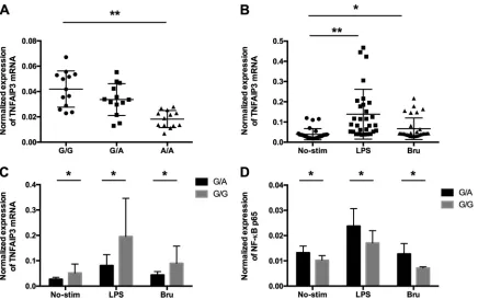
rs7749323 is associated with reduced expression of the
TNFAIP3
gene,
de-creased sensitivity to stimulations with
B. abortus
, and increased expression of
NF-
B1.
To assess the role of the rs7749323 variant in regulating the expression of the
TNFAIP3
gene, we isolated PBMCs from 39 healthy individuals with different genotypes
at the rs7749323 variant (13 G/G, 13 G/A, and 13 A/A) and determined the mRNA
expression levels of the
TNFAIP3
gene using real-time quantitative PCR assays. In line
with data from previously reported studies (25, 27), individuals carrying the protective
A allele of rs7749323 displayed significant reductions in mRNA levels of the
TNFAIP3
gene (as shown in Fig. 3A). We further assessed the expression of NF-
B1 in monocytes
with different genotypes at the rs7749323 variant. Our data showed that individuals
carrying the protective A allele demonstrated a significant increase in NF-
B1
expres-sion compared to that of carriers of the G allele (Fig. 3D). We further assessed mRNA
expression levels of the
TNFAIP3
gene in monocytes stimulated with lipopolysaccharide
(LPS) and
B. abortus. As shown in Fig. 3B, the mRNA levels of
TNFAIP3
were significantly
increased compared to those under unstimulated conditions. Interestingly, when
sam-ples were stratified by their genotypes at rs7749323, we observed that individuals
carrying the G/G genotype are more sensitive to such stimulations than are individuals
carrying the G/A genotype (Fig. 3C).
The brucellosis-associated protective A allele significantly upregulates the
expression of multiple cytokines in monocytes.
Our data showed that the protective
A allele of the rs7749323 variant reduces the expression of
TNFAIP3
and increases the
expression of NF-
B1. We further assessed expressions of multiple cytokines in
mono-cytes with various genotypes at the rs7749323 variant. As shown in Fig. 4, we detected
expressions of IL-1

, IL-6, and IL-10 in monocytes stimulated with
E. coli-derived LPS
FIG 3Protective allele A reduces mRNA expression levels ofTNFAIP3in PBMCs and monocytes. (A) The three different genotypes for the proxy variant are displayed on thexaxis. Theyaxis represents the mRNA expression level of theTNFAIP3gene. Each dot represents the expression level ofTNFAIP3for one individual; the individuals are grouped by genotype. (B) mRNA expression levels ofTNFAIP3in unstimulated monocytes (No-Stim), LPS-stimulated monocytes, andBrucella-stimulated monocytes (Bru). (C) mRNA expression levels of TNFAIP3in monocytes with G/A and G/G genotypes. (D) mRNA expression levels of NF-B1 in monocytes under resting, LPS-stimulated, andBrucella-stimulated conditions.*indicates aPvalue of⬍0.05;**indicates aPvalue of⬍0.01.
on May 16, 2020 by guest
http://jcm.asm.org/
[image:6.585.42.478.69.342.2]and
B. abortus. When comparing G/G and G/A genotypes, we observed that monocytes
with the G/A genotype showed significant increases in the expression levels of IL-1

,
IL-6, and IL-10. These data suggest that the brucellosis-associated protective A allele
reduces the gene expression level of
TNFAIP3, leading to the increased activity of NF-
B
signaling and increased expression levels of multiple cytokines in monocytes.
DISCUSSION
Increasing evidence shows that human diseases are influenced by genetic variants,
but our understanding of the mechanisms that link a DNA sequence to a disease
FIG 4Protein expression levels of IL-1, IL-6, and IL-10 are increased inB. abortus-stimulated monocytes from individuals carrying the protective A allele. Monocytes from 5 persons with the AG genotype and 5 persons with the GG genotype were separated and cultured in medium alone or withE. coli-derived LPS or heat-killedB. abortusfor 12 h. The cells were then collected, and the protein levels of IL-1(A), IL-6 (C), and IL-10 (E) were measured by intracellular staining; the IL-1 (B), IL-6 (D), and IL-10 (F) levels were higher for the TNFAIP3 AG genotype.*indicates aPvalue of⬍0.01. SSC, side scatter.
on May 16, 2020 by guest
http://jcm.asm.org/
[image:7.585.41.428.69.553.2]phenotype is limited. Large numbers of disease-susceptible nucleotide variants fall
within the noncoding region of the human genome, making it likely that they act by
modifying regulatory sequences for genes. In the present study, we investigated the
TT
⬎
A functional variants, 42 kbp downstream of the
TNFAIP3
gene, in our cohort of
1,209 healthy controls and 150 brucellosis patients. To our knowledge, this is the first
evidence that SLE-associated risk variants play a protective role in an infectious disease,
namely, brucellosis.
Brucellosis is a well-known condition with importance in animal husbandry and in
human disease in many regions of the world. Since
Brucella
species bacteria cause
intracellular infection, the robust immune response against them is mediated by
cell-mediated immunity, with a focus on macrophage activation (28).
TNFAIP3
encodes
A20, a ubiquitin-editing enzyme with a pivotal role in negatively regulating NF-
B
signaling downstream of multiple cell surface receptors (35). A20-deficient cells excrete
high levels of proinflammatory cytokines and immediately activate lymphoid and
myeloid cells. Genome-wide association studies (GWASs) of various autoimmune
dis-eases have reported significant associations with sequence variants in the vicinity of the
TNFAIP3
gene (36). We previously described a pair of functional variants (TT
⬎
A) located
in a distal enhancer of the
TNFAIP3
gene that were associated with SLE (25, 27). The
enhancer element in which the TT
⬎
A variants are found binds to the NF-
B
transcrip-tion factors p50, p65, and cREL and delivers them to the promoter region of the
TNFAIP3
gene, activating NF-
B-mediated transcription in multiple types of cells. The
SLE-associated risk A allele disrupts the binding of NF-
B transcription factors and
significantly reduces the expression of
TNFAIP3. In our previously reported study, we
also showed that TALEN-mediated TT
⬎
A enhancer knockout leads to reduced
expres-sion levels of
TNFAIP3
and significant increases in NF-
B signaling activity (26). These
data have positioned the TT
⬎
A variants as NF-
B signaling modifiers, and they might
play a role in infectious diseases. As shown in this study, the SLE-associated risk allele
was significantly associated with a decreased risk of brucellosis.
Toll-like receptor (TLR) activation leads to intracellular signaling via MyD88 and
IRAK-4, resulting in the activation of NF-
B and mitogen-activated protein kinases
(MAPKs) and the production of inflammatory cytokines (37). The TT
⬎
A functional
variants led to reduced expression levels of
TNFAIP3
and enhanced NF-
B signaling,
which predisposes individuals to autoimmune diseases while protecting them from
brucellosis. We therefore assessed the role of TT
⬎
A variants in regulating the gene
expressions of multiple cytokines. Our data showed that the increased activity of NF-
B
in monocytes results in significant increases in the expression levels of IL-1

, IL-6, and
IL-10.
IL-1

is produced by monocytes, tissue macrophages, and dendritic cells (38).
Increased levels of production of IL-1

were detected in the sera of untreated
brucel-losis patients and in the supernatants of
Brucella suis-infected macrophage cell cultures,
suggesting an important role of IL-1

in the pathogenesis of brucellosis (39, 40). After
treatment, the level of IL-1

in the serum of brucellosis patients was significantly
decreased and normalized (40). However, more studies are required to further
under-stand the mechanism by which IL-1

influences brucellosis. Although the function
of IL-6 in brucellosis is not clear, previous studies showed a significant association
between an IL-6 polymorphism and brucellosis patients in Turkey (41, 42). Further
evaluation of both the role of this IL-6 genetic variant and the function of IL-6 in
brucellosis is required. IL-10 is a cytokine synthesis inhibitory factor because of its
negative regulatory effect on cytokines, specifically IL-2 and gamma interferon (IFN-
␥
).
In a murine model, IL-10 has been shown to influence the development of brucellosis
by downregulating the response of CD4
⫹T helper cells and the secretion of IFN-
␥
(43,
44). On the other hand, IL-10 also promotes B cell antibody production. Furthermore,
a genetic variant of the IL-10 gene has been reported to influence susceptibility to
brucellosis (41, 42, 45, 46).
In addition to the TT
⬎
A polymorphic dinucleotide, the functional T
⬎
G (p.Phe127Cys)
variant rs2230926 at exon 3 of
TNFAIP3
has been reported in different populations (47).
on May 16, 2020 by guest
http://jcm.asm.org/
Follow-up studies showed that the coding variant rs2230926 reduces the activity of
A20, resulting in decreased NF-
B signaling (47). Further study of the role of this
TNFAIP3
gene coding variant in brucellosis is required for a better understanding of
how functional genetic variants influence susceptibility to brucellosis.
In summary, we demonstrated for the first time that autoimmune disease-associated
risk variants (TT
⬎
A) in the
TNFAIP3
gene play a protective role in brucellosis. The TT
⬎
A
variants are associated with reduced expression levels of
TNFAIP3
and increased NF-
B
activity. Functional studies revealed that individuals carrying the protective A allele
display increased expression levels of multiple inflammatory cytokines.
SUPPLEMENTAL MATERIAL
Supplemental material for this article may be found at
https://doi.org/10.1128/JCM
.01363-17
.
SUPPLEMENTAL FILE 1,
PDF file, 0.1 MB.
ACKNOWLEDGMENTS
We thank all individuals, including the brucellosis patients and the controls, who
participated in this study. We are grateful to the research assistants, coordinators, and
physicians who helped in the recruitment of participants.
L.L., W.B., and X.L. performed the genomic DNA isolation and SNP genotyping; L.L.,
H.S., and H.L. performed the functional studies; W.B., Y.W., K.Z., and W.G. participated in
obtaining the clinical samples and in data collection; S.W. and K.T. jointly directed this
project; S.W. wrote the manuscript, and all authors participated in proofreading.
We declare no competing financial interests.
This work was supported by the National Natural Science Foundation of China
(grants 81373143 and 81571535 to Z.T.). The funders had no role in the study design,
data collection and analysis, decision to publish, or preparation of the paper.
REFERENCES
1. Pappas G, Akritidis N, Bosilkovski M, Tsianos E. 2005. Brucellosis. N Engl J Med 352:2325–2336.https://doi.org/10.1056/NEJMra050570. 2. Dornand J, Gross A, Lafont V, Liautard J, Oliaro J, Liautard JP. 2002. The
innate immune response againstBrucellain humans. Vet Microbiol 90:383–394.https://doi.org/10.1016/S0378-1135(02)00223-7. 3. Golding B, Scott DE, Scharf O, Huang LY, Zaitseva M, Lapham C, Eller N,
Golding H. 2001. Immunity and protection againstBrucella abortus. Mi-crobes Infect 3:43– 48.https://doi.org/10.1016/S1286-4579(00)01350-2. 4. Baldwin CL, Goenka R. 2006. Host immune responses to the intracellular
bacteriaBrucella: does the bacteria instruct the host to facilitate chronic infection? Crit Rev Immunol 26:407– 442. https://doi.org/10 .1615/CritRevImmunol.v26.i5.30.
5. Zhang Q, Lenardo MJ, Baltimore D. 2017. 30 years of NF-kappaB: a blossoming of relevance to human pathobiology. Cell 168:37–57.https:// doi.org/10.1016/j.cell.2016.12.012.
6. Musone SL, Taylor KE, Nititham J, Chu C, Poon A, Liao W, Lam ET, Ma A, Kwok PY, Criswell LA. 2011. Sequencing ofTNFAIP3and association of variants with multiple autoimmune diseases. Genes Immun 12:176 –182.
https://doi.org/10.1038/gene.2010.64.
7. Lee YH, Bae SC, Choi SJ, Ji JD, Song GG. 2012. Associations between TNFAIP3gene polymorphisms and rheumatoid arthritis: a meta-analysis. Inflamm Res 61:635– 641.https://doi.org/10.1007/s00011-012-0455-5. 8. Song GG, Bae SC, Lee YH. 2013. Pathway analysis of genome-wide
association studies on rheumatoid arthritis. Clin Exp Rheumatol 31: 566 –574.
9. Suzuki T, Ikari K, Yano K, Inoue E, Toyama Y, Taniguchi A, Yamanaka H, Momohara S. 2013. PADI4 and HLA-DRB1 are genetic risks for radio-graphic progression in RA patients, independent of ACPA status: results from the IORRA cohort study. PLoS One 8:e61045.https://doi.org/10 .1371/journal.pone.0061045.
10. Hao G, Li Y, Liu J, Wo M. 2014.TNFAIP3rs2230926 polymorphisms in rheumatoid arthritis of southern Chinese Han population: a case-control study. Int J Clin Exp Pathol 7:8958 – 8961.
11. Zhu L, Wang L, Wang X, Zhou L, Liao Z, Xu L, Wu H, Ren J, Li Z, Yang L, Chen
S, Li B, Wu X, Zhou Y, Li Y. 2015. Characteristics of A20 gene polymorphisms and clinical significance in patients with rheumatoid arthritis. J Transl Med 13:215.https://doi.org/10.1186/s12967-015-0566-1.
12. Hegab MM, Abdelwahab AF, El-Sayed Yousef AM, Salem MN, El-Baz W, Abdelrhman S, Elshabacy F, Alhefny A, Abouraya W, Ibrahim SM, Ragab G. 2016. CD28 and PTPN22 are associated with susceptibility to rheu-matoid arthritis in Egyptians. Hum Immunol 77:522–526.https://doi.org/ 10.1016/j.humimm.2016.04.018.
13. Zhang MY, Yang XK, Pan HF, Ye DQ. 2016. Associations betweenTNFAIP3 gene polymorphisms and systemic lupus erythematosus risk: an updated meta-analysis. HLA 88:245–252.https://doi.org/10.1111/tan.12908. 14. Moaaz M, Mohannad N. 2016. Association of the polymorphisms of
TRAF1 (rs10818488) andTNFAIP3(rs2230926) with rheumatoid arthritis and systemic lupus erythematosus and their relationship to disease activity among Egyptian patients. Cent Eur J Immunol 41:165–175.
https://doi.org/10.5114/ceji.2016.60991.
15. Han JW, Wang Y, Li HB, Alateng C, Bai YH, Sun ZQ, Lv XX, Wu RN. 2016. Single nucleotide polymorphisms ofTNFAIP3are associated with sys-temic lupus erythematosus in Han Chinese population. Int J Immuno-genet 43:96 –100.https://doi.org/10.1111/iji.12250.
16. Elghzaly AA, Metwally SS, El-Chennawi FA, Elgayaar MA, Mosaad YM, El-Toraby EE, Hegab MM, Ibrahim SM. 2015. IRF5, PTPN22, CD28, IL2RA, KIF5A, BLK and TNFAIP3 genes polymorphisms and lupus susceptibility in a cohort from the Egypt Delta; relation to other ethnic groups. Hum Immunol 76:525–531.https://doi.org/10.1016/j.humimm.2015.06.001. 17. Kadota K, Mori M, Yanagimachi M, Miyamae T, Hara T, Kanetaka T,
Nozawa T, Kikuchi M, Hara R, Imagawa T, Kaneko T, Yokota S. 2013. Analysis of gender differences in genetic risk: association ofTNFAIP3 polymorphism with male childhood-onset systemic lupus erythemato-sus in the Japanese population. PLoS One 8:e72551.https://doi.org/10 .1371/journal.pone.0072551.
18. Cen H, Zhou M, Leng RX, Wang W, Feng CC, Li BZ, Zhu Y, Yang XK, Yang M, Zhai Y, Zhang M, Hu LF, Li R, Chen GM, Chen H, Pan HF, Li XP, Ye DQ. 2013. Genetic interaction between genes involved in NF-kappaB
on May 16, 2020 by guest
http://jcm.asm.org/
signaling pathway in systemic lupus erythematosus. Mol Immunol 56: 643– 648.https://doi.org/10.1016/j.molimm.2013.07.006.
19. Zhou XJ, Lu XL, Nath SK, Lv JC, Zhu SN, Yang HZ, Qin LX, Zhao MH, Su Y, Shen N, Li ZG, Zhang H. 2012. Gene-gene interaction of BLK, TNFSF4, TRAF1, TNFAIP3, and REL in systemic lupus erythematosus. Arthritis Rheum 64:222–231.https://doi.org/10.1002/art.33318.
20. Lee YH, Song GG. 2012. Associations betweenTNFAIP3gene polymor-phisms and systemic lupus erythematosus: a meta-analysis. Genet Test Mol Biomarkers 16:1105–1110.https://doi.org/10.1089/gtmb.2012.0096. 21. Fan Y, Tao JH, Zhang LP, Li LH, Ye DQ. 2011. The association between BANK1 andTNFAIP3 gene polymorphisms and systemic lupus erythe-matosus: a meta-analysis. Int J Immunogenet 38:151–159.https://doi.org/ 10.1111/j.1744-313X.2010.00990.x.
22. Shimane K, Kochi Y, Horita T, Ikari K, Amano H, Hirakata M, Okamoto A, Yamada R, Myouzen K, Suzuki A, Kubo M, Atsumi T, Koike T, Takasaki Y, Momohara S, Yamanaka H, Nakamura Y, Yamamoto K. 2010. The asso-ciation of a nonsynonymous single-nucleotide polymorphism inTNFAIP3 with systemic lupus erythematosus and rheumatoid arthritis in the Japanese population. Arthritis Rheum 62:574 –579. https://doi.org/10 .1002/acr.20194.
23. Kawasaki A, Ito I, Ito S, Hayashi T, Goto D, Matsumoto I, Takasaki Y, Hashimoto H, Sumida T, Tsuchiya N. 2010. Association ofTNFAIP3 poly-morphism with susceptibility to systemic lupus erythematosus in a Japanese population. J Biomed Biotechnol 2010:207578.https://doi.org/ 10.1155/2010/207578.
24. Graham RR, Cotsapas C, Davies L, Hackett R, Lessard CJ, Leon JM, Burtt NP, Guiducci C, Parkin M, Gates C, Plenge RM, Behrens TW, Wither JE, Rioux JD, Fortin PR, Graham DC, Wong AK, Vyse TJ, Daly MJ, Altshuler D, Moser KL, Gaffney PM. 2008. Genetic variants nearTNFAIP3 on 6q23 are associated with systemic lupus erythematosus. Nat Genet 40:1059 –1061.https://doi.org/10.1038/ng.200.
25. Wang S, Wen F, Wiley GB, Kinter MT, Gaffney PM. 2013. An enhancer element harboring variants associated with systemic lupus erythemato-sus engages theTNFAIP3promoter to influence A20 expression. PLoS Genet 9:e1003750.https://doi.org/10.1371/journal.pgen.1003750. 26. Wang S, Wen F, Tessneer KL, Gaffney PM. 2016. TALEN-mediated
enhancer knockout influencesTNFAIP3gene expression and mimics a molecular phenotype associated with systemic lupus erythematosus. Genes Immun 17:165–170.https://doi.org/10.1038/gene.2016.4. 27. Adrianto I, Wen F, Templeton A, Wiley G, King JB, Lessard CJ, Bates JS, Hu
Y, Kelly JA, Kaufman KM, Guthridge JM, Alarcon-Riquelme ME, Anaya JM, Bae SC, Bang SY, Boackle SA, Brown EE, Petri MA, Gallant C, Ramsey-Goldman R, Reveille JD, Vila LM, Criswell LA, Edberg JC, Freedman BI, Gregersen PK, Gilkeson GS, Jacob CO, James JA, Kamen DL, Kimberly RP, Martin J, Merrill JT, Niewold TB, Park SY, Pons-Estel BA, Scofield RH, Stevens AM, Tsao BP, Vyse TJ, Langefeld CD, Harley JB, Moser KL, Webb CF, Humphrey MB, Montgomery CG, Gaffney PM. 2011. Association of a functional variant downstream ofTNFAIP3with systemic lupus erythem-atosus. Nat Genet 43:253–258.https://doi.org/10.1038/ng.766. 28. Skendros P, Pappas G, Boura P. 2011. Cell-mediated immunity in human
brucellosis. Microbes Infect 13:134 –142.https://doi.org/10.1016/j.micinf .2010.10.015.
29. Barquero-Calvo E, Chaves-Olarte E, Weiss DS, Guzman-Verri C, Chacon-Diaz C, Rucavado A, Moriyon I, Moreno E. 2007.Brucella abortususes a stealthy strategy to avoid activation of the innate immune system during the onset of infection. PLoS One 2:e631.https://doi.org/10.1371/ journal.pone.0000631.
30. Wei P, Cui G, Lu Q, Yang L, Guan Z, Sun W, Zhao Y, Wang S, Peng Q. 2015. A20 promotesBrucellaintracellular growth via inhibition of macrophage cell death and activation. Vet Microbiol 175:50 –57.https://doi.org/10 .1016/j.vetmic.2014.11.006.
31. Zaitseva M, King LR, Manischewitz J, Dougan M, Stevan L, Golding H, Golding B. 2001. Human peripheral blood T cells, monocytes, and mac-rophages secrete macrophage inflammatory proteins 1alpha and 1beta
following stimulation with heat-inactivatedBrucella abortus. Infect Im-mun 69:3817–3826.https://doi.org/10.1128/IAI.69.6.3817-3826.2001. 32. Creery D, Angel JB, Aucoin S, Weiss W, Cameron WD, Diaz-Mitoma F,
Kumar A. 2002. Nef protein of human immunodeficiency virus and lipopolysaccharide induce expression of CD14 on human monocytes through differential utilization of interleukin-10. Clin Diagn Lab Immunol 9:1212–1221.
33. Sponaas AM, Moen SH, Liabakk NB, Feyzi E, Holien T, Kvam S, Groseth LAG, Stordal B, Buene G, Espevik T, Waage A, Standal T, Sundan A. 2015. The proportion of CD16(⫹)CD14(dim) monocytes increases with tumor cell load in bone marrow of patients with multiple myeloma. Immun Inflamm Dis 3:94 –102.https://doi.org/10.1002/iid3.53.
34. Wang Y, Li YX, Li HJ, Song HX, Zhai NC, Lou LX, Wang F, Zhang KY, Bao WG, Jin X, Su LS, Tu ZK. 2017.Brucelladysregulates monocytes and inhibits macrophage polarization through LC3-dependent autophagy. Front Immunol 8:691.https://doi.org/10.3389/fimmu.2017.00691. 35. Beyaert R, Heyninck K, Van Huffel S. 2000. A20 and A20-binding proteins
as cellular inhibitors of nuclear factor-kappa B-dependent gene expres-sion and apoptosis. Biochem Pharmacol 60:1143–1151.https://doi.org/ 10.1016/S0006-2952(00)00404-4.
36. Ma A, Malynn BA. 2012. A20: linking a complex regulator of ubiquityla-tion to immunity and human disease. Nat Rev Immunol 12:774 –785.
https://doi.org/10.1038/nri3313.
37. Oliveira SC, de Oliveira FS, Macedo GC, de Almeida LA, Carvalho NB. 2008. The role of innate immune receptors in the control ofBrucella abortusinfection: Toll-like receptors and beyond. Microbes Infect 10: 1005–1009.https://doi.org/10.1016/j.micinf.2008.07.005.
38. Dinarello CA, van der Meer JW. 2013. Treating inflammation by blocking interleukin-1 in humans. Semin Immunol 25:469 – 484.https://doi.org/ 10.1016/j.smim.2013.10.008.
39. Chen F, Ding X, Ding Y, Xiang Z, Li X, Ghosh D, Schurig GG, Sriranga-nathan N, Boyle SM, He Y. 2011. Proinflammatory caspase-2-mediated macrophage cell death induced by a rough attenuatedBrucella suisstrain. Infect Immun 79:2460 –2469.https://doi.org/10.1128/IAI.00050-11. 40. Rodriguez-Zapata M, Matias MJ, Prieto A, Jonde MA, Monserrat J,
San-chez L, Reyes E, De la Hera A, Alvarez-Mon M. 2010. Human brucellosis is characterized by an intense Th1 profile associated with a defective monocyte function. Infect Immun 78:3272–3279. https://doi.org/10 .1128/IAI.01385-09.
41. Karaoglan I, Pehlivan S, Namiduru M, Pehlivan M, Kilincarslan C, Balkan Y, Baydar I. 2009. TNF-alpha, TGF-beta, IL-10, IL-6 and IFN-gamma gene polymorphisms as risk factors for brucellosis. New Microbiol 32:173–178. 42. Budak F, Goral G, Heper Y, Yilmaz E, Aymak F, Basturk B, Tore O, Ener B, Oral HB. 2007. IL-10 and IL-6 gene polymorphisms as potential host susceptibility factors in brucellosis. Cytokine 38:32–36.https://doi.org/ 10.1016/j.cyto.2007.04.008.
43. Zhan Y, Kelso A, Cheers C. 1995. Differential activation ofBrucella-reactive CD4⫹T cells byBrucellainfection or immunization with antigenic
ex-tracts. Infect Immun 63:969 –975.
44. Scharf O, Agranovich I, Lee K, Eller NL, Levy L, Inman J, Scott DE, Golding B. 2001. Ontogeny of Th1 memory responses against aBrucella abortus conjugate. Infect Immun 69:5417–5422.https://doi.org/10.1128/IAI.69.9 .5417-5422.2001.
45. Rasouli M, Kiany S, Behbin M. 2008. Interleukin-10 gene polymorphisms and susceptibility to brucellosis in Iranian patients. Iran J Immunol 5:131–135.
46. Bravo MJ, de Dios Colmenero J, Alonso A, Caballero A. 2003. Polymor-phisms of the interferon gamma and interleukin 10 genes in human brucellosis. Eur J Immunogenet 30:433– 435. https://doi.org/10.1111/j .1365-2370.2003.00419.x.
47. Musone SL, Taylor KE, Lu TT, Nititham J, Ferreira RC, Ortmann W, Shifrin N, Petri MA, Kamboh MI, Manzi S, Seldin MF, Gregersen PK, Behrens TW, Ma A, Kwok PY, Criswell LA. 2008. Multiple polymorphisms in the TNFAIP3region are independently associated with systemic lupus ery-thematosus. Nat Genet 40:1062–1064.https://doi.org/10.1038/ng.202.