www.impactjournals.com/oncotarget/ Oncotarget, Vol. 5, No. 6
Capturing the biological impact of
CDKN2A
and
MC1R
genes as
an early predisposing event in melanoma and non melanoma
skin cancer.
Joan Anton Puig-Butille1,2, María José Escámez3,4,5, Francisco Garcia-Garcia6,7, Gemma Tell-Marti 1, Àngels Fabra8, Lucía Martínez-Santamaría3,4,5, Celia Badenas1,2, Paula Aguilera1,2, Marta Pevida5,9, Joaquín Dopazo6,7,10, Marcela del Río3,4,5, Susana Puig1,2
1 Melanoma Unit, Hospital Clinic & IDIBAPS (Institut d’Investigacions Biomèdiques Agustí Pi i Sunyer), Barcelona, Spain 2 Centro de Investigación Biomédica en Red de Enfermedades Raras (CIBERER), Barcelona, Spain.
3 Regenerative Medicine Unit. Epithelial Biomedicine Division. Centro de Investigaciones Energéticas, Medioambientales y Tecnológicas (CIEMAT), Madrid, Spain.
4 Department of Bioengineering. Universidad Carlos III (UC3M), Madrid, Spain.
5 Centro de Investigación Biomédica en Red de Enfermedades Raras (CIBERER), Madrid, Spain 6 Functional Genomics Node, National Institute of Bioinformatics, CIPF Valencia, Spain
7 Department of Bioinformatics, Centro de Investigación Príncipe Felipe, Valencia, Spain
8 Biological Clues of the Invasive and Metastatic Phenotype Group. Molecular Oncology Lab, IDIBELL, Barcelona, Spain 9 Tissue Engineering Unit. Centro Comunitario de Sangre y Tejidos del Principado de Asturias (CCST), Oviedo, Spain 10 Centro de Investigación Biomédica en Red de Enfermedades Raras (CIBERER), Valencia, Spain.
Correspondence to: Susana Puig, email: susipuig@gmail.com
Keywords: Familial Melanoma, CDKN2A, MC1R, gene expression, p16, mutations
Received: September 25, 2013 Accepted: December 16, 2013 Published: December 16, 2013
This is an open-access article distributed under the terms of the Creative Commons Attribution License, which permits unrestricted use, distribution, and reproduction in any medium, provided the original author and source are credited.
ABSTRACT:
Germline mutations in CDKN2A and/or red hair color variants in MC1R genes are associated with an increased susceptibility to develop cutaneous melanoma or non melanoma skin cancer.
We studied the impact of the CDKN2A germinal mutation p.G101W and MC1R
variants on gene expression and transcription profiles associated with skin cancer.
To this end we set-up primary skin cell co-cultures from siblings of melanoma prone-families that were later analyzed using the expression array approach.
As a result, we found that 1535 transcripts were deregulated in CDKN2A mutated cells, with over-expression of immunity-related genes (HLA-DPB1, CLEC2B, IFI44, IFI44L, IFI27, IFIT1, IFIT2, SP110 and IFNK) and down-regulation of genes playing a role in the Notch signaling pathway. 3570 transcripts were deregulated in MC1R variant carriers. In particular, genes related to oxidative stress and DNA damage pathways were up-regulated as well as genes associated with neurodegenerative diseases such as Parkinson’s, Alzheimer and Huntington.
Finally, we observed that the expression signatures indentified in phenotypically
INTRODUCTION
The worldwide incidence of skin cancer, including non melanoma skin cancer (NMSC) and cutaneous melanoma (CM), is rapidly increasing, being the most common human cancer[1]. While, both entities are
influenced by the carcinogenic effect of UV exposure, their
incidence and tumor aggressiveness differ considerably, CM being the least frequent and most aggressive form[2] .
Approximately, 10% of total melanoma cases
originate in individuals belonging to high-risk melanoma pedigrees. To date, CDKN2A is the major gene associated with the high risk inherited in melanoma prone families [3] and in multiple primary melanoma patients [4].
CDKN2A acts as a tumor suppressor gene, negatively regulating cell cycle progression and promoting cellular senescence. Recently, a role for CDKN2A in cellular
oxidative stress regulation has been suggested [5, 6].
Recurrent CDKN2A mutations have been identified in melanoma families [7-11], mutation p.G101W being the
most frequent one detected in Mediterranean pedigrees [12]. Although heterozygous loss of CDKN2A is sufficient to confer a 67% lifetime risk of melanoma [13], the
mechanisms responsible for tumor enhancement still have
to be clarified [14]. In contrast, the role of high-penetrance CDKN2A mutationsin NMSC susceptibility has not been
clearly elucidated [15, 16].
Skin cancer epidemiology is complex due to the multigenic nature of the disease. In particular, the MC1R gene is a key regulator of skin pigmentation and melanocyte differentiation, playing a central role in determining the pigmentation phenotype, sun sensitivity and tanning ability[17]. Certain MC1R polymorphisms are responsible for the red hair color (RHC) phenotype (red hair, fair skin and poor tanning response)[18] which
is associated with high UV-radiation sensitivity and skin cancer susceptibility (CM and NMSC)[19, 20].
Furthermore, MC1R variants are a modifying factor for melanoma risk in CDKN2A mutation carriers, [21-23] suggesting that multiple medium- and low-penetrance
genes may influence the risk conferred by high penetrance
melanoma genes.
To date, there is a lack of information about the mechanisms underlying the increased skin cancer risk in carriers of CDKN2A mutations in association with MC1R
variants. The effect of both genes has been separately investigated using murine-derived melanocytes and/
or focused on mono-cultured melanocytes [6, 24-26]. However, these mono-cultures exhibit phenotypic
characteristics closely mimicking those observed in transformed melanocytes, such as elevated growth rate
[27, 28] and acquired expression of melanoma-associated antigens[29, 30] which are abolished when they are co-cultured with keratinocytes[30-32]. This data indicates
that melanocyte proliferation and behavior is controlled by surrounding basal keratinocytes which regulate epidermal
homeostasis [33, 34]. Thus, the establishment and characterization of a melanocyte–keratinocyte co-culture system is essential for elucidating the early molecular events leading to skin cancer.
The aim of the present study was to investigate the global molecular effect of germinal CDKN2A mutations
(p.G101W) and MC1R RHC variants in the transcriptome of primary skin cells from individuals belonging to skin cancer prone families (familial melanoma pedigrees).
RESULTS
Impact of CDKN2A germinal mutation p.G101W on Global Transcript Profiles of skin primary cultures:
Whole genome expression profiling was performed
on melanoctye-keratinocyte co-cultures from CDKN2A
mutation carriers (samples A1 and B1 described in Table 1) and wild-type CDKN2A individuals (samples A2 and
B2). After stringent microarray data filtering, 1536 of the initial 19596 transcripts on the array were found to be differentially expressed in CDKN2A mutated cultured
cells. Specifically, 60.7% of transcripts were found to be up-regulated (933/1536) and 39.3% of them down-regulated (603/1536).
Class comparison generated a signature of 108 unique genes that significantly discriminated between CDKN2A mutation and non-mutation (p-value <0.0001,
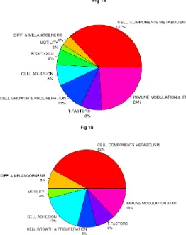
listed in Table S1). Functional categorization of those transcripts belonging to known genes (Figure 1a, Figure 1b) showed that the mutant CDKN2A signature included deregulation of a vast number of genes involved in cell
component and metabolism (37% up-regulated and 42%
down-regulated,). Notably, immunomodulation and interferon response genes represented the second most
prevalent subset of up-regulated transcripts (24%) and third down-regulated subgroup (13%) which may reflect a constitutively altered cytokine and chemokine profile
in mutant cells. The set of deregulated genes involved in immunomodulation and interferon response includes:
CLEC2B, IFI44L, IFIT1, IFI44, IFI27, IFNK, DDX58, RNF182, IFIT2, IL15, SP110, BTN3A2, RFX2, IL17D and
HLA-DPB1 (Figure 2). The molecular profile also included
deregulation of genes which may confer a phenotype with the capability for malignant transformation since they are involved in cell adhesion, cell growth and proliferation.
Over-expression of apoptosis related genes CASP3, XAF1
and SAMD9 was also detected. Transcription factors such as CEBPZ, LEF1, MACC1, TBX1, ZNF404 and ZNF8,
epidermal differentiation and melanogenesis genes such as KRT2, TYPR1, LIMS3, EEA1 and MFI2 were also components of the mutant CDKN2A signature.
analysis revealed an over-representation of
down-regulated genes from the Notch signaling pathway (hsa_04330; p=0.038) in mutant including NCOR2, PSEN, DLLC1, DVL3, NOTCH3, CDKN2A skin cells
Figure 1: Impact of germinal CDKN2A mutation on global transcript profile in melanocyte-keratinocyte co-culture system. Functional classification of the set of 78 of 108 deregulated genes in CDKN2A mutants are indicated in Fig.1a Up-regulated genes and Fig.1b Down-regulated genes. T. Factors (transcription factors), Immune modulation & IFN (Immunomodulation and interferon related genes); Cell. Components and metabolism (Cellular components and metabolism), Diff & Melanogenesis (differentiation and
[image:3.612.203.465.331.662.2]melanogenesis)
Table 1: Genotype and phenotype features of four individuals from
two melanoma pedigrees.
Pedigree A
Individual MM* CDKN2A MC1R Skin phototype Eye color Hair color
A1 5 p.G101W p.R160W, p.R151C II Green Red
A2 0 Wild-type p.R160W, p.R151C II Green Red Pedigree B
Individual MM* CDKN2A MC1R Skin phototype Eye color Hair color B1 0 p.G101W Wild-type II Blue Black
B2 0 Wild-type Wild-type II Blue Brown
Two siblings from each pedigree (A and B) were included. Each individual was considered as a “genomic condition” in the study. A total of two and three melanoma cases have been found in pedigree A and B, respectively. Abbreviations: CDKN2A: cyclin-dependent kinase inhibitor 2A; MC1R: melanocortin 1
RFNG andthe co-activator MAML1 which controls the growth-promoting effect of Notch1.
Impact of non-functional MC1R gene on Global Transcript Profiles of skin primary cultures:
MC1R analysis was carried out following the
same workflow used in the CDKN2A analysis. Whole
genome expression profiling displayed 3570 transcripts differentially expressed in RHC variant (A1 and A2) vs. wild-type MC1R skin cells (B1 and B2). Specifically, 54% of transcripts (1954/3570) were up-regulated while 46% (1616/3570) were down-regulated. By class comparison, 159 genes showed highly statistically significant differences (p < 0.0000001) (listed in Table
S2). RHC skin cells showed a down-regulation of a subset of genes categorized as melanocyte differentiation and pigmentation genes. The transcription of most of
these genes is regulated directly via the MC1R signaling pathway, or indirectly through keratinocyte differentiation genes (TYRP1, MLANA, TYR, CRNN, PADI1, GDF15, MLPH, KIT, INHBB, DAB2, KLK6, FOXC1, and NDRG4).
Furthermore, RHC variant skin cells exhibited
up-regulation of DNA damage and/or DNA repair response genes (BUB1, CHEK1, POLQ, RAD51AP1, RAD54B, RAD51C, BLM, UBE2T, GINS2, EXO1, FAM33A,PTTG2, PTTG1, RAD54L, DTL), cell cycle and proliferation genes (CDKN3, CDC25, CDK1, CCNA2, CDC20, CCNB2, CCNB1, CKS, CDCA3, PLK1,PRIM, CDCA8, GMNN, CDC6, VRK1, CKS1B, RFC4, CDC45, CENPN, HAUS8, PSRC, MCM6, MELK, AURKA, NCAPG) and microtubule-based motors kinesin superfamily genes
(KIF11, KIF15, KIF2C, KIF4A,KIF23).
The functional enrichment analysis showed a considerably higher number of altered pathways in non-functional MC1R cells than in CDKN2A mutant cells. The
[image:4.612.126.491.289.470.2]RHC skin cells displayed over-expression of genes playing
Figure 2: Set of immunomodulation and interferon response deregulated in mutant CDKN2A co-cultures. The logarithm of the Fold change (LogFC) for the set of genes is indicated for wild-type MC1R co-cultures (wt_MC1R; green) and for Red Hair Colour
MC1R variants co-cultures (RHC_MC1R; yellow).
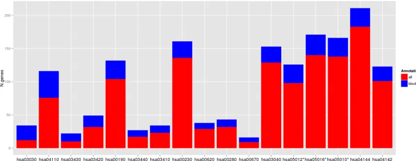
Figure 3: Deregulated pathways in RHC MC1R melanocyte-keratinocyte co-culture system. In blue are the number of total
genes annotated within the pathway, in red are the number of deregulated genes in the RHC MC1R co-cultures. The KEGG term is indicated
[image:4.612.88.517.513.679.2]a role in major DNA repair and cell homeostasis pathways
such as DNA replication, cell cycle and also oxidative
phosphorylation which leads to production of reactive
oxygen species (Figure 3 and Table 2). The lysosome and
endocytosis pathways, which are intrinsically related to melanosome transfer from melanocytes to keratinocytes, were over-represented in the set of down-regulated transcripts as a direct consequence of low pigment
production in RHC skin cells. Interestingly, RHC skin
cells also exhibited an up-regulation of genes involved
in neurodegenerative disorders such as Parkinson´s
disease (hsa05012), Huntington´s disease (hsa5016) and Alzheimer´s disease (hsa05010). Although most of the
genes were involved in several of these pathways, some of them were restricted to Huntington’s disease (AP2B1, CREB3L4, POLR2H and SOD1), to Alzheimer´s disease (NAE1, FAS) or to Parkinson’s disease (PARK7).
Besides the lack of functionality caused by p.R151C and p.R160W variants, we also analyzed a possible
effect on MC1R levels, by the relative quantification of MC1R expression. We observed a statistically significant
reduction in MC1R expression in RHC skin cells (ddCt=0.06±0.02) compared to wild-type MC1R skin
cells (ddCt=0.50±0.07; P-value<0.0001). Furthermore, the expression of FARP1, SLFN11, GFPT2, COL5A3, TYRP1, LEF1, KRT2, ST6GALNAC3, MLANA, MSMB, SILV, A2M,
and ALOX5 was evaluated by RT-PCR confirming the
results obtained in the array (data not shown).
We further examined the overlap between genes differentially expressed in both mutant CDKN2A (n=1536) and in non functional MC1R cells (n=3570). A total of 485 genes were altered in the same fashion in both
mutant CDKN2A and non functional MC1R cells and the
expression of 136 genes was found inversely correlated between both groups. Of these 136 genes (listed in Table S3), 30 were up-regulated in mutant CDKN2A and
down-regulated in non functional MC1R cells and 106
genes were down-regulated in mutant CDKN2A and up-regulated in non functional MC1R cells (Figure 4). The
functional analysis of these 136 genes did not reveal over-representation of any specific pathway.
In silico model evaluation
We hypothesized that if the altered gene signatures observed in non-lesional skin from CDKN2A mutation or RHC MC1R variant carriers are critical for malignant transformation, these patterns will be maintained across the carcinogenic process. To support our hypothesis,
the expression pattern of the most deregulated genes in
mutant CDKN2A or RHC MC1R variant skin cells was
compared to their expression observed in two previously published skin cancer datasets. The GSE2503 dataset contained the whole genome expression of five squamous cell carcinomas (SCCs) and six healthy skin samples[35]
and the GSE12391 dataset included 23 melanomas (CMs) at different stages (radial growth phase or vertical growth
phase) and 18 common nevi[36]. Due to differences
in the array platforms used among the studies, only the
expression of genes present in all datasets was evaluated (Figure 5). Thus, MC1R expression pattern analyses included 76.1% (121/159) of the most deregulated
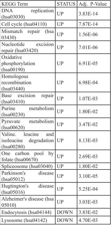
Table 2: Deregulated pathways in RHC
MC1R melanocyte-keratinocyte co-culture
system.
KEGG Term STATUS Adj. P-Value
DNA replication
(hsa03030) UP 3.83E-14 Cell cycle (hsa04110) UP 7.87E-14 Mismatch repair (hsa
03430) UP 1.56E-06
Nucleotide excision
repair (hsa03420) UP 7.01E-06 Oxidative
phosphorylation
(hsa00190) UP 6.91E-05
Homologous recombination
(hsa03440) UP 6.98E-04 Base excision repair
(hsa03410) UP 1.07E-03
Purine metabolism
(hsa00230) UP 1.80E-02
Pyruvate metabolism
(hsa00620) UP 3.47E-02 Valine. leucine and
isoleucine degradation
(hsa00280) UP 8.13E-03
One carbon pool by
folate (hsa00670) UP 2.69E-03 Spliceosome (hsa03040) UP 1.80E-02
Parkinson's disease
(hsa05012) UP 3.10E-05
Hugtington's disease
(hsa05016) UP 5.25E-04
Alzheimer's disease (hsa
05010) UP 3.03E-03
Endocytosis (hsa04144) DOWN 3.83E-02 Lysosome (hsa04142) DOWN 4.70E-03
The KEGG term is indicated for each pathway (X-axis). Status: UP:
pathway overrepresented in the group of up-regulated transcripts in
RHC MC1R; DOWN: pathway over-represented in the group of
[image:5.612.59.273.90.528.2]genes. Primary co-cultures with the non-functional
MC1R gene showed a statistically significant expression pattern (p-value <0.005) similar to those detected in SCCs (Pearson correlation=0.44) and CMs (Pearson correlation=0.25) (Figure 5a).
Because of platform disparities, the mutant
CDKN2A expression pattern was evaluated analyzing
53.7% of the most deregulated transcripts (58/108). The expression pattern correlated to those observed in SCCs (Pearson correlation=0.35) and CMs (Pearson correlation=0.28) showing a statistical tendency among datasets (p-value <0.06), in which immunomodulation
[image:6.612.116.512.145.342.2]and interferon response genes were also up-regulated (SP110, BTN3A2, IL15, IFIT2, IFI44, IFIT1, IFI44L and
Figure 4: Genes inversely expressed in mutant CDKN2A and RHC MC1R. 136 genes were found inversely expressed between
mutant CDKN2A and RHC MC1R genes (listed in Table S3). A: RHC MC1R vs wt MC1R (both mutant CDKN2A); B: mutant CDKN2A vs wt CDKN2A (both RHC MC1R); C: mutant CDKN2A vs wt CDKN2A (both wt MC1R); D: RHC MC1R vs wt MC1R (both wt CDKN2A).
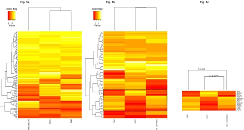
Figure 5: In-silico model evaluation. Comparison of the expression patterns detected in our study to those observed in Squamous cell Carcinomas (SSC; GSE2503 dataset) and Melanomas (MM; GSE12391 dataset).
Fig. 5A: Heatmap MC1R expression pattern showed a positive correlation among datasets (p-value <0.005).
Fig. 5B: Heatmap CDKN2A expression pattern showed a positive correlation among datasets (p-value <0.06).
Fig. 5C: Subset of genes with an inverse expression pattern between mutant CDKN2A skin cells and SSC and MM. INV: inversely correlated
pattern.
P=0.027INV
[image:6.612.61.549.390.651.2]P=0.009
CLEC2B; Figure 5b). A correlated expression pattern
was found within a subset of genes between CMs and mutant CDKN2A skin cells (Pearson Correlation=0.66, P-value = 0.009; Figure 5c). Remarkably, this subset of genes presented an expression pattern that inversely correlated between SCCs and CMs (Pearson correlation= -0.88, p-value<0.001) and also between SCCs and
mutant CDKN2A skin cells (Pearson correlation= -0.58, p-value=0.027). Specifically, the expression pattern of TBX1, CLDN17, RFX2, COL5A3, IFIT2, CEACAM1, WDR78, LXN, SLC39A8, ZNF83, RPS27L, LSM3, CTSK, and PMP22 was exclusively correlated to MM and mutant CDKN2A MM prone skin cells, and inversely with NMSC.
DISCUSSION
The aim of the study was to determine the constitutional effect of germline CDKN2A mutations or
MC1R variants in skin cancer, in order to identify early critical molecular targets implicated in the disease. To do so we analyzed co-cultured melanocyte-keratinocyte systems derived from two siblings belonging to two melanoma prone families with a founder CDKN2A
mutation and/or carrying non-functional MC1R alleles to
compare global gene expression profiles.
To date, CDKN2A is the major gene responsible for increased melanoma susceptibility in high-risk pedigrees. Germline high-penetrance mutations are
found in 10% of melanoma prone families and somatic CDKN2A alterations are also recurrent events in primary melanomas[37] and melanoma cell lines[38]. Alterations
in gene expression in as yet phenotypically normal cells
but bearing single-hit mutations in tumor suppressor genes appear to be the earliest molecular change during cancer development[39].
Altered baseline expression signatures associated
with CDKN2A mutations in cultured normal skin
fibroblasts from familial melanoma patients have already been reported [40]. However, the transcriptome
deregulation within keratinocytes and melanocytes where cutaneous carcinomas and melanomas arise have not been previously assessed. We found that transcriptome signatures were altered by single hit CDKN2A mutations in co-cultured melanocyte-keratinocyte systems. CDKN2A mutant cells exhibit mainly baseline differences in genes
related to cell components, metabolism and immune response. Melanoma cells are addicted to oncogene-driven energy production which can be mediated by somatic BRAF mutations [41], our findings indicate that
non transformed skin cells already have a deregulated
metabolic profiling which may be necessary for the skin
cancer initiation.
Previous studies suggest that genetic variation of
some cytokines or their receptors influence melanoma
susceptibility [42, 43] and even modify the risk in melanoma-prone families with CDKN2A mutations
[44]. Consistent with this hypothesis, our data reflects
the important role of innate immunity and immune response pathways which may be deregulated in early steps of melanoma development. Skin cells also
exhibited deregulation of epidermal differentiation and
melanogenesis genes. Thus, we found down-regulation of the TYRP1 gene, a melanocyte differentiation marker
and over expression of MFI2 gene, a cell-surface glycoprotein playing a role in melanoma cell proliferation
and tumorigenesis [45] and EEA1 gene, a marker of
early endosomes [46]. Interestingly, EEA1 interacts with
MFI2 regulating endosome fusion and trafficking [47].
Early endosomes are direct precursors of melanosomes, which are the specialized organelles for the biosynthesis
and storage of melanins[46]. These findings may indicate
that constitutive disorganized melanosomal structures and changes in pigment production may favormalignant transformation which in turn, could be related to autoimmune response deregulation [48, 49].
Via functional analyses, we observed a
down-regulation of the Notch signaling pathway in CDKN2A
mutant cells. This pathway is essential in
epidermal-melanocyte interactions [50, 51] and recent evidence
suggests Notch pathway as a link between the control of epidermal differentiation and proliferation and skin
homeostasis [52, 53]. Notch signaling has a dual action (as
oncogene or as tumor suppressor) in skin cancer. While, up-regulation of the Notch pathway is observed in CMs or
in SCCs at sun-protected sites, it is down-regulated in
UV-related solar keratoses SCCs or in common melanocytic
nevi (reviewed in Panelos and Massi, 2009 [54]). A
high melanocytic nevi count is the strongest known risk
factor for melanoma[55] and a potent predictor of mutant CDKN2A gene carrier status[56]. Thus, the baseline
down-regulation of the Notch signaling pathwayin CDKN2A
mutation carriers may proffer a melanocyte proliferation advantage, which can trigger common nevi development in human skin.
The pigmentation related gene MC1R acts as a moderate melanoma risk gene and variants in this gene are modifying factors for melanoma risk in CDKN2A
mutation carriers [21-23]. Furthermore, MC1R variants have also been clearly associated with elevated NMSC
susceptibility [19, 20]. We observed that the number of genes differentially expressed in non-functional MC1R
was considerably higher than in mutant CDKN2A. Both
p.R151C and p.R160W alleles halted MC1R activity and
also induced an altered cell surface molecule expression due to impaired export traffic [57] that could lead to a
reduction in MC1R gene expression levels as observed in
increased DNA damage without UV radiation exposure
were up-regulated, indicating that RHC variants contribute to cancer risk in humans increasing the DNA damage
mediated by intrinsic UV-independent mechanisms as recently observed in mice models [58]. Notably, RHC MC1R skin cells also showed up-regulated pathways related to neurodegenerative diseases. The MC1R gene is
expressed in cells of the nervous system and its activation decreases oxidative stress and has anti-inflammatory and immunomodulatory effects [59]. Co-occurrence of Parkinson’s disease (PD) and CMs [60, 61] has been
reported in epidemiological studies. The diagnosis of CM
is associated with an approximate 50% increased risk of subsequent PD[61] development whereas individuals
with PD have a two-fold increase in risk of subsequent
CM[62] development. Previous data suggests that this relation is associated with pigment-related genes[63] rather than Parkinson’s-related genes[64, 65]. Our study
further supports that MC1R is involved in the cross-link between both diseases. Thus, MC1R variants may
increase the oxidative damage in brain cells and deregulate inflammatory processes which consequently, increase the
susceptibility to neurodegenerative disorders.
Previous studies showed that many of the alterations associated with transcriptome and proteome signatures of as yet phenotypically normal cells bearing inherited alterations are also present in the corresponding form
of cancer [39]. Thus, in our study, expression patterns
detected in MM prone cells from non-lesional areas of the skin were compared with those observed in skin cancer
tumors (SCCs and CMs) [35, 36]. Although differences in the experimental designs among studies (normal skin cells vs tumoral tissue or cultured cells vs fresh-frozen tissue)
may interfere in the analysis, the expression patterns
detected in our study correlate to those observed in skin
tumors. Interestingly, the MC1R expression pattern was
more similar among studies than the CDKN2A pattern
(p-value<0.005 and p-value<0.06, respectively). These
differences are caused in part by a subset of genes showing a converse pattern which allows us to distinguish between SSCs versus CMs and mutant CDKN2A skin cells (MM prone cells). Accordingly, previous studies have detected
the inverse expression of two genes from this subset
(CEACAM1 and CTSK) between SSCs and MMs [66, 67]. Such differences may underlie the epidemiological
differences observed between MC1R and CDKN2A and skin cancer susceptibility since MC1R polymorphisms are involved in both CMM and NMSC susceptibility while CDKN2A mutations are closely related to CM susceptibility rather than SCC susceptibility.
In conclusion, we have identified baseline expression signatures in skin cells carrying germline CDKN2A mutations and RHC MC1R variants which are maintained in skin tumors (melanoma and squamous cell
carcinomas). The study identified a large catalogue of
genes in CDKN2A mutant skin cells that are closely related
to skin cancer, highlighting the role of genes involved in immune response, in melanosome biogenesis and the Notch signaling pathway Also, our data indicates that non functional MC1R variants promote DNA damage by
intrinsic UV-independent mechanisms in human skin cells.
Furthermore, our study revealed a role for MC1R in the susceptibility to neurodegenerative diseases which may
be related to its role in oxidative stress and inflammatory
processes.
METHODS
Study Design
To detect the effect of CDKN2A and MC1R genes,
two melanoma families (A and B) were selected (Table I).
The most frequent mutation,p.G101W, resulting from a common ancestor in Mediterranean pedigrees, was chosen in relation to the CDKN2A status. Regarding MC1R status, the presence of two red hair variants in each individual
(RHC: p.R151C and p.R160W) was analyzed. To reduce
the effect of intra-individual variability, two siblings from each family were selected, resulting in four different
extreme genotypic conditions: a carrier of variants in both
genes (sample A1), an individual without variants in any gene (sample B2) and two carriers of variants in one of these genes (samples A2 and B1), respectively.
The study was approved by the IRB and signed
informed consents were obtained from all individuals.
Primary keratinocyte and melanocyte co-culture
Skin biopsies from non-lesional areas were taken and human keratinocytes and melanocytes were obtained by mechanical fragmentation and enzymatic digestion
[68]. Briefly, skin biopsy fragments were digested with collagenase type I solution (2mg/ml) (Sigma–Aldrich, Gillingham, UK) for 90 minutes. The collagenase solution
was then completely eliminated and the remaining skin
biopsy was introduced into a mixture of 0.05% trypsin /0.02% EDTA (T/E) (Sigma). Every 30 min, T/E was changed for a fresh T/E mixture. Collected T/E was
inactivated with serum containing culture medium and
was centrifuged at 1400 rpm for 10 minutes. Primary
keratinocytes and melanocytes obtained by this method were cultured on a feeder layer of lethally irradiated
culture medium was changed every two days. This
co-culture system has been extensively proven to preserve stemness of keratinocytes[69-71] as well as to maintain the physiological melanocyte:keratinocyte ratio (1:40)[72,
73]. Moreover, by using the skin cells from this type of
co-culture, the donor’s phenotypic pigmentation and
UV-response features are preserved in vivo on a humanized skin mouse model[74].
RNA extraction
Total RNA isolation from primary cultures on
passage 3-4 was performed using the Trizol extraction method (Invitrogen Life Technologies, Carlsbad, CA) followed by purification in commercial columns (Qiagen, Valencia, CA). Total isolated RNA was further purified using an RNeasy kit (Qiagen, Valencia, CA).
RNA concentration was determined using a NanoDrop
Spectrophotometer (Thermo Scientific) and integrity of the RNA was verified by Bioanalyzer 2100 (Agilent, USA). The RNA integrity number was in all cases higher
than 8.
Expression array
Analysis of global expression was performed using the Whole Human Genome (4x44k) Oligo Microarray kit (G4112F, Agilent, US). The microarray contains probes from over 41,000 unique human genes and transcripts, all with public OMIM annotations (RefSeq, Goldenpath, Ensembl, Unigene Human Genome and GenBank databases). Overall, 50 ng of RNA were labeled using Low input Quickamp Labeling kit (Agilent, US). In all samples 10 commercial controls probes were added
in order to standardize the results (RNA Spike-in kit,
one color, Agilent, US). The arrays were scanned using the DNA Microarray Scanner G2565CA (Agilent, US). Finally, Feature Extraction Software (FES, Agilent, USA)
was used both to perform the quality control process and
to extract the information. Three replicates from each
primary culture were analyzed.
Quantitative real-time reverse transcriptase polymerase chain reaction (qRT-PCR) of selected genes
To confirm the microarray results, the expression
of 13 genes (FARP1, SLFN11, GFPT2, COL5A3, TYRP1, LEF1, KRT2, ST6GALNAC3, MLANA, MSMB, SILV, A2M, and ALOX5) was validated by RT-PCR. Also, MC1R expression was evaluated. The ACTB gene was used for normalization. RNA derived from donors’ healthy skin
was used as a calibrator in each reaction. A total of 200 ng
of RNA was retrotranscribed to cDNA using Taqman PCR
Core Reagent kit (Roche Applied Science, Penzbergf, Germany). Real-time PCR was performed using Taqman
Universal PCR master Mix (Roche Applied Science, Penzbergf, Germany). Reaction was performed in an ABI
Step One plus RT-PCR sequence detection instrument
(Applied Biosystems, CA, US). Primer design and gene-specific probe selection were carried out by the Universal Probe Library software (UPL, Roche, Mannheim,
Germany).
Data was evaluated using the relative quantification method of ddCt[75]. Expression values were evaluated by T-test for equality means using the SPSS 17.0. P-values less than or equal to 0.05 were considered statistically significant.
Microarray data and statistical analyses
The Agilent Processed Signal was standardized
across arrays using quantile normalization[76]. Differential gene expression analysis was carried out using
the limma package from Bioconductor. Multiple testing adjustments of p-values were performed according to Benjamini and Hochberg methodology[77].
Gene set analysis was carried out for the Kyoto Encyclopedia of Genes and Genomes pathways (KEGG) using FatiScan[78] in Babelomics[79]. This tool detects
significantly up- or down-regulated blocks of functionally
related genes in lists of genes ordered by different criteria
such as differential expression, KEGG pathways and
others. The core of the method is based on an algorithm to test whether a set of genes, labeled with terms (biological
information), contain significant enrichments of one
or several of these terms with respect to another set of
reference genes. FatiScan uses a Fisher’s exact test for
2×2 contingency tables for comparing two groups of genes
and extracting a list of KEGG terms whose distribution among the groups is significantly different. Given that
many KEGG terms are simultaneously tested, the results of the test are corrected for multiple testing to obtain an adjusted p-value. FatiScan returns adjusted p-values based on the False Discovery Rate (FDR) method[77]. KEGG Pathways for the genes in the microarray where taken from the KEGG web.
In order to gain insight into the co-regulation patterns on transcript expression profiles, data from our microarray experiments were clustered together with expression data obtained from two previous experiments focused on NMSC (GSE2503 dataset[35]) and MM (GSE12391 dataset[36]). Raw data were obtained from the Gene Expression Omnibus. For each dataset we analyzed the differential expression and determined the
fold-changes. We selected two subsets: genes differentially
expressed in our study for the MC1R comparison (RHC alleles vs. wild-type MC1R) and a second group of genes
scenario, fold-changes were normalized using the quantile
method[76] to compare differential expression results in all experiments. We performed a hierarchical clustering analysis of differentially expressed transcripts and the
graphical representation showed the relationship between
experiments. The correlation analysis quantified the linear
relationship between the different studies m
Supplemental Data Description
Supplemental Data includes two tables with the list of the most deregulated genes associated with CDKN2A
mutations or MC1R variants; and one table containing the
list of genes inversely deregulated between CDKN2A and MC1R.
ACKNOWLEDGMENTS
The present project was founded by a grant from
CIBER de Enfermedades Raras of the Instituto de Salud Carlos III, Spain (INTRA/07/726.2). The research at the Melanoma Unit in Barcelona is partially funded by Grants from Fondo de Investigaciones Sanitarias (P.I. 09/01393 and P.I. 12/00840, co-funded by “Fondo Europeo de Desarrollo Regional (FEDER). Unión Europea. Una manera de hacer Europa”); by the AGAUR 2009 SGR 1337 of the Catalan Government, Spain; by the European Commission under the 6th Framework Programme, Contract nr: LSHC-CT-2006-018702 (GenoMEL) and by the National Cancer Institute (NCI) of the US National Institute of Health (NIH) (CA83115). This work is also partly supported by grants BIO2008-04212 and SAF2010-16976 from the Spanish Ministry of Science and Innovation (MICINN), PROMETEO/2010/001 from the GVA-FEDER and S2010/BMD-2420 (CELLCAM) from
Comunidad de Madrid.
Web Resources:
Bioconductor (http://www.bioconductor.org) Babelomics (http://babelomics.bioinfo.cipf.es/ ) Kyoto Encyclopedia of Genes and Genomes pathways (KEGG) (http://www.genome.jp/kegg/)
Ensembl 56 release (http://www.ensembl.org) Gene Expression Omnibus (http://www.ncbi.nlm.
nih.gov/geo/)
Accession Numbers The GEO accession number for
the expression microarray data reported in this paper is GSE44805.
http://www.ncbi.nlm.nih.gov/geo/query/acc.cgi?tok
en=ptorruosgomwqds&acc=GSE44805
Conflict of Interest:
The authors state no conflict of interest.
REFERENCES
1. Siegel R, Naishadham D and Jemal A. Cancer statistics, 2012. CA Cancer J Clin. 2012; 62(1):10-29.
2. Thompson JF, Scolyer RA and Kefford RF. Cutaneous melanoma in the era of molecular profiling. Lancet. 2009; 374(9687):362-365.
3. Goldstein AM, Chan M, Harland M, Hayward NK, Demenais F, Bishop DT, Azizi E, Bergman W,
Bianchi-Scarra G, Bruno W, Calista D, Albright LA, Chaudru V,
Chompret A, Cuellar F, Elder DE, et al. Features associated with germline CDKN2A mutations: a GenoMEL study of
melanoma-prone families from three continents. J Med Genet. 2007; 44(2):99-106.
4. Puig S, Malvehy J, Badenas C, Ruiz A, Jimenez D, Cuellar F, Azon A, Gonzalez U, Castel T, Campoy A, Herrero J, Marti R, Brunet-Vidal J and Mila M. Role of the CDKN2A locus in patients with multiple primary melanomas. J Clin Oncol. 2005; 23(13):3043-3051.
5. Liontos M, Pateras IS, Evangelou K and Gorgoulis VG. The Tumor Suppressor Gene ARF as a Sensor of Oxidative Stress. Curr Mol Med. 2012.
6. Jenkins NC, Liu T, Cassidy P, Leachman SA, Boucher
KM, Goodson AG, Samadashwily G and Grossman D. The
p16(INK4A) tumor suppressor regulates cellular oxidative stress. Oncogene. 2011; 30(3):265-274.
7. Lang J, Boxer M and MacKie RM. CDKN2A mutations
in Scottish families with cutaneous melanoma: results
from 32 newly identified families. Br J Dermatol. 2005; 153(6):1121-1125.
8. Larre Borges A, Cuellar F, Puig-Butille JA, Scarone M, Delgado L, Badenas C, Mila M, Malvehy J, Barquet V, Nunez J, Laporte M, Fernandez G, Levrero P,
Martinez-Asuaga M and Puig S. CDKN2A mutations in melanoma
families from Uruguay. Br J Dermatol. 2009; 161(3):536-541.
9. Yakobson E, Eisenberg S, Isacson R, Halle D, Levy-Lahad E, Catane R, Safro M, Sobolev V, Huot T, Peters G, Ruiz A, Malvehy J, Puig S, Chompret A, Avril MF, Shafir R, et al. A single Mediterranean, possibly Jewish, origin for the Val59Gly CDKN2A mutation in four melanoma-prone families. Eur J Hum Genet. 2003; 11(4):288-296.
10. Majore S, De Simone P, Crisi A, Eibenschutz L, Binni F, Antigoni I, De Bernardo C, Catricala C and Grammatico P. CDKN2A/CDK4 molecular study on 155 Italian subjects
with familial and/or primary multiple melanoma. Pigment
Cell Melanoma Res. 2008; 21(2):209-211.
11. Eliason MJ, Larson AA, Florell SR, Zone JJ,
in Utah melanoma families. J Invest Dermatol. 2006; 126(3):660-666.
12. Ciotti P, Struewing JP, Mantelli M, Chompret A, Avril
MF, Santi PL, Tucker MA, Bianchi-Scarra G, Bressac-de Paillerets B and Goldstein AM. A single genetic origin for
the G101W CDKN2A mutation in 20 melanoma-prone families. Am J Hum Genet. 2000; 67(2):311-319.
13. Bishop DT, Demenais F, Goldstein AM, Bergman W,
Bishop JN, Bressac-de Paillerets B, Chompret A, Ghiorzo P, Gruis N, Hansson J, Harland M, Hayward N, Holland EA, Mann GJ, Mantelli M, Nancarrow D, et al. Geographical
variation in the penetrance of CDKN2A mutations for
melanoma. J Natl Cancer Inst. 2002; 94(12):894-903. 14. Talantov D, Mazumder A, Yu JX, Briggs T, Jiang Y,
Backus J, Atkins D and Wang Y. Novel genes associated
with malignant melanoma but not benign melanocytic
lesions. Clin Cancer Res. 2005; 11(20):7234-7242. 15. Jefferies S, Edwards SM, Hamoudi RA, A’Hern R, Foulkes
W, Goldgar D and Eeles R. No germline mutations in
CDKN2A (p16) in patients with squamous cell cancer of the head and neck and second primary tumours. Br J Cancer. 2001; 85(9):1383-1386.
16. Cabanillas R, Astudillo A, Valle M, de la Rosa J, Alvarez R, Duran NS and Cadinanos J. Novel germline CDKN2A
mutation associated with head and neck squamous cell
carcinomas and melanomas. Head Neck. 2011.
17. Rees JL. The genetics of sun sensitivity in humans. Am J Hum Genet. 2004; 75(5):739-751.
18. Valverde P, Healy E, Jackson I, Rees JL and Thody AJ. Variants of the melanocyte-stimulating hormone receptor
gene are associated with red hair and fair skin in humans.
Nat Genet. 1995; 11(3):328-330.
19. Bastiaens MT, ter Huurne JA, Kielich C, Gruis NA, Westendorp RG, Vermeer BJ and Bavinck JN.
Melanocortin-1 receptor gene variants determine the risk of nonmelanoma skin cancer independently of fair skin and
red hair. Am J Hum Genet. 2001; 68(4):884-894.
20. Box NF, Duffy DL, Irving RE, Russell A, Chen W,
Griffyths LR, Parsons PG, Green AC and Sturm RA. Melanocortin-1 receptor genotype is a risk factor for basal
and squamous cell carcinoma. J Invest Dermatol. 2001; 116(2):224-229.
21. Box NF, Duffy DL, Chen W, Stark M, Martin NG, Sturm RA and Hayward NK. MC1R genotype modifies risk of
melanoma in families segregating CDKN2A mutations. Am
J Hum Genet. 2001; 69(4):765-773.
22. Goldstein AM, Chaudru V, Ghiorzo P, Badenas C, Malvehy J, Pastorino L, Laud K, Hulley B, Avril MF, Puig-Butille JA, Miniere A, Marti R, Chompret A, Cuellar F, Kolm I,
Mila M, et al. Cutaneous phenotype and MC1R variants as modifying factors for the development of melanoma in
CDKN2A G101W mutation carriers from 4 countries. Int J Cancer. 2007; 121(4):825-831.
23. Demenais F, Mohamdi H, Chaudru V, Goldstein AM,
Newton Bishop JA, Bishop DT, Kanetsky PA, Hayward
NK, Gillanders E, Elder DE, Avril MF, Azizi E, van Belle P, Bergman W, Bianchi-Scarra G, Bressac-de Paillerets B, et al. Association of MC1R variants and host phenotypes with melanoma risk in CDKN2A mutation carriers: a
GenoMEL study. J Natl Cancer Inst. 2010; 102(20):1568-1583.
24. Herraiz C, Jimenez-Cervantes C, Zanna P and Garcia-Borron JC. Melanocortin 1 receptor mutations impact
differentially on signalling to the cAMP and the ERK mitogen-activated protein kinase pathways. FEBS Lett.
2009; 583(19):3269-3274.
25. Le Pape E, Passeron T, Giubellino A, Valencia JC, Wolber R and Hearing VJ. Microarray analysis sheds light on the
dedifferentiating role of agouti signal protein in murine
melanocytes via the Mc1r. Proc Natl Acad Sci U S A. 2009; 106(6):1802-1807.
26. Sviderskaya EV, Hill SP, Evans-Whipp TJ, Chin L, Orlow SJ, Easty DJ, Cheong SC, Beach D, DePinho RA and Bennett DC. p16(Ink4a) in melanocyte senescence and differentiation. J Natl Cancer Inst. 2002; 94(6):446-454.
27. Eisinger M and Marko O. Selective proliferation of normal human melanocytes in vitro in the presence of phorbol
ester and cholera toxin. Proc Natl Acad Sci U S A. 1982; 79(6):2018-2022.
28. Herlyn M, Rodeck U, Mancianti M, Cardillo FM, Lang A, Ross AH, Jambrosic J and Koprowski H. Expression of
melanoma-associated antigens in rapidly dividing human
melanocytes in culture. Cancer Res. 1987; 47(12):3057-3061.
29. Elder DE, Rodeck U, Thurin J, Cardillo F, Clark WH, Stewart R and Herlyn M. Antigenic profile of tumor
progression stages in human melanocytic nevi and
melanomas. Cancer Res. 1989; 49(18):5091-5096. 30. Valyi-Nagy IT, Hirka G, Jensen PJ, Shih IM, Juhasz I and
Herlyn M. Undifferentiated keratinocytes control growth, morphology, and antigen expression of normal melanocytes through cell-cell contact. Lab Invest. 1993; 69(2):152-159. 31. Hsu MY, Meier FE, Nesbit M, Hsu JY, Van Belle P, Elder
DE and Herlyn M. E-cadherin expression in melanoma cells
restores keratinocyte-mediated growth control and
down-regulates expression of invasion-related adhesion receptors. Am J Pathol. 2000; 156(5):1515-1525.
32. Phillips J, Gawkrodger DJ, Caddy CM, Hedley S, Dawson
RA, Smith-Thomas L, Freedlander E and Mac Neil S.
Keratinocytes suppress TRP-1 expression and reduce cell
number of co-cultured melanocytes - implications for
grafting of patients with vitiligo. Pigment Cell Res. 2001; 14(2):116-125.
33. Haass NK, Smalley KS, Li L and Herlyn M. Adhesion, migration and communication in melanocytes and
melanoma. Pigment Cell Res. 2005; 18(3):150-159.
mammalian epidermal melanocytes. Pigment Cell Res.
2005; 18(1):2-12.
35. Nindl I, Dang C, Forschner T, Kuban RJ, Meyer T, Sterry W and Stockfleth E. Identification of differentially expressed genes in cutaneous squamous cell carcinoma by microarray expression profiling. Mol Cancer. 2006; 5:30. 36. Scatolini M, Grand MM, Grosso E, Venesio T, Pisacane A,
Balsamo A, Sirovich R, Risio M and Chiorino G. Altered
molecular pathways in melanocytic lesions. Int J Cancer. 2010; 126(8):1869-1881.
37. de Snoo FA and Hayward NK. Cutaneous melanoma
susceptibility and progression genes. Cancer Lett. 2005; 230(2):153-186.
38. Ogbah Z, Puig-Butille JA, Simonetta F, Badenas C, Cervera R, Mila J, Benitez D, Malvehy J, Vilella R and Puig S.
Molecular characterization of human cutaneous
melanoma-derived cell lines. Anticancer Res. 2012; 32(4):1245-1251. 39. Herbert BS, Chanoux RA, Liu Y, Baenziger PH, Goswami
CP, McClintick JN, Edenberg HJ, Pennington RE, Lipkin
SM and Kopelovich L. A molecular signature of normal breast epithelial and stromal cells from Li-Fraumeni
syndrome mutation carriers. Oncotarget. 2012;
1(6):405-422.
40. Fan M, Pfeffer SR, Lynch HT, Cassidy P, Leachman S,
Pfeffer LM and Kopelovich L. Altered transcriptome
signature of phenotypically normal skin fibroblasts
heterozygous for CDKN2A in familial melanoma: relevance
to early intervention. Oncotarget. 2013; 4(1):128-141.
41. Hall A, Meyle KD, Lange MK, Klima M, Sanderhoff M,
Dahl C, Abildgaard C, Thorup K, Moghimi SM, Jensen PB, Bartek J, Guldberg P and Christensen C. Dysfunctional oxidative phosphorylation makes malignant melanoma cells addicted to glycolysis driven by the (V600E)BRAF oncogene. Oncotarget. 2013; 4(4):584-599.
42. Gu F, Qureshi AA, Niu T, Kraft P, Guo Q, Hunter DJ and Han J. Interleukin and interleukin receptor gene
polymorphisms and susceptibility to melanoma. Melanoma
Res. 2008; 18(5):330-335.
43. Howell WM, Turner SJ, Bateman AC and Theaker JM. IL-10 promoter polymorphisms influence tumour development in cutaneous malignant melanoma. Genes Immun. 2001; 2(1):25-31.
44. Yang XR, Pfeiffer RM, Wheeler W, Yeager M, Chanock S, Tucker MA and Goldstein AM. Identification of modifier
genes for cutaneous malignant melanoma in
melanoma-prone families with and without CDKN2A mutations. Int J Cancer. 2009; 125(12):2912-2917.
45. Guo J, Wen DR, Huang RR, Paul E, Wunsch P, Itakura E and Cochran AJ. Detection of multiple
melanoma-associated markers in melanoma cell lines by RT in situ
PCR. Exp Mol Pathol. 2003; 74(2):140-147.
46. Raposo G and Marks MS. The dark side of
lysosome-related organelles: specialization of the endocytic pathway
for melanosome biogenesis. Traffic. 2002; 3(4):237-248.
47. Ramanathan HN and Ye Y. The p97 ATPase associates
with EEA1 to regulate the size of early endosomes. Cell
Res. 2011; 22(2):346-359.
48. Sakai C, Kawakami Y, Law LW, Furumura M and Hearing VJ, Jr. Melanosomal proteins as melanoma-specific immune targets. Melanoma Res. 1997; 7(2):83-95.
49. Hearing VJ. Biochemical control of melanogenesis and melanosomal organization. J Investig Dermatol Symp Proc. 1999; 4(1):24-28.
50. Schouwey K, Delmas V, Larue L, Zimber-Strobl U, Strobl LJ, Radtke F and Beermann F. Notch1 and Notch2 receptors influence progressive hair graying in a dose-dependent manner. Dev Dyn. 2007; 236(1):282-289.
51. Okuyama R, Tagami H and Aiba S. Notch signaling: its role
in epidermal homeostasis and in the pathogenesis of skin
diseases. J Dermatol Sci. 2008; 49(3):187-194.
52. Xin Y, Lu Q and Li Q. IKK1 control of epidermal differentiation is modulated by notch signaling. Am J Pathol. 2011; 178(4):1568-1577.
53. Lin HY, Kao CH, Lin KM, Kaartinen V and Yang
LT. Notch signaling regulates late-stage epidermal differentiation and maintains postnatal hair cycle
homeostasis. PLoS One. 2011; 6(1):e15842.
54. Panelos J and Massi D. Emerging role of Notch signaling in
epidermal differentiation and skin cancer. Cancer Biol Ther.
2009; 8(21):1986-1993.
55. Chang YM, Newton-Bishop JA, Bishop DT, Armstrong BK, Bataille V, Bergman W, Berwick M, Bracci PM, Elwood JM, Ernstoff MS, Green AC, Gruis NA, Holly EA, Ingvar C, Kanetsky PA, Karagas MR, et al. A pooled
analysis of melanocytic nevus phenotype and the risk of
cutaneous melanoma at different latitudes. Int J Cancer. 2009; 124(2):420-428.
56. Bishop JA, Wachsmuth RC, Harland M, Bataille V, Pinney E, Mac KP, Baglietto L, Cuzick J and Bishop DT.
Genotype/phenotype and penetrance studies in melanoma
families with germline CDKN2A mutations. J Invest Dermatol. 2000; 114(1):28-33.
57. Sanchez-Laorden BL, Herraiz C, Valencia JC, Hearing VJ, Jimenez-Cervantes C and Garcia-Borron JC. Aberrant trafficking of human melanocortin 1 receptor variants
associated with red hair and skin cancer: Steady-state
retention of mutant forms in the proximal golgi. J Cell Physiol. 2009; 220(3):640-654.
58. Mitra D, Luo X, Morgan A, Wang J, Hoang MP, Lo J, Guerrero CR, Lennerz JK, Mihm MC, Wargo JA, Robinson KC, Devi SP, Vanover JC, D’Orazio JA, McMahon M,
Bosenberg MW, et al. An ultraviolet-radiation-independent pathway to melanoma carcinogenesis in the red hair/fair
skin background. Nature. 2012; 491(7424):449-453. 59. Catania A. Neuroprotective actions of melanocortins: a
therapeutic opportunity. Trends Neurosci. 2008; 31(7):353-360.
relationship between Parkinson disease and melanoma.
Neurology. 2011; 76(23):2002-2009.
61. Olsen JH, Friis S and Frederiksen K. Malignant melanoma
and other types of cancer preceding Parkinson disease.
Epidemiology. 2006; 17(5):582-587.
62. Olsen JH, Friis S, Frederiksen K, McLaughlin JK,
Mellemkjaer L and Moller H. Atypical cancer pattern in
patients with Parkinson’s disease. Br J Cancer. 2005; 92(1):201-205.
63. Gao X, Simon KC, Han J, Schwarzschild MA and Ascherio
A. Genetic determinants of hair color and Parkinson’s
disease risk. Ann Neurol. 2009; 65(1):76-82.
64. Meng S, Song F, Chen H, Gao X, Amos CI, Lee JE, Wei Q, Qureshi AA and Han J. No association between Parkinson
disease alleles and the risk of melanoma. Cancer Epidemiol
Biomarkers Prev. 2011; 21(1):243-245.
65. Hassin-Baer S, Laitman Y, Azizi E, Molchadski I,
Galore-Haskel G, Barak F, Cohen OS and Friedman E. The
leucine rich repeat kinase 2 (LRRK2) G2019S substitution
mutation. Association with Parkinson disease, malignant
melanoma and prevalence in ethnic groups in Israel. J Neurol. 2009; 256(3):483-487.
66. Shinozuka K, Uzawa K, Fushimi K, Yamano Y, Shiiba M, Bukawa H, Yokoe H and Tanzawa H. Downregulation of
carcinoembryonic antigen-related cell adhesion molecule 1 in oral squamous cell carcinoma: correlation with
tumor progression and poor prognosis. Oncology. 2009; 76(6):387-397.
67. Quintanilla-Dieck MJ, Codriansky K, Keady M, Bhawan J and Runger TM. Cathepsin K in melanoma invasion. J Invest Dermatol. 2008; 128(9):2281-2288.
68. Rheinwald JG and Green H. Serial cultivation of strains
of human epidermal keratinocytes: the formation of
keratinizing colonies from single cells. Cell. 1975; 6(3):331-343.
69. Llames SG, Del Rio M, Larcher F, Garcia E, Garcia M, Escamez MJ, Jorcano JL, Holguin P and Meana A.
Human plasma as a dermal scaffold for the generation of a completely autologous bioengineered skin. Transplantation.
2004; 77(3):350-355.
70. Del Rio M, Larcher F, Serrano F, Meana A, Munoz M, Garcia M, Munoz E, Martin C, Bernad A and Jorcano JL. A preclinical model for the analysis of genetically modified human skin in vivo. Hum Gene Ther. 2002; 13(8):959-968.
71. Larcher F, Dellambra E, Rico L, Bondanza S, Murillas
R, Cattoglio C, Mavilio F, Jorcano JL, Zambruno G and
Del Rio M. Long-term engraftment of single genetically
modified human epidermal holoclones enables safety pre-assessment of cutaneous gene therapy. Mol Ther. 2007; 15(9):1670-1676.
72. De Luca M, Franzi AT, D’Anna F, Zicca A, Albanese
E, Bondanza S and Cancedda R. Coculture of human keratinocytes and melanocytes: differentiated melanocytes are physiologically organized in the basal layer of the
cultured epithelium. Eur J Cell Biol. 1988; 46(1):176-180.
73. Guerra L, Capurro S, Melchi F, Primavera G, Bondanza S, Cancedda R, Luci A, De Luca M and Pellegrini G. Treatment of “stable” vitiligo by Timedsurgery and transplantation of cultured epidermal autografts. Arch
Dermatol. 2000; 136(11):1380-1389.
74. Garcia M, Llames S, Garcia E, Meana A, Cuadrado N,
Recasens M, Puig S, Nagore E, Illera N, Jorcano JL, Del Rio M and Larcher F. In vivo assessment of acute UVB responses in normal and Xeroderma Pigmentosum (XP-C) skin-humanized mouse models. Am J Pathol. 2010; 177(2):865-872.
75. Livak KJ and Schmittgen TD. Analysis of relative gene expression data using real-time quantitative PCR and the 2(-Delta Delta C(T)) Method. Methods. 2001; 25(4):402-408.
76. Bolstad BM, Irizarry RA, Astrand M and Speed TP. A
comparison of normalization methods for high density oligonucleotide array data based on variance and bias.
Bioinformatics. 2003; 19(2):185-193.
77. Benjamini Y, Drai D, Elmer G, Kafkafi N and Golani I.
Controlling the false discovery rate in behavior genetics
research. Behav Brain Res. 2001; 125(1-2):279-284. 78. Al-Shahrour F, Arbiza L, Dopazo H, Huerta-Cepas J,
Minguez P, Montaner D and Dopazo J. From genes to
functional classes in the study of biological systems. BMC
Bioinformatics. 2007; 8:114.
79. Medina I, Carbonell J, Pulido L, Madeira SC, Goetz S, Conesa A, Tarraga J, Pascual-Montano A, Nogales-Cadenas R, Santoyo J, Garcia F, Marba M, Montaner D and Dopazo J. Babelomics: an integrative platform for the analysis
of transcriptomics, proteomics and genomic data with