R E S E A R C H
Open Access
Developmental dynamics of gene
expression and alternative polyadenylation
in the
Caenorhabditis elegans
germline
Sean M. West
1†, Desirea Mecenas
1†, Michelle Gutwein
1†, David Aristizábal-Corrales
1, Fabio Piano
1,2*and Kristin C. Gunsalus
1,2*Abstract
Background:The 3′untranslated regions (UTRs) of mRNAs play a major role in post-transcriptional regulation of gene expression. Selection of transcript cleavage and polyadenylation sites is a dynamic process that produces multiple transcript isoforms for the same gene within and across different cell types. Using LITE-Seq, a new quantitative method to capture transcript 3′ends expressed in vivo, we have characterized sex- and cell type-specific transcriptome-wide changes in gene expression and 3′UTR diversity inCaenorhabditis elegansgermline cells undergoing proliferation and differentiation.
Results:We show that nearly half of germline transcripts are alternatively polyadenylated, that differential regulation of endogenous 3′UTR variants is common, and that alternative isoforms direct distinct spatiotemporal protein expression patterns in vivo. Dynamic expression profiling also reveals temporal regulation of X-linked gene expression, selective stabilization of transcripts, and strong evidence for a novel developmental program that promotes nucleolar dissolution in oocytes. We show that the RNA-binding protein NCL-1/Brat is a posttranscriptional regulator of numerous ribosome-related transcripts that acts through specific U-rich binding motifs to down-regulate mRNAs encoding ribosomal protein subunits, rRNA processing factors, and tRNA synthetases.
Conclusions:These results highlight the pervasive nature and functional potential of patterned gene and isoform expression during early animal development.
Background
Spatio-temporal regulation of gene activity is critical for the development of multicellular organisms and occurs at multiple levels. Post-transcriptional regulation influ-ences messenger RNA (mRNA) processing [1], stability [2], localization [3, 4], and translation [5–8]. An mRNA’s fate depends on internal cis-regulatory sequences that are most commonly located in their 3′untranslated re-gions (3′UTRs), and mutations within these have been associated with cancers and other diseases [9]. Func-tional elements in transcripts are recognized by trans-acting factors, including both RNA binding proteins (RBPs) and small regulatory RNAs [10, 11]. In the C.
elegans germline, a complex network of RBPs and microRNAs controls germ cell proliferation and differ-entiation by regulating the expression patterns of nu-merous genes through their 3′UTRs [5, 12–15].
Germ cells are produced in assembly-line fashion in the gonad, progressing from mitotic proliferation in the distal region through meiosis and differentiation into oocytes and sperm in the proximal region. This develop-mental program depends on the spatio-temporal restric-tion of RBP activity, which is mediated largely by translational control of their own mRNAs through their 3′UTRs [14, 16, 17]. For example GLD-1, which is in-volved in the transition from mitosis to meiosis and oo-cyte differentiation [18–20], binds multiple targets [21] to both repress translation [22] and stabilize transcripts [17, 18, 23]. Translation of GLD-1 mRNA is negatively regulated by another RBP, FBF-1, which promotes mi-totic proliferation and represses transcripts required for * Correspondence:fp1@nyu.edu;kcg1@nyu.edu
†Equal contributors
1Center for Genomics & Systems Biology, Department of Biology, New York
University, New York, NY 10012, USA
Full list of author information is available at the end of the article
meiotic entry [24, 25]. Thousands of maternal tran-scripts are deposited into the embryo [26, 27], where a combinatorial code of translational regulators controls key developmental patterning events [8, 17, 28, 29].
The selection of cleavage and polyadenylation (CPA) sites during mRNA processing determines the sequence content in 3′UTRs, and thus the landscape of cis-regulatory elements available to trans-acting factors. Since longer 3′UTRs have more regulatory potential than shorter 3′UTRs, alternative polyadenylation (APA) can have significant functional consequences [30, 31]. 3′ UTR variation during development and in different cell types is most commonly achieved by regulated selection of proximal or distal APA sites downstream of a shared terminal coding region, but can also affect coding se-quences [31–39]. About half of genes in yeast, plants, worms, flies, zebrafish, mice, and humans produce mul-tiple 3′UTR isoforms through APA, indicating that regu-lated isoform selection is a deeply conserved and widespread gene regulatory mechanism [34, 37, 40–47].
The production of alternative 3′UTR isoforms is coor-dinated with cell state and is influenced by cell-type-specific differences in chromatin structure, epigenetic marks, transcriptional rates, splicing and 3′-end process-ing factors, and competitive bindprocess-ing of regulatory RBPs [31, 48]. Differentiated cells, and neurons in particular, tend to display longer 3′UTRs [36], and progressive shifts toward longer 3′UTR lengths are observed during development in both worm and mouse [35, 43]. Con-versely, proliferating cells generally have shorter 3′UTRs [32] and inducing pluripotency correlates with 3′UTR shortening [49]. In C. elegans, genome-wide 3′UTR
studies have documented widespread APA in different developmental stages [43, 44] and tissues [38, 39]. The body of evidence thus suggests that target site accessibil-ity for factors that recognize functional elements in 3′ UTRs is an important regulatory platform for mRNAs that is integrated into developmental programs control-ling specialized cellular functions [50, 51].
Because post-transcriptional regulation of maternal mRNAs plays a key role in the C. elegans germline and early embryo [8, 16, 17, 29, 52, 53], we hypothesized that APA may serve as a mechanism to modulate gene ex-pression between progressive cellular stages within the gonad. However, previous studies have lacked the reso-lution to examine transcriptome-wide spatio-temporal dynamics during gametogenesis [27, 32, 43, 54–57]. Here, we present a map of 3′UTR isoform expression across different regions of the adult hermaphrodite germline, based on a novel method we developed that quantifies transcript levels and precise 3′ termini using small samples of RNA. By analyzing 3′UTR isoforms in dissected samples, we characterize the dynamics of gene expression and APA throughout gametogenesis and
demonstrate isoform- and cell-type-specific variation be-tween sexes and within distinct regions of the female germline. We show that this can give rise to differential protein expression patterns, using a dual-color in vivo reporter system that simultaneously monitors the cis-regulatory influence of alternative 3′UTR isoforms. In addition, we identify a new developmental program in-volving coordinated post-transcriptional regulation of nucleolar biogenesis by a conserved interaction between NCL-1/Brat and UUGUU motifs within ribosome-related transcripts.
Results
A Quantitative 3′-end Capture Sequencing Method for Low Input RNA Samples
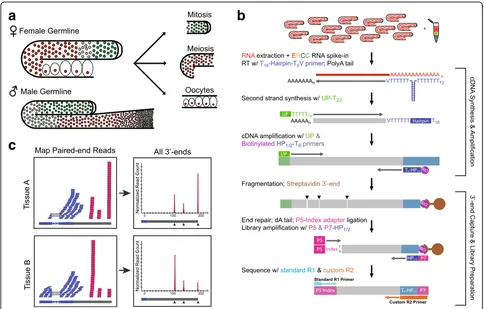
To monitor gene expression and 3′UTR dynamics using small amounts of RNA isolated from manually dissected germline tissue, we developed a new Low-Input 3′-Ter-minal sequencing method (LITE-Seq) that is both highly sensitive and quantitative (Fig. 1). We isolated mRNA from mixed-stage whole animals, intact gonads dissected from adult males and hermaphrodites with the sperma-theca removed (“female”germline), and three distinct re-gions of the adult hermaphrodite germline: the distal mitotic proliferation zone (partially including the early transition zone), the meiotic region, and developing oo-cytes (Fig. 1a). These samples allowed us to characterize variation in both mRNA expression and cleavage and polyadenylation (CPA) sites between sexes and as germ cells progress through the germline.
LITE-Seq targets mRNA transcripts using a poly-dT hairpin primer that precisely anchors at the beginning of the poly-A tail (Fig. 1b). First-strand cDNA synthesis is followed by linear cDNA amplification and pull-down using a biotinylated primer that specifically enriches for mRNA fragments containing 3′ends. Reverse transcrip-tion is performed using a primer that anchors to the be-ginning of a polyA tail, so the first base of Read 2 corresponds to the CPA site and thus defines the 3′-end of the transcript. Subsequent paired-end sequencing captures the precise position of CPA sites and produces a quantitative readout of transcript abundance.
mapping to genomic regions containing downstream polyA stretches and with no identifiable upstream PAS site (8,375 3′ ends) were removed to filter out internal mispriming events, resulting in the final set of 48,092 distinct CPA sites we report in this study.
For comparative analysis between samples, it is cru-cial to understand the relationship of reads obtained from LITE-Seq with molecular copy number. We used ERCC spike-in standards to confirm a linear relation-ship between input RNA and read counts (Additional file 1: Figure S4). LITE-Seq provides quantitative read-outs with high biological reproducibility for both gene expression (Additional file 1: Figure S5A) and CPA sites (Additional file 1: Figure S5B), and has the added ad-vantage that it captures the precise variation in 3′UTR isoforms across samples.
LITE-Seq shows similar sensitivity to other 3′-end pro-filing methods. LITE-Seq of germline samples recovered a
majority of CPA sites identified in previous surveys [43, 44] (Additional file 1: Figure S6, left) and revealed 17,129 previ-ously unannotated 3′UTR isoforms enriched in specific cell types. The detection rate for CPA sites previously identified in staged whole worm samples correlated with reported transcript abundance (Additional file 1: Figure S6, right), as anticipated from the underrepresentation of somatic tissues in our study and the lower reproducibility of sites with fewer supporting reads. Thus, the LITE-Seq method can detect and accurately amplify low amounts of input RNA to provide quantitative readouts of both transcript abundance and precise locations of cleavage and polyadenylation.
Gene expression in the germline varies in a sex- and developmentally-specific manner
In total, we observed 14,303 germline-expressed genes: 12,555 in male and 11,394 in female (9,573 in isolated
a
c
b
[image:3.595.56.541.87.396.2]gonads, 8,228 in the mitotic region, 9,008 in the early meiotic region, and 9,067 in developing oocytes). Based
on normalized read counts, 8,519 genes were
expressed in both sexes, of which 4,929 were present at similar levels (Additional file 2: Table S1). About twice as many differentially expressed genes were present in male (3,338) as in female (1,756) germline, suggesting that spermatogenesis is a highly specialized developmental program.
LITE-Seq compared favorably to several published transcriptome datasets, demonstrating its sensitivity relative to previous studies. We detected 85-95% of germline-enriched genes identified using microarrays [55] (1022/1220 intrinsic germline, 984/1013 oocyte-enriched, and 663/689 sperm-enriched genes) and 85– 95% of germline-expressed genes found using RNA-seq [57] (2,415/2,748 of spermatogenic gonad and 1,647/ 1,732 of oogenic gonad genes). Comparing our data to two recent transcriptome analyses of mature gametes, we recovered 85% (3,175/3,731) of reported sperm tran-scripts [58], and the majority of reported genes identified in isolated sperm (77%) [59] and oocytes (99%) [27] (Additional file 1: Figure S7). These comparisons indi-cate that our 3′-end capture method detects gene ex-pression with high sensitivity.
Gene Ontology term enrichment analysis of germline-expressed genes (Additional file 3: Table S2) reflected both shared and distinct biological functions during gametogenesis in the two sexes. A majority of expressed genes were enriched for sexually non-dimorphic func-tions associated with DNA synthesis, transcription, and translation, evidencing the shared requirements for basic cell biological processes during mitosis and meiosis. Genes more highly expressed in the male germline were enriched for functions associated with phosphatase and kinase activities, as previously reported [58], and the regulation of cell shape, highlighting the importance of post-translational signaling and morphological changes in later stages of spermatogenesis [60]. In contrast, genes with higher expression in the female germline were enriched for functions characteristic of maternal devel-opmental processes: reproduction, embryo development, gene regulation, and metabolism.
Pairwise comparisons of the sectional germline tran-scriptomes (Additional file 3: Table S2) generally reflected the high biosynthetic activities of the mitotic and meiotic regions, which were enriched for mRNAs with functions related to gene expression and translation relative to oo-cytes. The meiotic region showed a high level of tran-scripts related to genome packaging and oogenesis relative to mitosis, and to RNA binding relative to oocytes. The differential expression of transcripts encoding RBPs indi-cates that the majority are transcribed during pachytene and suggest that at least some of these may be eliminated
or deadenylated in oocytes. Taken together, these func-tional changes reflect the progression of germ cell nuclei through meiosis and the transition to oogenesis and early embryogenesis, which rely heavily on post-transcriptional control by maternal factors.
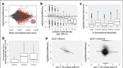
Genes with higher expression levels in oocytes also showed some notable characteristics. Oocyte-enriched transcripts are depleted for a wide range of biosynthetic functions and are enriched for genes involved in cellular and developmental processes, including signaling, mor-phogenesis, and nervous system development. These functional trends are consistent with maternal loading of embryos with numerous transcripts required for embry-onic patterning, gastrulation, and cellular differentiation [17]. LITE-Seq data also reflect a strong temporal asym-metry in X-linked gene expression (Fig. 2c): transcripts expressed from the X were strongly depleted in mitosis, began to rise in the meiotic region, and increased dra-matically in oocytes, but remained lower overall than autosomal expression (Additional file 1: Figure S8). This pattern confirms and extends previous work showing that X-linked genes are transcriptionally silenced in the germline prior to late pachytene, resulting in a short burst of expression before transcriptional activity is shut down globally in developing oocytes [56, 61].
When we looked for highly represented sequence mo-tifs in different samples (Additional file 4: Table S3), the most significant motifs, (A)AURAA, contained the core of the two most abundant PAS sites, which are bound by the cleavage and polyadenylation stimulating factor CPSF (accounting for 48% of all PAS sites). We also ob-served sequences resembling consensus Puf binding mo-tifs (UGUR core). For example, the mitotic region was depleted of transcripts with binding sites for FBF pro-teins that were enriched in meiosis (UGUAYW) and oo-cytes (UGUAHWU) [25], which prevent premature meiotic entry by down-regulating transcripts in the distal germline [24]. Transcripts that accumulate in oocytes were enriched for a polyC motif that directs the degradation of many maternal mRNAs in the embryo shortly after fertilization [27] and for UA[A/U]-rich motifs (UAUDUAU and UAAUUUAU) that could be bound by the oocyte mat-uration regulators OMA-1 and OMA-2 [62]. Several motifs resemble sites recognized by various other mRNA process-ing factors involved in transcript biogenesis.
many of these transcripts in conjunction with CGH-1 heli-case [23], and our data support this trend in meiosis and oocytes (Fig. 2e and Additional file 1: Figure S9).
In analyzing the dynamics of gene expression, we no-ticed that a specific subset of highly abundant transcripts in mitosis and meiosis appears to be abruptly cleared in developing oocytes (Fig. 2a, Additional file 1: Figure S10A, and Additional file 5: Table S4). These transcripts encode ribosomal proteins (Additional file 1: Figure S10B), and almost all contain one or more characteristic UUGUU motifs in their 3′UTR. Moreover, the density of UUGUU repeats inversely correlates with their rela-tive depletion in oocytes (Fig. 2b and Additional file 1: Figure S11). In Drosophilathe UUGUU motif is recog-nized by Brat, an RNA-binding protein that directs clearance of transcripts containing this motif during the maternal to zygotic transition (MZT) [63]. The C. ele-gans Brat homolog, NCL-1, also binds RNA
contain-ing the Brat motif [64]; moreover, ncl-1 mutant
animals are rescued by Brat [65], indicating functional
conservation between these proteins in two distantly related animal lineages.
Both NCL-1 and Brat are negative regulators of cell growth, nucleolar size, and ribosomal RNA (rRNA) levels [65–67]. Animals with mutations inncl-1 are lar-ger than normal, contain more total protein, and exhibit increased transcription of rRNA genes by RNA Polymer-ases I and III [67]. Within the adult germline, NCL-1 ac-cumulates to high levels in developing oocytes in parallel with the gradual disappearance of nucleoli [67]. Whereas NCL-1 has no known role in the post-transcriptional regulation of ribosomal subunit genes, NCL-1 and Puf family proteins have recently been implicated as co-operating partners in the translational repression of fibrillarin (fib-1), which encodes a ribosomal RNA processing factor [68]. In that study the PUF binding site in the fib-1 3′UTR was shown to directly mediate
regulation, whereas regulation by NCL-1 was inferred genetically based on increased FIB-1 expression in an
ncl-1 mutant background.
a
b
d
e
c
[image:5.595.56.539.88.357.2]We observe that the fib-1 transcript contains eight UUGUU motifs (six of which are located in the 3′UTR), and its mRNA abundance decreases around nine-fold in oocytes from its level in the mitotic region. Transcripts encoding several other ribosomal and tRNA biogenesis factors also show decreased expression in oocytes (Add-itional file 1: Figure S10B) and enrichment for the UUGUU motif in their 3′UTRs–includingpro-1, pro-2, W07E6.2, T04A8.6, K01C8.9,and rbd-1 [69]; dao-5[70]; and nol-6 [71]. The concomitant depletion of multiple UUGUU-bearing transcripts and accumulation of NCL-1 protein in oocytes raises the possibility that NCL-NCL-1 promotes dissolution of the nucleolus in the -1 oocyte, at least in part, by coordinated translational repression, deadenylation, and/or degradation of transcripts in-volved in nucleolar biogenesis.
Identification of 3′UTR isoforms in the germline
LITE-Seq provides data on the precise location of 3′-end processing in mRNA transcripts. To define putative 3′UTR isoforms of protein-coding genes, paired-end reads comprising clustered CPA sites were assigned to genes based on unambiguous overlap with exons (84% of total CPA sites (40,630/48,092)) or, for those with no reads overlapping exons, based on the nearest exon ≤1500 nt upstream (4,634 or 10% of CPA sites). Using this method, we identified a total of 43,725 3′UTRs (91% of CPA sites) for 14,946 protein-coding genes. In addition, we identified 1,485 transcript 3′ ends for 845 annotated non-coding genes and 2,828 that were not near any annotated features and may represent novel unannotated transcripts, which we did not analyze fur-ther. After filtering low abundance 3′UTR isoforms (representing <5% of clustered reads per gene) within each sample type, we report a total of 25,450 3′UTR iso-forms in the germline.
Around 30% of protein-coding genes in individual samples and 42% cumulatively showed alternative 3′ UTR isoforms, revealing tissue-specific selection of 3′ UTR isoforms for genes with multiple CPA sites (Add-itional file 1: Figure S12). The vast majority of 3′-end variation (97%) represented tandem alternative polyade-nylation (APA) downstream of the 3′-most CDS stop codon (Additional file 5: Table S4). Annotated functions for genes with multiple isoforms were enriched for pro-tein and nucleic acid binding and for processes involving sexual differentiation and gonadal, embryonic, and larval development. Thus, changes in the germline 3′UTR iso-form landscape likely play a role in post-transcriptional regulation of developmental processes.
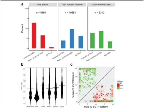
Most mRNAs contain polyadenylation signal (PAS) motifs in their 3′UTRs that, together with sequences downstream of the polyA addition site, serve to position the CPA machinery [30]. 81.5% of 3′UTRs contained
one of the 15 most common PAS motifs, which were typically located 19 nucleotides upstream of the CPA site [43, 44] (Additional file 1: Figure S13). The majority (61%) of cleavage events for a particular 3′UTR isoform occurred at the same nucleotide position and over 90% of reads were within ±5 nucleotides, though different PAS sequences showed some variation in cleavage preci-sion (Additional file 1: Figure S2B). Genes with a single 3′UTR isoform (8,586) largely use the canonical PAS se-quence AAUAAA (64%) (Fig. 3a). For genes with mul-tiple 3′UTRs (6,310), the canonical PAS motif was twice as likely to associate with distal (39%) vs. proximal (20%) CPA sites. Non-canonical PAS motifs constituted a plur-ality of PAS sites in genes with multiple 3′UTRs, but showed only a marginal preference for proximal (49%) vs. distal (43%) isoforms. In contrast, 3′UTRs with none of the 15 most common PAS motifs occurred at very low frequency among single-UTR genes, and were more often associated with proximal (32%) vs. distal (18%) CPA sites in genes with multiple isoforms. Since we per-formed stringent filtering of potential CPA sites, we think these results are largely representative of the true distribution of PAS sites. Previous studies also identified a bias toward the canonical PAS motif in distal isoforms and“weaker”CPA signals in proximal isoforms [31, 72].
The 3′UTR isoform regulatory landscape varies in a cell-type specific manner
3′UTR isoforms were highly dynamic in the germline transcriptome. The distribution of 3′UTR lengths showed large shifts between samples (Fig. 3b), revealing global trends that could significantly impact the tissue-specific cis-regulatory landscape. Overall, 3′UTRs were notably shorter in the male germline, with a median length (72 nt) less than half that in the female germline. These trends remained consistent even excluding the 10% most highly expressed isoforms in each sample and thus are not driven by a small number of short isoforms (Additional file 1: Figure S14). The median 3′UTR length increased as cells progressed through the fe-male germline, from ~130 nt in mitosis to ~157 nt in oocytes. While not extreme, this shift is in line with previously observed trends for proliferating vs. differ-entiated cells [73].
(221/310) (Fig. 3c). While most genes showed the same dominant 3′UTR isoform throughout the female germ-line, 461 of 9,435 genes expressed in at least two regions showed a shift as gametogenesis progressed (Additional file 1: Figure S15). Usage of proximal or distal isoforms was mixed: roughly half (106/188) of 3′UTRs that switched in length between meiosis and mitosis were lon-ger in the meiotic region; just under two thirds (108/172) were longer in oocytes compared to meiotic nuclei; and about half (157/285) were longer in oocytes vs. mitotic nu-clei. Genes whose major isoform changed across germline regions showed no strong bias in functional annotations compared to the full germline transcriptome; however,
chromatin organization was overrepresented among genes with both longer (19/157) and shorter (16/128) 3′UTRs in oocytes relative to mitosis, indicating that such transcripts may be important targets for APA and post-transcriptional regulation.
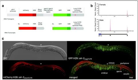
3′UTR isoform selection influences protein expression patterns in vivo
[image:7.595.57.538.87.447.2]which mCherry and GFP genes flanked by distinct down-stream sequences are transcribed from a single polycis-tronic operon locus and spliced into individual mRNAs (Fig. 4a, Additional file 1: Figure S16A). Because both re-porters are driven by the same promoter, any differences in expression patterns should be dictated exclusively by their 3′UTRs.
Controls with identical 3′UTRs displayed ubiquitous ex-pression in the hermaphrodite germline (Additional file 1: Figure S16C,D), verifying that thegld-1 promoter is per-missive for expression throughout. However mCherry was stronger in mature sperm and also became enriched over GFP in L4 and male germlines as spermatocytes matured. The intensity of both fluorophores in embryos was vari-able, with thegpd-2control 3′UTR appearing to be more
permissive than the rpl-22control in embryos. Observed biases in expression patterns of control reporters were fac-tored into the interpretation of experimental assays.
As proof of principle, we examined the effect of alterna-tive 3′UTR isoforms on spatiotemporal protein expression
using reporters for three genes that showed APA in the germline by LITE-Seq. For all three genes (air-2,aly-3, and
cap-2), the major isoform was short in male and long in fe-male germline (Fig. 4b; Additional file 1: Figure S17A), and mCherry (linked to the short 3′UTRs) showed expression patterns similar to controls (Fig. 4c, Additional file 1: Figure S17B,C; compare with Additional file 1: Figure S16C,D). The long isoforms ofair-2and aly-3directed distinct de-velopmental patterns of GFP expression in the hermaphro-dite germline that reappeared very robustly in early embryos. Embryonic expression was maternal, as crosses of wild-type hermaphrodites with transgenic males carrying these reporters showed no GFP signal (data not shown). The GFP signal for the Aurora B kinase homologair-2was brightest in the mitotic region, dropped dramatically in pachytene and oocytes, and reappeared in embryos at the one- or two-cell stage (Fig. 4c). This suggests that AIR-2 protein– which functions in chromosome segregation by regulating the release of chromatid cohesion and has an in-dependent role in cytokinesis [74, 75]–is produced during
[image:8.595.57.539.345.625.2]mitosis in preparation for meiosis and is reactivated dur-ing mitotic divisions in the embryo. The aly-3 gene en-codes an RRM protein whose homologs recruit mRNA nuclear export factors [76] and can also influence tran-scription and RNA Pol II occupancy [77]. LITE-Seq de-tected analy-3 transcript with a 205 nt 3′UTR that is a
known GLD-1 target [21] and a short (36 nt), previously unannotated 3′UTR isoform lacking the GLD-1 binding motif (Additional file 1: Figure S17A). GFP was brightest in the hermaphrodite mitotic region and attenuated in pachytene relative to mCherry. This pattern is consistent with negative regulation by GLD-1, which accumulates to high levels in pachytene [13, 16, 52]. In addition, GFP ex-pression was strongly suppressed in 1- and 2 cell-stage embryos, reappearing robustly in embryos from the 8-cell stage onward. These results show that the long 3′UTR isoform is needed for repression ofaly-3 in the germline and early embryogenesis. Finally, CAP-2 is the beta sub-unit of F-actin capping protein and is important for
cyto-skeletal regulation throughout development [78].
Although cap-2 isoforms shifted in the female germline, in vivo expression patterns for both reporters were fairly similar to controls (Additional file 1: Figure S17C; com-pare with Additional file 1: Figure S16C,D).
Collectively, these reporter assays show that forced pression of alternative isoforms can drive different ex-pression patterns in the germline and early embryo, but that some APA events could be benign variants due to cell type-specific differences in 3′-end processing factors. Distinct spatiotemporal patterns indicate that longer 3′UTR isoforms contain negative regulatory se-quences that are absent from their short counterparts. While consistent with known examples of translational regulation of maternal products, further work will be re-quired to assess the broader functional consequences of APA in the germline.
NCL-1/Brat negatively regulates ribosome-related mRNAs through UUGUU motifs
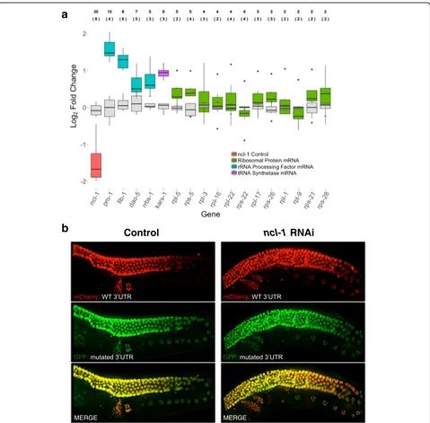
The combined expression dynamics of NCL-1 protein and UUGUU-containing transcripts led us to hypothesize that binding of NCL-1 to this motif contributes to mRNA deg-radation and/or deadenylation during oogenesis inC. ele-gans. In addition to the large decrease in ribosomal subunit mRNAs (~32-64 fold), we noticed that transcripts for other nucleolar factors, while generally expressed at much lower levels, also decreased by ~2-9-fold in oocytes (Additional file 1: Figure S10B). Coordinated post-transcriptional targeting of ribosomal protein subunits, rRNA processing factors, and tRNA synthetases by NCL-1 could therefore contribute to the rapid disappearance of the nucleolus that is seen in the -1 oocyte.
To test whether NCL-1 activity influences these mRNAs, we performed quantitative RT-PCR (qRT-PCR)
using embryos derived from WT and
ncl-1(RNAi)-treated animals, reasoning that maternal stores in oo-cytes should be reflected in early embryos. We found that reducingncl-1levels by around 65% leads to higher levels not only of fib-1, but also of several other tran-scripts related to nucleolar activity, and the effect was proportional to the number of UUGUU motifs contained in these transcripts (Fig. 5a). We also used our two-color in vivo reporter system to test whether 3′UTRs with either intact (UUGUU) or mutated (UUCUU) NCL-1/Brat bind-ing motifs are responsive to varybind-ing NCL-1 levels in WT andncl-1(RNAi) germlines. This mutation affects the core of the binding motif [64] and hence should disrupt NCL-1 binding. Again, genes with the largest number of UUGUU motifs in their 3′UTRs (pro-1andfib-1) showed the great-est difference in expression in oocytes and/or early
em-bryos when comparing WT and ncl-1(RNAi) animals
(Fig. 5b and Additional file 1: Figure S18A-F). No differ-ence in expression between WT andncl-1(RNAi)was ob-served for the reporter with mutated UUGUU motifs in the 3′UTR, confirming that these are required for ncl-1
mediated regulation.
In contrast to the germline LITE-Seq data, which re-vealed large fold-changes in oocytes for endogenous large (rpl) and small (rps) ribosomal subunit mRNAs, these showed much more modest and highly variable re-sponses to ncl-1(RNAi) in both qRT-PCR (Fig. 5a) and 3′UTR reporter assays (Additional file 1: Figure S18D-F). Thus it would appear that partial knock-down of ncl-1 is still sufficient to down-regulate these transcripts in oocytes. A logical explanation for this discrepancy, based on stoichiometric considerations, is that the extreme abundance of these mRNAs in meiosis could drive their depletion by NCL-1 as it reaches a very high concentra-tion in oocytes – whereas lower levels of 3′UTR re-porter expression driven by the gld-1 promoter, or partial depletion of ncl-1 by RNAi, might not be suffi-cient to elicit a robust post-transcriptional response. An-other possibility is that ncl-1 may cooperate with other factors to regulate these transcripts, as has been shown forfib-1. The apparent increase in the GFP-linkedrpl-22
reporter with mutated UUGUU motifs relative to the GFP-linked control rpl-22 3′UTR (compare Additional file 1: Figure S18E with Figure S16D and S18F) supports the idea that the motif is necessary but not sufficient for repression when driven at low levels. Notably, thencl-1
transcript itself contains 20 UUGUU motifs (8 in the 3′ UTR), raising the possibility of a negative NCL-1 self-regulatory feedback loop. Further study will be needed to resolve these outstanding questions.
Discussion
of gene expression and the precise identification of tran-script 3′ends using limited quantities of RNA. We showed that LITE-Seq accurately reflects isoform-specific expres-sion levels across several orders of magnitude and is highly
reproducible, enabling meaningful comparative analyses be-tween different tissues and developmental stages. We used LITE-Seq to characterize the dynamics of gene expression and 3′UTR isoform usage within the C. elegans germline mCherry::
GFP::
MERGE
mCherry::
GFP::
MERGE
Control
ncl-1
RNAi
a
b
Fig. 5Ribosome-related transcripts containing UUGUU motifs are negatively regulated byncl-1in the germline and embryo.aRNAi ofncl-1
leads to increased expression of ribosome-related transcripts. Log2fold-change in mRNA expression between WT and andncl-1(RNAi)-treated early embryos for ribosome-related genes, as measured by qRT-PCR (n = 8). The median and interquartile range (IQR) for transcripts encoding ribosomal subunits (green), rRNA processing factors (teal), and a tRNA synthetase (purple) uponncl-1(RNAi) (salmon) are shown in comparison with the variation among controls (gray). Whiskers denote 1.5 times the IQR. On average, a 65% knockdown ofncl-1mRNA was observed in
[image:10.595.59.538.86.557.2]and identified numerous examples of alternative 3′UTR iso-forms. We demonstrated that naturally occurring 3′UTR variants drive different spatiotemporal expression patterns in vivo and are thus used endogenously to confer differential post-transcriptional regulation in the developing germline and early embryo.
Germline-expressed genes show sex-specific and spatio-temporal variation
Comparisons of samples between sexes and different de-velopmental zones in the female germline revealed evi-dence for widespread spatiotemporal variation in both expression levels and 3′UTR isoform selection during developmental transitions in female gametogenesis. Functional annotations revealed large-scale changes in gene expression consistent with known genetic require-ments for mitotic proliferation, meiosis, differentiation of male and female gametes, and maternal contributions to early embryogenesis.
Our temporally resolved transcriptome profiling ex-tends previous studies of X-linked expression in the germline. The X chromosome is largely silenced in male and distal hermaphrodite gonads and is activated during a short window in late pachytene and diplotene, before Pol II transcription shuts down in diakinesis [55, 56, 61, 79]. Transcriptome-wide comparisons of LITE-Seq data revealed a small subset of X-linked transcripts that es-cape silencing in the distal germline; in addition, we found that maximal expression reaches only around half that of autosomal genes, as seen in microarray data for 258 oocyte-enriched genes [61]. X-linked expression in the germline is biased against genes essential for early germline development and toward genes required later during oogenesis or embryogenesis, similar to observa-tions from microarray studies [55].
We investigated mRNA targets of GLD-1, an import-ant post-transcriptional regulator of both entry into mei-osis and oocyte differentiation that is expressed at high levels only in the pachytene region [13, 52]. Transla-tional repression of specific transcripts by GLD-1 at this stage is important to delay expression of their protein products until they are needed in developing oocytes or the early embryo, and conversely GLD-1 repression must subsequently be lifted to allow their translation. We ob-served that transcripts with increased expression in pachytene contained more predicted binding sites for GLD-1 [80], and these were largely retained in oocytes. We also observed a correlation between mRNA levels and the presence of both GLD-1 and CGH-1 binding sites, which have been shown to co-stabilize certain transcripts in association with P granules and P body-like storage granules in the germline [23]. Thus, accu-mulation of a large body of GLD-1 targets is coordinated with the onset of GLD-1 translation during meiosis, and
these transcripts are selectively maintained in oocytes and loaded into the early embryo.
Post-transcriptional regulation of nucleolar biogenesis reveals a new developmental program inC. elegans oocytes
Our germline transcriptome data showed that polyade-nylated ribosomal subunit mRNAs abruptly disappear in developing oocytes, and that the vast majority of these contain multiple copies of the short sequence UUGUU. We found that many transcripts encoding rRNA and tRNA biogenesis factors are similarly down-regulated at this stage and also contain UUGUU repeats. This motif is known to mediate maternal transcript degradation by the tumor suppressor Brat during the MZT in Drosoph-ila embryos [63]. Brat and its C. elegans homolog NCL-1, which also binds UUGUU in vitro [64], are both negative regulators of nucleolar size [65–67], and Brat rescuesncl-1mutant phenotypes inC. elegans[65].
In theC. elegansgermline, nucleoli disappear in the -1 oocyte where NCL-1 levels are highest [67], raising the possibility that this is coordinated with oocyte matur-ation signals from the sperm. Intriguingly, mouse oo-cytes also show a decrease in ribosomal protein transcripts and total rRNA as part of a broad mRNA decay program that takes place during meiotic matur-ation [81, 82]. Thus, the suspension of ribosomal bio-genesis in preparation for the completion of meiosis upon egg activation appears to be conserved between nematodes and mammals and represents a previously unrecognized developmental event in the C. elegans
oocyte-to-embryo transition. This regulatory program precedes the MZT inC. elegans[26] and is independent of the poly-C cis-regulatory motif that contributes to maternal mRNA turnover in the fertilized zygote [27].
Our data provide a new angle on how NCL-1 likely regulates nucleolar activity. Prior to this study know-ledge ofncl-1function was restricted to genetic evidence that it works together with Puf family proteins to repress translation of a single transcript, encoding the rRNA processing factor fibrillarin (FIB-1) [68]. However, fib-1
regulation cannot fully account for the nucleolar func-tions ofncl-1, as evidenced by the fact thatfib-1 knock-down does not rescue the increase in nucleolar size observed inncl-1 mutants [68]. Our data provide a mo-lecular basis for this and indicate that NCL-1 acts in a much broader fashion to negatively regulate dozens of genes related to nucleolar biogenesis through direct binding of UUGUU motifs present in these transcripts.
ribosome-related transcripts that we observed in oocytes and em-bryos uponncl-1depletion implies that nucleolar pheno-types arise from defects in deadenylation and/or degradation of maternal transcripts. Sincencl-1mutants appear to have no effect on transcription by RNA Pol II [67], a post-transcriptional mechanism that broadly tar-gets ribosomal proteins, rRNAs and tRNAs would pro-vide an efficient mechanism to simultaneously shut down multiple facets of nucleolar biogenesis, in turn leading to reduced rRNA transcription and nucleolar size [67]. Deciphering its primary mechanism of ac-tion awaits future studies of its effects on endogenous mRNA targets to help clarify whether it binds co-operatively to multiple motifs or with other factors, and how this may influence mRNA translation, deade-nylation, and/or stability.
It is interesting that NCL-1 itself is developmentally regulated, showing lowest levels in cells with higher metabolic activity, such as the intestine [67]. The fairly
low levels of ncl-1 mRNA throughout the germline
(around 80-100 counts) could be maintained by negative self-regulatory feedback, as it contains 20 NCL-1 bind-ing motifs (the most we found in any germline tran-scripts). The transcript must also be under translational control in the germline, as it accumulates to extremely high levels in oocytes whereas the mRNA does not. Its 3′UTR additionally contains a functional binding site for the heterochronic miRNAlet-7[68], providing a poten-tial mechanism to coordinate its activity with
develop-mental growth cycles. As nucleoli link overall
biosynthetic capacity with the cell cycle, cell growth, nu-tritional status, and stress response [84], it is tempting to speculate that NCL-1 may be a master regulator of nucleolar function that senses metabolic and develop-mental cues and responds by titrating global biosynthetic activity to promote cellular homeostasis at all stages of development.
The 3′UTR isoform regulatory landscape varies in a cell-type specific manner
Analysis of our sequencing data highlights the advan-tages of the LITE-Seq method for identifying and quanti-fying expression of distinct 3′UTR isoforms for any given gene. By analyzing global dynamics of the 3′UTR landscape, we found that 40% of germline-expressed genes are alternatively polyadenylated, and we uncovered numerous 3′UTR isoforms not identified in previous studies. We found that most cleavage and polyadenyla-tion sites are very sharp, i.e. individual transcripts show 3′ ends that cluster tightly around a preferred position. Similar to previous studies [43, 44], we observed that C. elegans uses a wide variety of signal sequences. Whereas in humans the canonical PAS is strongly preferred in all 3′UTRs [85], C. elegans seems to have much looser
requirements, particularly in genes that undergo alterna-tive polyadenylation. This suggests either that the CPA machinery is less stringent or that additional sequence features and/or cleavage factors confer specificity to these sites. Comparisons of 3′UTR usage between differ-ent samples allowed us to iddiffer-entify numerous genes that show differential APA between sexes and different stages of germ cell development. Given the widespread nature of APA in the germline, it appears to be regulated in a cell-type-specific manner to ensure the inclusion or clusion of specific cis-regulatory motifs. We cannot ex-clude the possibility, however, that some of the observed variation in transcript termination is a benign byproduct of cell-type-specific differences in transcriptional activity or the complement of mRNA processing factors, which can all contribute to the global APA profile [86].
The length of 3′UTRs was generally much shorter in males, as also observed in D. melanogaster [37]. This suggests that the spermatogenic program may rely more heavily on transcriptional regulation, though some key events depend on translational control [87]. In C. ele-gans, the shift to shorter 3′UTR isoforms may
poten-tially be linked to the RNA cleavage factor CFIm, which upon knockdown in mammals causes preferential usage of proximal sites [88]. We detected significantly lower expression of CFIm-1 and CFIm-2 in the male germline, indicating that this may be an operant factor in the bias toward shorter 3′UTRs. Mammals also show testis-specific patterns in APA and expression of CPA factors [89]. Notably, 3′UTRs undergo progressive shortening during mouse spermatogenesis, and this is associated with changes in chromatin states and transcriptional ac-tivity [90, 91]. Shortened 3′UTRs tend to exclude desta-bilizing elements – such as U-rich elements – and lengthened 3′UTRs add sequences that may help stabilize transcripts [91]. The similarity of trends across distantly related species suggests the intriguing possibil-ity that regulated APA selection favoring shorter 3′UTRs may be a deeply conserved property of male germline development.
during later stages of oogenesis, consistent with well-documented genetic requirements for translational regu-lation of maternal products during oocyte development and early embryogenesis [16, 29]. Transcripts with shorter 3′UTR isoforms may tend to be translated more efficiently, since many RBPs act as translational repres-sors at these stages [5, 17]. The regulated production of multiple isoforms may thus allow expression levels of in-dividual proteins to be tuned depending on the specific needs of cells at different stages of development.
3′UTR isoform selection directs differential protein expression patterns in vivo
3′UTRs have been shown to be the main drivers of spa-tiotemporal protein expression patterns for numerous genes in the germline [14]. To test the biological effects of endogenous 3′UTR variants that we identified, we ex-amined patterns of reporter gene expression in vivo for three genes whose dominant isoforms differ between sexes or in different regions of the female germline. For two genes, reporters for short and long 3′UTR isoforms displayed different localization patterns when driven by a ubiquitous germline promoter. Unlike their shorter counterparts, long isoforms experienced negative regula-tion in the female germline and showed reactivaregula-tion of expression in early embryos. Thus, selective inclusion of specific post-transcriptional regulatory elements in lon-ger 3′UTRs can allow for more nuanced patterns of reg-ulated expression in the germline and early embryo.
More generally, the observed sex-specific and spatiotem-poral differences in isoform prevalence and protein expres-sion driven by alternative 3′UTRs point to isoform selection as a functionally important feature of both the spermatogenic and oogenic differentiation programs. Post-transcriptional regulation is specified by a combination of RNA-binding proteins and small RNAs and is achieved through selective translation, stabilization, deadenylation, and/or degradation of mRNAs bearing alternative 3′UTR isoforms. Since LITE-Seq detects only polyadenylated tran-scripts, future studies will be required to distinguish be-tween these alternative mechanisms in the germline. This work sets the stage to learn more about how cytoplasmic and nuclear processes such as epigenetics, transcription, splicing, APA, and post-transcriptional regulation are inte-grated to govern the lifecycle of mRNAs that drives pat-terned gene expression during development.
Conclusions
We have developed a new experimental method, LITE-Seq, which quantifies the 3′ends of all polyadenylated mRNAs expressed in vivo using small amounts of input sample. LITE-Seq provides a new tool to probe 3′UTR variation in different cellular and developmental con-texts. Spatiotemporal profiling of theC. elegansgermline
with LITE-seq revealed dynamic changes in both the ex-pression and diversity of 3′UTRs during gametogenesis at unprecedented resolution. Nearly half of germline-expressed genes are alternatively polyadenylated, sug-gesting that differential post-transcriptional control is pervasive in the developing germline. Shorter transcripts are expressed in males and in rapidly proliferating mi-totic cells, while longer transcripts expressed during meiotic differentiation in the female germline expose a greater repertoire of putative target elements for trans-acting factors. The transcripts expressed later in gameto-genesis, which are expected to be under more complex regulatory control, encode proteins involved in gene regulation, development and morphology. We show that alternative 3′UTRs can drive distinct protein expression patterns across the male and female germline, demon-strating isoform-specific regulation of endogenous tran-scripts. The broader functional significance of these differences awaits further study.
We discovered a post-transcriptional developmental program that down-regulates mRNAs involved in ribosome, rRNA, and tRNA biosynthesis in oocytes, coincident with the dissolution of the nucleolus just prior to fertilization. We demonstrate that the RBP NCL-1/Brat is a regulator of this transition and show that it acts through UUGUU motifs present in these transcripts. We propose that NCL-1 acts as a master regulator of the nucleolus by promoting the coordinated down-regulation of a broad spectrum of transcripts related to ribosomal biogenesis and nu-cleolar function.
trans-acting factors integrate these levels of regulation into dy-namic developmental programs.
Methods
C. elegansworm strains
The wild-type N2 Bristol strain was used for all sequen-cing experiments. Worms were maintained at room temperature on NGM-agar plates seeded with E. coli
strain OP50-1. N2 males were generated by heat-shocking L4 hermaphrodite worms at 30 °C for 6 hours to induce nondisjunction of the X chromosome and were maintained by continuous crossing. Transgenic lines were created using the EG6699 strain for MosSci [93] insertions onto chromosome II and maintained at 25 °C on NGM agar plates seeded with
E. coli strain HB101. Transgenic worms expressing the two-color reporter constructs were generated at Knudra Transgenics (Murray, UT) or injected in our lab as described in [93] using the MosSCI strain EG6699. Constructs were injected into young adults and recovered at room temperature. Plates containing rescued Unc worms were screened seven days later for the absence of negative selection markers. Worms were passaged to new plates, and homozygotes ana-lyzed for expression of mCherry and GFP in the germline. Additional file 7: Table S6 lists all trans-genic strains constructed for this study.
RNA sample collections for LITE-Seq
Worms were synchronized by plating arrested L1 larvae and allowed to grow to young adults. N2 hermaphro-dites from non-mated plates or N2 males were dissected in a depression slide containing 0.5 mM levamisole in M9 buffer. Heads were severed below the pharynx with a 25G1½ needle, and intact extruded germ lines were separated from the body. The ‘female’germline was cut to include everything except the spermatheca, while the male germline was cut to include spermatids, although some were released into the buffer when dissected. Germline pieces were visually assessed and cut under
dissecting microscope. Samples were immediately
immersed in RNALater buffer (Qiagen, Germantown, MD) to preserve the RNA and then transferred using a mouth pipet to the cap of a 0.2 mL microcentrifuge tube containing additional RNALater. Samples were pooled in the same cap until sufficient quantities were col-lected and then stored at -80 °C. Once samples had been collected in triplicate, RNA was extracted using the Qiagen RNeasy Micro kit according to the manu-facturer’s protocol with an on-column DNase I treat-ment and eluted in RNase-free water. The quantity and quality of RNA recovered were assessed on an Agilent Bioanalyzer (Santa Clara, California).
cDNA Amplification, Library Preparation and Sequencing
To generate cDNA, we used 65 ng or 50 ng of total RNA from each sample pool. The ERCC RNA spike-in mix (Invitrogen, Carlsbad, California) was added in quantities recommended by the manufacturer. RNA was added to lysis mixture (0.9X PCR buffer, 3 mM MgCl2, 0.45% NP40, 4.5 mM DTT, 0.18U/ul SUPERase-In, 0.36U/ul RNase Inhibitor, 0.125uM dNTP) and heated to 70 °C for 90 seconds. First strand cDNA synthesis was performed using SuperScript III reverse transcript-ase (Invitrogen) along with a modified poly-dT primer in reactions containing 13.2U/ul SuperScript III, 0.5uM poly-dT hairpin primer, 0.4U/ul RNase Inhibitor, and 0.07ug/ul T4 gene 32 protein (Invitrogen). The primer
(5′-T18TGGAATTCTCGGGTGCCAACCCTTGGCAC
CCGAGAATTCCAT6V-3′) contains a single non-T
nucleotide at the 3′ end, which serves to anchor it to the transcript immediately upstream of the polyA tail, and an internal sequence that forms a hairpin loop. Prior to the RT reaction, the primer was heated to 98 °C for 10 minutes and allowed to slowly cool to room temperature in order to ensure proper formation of the hairpin. Following first strand cDNA synthesis, ExoSAP (Affymetrix, Santa Clara, California) was added to remove all remaining free primer. The cDNA was polyadenylated using a terminal transferase and treated with RNaseH to remove the RNA strand (New England Biolabs, Ipswitch, Massachusetts). Purification of the single-stranded cDNA was done using Ampure beads (Agencourt, Lynn, Massachusetts). The second strand of cDNA was synthe-sized using a UP2-polydT primer (5′-ATATCTCGAGGGC
GCGCCGGATCCT22-3′) and Takara Taq polymerase
(Nojihigashi, Japan). Eighteen rounds of cDNA amplifica-tion were performed using a UP2 primer (5′-ATATCTC-GAGGGCGCGCCGGATCC-3′) and a biotinylated primer containing only a part of the hairpin sequence
(5′-Bio-CAACCCTTGGCACCCGAGAATTCCAT6-3′) in order
to limit residual polyA tail sequences to 6 nt in the final product. Half of the cDNA was re-amplified by another six cycles of PCR to generate sufficient cDNA for library prep-aration, repurified with Ampure beads, and examined on the Bioanalyzer to assess quality and quantity of the PCR products.
to beads, the captured cDNA was end-repaired and an ad-enosine was added to the 3′end (NEBNext Ultra End Re-pair/dA-Tailing module). Illumina P5 adapters carrying a six base barcode were T/A-ligated to the free end of the
DNA fragments (5′-AATGATACGGCGACCACCGA
GATCTACACTCTTTCCCTACACGACGCTCTTCCGA TCT-XXXXXX-T-3′). The library was amplified from the beads for twelve cycles with KAPA HiFi HotStart ReadyMix (Kapa Wilmington, Massachusetts) using a P5 primer (5′-AATGATACGGCGACCACCGAGATC T-3′) and a P7-half-hairpin primer (5′- CAAGCA-GAAGACGGCATACGAGATCCAACCCTTGGCACC CGAGAATTCCA -3′). During PCR amplification, the beads easily settle to the bottom. To ensure proper amplification of the library, the tubes were quickly vortexed after each denaturing step. Libraries were cleaned with Ampure beads and checked on a Bioanalyzer for quality and quantity before storage in−20 °C. Prior to sequencing libraries were quantified using the KAPA Library Quantification Kit, diluted to a concentration of 2nM, and pooled. A PhiX library was added at approxi-mately 20% to increase the base diversity. A paired-end 100 bp rapid run was performed on the Illumina HiSeq 2500 platform (San Diego, California) using the standard Read 1 primer and a custom sequencing primer for Read 2 (5′-AGCAGAAGACGGCATACGAGATCCAACCCTT
GGCACCCGAGAATTCCAT6-3′), which is designed to
provide sequences starting from the last base prior to the poly(A) tail.
Bioinformatic Analysis
Sequencing reads were de-multiplexed into sample li-braries according to the barcode encoded in the first six bases of Read 1. Reads were trimmed of the barcode and Illumina adaptor sequences. Short (<50 nt) or low qual-ity reads (≤95% identity) were excluded from further analysis. Reads were mapped to theC. elegansreference genome (WS250) using paired-end reads with TopHat 2 in an iterative manner with parameters “–b2-very-sensitive –min-segment-intron 14 –min-coverage-intron 14 –min-intron-length 14–max-segment-intron 24000 –max-cover-age-intron 24000–max-intron-length 24000–read-edit-dist 5 –read-realign-edit-dist 0–segment-mismatches 3 –read-mismatches 5 –max-deletion-length 5 –max-insertion-length 5 –no-discordant”, mate-inner-distance and mate-std-dev were determined using Qualimap 2 [94]. Read 2 of unmapped paired-reads were remapped after trimming any stretches of thymidines at the be-ginning of the sequence, which occasionally occurred due to mis-anchoring of the hairpin primer within polyA tails during the RT reaction. Any mapped reads potentially generated by internal priming of the hair-pin primer to A-rich regions within transcripts (i.e. that matched genomic sequence and thus most likely
did not represent polyA tails) were filtered. Finally, remaining unmapped Read 2 sequences were re-mapped after trimming to 50 nt to remove occasional palindromic sequences that were occasionally ob-served among the unmapped reads and which pre-sumably arose from technical errors during PCR amplification. Any CPA sites for which Read 2 began with an adenosine, was followed by 3 adenosines in the next 4 bases, and did not contain a known PAS site 8–25 nucleotides upstream of the putative CPA site were removed as potential false positives; those containing a PAS site were retained as a separate set of potential true positives but were not included in our final analysis. Raw and mapped reads were depos-ited at NCBI Sequence Read Archive, accession num-ber SRP096640.
We assigned read-pairs to an annotated gene if reads overlapped a known exon using HTSeq [95] or, if no exons overlapped the read, to the closest annotated gene within 1,500 nucleotides upstream. To identify differen-tially expressed genes, libraries were first normalized using the size factor calculation from DESeq2 [96] to allow comparisons between libraries. GO term enrich-ment analysis for differentially expressed genes was per-formed using PantherDB [97].
In order to define 3′UTR isoforms detected within each sample, a ‘universe’ of all possible cleavage and polyadenylation (CPA) sites was established by combin-ing Read 2 data from all 14 libraries. Reads startcombin-ing at a given position (chromosome and strand) found in at least two samples were summed and sorted in descend-ing order of abundance, if a position was not supported by at least one read in two samples, read counts sup-porting this position were not included. CPA sites were defined by clustering reads within a ±12 nucleotide win-dow of the position with the highest number of support-ing reads. If two sites within a given window were supported by the same number of reads, we selected the 3′most position.
To determine 3′UTR isoform switching between sam-ples, potential CPA sites were identified for each gene in every sample. Major isoforms with the highest usage were identified in each sample. Genes that switched usage from one major 3′UTR isoform to another in a given set of samples were identified using an algorithm for calculating Significance Analysis of Alternative Polya-denylation [98]; to allow for cases with more than two isoforms, relative expression for each given isoform was compared to collective usage of all other isoforms.
Two-color Reporter System
S16. Additional file 7: Table S6 lists all primer sequences and plasmids used for cloning and resulting vectors. To generate the final reporter constructs for each pair of 3′ UTR isoforms, three Entry clones were generated as de-scribed below and combined together with LR+ clonase into Gateway Destination vector pCFJ150 for MosSCI recombination.
Entry Construct 1, containing thegld-1promoter, was PCR amplified from C. elegans N2 genomic DNA using primers DM126 and DM127. The cassette spans 530 bp of genomic DNA, beginning immediately downstream of the T23G11.4 CDS stop codon (DM126) and ending
with the gld-1 CDS start codon (DM127). The PCR
product was recombined into pDONR P4-P1R using BP clonase (Invitrogen).
Entry Construct 2, the operon-reporter cassette, con-tains the gpd-2/gpd-3 intergenic region from the mai-1
operon [99] flanked by GFP:H2B and mCherry:H2B re-porters. The operon cassette was made in several steps using a pDONR 221 vector modified to include part of the multiple cloning region (MCR) from pSL1180 (Addgene, Cambridge, MA) that spans 17 restriction sites from SpeI to BglII. A 244 bp fragment containing the entiregpd-2/gpd-3intergenic region was PCR ampli-fied from N2 genomic DNA with primers DM144 and DM145. The genomic sequence begins immediately downstream of thegpd-2 CDS stop codon (DM144), in-cludes the full 133 nt gpd-2 3′UTR, and ends
immedi-ately upstream of the gpd-3 CDS start codon (DM145). The resulting PCR product was cloned between the SacII and NotI sites in the MCR. Fluorescent reporter genes were then cloned between SpeI-SacI (upstream gene) and NotI-BglII (downstream gene) sites. We gen-erated constructs containing GFP:H2B and mCher-ry:H2B reporters in both orientations (i.e. mCherry upstream and GFP downstream, and vice versa). GFP::H2B and mCherry:H2B were amplified from plas-mids pGC468 and pGC544, gifted by the Nance
labora-tory, using primers containing the corresponding
restriction sites flanked by gene-specific sequences. For gene-specific 3′UTR reporter constructs, short and long 3′UTR isoforms were PCR amplified from N2 genomic DNA using primers targeting the regions of interest and flanked by appropriate cloning sites (described below). For reporters with UUGUU motifs,
WT and mutated (UUGUU to UUCUU) 3′UTR oligos
were chemically synthesized (IDT). Short 3′UTR iso-forms and WT UUGUU 3′UTRs were cloned into Entry Construct 2 between SacI and SacII to generate gene-specific variants. Cassettes with long 3′UTR iso-forms and 3′UTRs with mutated UUCUU motifs flanked by Gateway sites were used to generate a gene-specific Entry Construct 3 in pDONR P2R-P3. To ensure correct CPA site selection for long 3′UTR
isoforms, any upstream PAS sites were eliminated using site-directed mutagenesis. PAS sites were al-tered by 25 cycles of “around-the-world” PCR with Phusion polymerase (New England Biolabs), using complementary primers containing mismatches within the motif and flanked by perfect matches of 20 nt up-stream and downup-stream. PCR products were cleaned, concentrated, and then digested with DpnI to degrade the original template.
Quantitative RT-PCR
To synchronize animals for RNAi treatment and RNA extraction, gravid N2 worms were bleached, re-leased embryos were grown overnight in M9, and ~25,000 L1 larvae were plated onto two extra large NGM-IPTG plates seeded with E. coli containing ei-ther ncl-1 or empty control (L4440) RNAi vectors. These plates were left at 20 °C until the worms were gravid and some embryos were visible on the plate. Several worms from each experimental plate were checked for ncl-1 RNAi phenotypes (e.g. presence of a nucleolus in the -1 oocyte). Once the presence of these phenotypes was confirmed (Additional file 1: Figure S18G), the worms were bleached and any remaining carcasses removed via vacuum filtration. Aliquots of bleached embryos were examined under 100x magnification to ensure a good representation of early embryos (1-64 cells). Total RNA was ex-tracted from these embryos using RNeasy Mini Kit (Qiagen), and any remaining DNA was digested with EZ DNAse (ThermoFisher, Waltham, MA). Each collection yielded ~40-50ug of RNA. 20ug of RNA
was then reverse transcribed into cDNA using
Superscript IV reverse transcriptase (ThermoFisher). qPCR was performed on a LightCycler 480 (Roche, Indianapolis, IN) using LightCycler 480 Sybr Green 1 Master (Roche). Results were analyzed using the ΔΔCT method [100]. For each gene, eight
independ-ent biological replicates were assayed using two
technical replicates per assay. For comparison,
variation among control replicates was calculated by normalizing to the mean of the replicates and calcu-lating the log2 fold-change.
Microscopy
Z-stacks spanning approximately 20um were acquired at 20x or 40x with a Leica DFC365 FX digital camera at-tached to a Leica DM5500 B microscope (Leica, Wetzlar, Germany) using filters for GFP (Ex:450-490, DC:495,
EM:500-550) and mCherry (Ex:540-580, DC:595,
Acknowledgements
We are grateful to Katherine Jimenez for gonad dissections and cloning of the reporter constructs, Olivia Bay for assistance with gonad dissections and imaging, Jola Polanawska for assistance with experimental troubleshooting, Lena Strett for injection of some of the UUGUU reporters, and Jola Polanowska and Hin Hark Gan for valuable discussions. We thank the Nance laboratory for gifting constructs and the Caenorhabditis Genetics Center (CGC) for providing strains.
Funding
This work was supported by grants from the National Institutes of Health to SW (NIGMS NHRA 5F32GM100614) and to FP and KCG (NHGRI U01 HG004276, NICHD R01 HD046236) and by research funding from New York University Abu Dhabi to FP and KCG.
Availability of data and materials
[image:17.595.302.537.130.734.2]Raw and mapped reads were deposited at NCBI Sequence Read Archive, accession number SRP096640 (https://trace.ncbi.nlm.nih.gov/Traces/sra/ sra.cgi?study=SRP096640) [101]. Additional file 1: Figures S1-S18 (https://doi.org/ 10.6084/m9.figshare.5561983) [102], Additional file 2: Table S1, Additional file 3: Table S2, Additional file 4: Table S3, Additional file 5: Table S4, Additional file 6: Table S5, Additional file 7: Table S6 (https://doi.org/10.6084/m9.figshare.5562007) [103], and Additional files 8 and 9 (https://doi.org/10.6084/m9.figshare.5567806) [104], containing CPA and 3′UTR sites are available on Figshare.
Other data sources include: germline expressed genes from microarray [55], Supplemental Data S1 (https://doi.org/10.1242/dev.00914); germline expressed genes from RNA-seq [57], Supplemental Table S1 (https://doi.org/ 10.1534/g3.114.012351); gene expression from isolated sperm [58], Additional file 4 (https://doi.org/10.1186/1471-2164-15-168), and [59], Additional file 4: Table S3 (https://doi.org/10.15252/embj.201488117); gene expression from isolated oocytes [27], Additional file 2: Table S1 (https://doi.org/10.15252/embj.201488769); differential gene expression data forcgh-1mutant [23], Additional file 2: Table S1 (https://doi.org/10.1371/journal.pgen.1002742.s008); previousC. elegans3′UTR stud-ies [43] (http://intermine.modencode.org/release-33/experiment.do?experimen- t=Encyclopedia%20of%20C.%20elegans%203%27UTRs%20and%20their%20re-gulatory%20elements), and [44], Supplementary Dataset 2 (https:// www.ncbi.nlm.nih.gov/geo/query/acc.cgi?acc=GSE24924).
Authors’contributions
SMW analyzed and interpreted data and results, and drafted the manuscript. DM contributed to experimental design, data collection, and interpretation of results. MG contributed to experimental design, data collection, and interpretation of results. DA-C performed and analyzed experiments with UUGUU reporters. FP was involved in the conception and design of experiments and analyses. KCG was in-volved in conception and design of experiments and analyses, results interpretation, and drafting the manuscript. All authors contributed manuscript revisions. All au-thors read and approved the final manuscript.
Ethics approval and consent to participate
Not applicable.
Consent for publication
Not applicable.
Competing interests
The authors declare that they have no competing interests.
Publisher’s Note
Springer Nature remains neutral with regard to jurisdictional claims in published maps and institutional affiliations.
Author details
1
Center for Genomics & Systems Biology, Department of Biology, New York University, New York, NY 10012, USA.2Center for Genomics & Systems
Biology, NYU Abu Dhabi, P.O. Box 129188, Saadiyat Island, Abu Dhabi, United Arab Emirates.
Received: 21 March 2017 Accepted: 3 December 2017
References
1. Gruber AR, Martin G, Keller W, Zavolan M. Means to an end: mechanisms of alternative polyadenylation of messenger RNA precursors. Wiley Interdiscip Rev RNA. 2014;5:183–96.
2. Cheadle C, Fan J, Cho-Chung YS, Werner T, Ray J, Do L, Gorospe M, Becker KG. Control of gene expression during T cell activation: alternate regulation of mRNA transcription and mRNA stability. BMC Genomics. 2005;6:75. 3. Holt CE, Bullock SL. Subcellular mRNA localization in animal cells and why it
matters. Science. 2009;326:1212–6.
4. Martin KC, Ephrussi A. mRNA localization: gene expression in the spatial dimension. Cell. 2009;136:719–30.
5. Thompson B, Wickens M, Kimble J. 19 Translational Control in Development. Cold Spring Harbor Monograph Archive. 2007;48:507–44.
6. Iwakawa HO, Tomari Y. The Functions of MicroRNAs: mRNA Decay and Translational Repression. Trends Cell Biol. 2015;25:651–65.
7. Wilczynska A, Bushell M. The complexity of miRNA-mediated repression. Cell Death Differ. 2015;22:22–33.
8. Evans TC, Hunter CP. Translational control of maternal RNAs. (November 10, 2005), WormBook, ed. TheC. elegansResearch Community, WormBook. https://doi.org/10.1895/wormbook.1.34.1. http://www.wormbook.org. 9. Li J, Lu X. The emerging roles of 3′untranslated regions in cancer. Cancer
Lett. 2013;337:22–5.
10. Lai EC. Micro RNAs are complementary to 3′UTR sequence motifs that mediate negative post-transcriptional regulation. Nat Genet. 2002;30(4):363–4. 11. Kuersten S, Goodwin EB. The power of the 3′UTR: translational control and
development. Nat Rev Genet. 2003;4:626–37.
12. Kimble J. Molecular regulation of the mitosis/meiosis decision in multicellular organisms. Cold Spring Harb Perspect Biol. 2011;3:a002683. 13. Kim S, Spike C, Greenstein D. Control of oocyte growth and meiotic
maturation inCaenorhabditis elegans. In Germ Cell Development in C elegans. Springer; 2013: 277-320
14. Merritt C, Rasoloson D, Ko D, Seydoux G. 3′UTRs are the primary regulators of gene expression in theC. elegansgermline. Curr Biol. 2008;18:1476–82. 15. Bukhari SI, Vasquez-Rifo A, Gagne D, Paquet ER, Zetka M, Robert C, Masson
JY, Simard MJ. The microRNA pathway controls germ cell proliferation and differentiation inC. elegans. Cell Res. 2012;22:1034–45.
16. Nousch M, Eckmann CR. Translational Control in theCaenorhabditis elegans
Germ Line. In: Schedl T, editor. Germ Cell Development in C elegans. Springer: New York; 2013. p. 205–47.
17. Lee MH, Schedl T. RNA-binding proteins. WormBook 2006:1-13 18. Crittenden SL, Eckmann CR, Wang L, Bernstein DS, Wickens M, Kimble J.
Regulation of the mitosis/meiosis decision in theCaenorhabditis elegans
germline. Philos Trans R Soc Lond B Biol Sci. 2003;358:1359–62.
19. Jones AR, Francis R, Schedl T. GLD-1, a Cytoplasmic Protein Essential for Oocyte Differentiation, Shows Stage-and Sex-Specific Expression duringCaenorhabditis elegansGermline Development. Dev Biol. 1996;180:165–83.
20. Francis R, Barton MK, Kimble J, Schedl T. gld-1, a tumor suppressor gene required for oocyte development inCaenorhabditis elegans. Genetics. 1995;139:579–606.
21. Jungkamp AC, Stoeckius M, Mecenas D, Grun D, Mastrobuoni G, Kempa S, Rajewsky N. In vivo and transcriptome-wide identification of RNA binding protein target sites. Mol Cell. 2011;44:828–40.
22. Brummer A, Kishore S, Subasic D, Hengartner M, Zavolan M. Modeling the binding specificity of the RNA-binding protein GLD-1 suggests a function of coding region-located sites in translational repression. RNA. 2013;19:1317–26.
23. Scheckel C, Gaidatzis D, Wright JE, Ciosk R. Genome-wide analysis of GLD-1-mediated mRNA regulation suggests a role in mRNA storage. PLoS Genet. 2012;8:e1002742.
24. Suh N, Crittenden SL, Goldstrohm A, Hook B, Thompson B, Wickens M, Kimble J. FBF and its dual control of gld-1 expression in theCaenorhabditis elegansgermline. Genetics. 2009;181:1249–60.
25. Kershner AM, Kimble J. Genome-wide analysis of mRNA targets for
Caenorhabditis elegansFBF, a conserved stem cell regulator. Proc Natl Acad Sci U S A. 2010;107:3936–41.